DOI 10.1393/ncc/i2020-20008-5 Colloquia: FATA 2019
Imaging is believing: The future of human total-body molecular
imaging starts now
Stanislaw Majewski
University of California Davis - Davis, CA, USA received 2 March 2020
Summary. — Explorer brings revolution to medical imaging. The wealth of molec-ular/functional information provided by a single scan is overwhelming. Beyond the obvious issues of how to store and analyze this vast amount of data, and how to fuse the PET images from the almost 200 cm long total-body PET imager with MRI images, the imaging scientists work on improving spatiotemporal resolution of the scanner. But how can we obtain better spatial and time (time of flight) resolutions at the same time? Efforts to push timing resolution down to 50 ps and potentially even down to 10 ps were initiated. While attaining∼ 1 mm resolution in the total-body Explorer imager is not immediately practically possible or even justifiable, due to other limiting factors such as large amount of recorded coincident events (“statis-tics”) necessary to produce good quality∼ 1 mm resolution images in the human body and not just as before in the small animal body, one of the high-resolution scenarios is to imagine and start planning magnifying attachments-inserts to the Explorer scanner and a dedicated very high-performing (∼ 1 mm resolution, 100 ps or better TOF resolution and∼30% efficiency) compact brain imager. This (Ex-plorer + Brain) Tandem PET scanner may be closer to the ideal optimal human PET imager, if there is small interference between the two imager components to image body and brain, respectively. Other options such as 100 cm long extended Torso Explorer plus Brain Imager are also being discussed.
1. – Introduction: What is Explorer?
The total-body Explorer imager [1-11] (fig. 1) enables revolutionary paradigm-changing transformative clinical research by providing total-body kinetics post-injection, instead of just catching typically one shot at a time of the uptake distribution under standard PET imaging protocol [6, 12-14]. Total-body, model-based kinetic, parametric images of all tissues/organs imaged simultaneously contain substantially more biological information than standard static images. In addition, due to the highly increased angular coverage, the imager delivers compared to standard PET scanner an over 40× gain in effective sensitivity for total-body imaging, low-dose 4–5× gain in sensitivity for single-organ imaging, enabling low-dose and high-accuracy dynamic imaging that typically lacks for event statistics.
(A) A conventional PET and (B) total-body PET (TB-PET) scanner.
Fig. 1. – 200 cm long Explorer scanner (center) as opposed to the standard clinical PET scanner with about 25 cm axial length (left). Right: total-body uExplorer PET with ∼195 cm axial length and the inventors: Ramsey Badawi, Terry Jones, Simon Cherry.
2. – Rationale for a large axial size PET imager
Molecular imaging offers a unique opportunity to visualize and evaluate any alter-ations to biological processes that happen at molecular and cellular levels in response to pathophysiological conditions. Therefore, targeting biologics either through specific events or through observing metabolism is a path to obtain real-time in vivo information via dynamic 3D image acquisition, defining intra/inter-cellular, inter-organ correlation through dynamic acquirement of signal, processing and confirming origination of signal from images.
PET modality amongst the imaging modalities is intrinsically advantageous in terms of producing active timely signal (proportional to the local distribution of the system-ically injected imaging agent) in real time and in a dynamic way in all distinct loca-tions (multiple tissue/organs) in a human body at any given time frame and contains intrinsically information about correlations of observed physiological processes towards evaluation and diagnosis of pathological conditions. It is especially important in acute in-flammatory diseases such as stroke and myocardial infarction wherein diagnosis through a variety of molecular and cellular events plays a significant role in determining the patho-physiological condition and seeking appropriate care. The accurate and timely evaluation of biological processes that happens at a time in response to distress signal (as in case of ischemic-reperfusion injury) is of essence for clinicians to provide the patients with the best care possible.
One of the key limitations of the scanning type device used in the standard PET scan-ners is its inability to produce images (snapshots) of the whole patient body at different times post-injection. However, once the PET imaging probe is injected systemically it gets distributed quickly throughout the body and its concentration varies a lot in various organs already in the first seconds post injection. Observing this dynamic information is highly essential to diagnosis of pathologies associated with acute and chronic diseases. With time this vital PET signal information will be lost because of no active coverage at all times by the standard small (∼25 cm) axial field of view scanner.
Only a partial list of studies that benefit from such a revolutionary device: • Systemic diseases and therapies
- Cancer: Ultra-staging and micro-metastasis - Inflammation
- Infection
- Cellular therapy and trafficking - Mind-body interactions
• Total-body biodistribution and pharmacokinetics
- Development of radio-labelled imaging biomarkers with whole body biodistribution
- Drug development, studies of new drug deliveries with extended time courses - Toxicology
- Biomarker discovery
• Studying real-time interactions between the body’s organs (gut-brain, spleen-brain, etc.)
- Anxiety/Depression - Alzheimer’s Disease
- Metabolic syndrome/obesity - Central-peripheral synapse - Neuro regulation of inflammation • Low dose opens up new protocols
- Repeat studies
- Multi-parameter studies • Low dose opens up new populations
- Expanded use in pediatrics, young patients - Use in chronic disease
- Studies of normal healthy biology, normal subjects
– Maternal-Fetal joint imaging 3. – Dynamic/kinetic analysis
Dynamic PET allows for sampling of the time course of the spatial distribution of tracers in the blood (input function) and tissues to enable 4-dimensional (4D) in vivo imaging for a range of molecular biomarkers. Subsequently, the acquired 4D data may be fitted to a kinetic model to enable quantification of physiological parameters of interest at the individual voxel level, known as parametric PET imaging. Unlike static SUV PET imaging, which only provides a temporal “snapshot” of the tracer dynamic distribution, parametric PET imaging enables a more objective characterization of the underlying physiology. Thus, the clinical translation of total-body dynamic PET imaging may fa-cilitate significant quantitative enhancements in diagnostic, prognostic and theranostic assessments for various oncology, cardiology and neurology diseases.
The dynamic/kinetic information is already there in terms of PET probe injection and its bio-distribution in various organs, however, because of detector coverage cannot be accounted for. And the cost of development of efficient functional PET molecular imaging probes is much higher than the development of larger improved detectors. If we allow this information to be collected and have better diagnostics, this will promote development of new innovative, sensitive molecular imaging probes and it is of much more value than the additional cost of the new detector.
There were several efforts to adapt the standard whole-body (WB) PET scanner to perform whole-body dynamic PET scan, and its potential in translating the quantita-tive benefits of parametric imaging to the clinic. Post-reconstruction standard Patlak (sPatlak) WB graphical analysis utilizing multi-bed multi-pass PET acquisition to pro-duce quantitative WB images of the tracer influx rate Ki as a complementary metric to the semi-quantitative standardized uptake value (SUV). However, the resulting Ki images suffer from high noise due to the need for short acquisition frames. Meanwhile, a generalized Patlak (gPatlak) WB post-reconstruction method had been suggested to limit Ki bias of sPatlak analysis in regions with non-negligible 18F-FDG uptake re-versibility; however, gPatlak analysis is non-linear and thus can further amplify noise. In a recent study, within the open-source software for tomographic image reconstruc-tion platform, a clinically adoptable 4D WB reconstrucreconstruc-tion framework enabling efficient estimation of sPatlak and gPatlak images directly from dynamic multi-bed PET raw data with substantial noise reduction was implemented. The novel gPatlak 4D method was initialized from an optimized set of sPatlak ML-EM iterations to facilitate EM con-vergence. Initially, realistic simulations were conducted utilizing published 18F-FDG kinetic parameters coupled with the XCAT phantom. Quantitative analyses illustrated enhanced Ki target-to-background ratio (TBR) and especially contrast-to-noise ratio (CNR) performance for the 4D vs. the indirect methods and static SUV. Furthermore, considerable convergence acceleration was observed for the nested algorithms involving 10–20 subiterations. Moreover, systematic reduction in Ki % bias and improved TBR were observed for gPatlak vs. sPatlak. Finally, validation on clinical WB dynamic data demonstrated the clinical feasibility and superior KiCNR performance for the proposed 4D framework compared to indirect Patlak and SUV imaging.
Despite the above efforts, the lower sensitivity of the standard PET scanner cannot be entirely compensated and the mechanical fast scan cannot well catch the initial dynamic uptake phase, that according to the recent studies, provides very important biological information. Also, the efficient translation to the clinic of these modified PET protocols to enhance quantification to achieve improved diagnostic and theranostic applications in molecular imaging still needs to be demonstrated. In contrast the extended field of view system will provide as standard the increased dynamic/kinetic capabilities.
4. – Examples of what Explorer can uniquely do
4.1. Immuno-therapy. – Immuno-therapy of cancer is an example of the great oppor-tunity area for total-body PET to help with development. The activated T-cells act as the “drug”. It is understood that when the therapy is delivered it mobilizes T-cells, which in effect are the drug, from the stores within the body. These stores include the spleen and the bone marrow. The scans in the past taken with a conventional PET scanner (as shown below), show the locations of the T-cells in the body. There is a large pool in the pelvis extending down in the femurs. The optimal length of the scanner should include the head and the pelvis with comfortable margins at top and bottom to assure
good sensitivity in the regions of interest. (Information kindly provided by Professor Terry Jones).
4.2. Imaging cancer metastasis. – Another example is that in the case of cancer metas-tasis multiple organs might have got affected by cancer cells migration, tumorigenesis and disruption of organ function, which can be obtained from static scan in diagnosis, but which tumor in which organ is most active cannot be assessed as this information is lost without dynamic scan. The maximum body coverage will provide not only dynamic bio-distribution map but also dynamic correlations between organ kinetics in different organs. A known limitation of the current PET-CT is the identification of liver metas-tases due to the normal high metabolic background of the normal hepatic parenchyma. Very often, patients will undergo a liver MRI due to equivocal findings on a PET study. By including the whole chest and upper abdomen, one could study the dynamic uptake in the whole lung parenchyma, mediastinum, and liver.
4.3. Effects of cancer on brain. – Another cancer-related application is a clinical re-search study examining the brain function of cancer patients who have finished treatment with chemotherapy. Cancer chemotherapy has many toxic effects and is known to cause memory problems, but the actual effects in different regions of the brain is not well understood. The extended PET scanner can be naturally used to acquire at the same time brain PET images in cancer patients who are undergoing whole-body torso FDG PET as part of their cancer follow-up care to identify and quantify the brain regions affected by the cancer chemotherapy. Since cancer is the second leading cause of death in industrialized nations, such a study may also demonstrate the potential clinical value of a dual-PET device capable of synchronized brain-body imaging using standard PET scanners paired with dedicated brain imagers.
4.4. Lung imaging —the potential in the dynamic/kinetic protocol . – One of the im-portant cases is lung imaging. Lung cancer is a deadly disease that impacts millions of people. Early detection requires efficient diagnostic tools. While standard static FDG PET as a molecular imaging modality is a sensitive device assisting with detection and staging of lung cancer, it has several limitations such as: 1) poor sensitivity, 2) less cover-age (it cannot see whole lungs in one scanner position) and 3) poor differentiation between cancer and inflammation when using static FDG uptake imaging. Limitation 1) requires new high-efficiency imager and 2) requires dynamic imaging of the FDG uptake and fol-lowing kinetic models, to assist with potential separation of inflammation from cancer. And accurate staging is essential to implement the most effective treatment with the best positive estimate of prognosis. FDG PET plays a crucial role in the staging of lung can-cer. It also demonstrates an excellent performance in classifying large solitary pulmonary nodules as benign or malignant. However, due the relatively low sensitivity (and resolu-tion) of current scanners, lesions smaller than about 8 mm cannot be accurately evaluated in static imaging. And, unfortunately, those lesions account for a significant portion of nodules that are detected on screening CT scans or in follow-up scans in patients with known malignancies. In fact, due to the low sensitivity of current PET scanners, the CT study portion of the combined PET/CT study is needed to properly locate a focus of uptake. Therefore, in patients with known lung cancer, small sub-centimeter hilar or mediastinal lesions that are indeterminate on CT cannot be characterized with PET; it thus limits the staging of a significant portion of patients with lung cancer. Due to the lack of a non-invasive efficient tool to characterize such smaller suspicious lesions,
invasive procedures such as guided imaging biopsy or surgical resection are often needed. Those are expensive and can result in significant morbidity.
In addition, increased sensitivity by means of a larger field of views would allow for faster image acquisition and thus for respiratory gating. The combination of gating to correct for motion would permit to push the limits of the scanner to detect even smaller lesions, and more importantly would allow the study of the dynamic rate of uptake of the whole chest, which would provide additional means to further refine the utility of PET to distinguish between tumor and inflammation, and as a tool of clinical outcome.
4.5. Imaging inflammation. – One of the important proposed applications of the extended field of view PET imagers will be to image simultaneous inflammation map in the different organs of the body. The initial evidence obtained from small animal models and in pilot human studies shows connection between inflammation and many diseases or conditions such as cancer, dementia, brain injury (TBI, stroke, etc.), lung injury and post-transplant, and suggests that a low-dose molecular imager can become a powerful diagnostic tool either used alone or in conjunction with other modalities, primarily MRI. Neuroinflammation is the inflammation of the nervous system and is observed in diseases of the central nervous system, including stroke, MS, Alzheimer’s (AD) and Parkinson’s disease (PD), neurotrophic viral infections, neoplasias, head traumas, and even excess ethanol absorption. One can for example perform leukocyte imaging for neu-roinflammation in stroke. Neuneu-roinflammation is a physiological defense process in which resident microglia and infiltrating immune cells from the periphery interact, assess the damaged brain tissue and perform rescue by cleaning/recycling dead cells and repair the tissue by achieving homeostasis of cells. Therefore, understanding of the stimulation, interaction and functional role these immune cells play as it happens in real time in the affected areas of the brain will have significant impact in seeking therapeutic treat-ment for neuroinflammation. Non-invasive imaging of immune cells has contributed and continues to contribute invaluable insight into neuroinflammation mechanisms through a variety of animal models. However, the current imaging targets are limited to local vasculature biomarkers such as increased blood flow and vascular permeability, or acti-vation of resident microglial cells. Evidently there is a lack of an imaging method that can investigate the important roles played by the infiltrating leukocytes (neutrophils and monocytes/macrophages, etc.) from circulation.
For example, it has been now established that in the case of myocardial infarction (heart attack) or in brain ischemic stroke, spleen plays a critical role in responding to in-flammatory signal originating from the heart or brain when injured. It is also established that attenuation of these ischemic-reperfusion injuries can be possible through spleen re-sponse when the splenocytes are released from spleen and migrate to the reperfused heart and cause reperfusion injury, and in vivo understanding of this critical process is not yet accurate and attenuation or suppressions of this damaging splenic response is not yet possible. Obtaining this dynamic information at the time of reperfusion (removal of blood clot) will be vital for preventing patient from severe injuries. The same is true for ischemic brain-stroke pathology. In yet another case of acute hypertension, inter-organ correlation between kidneys and heart is of high priority and could be visualized with axial extended PET imaging. Indeed, having such an extended view imager covering heart and spleen or brain and spleen in one field of view in a dynamic PET imaging protocol will enable to observe, analyze and define appropriate therapeutic strategy.
Fig. 2. – Left: illustration of inter-organ functional correlation (brain and spleen) by dynamic PET imaging. Right: expected dynamics of the inter-organ correlation for prognostics and physiological function observation and intervention.
5. – Clinical rationale for the brain-organ correlation(1)
The clinical and research benefits when operating extended field of view PET system with brain and another organ stem from the ability to acquire high-quality brain PET scans obtained with optimized geometry for brain scanning while other sectors of the imager acquire simultaneous PET scans of other organs. This “tandem” operation is expected to enhance clinical and research applications of molecular imaging in a broad number of medical specialties, including neurology, oncology, hepatology, and cardiovas-cular disease/stroke. Many diseases affect the body and brain simultaneously. However, it is difficult with current imaging techniques to quantify these joint brain-body effects. PET can be used to study the molecular effects of disease throughout the body, however most conventional PET/CT devices allow only one part of the body to be imaged at one time. The ability to simultaneously measure the time-activity course of a PET tracer across brain and other organs may enhance the understanding of how brain-body are related in disease (fig. 2).
5.1. Examples of specific clinical applications for the brain-organ correlation. –
5.1.1. Simultaneous brain and whole-body PET workups. High-quality brain PET images often take a long time to acquire to achieve good signal-to-noise characteristics. Because many patients have difficulty tolerating imaging for more than an hour, ded-icated brain scans are usually not performed in conjunction with a body scan. Using a simultaneous brain/body PET imaging protocol allows high-quality brain images to be acquired without additional time, thus minimizing patient fatigue and preserving the clinical throughput of the PET center.
5.1.2. Improved brain PET quantitation. Brain FDG PET imaging is useful for eval-uating dementia, seizures, and brain tumors. However, absolute quantitation of brain PET cannot be performed without arterial sampling to measure the concentrations of
Fig. 3. – uExplorer: maximum intensity projection (MIP) of selected dynamic OSEM recon-structed images (1 s and 120 s frames). The SUV images are shown in inverse gray scale with the maximum set to 100 and 10, respectively [7-10].
PET tracer being delivered through the blood. Using the extended long PET scanner to dynamically image the brain while simultaneously performing cardiac PET to dynami-cally image FDG levels in the blood will not only allow non-invasive quantitation of brain perfusion using FDG/PET, but also enable PET kinetic analysis using an image-derived arterial input function obtained from imaging the aortas. In addition to FDG, there are neurologic PET tracers which may benefit from this approach to non-invasive brain PET quantitation. For example, it may be possible to study the kinetics of amyloid-like proteins throughout the body and brain using 18F-fluorobetapir (for example Amyvid, approved by FDA for dementia PET), or study Parkinson’s disease (a common neurologic disease known to affect both the brain and heart) using dopaminergic PET tracers.
5.1.3. Effects of organ dysfunction on brain function. Imaging the brain and liver simultaneously with PET would be useful in understanding how chronic liver disease af-fects brain function. For example, brain function can be affected by the reduced capacity of the liver to remove ammonia from the blood (a condition called hepatic encephalopa-thy). Performing dual-region PET over the brain and abdomen using the FDA-approved PET tracer 13N-ammonia will allow the brain/liver pharmacokinetics of ammonia to be measured simultaneously to examine the relationship of liver dysfunction to ammonia toxicity in different brain regions.
6. – Experimental studies at Davis(2)
Parametric imaging from dynamic PET has the benefit of improving the accuracy of tracer kinetic assays of biological and pharmacologic processes, which from static uptake PET images can only be inferred indirectly. uExplorer offers the highest sensitivity achieved so far in any PET scanner. Total-body dynamic PET can significantly improve the counting statistics because all organs are imaged during the entire scan (fig. 3). The
Fig. 4. – Schematically regional tissue kinetics and arterial blood input functions with high statistical quality.
second major advantage of uExplorer is the simultaneous total-body coverage (fig. 4). Dynamic PET imaging using existing “whole-body” scanners is often limited to one-bed position, which is incompatible with the standard whole-body imaging protocol. While the multi-bed multi-pass approach can provide dynamic imaging with an extended axial field of view, its temporal resolution is limited due to bed motion and low sensitivity of the scanner, and therefore it cannot capture fast tracer dynamics (e.g., total-body perfusion with a highly diffusible tracer). Accurately tracking the time and position information of the bed motion is critical to obtain consistent results for parametric images. Either with the step-and-shoot or continuous-bed-motion mode, the whole-body multi-bed multi-pass approach loses sensitivity for photon detection and misses spatiotemporal information between passes. In contrast, the uExplorer scanner allows dynamic imaging of the entire body with high temporal resolution because there is no bed motion and the system has high sensitivity and can yield a high SNR in short time frames. With its total-body coverage, the uExplorer scanner simplifies the protocol for dynamic PET and every scan can be performed in a dynamic fashion without any sacrifice in the axial coverage or need for additional scan time (figs. 5, 6). The image-derived accurate arterial input function (AIF) can be determined from the aorta regardless of the organ of interest, which is impossible to perform on an existing whole-body PET scanners except for regions near the heart. The AIF is dependent on the sampling ROI position, and simultaneous measurement of AIF at various sampling ROI sites is possible unlike in the current clinical PET scanners. The results with uExplorer show indeed differences between the AIFs. Considering motion and partial volume effects (PVE), the AIF from the aorta provided the most accurate results for all parametric analyses. The combination of the total-body coverage and high sensitivity allows dynamic imaging with much higher temporal resolution than possible on existing PET scanners. To achieve accurate quantitation, motion correction is required.
7. – New fresh look at TOF advantages in PET
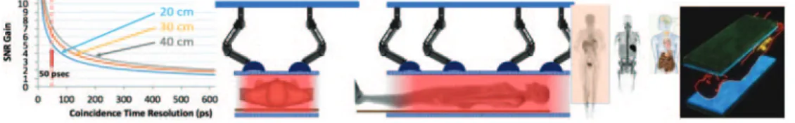
The traditional discussion about the advantages that “good” TOF brings to PET is about the improved S/N due to available information about the location of the positron annihilation point along the Line of Response (LOR). This S/N increase is equivalent to an increase in detection sensitivity and can translate into a non-traditional TOF benefit, with great practical impact. Instead of benefiting from the increase in the overall
sen-Fig. 5. – OSEM image-derived ROI-based blood input functions and major organs/tissues time activity curves. Left: arterial input functions from different ROIs during the first 2 min; middle: arterial input functions during the whole 1 h scan zoomed in on 0–12 kBq/mL scale; right: time activity curves of major organs/tissues of interest [7-10].
sitivity with the additional TOF-delivered sensitivity boost, the scintillator —typically the most expensive detector component— can be made thinner and, therefore, its cost can be lowered by simply using less scintillator, while maintaining the same or even im-proving excellent PET scanner performance (fig. 7). Using less scintillator and operating at better TOF settings may be in fact a way to overcome the current dissemination bar-rier to the total-body (long axial length) very expensive PET scanner. As an example, uExplorer is using 560 kG of LYSO. In addition, in a positive feedback, thinner scintilla-tor will improve timing, by decreasing the average scintillation light path and time jitter on a way to a photodetector. Also, depending on the achieved thickness, the need for the DOI measurement may disappear (say at 5 mm thickness), as opposed to the standard 20 mm.
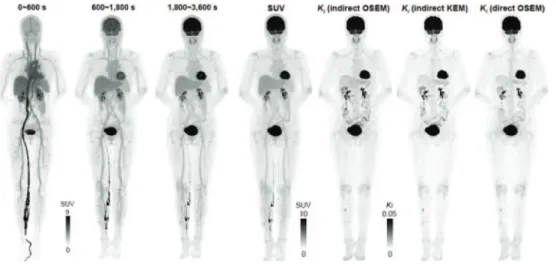
Fig. 6. – Left: reconstructed dynamic composite SUV images (OSEM MIP) of early 10 min, mid 20 min, late 30 min scans. The images were used to extract features to construct the kernel matrix. The SUV images are shown in inverse gray scale with the maximum set to 9. Right: maximum intensity projection (MIP) using last 30 min of data left to right: SUV; indirect OSEM Patlak slope Ki (3 iterations 10 subsets); indirect kernel-EM Patlak slope Ki (3 iterations 10
subsets); direct OSEM Patlak slope Ki(12 iteration 10 subsets, 10 sub-iterations of parametric
Fig. 7. – One theoretical approach to the cost issue is to benefit from the TOF delivered addi-tional sensitivity throuh SNR (S/N) boost and to decrease the amount of (expensive) scintillator, while maintaining key performance parameters at the same high level. Here a shorter and more compact version is shown (for immunotherapy focus) using planar modules, benefiting from TOF-provided compensation for limited angle tomography. (The immune system’s grey scale image and planar PET design sketch from 1996 shown in the right panel were kindly provided by Professor Terry Jones).
And still further, potentially less expensive thin monolithic scintillators can replace the more expensive scintillator arrays, due to no need for precise machining of thousands of scintillator pixels. To minimize the impact of the optical and mechanical discontinuities at the edges of the individual monolithic pieces, the gluing scheme can be implemented, as originally proposed by Craig Levin from Stanford [15], with the neighboring monolithic pieces in the array which are optically coupled, lowering the optical barriers (reflective and absorbing) for the scintillation photons, and suppressing reconstruction image artifacts coming from these edge discontinuities.
In addition, the already discussed excellent CRT TOF resolution of 50 ps FWHM [12, 16-18] would enable artifacts-free reconstructions using simpler, less ex-pensive detector schemes with limited angular coverage, built for example with more economical planar modules, both in pixelated and monolithic versions of the individual scintillation modules. Flexible-geometry designs with robust planar detector assemblies operated with sets of robotic arms could be a potential future solution to replace the one-size-fits-all current expensive cylindrical design with a lot of underutilized sensor material and electronics in the detector. The examples of some concepts are shown below.
8. – The future of PET devices —the personal view
From the perspective of a close witness to the enormous diagnostic opportunities created by the Explorer scanner, the progress with the kinetic parametric imaging rev-olution enabled with simultaneous total-body PET imager, totally depends in the near (and distant) future on the success of dissemination of this revolutionary concept. And this in turn depends on our ability as the medical imaging instrumentation commu-nity to develop economical yet high-performing options for the scanner. At present (December 2019) there are only two ∼2 m long uExplorer imagers in the world (both produced by United Imaging), one in Shanghai and one in Davis, California, with an-other 1.4 m long one based on Philips technology and built at UPenn and with some on-going efforts on similar but shorter scanners in Europe, and at least one in Beijing, China (to be built by another consortium not involving United Imaging). In our opinion, only a spectacular decrease in the price, while maintaining the key parameters such as total-body dynamic/kinetic imaging, will accelerate in a meaningful way the dissemina-tion process of the total-body Explorer approach.
Fig. 8. – Still valid reference for high-sensitivity BGO-based PET/CT EXACT3D imager (1996) (kindly provided by Professor Terry Jones).
of (expensive) scintillator, while maintaining the same sensitivity performance. The estimates of the TOF-delivered boost to the sensitivity and S/N at 30–50 ps FWHM CRT stipulate that the amount of LSO/LYSO can be decreased by 60–70% with substantial cost savings on the multimillion-dollar scanner, assuming that the cost of readout will not substantially increase, due to the required high-level timing performance. In fact, this is what happens in high-TOF systems under development pushing down to 100 ps and below, that the cost of electronics becomes dominating over the cost of scintillator and nullifies the cost advantage of using less scintillator and/or monolithic blocks instead of pixelated arrays.
However, upon critical review of the options and considering first very fast 30–50 ps TOF with generated boost in sensitivity to compensate the thinner scintillator (with the associated decrease in the cost, as mentioned above), it is our conclusion that the only practical option at this time to lower substantially the cost (beyond making the scanner shorter and therefore defeating its initial parametric total-body imaging purpose and strength) is to use BGO as the scintillator material (fig. 8). This is estimated to result in a threefold cost reduction for a set of fully executed scintillator arrays for a 2 m long scanner, as compared to LYSO. In principle, as shown in recent studies, Cherenkov ra-diation generated in BGO [19-21] can be used to boost the TOF performance. However, the sophisticated additional electronics needed to exploit this feature will potentially substantially raise the cost of the scanner. Therefore, the preferred approach seems to be to develop a “traditional” BGO scanner with optimized detection of its scintilla-tion light, and benefiting from its higher-than-LYSO stopping power and higher photo-fraction, and, additionally, from no radiation background like the one produced by 176-Lu in LYSO.
Assuming that by these steps the dissemination of the Explorer scanners will become much easier, beyond a few scanners considered by the few selected prime imaging cen-ters, it still remains to be seen how the Explorers will compete with standard PET/CT scanners of the new generation, such as Siemens Biograph Vision, and the PET/MR scanners such as GE Signa.
The next issue to look into in the near future is imaging with high resolution, ap-proaching the limits of the PET technique (due to positron range and non-collinearity of the two emitted annihilation gammas), and after implementing DOI corrections. Fi-nally, motion correction may in practical clinical setting be the limiting factor of achieved spatial resolution, if not implemented accurately, especially during long dynamic scans.
Fig. 9. – The concept of adding high-resolution AND high-sensitivity compact brain helmet to the Explorer total-body imager. Additional robotic arms may support high-resolution mag-nifying inserts. Many implementation options can be considered with small-size technological insert/entrance ports, as well as in the gaps made temporarily in the system, as shown above in the center, in this rough conceptual example for prostate imaging. To increase patient comfort, patient may use VR glasses. Other shorter (torso +) Explorer designs with or without brain imager and without robotic arms can be envisaged, as shown at right.
Implementing high resolution in the total-body PET will be again expensive even when using BGO. Therefore, a practical approach could be to apply locally the PET mag-nification technique proposed by Tai [18] with additional high-resolution inserts/outserts placed close to the organ or tissue to be imaged with higher resolution. This approach requires development of flexible-geometry limited-angle tomography algorithms, however it was already demonstrated to work in simulations and in phantom studies with standard PET scanners. An interesting facilitating option is to operate the inserts with robotic arms placing the inserts in required imaging positions close to the body to obtain the magnified PET images of selected tissues (fig. 9).
However, high-resolution imaging of the brain needs to be and can be treated differ-ently. In fact, the optimal solution here may be to use a separate compact helmet-style brain imager operating in a tandem with the total-body PET (see fig. 10). Based on the solutions initially developed and implemented in small animal PET scanners, recon-structed spatial resolution approaching 1 mm seems to be possible.
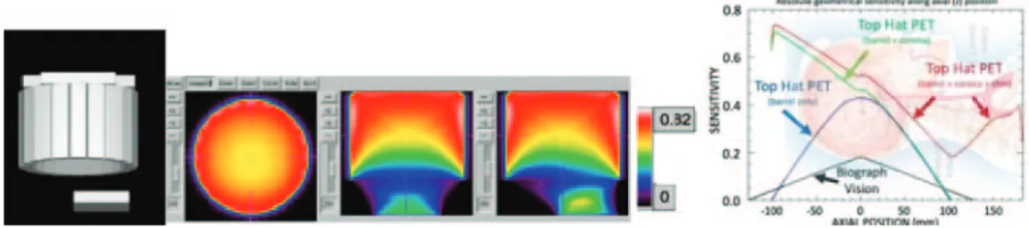
One of the many discussed dedicated PET brain imager designs is a robust Top Hat PET structure composed of a cylinder and two flat panels: one at the top and one at the bottom of the cylinder, under the chin. Comparative geometric sensitivity of up to triple the sensitivity of the Biograph Vision and spatial resolution down to half of the Biograph
Fig. 10. – Left: graphical rendering of a simulated model for the Top Hat PET scanner. Central panel: three views (top, front, and side) of preliminary sensitivities with a color scale shown in the right-most bar. Right: geometrical sensitivities of three Top Hat variants: barrel only (blue), barrel with corona (green), and complete Top Hat (red), compared with Siemens’ Biograph Vision (black). It can be noted that there is high sensitivity of detection in the carotid arteries, enabling extraction of the cardiac input function required for the dynamic studies and kinematic modeling (kindly provided by Professor Johan Nuyts, Loeven).
Vision’s are predicted, depending on the fine details of the selected implementation such as stopping power, pixelation and DOI resolution.
One of the areas of application of PET that is not discussed here is the field of dedicated PET scanners assisting with treatments, such as mobile systems applied in the surgical suit, OR and ICU, or assisting with radiation therapy delivery (such as proton therapy, brachytherapy or focused ultrasound therapy —HIFU).
9. – Conclusions and predictions
Explorer brings revolution to medical imaging. The wealth of molecular/functional information provided by a single dynamic total-body scan is overwhelming. However, the cost of the currently available PET technology creates a dissemination barrier (and limits the worldwide purchases to only few scanners, mostly in China), beyond the obvi-ous issues of how to store and analyze this vast amount of data, and how to fuse the PET images from this almost 200 cm long total-body PET imager with MRI images. But, as always, the imaging scientists already think about improving spatiotemporal resolution (NIH term) of the current model of the Explorer scanner. How to obtain better spatial and timing (time of flight) resolutions is always on their mind. Efforts to push timing resolution down to 50 ps and potentially even down to 10 ps were initiated. While at-taining∼1 mm spatial resolution in the total-body Explorer imager is not immediately practically possible or even justifiable, due to other limiting factors such as large amount of recorded coincident events (“statistics”) necessary to produce good quality ∼1 mm resolution images in the human body and not just as before in a small animal body, it is possible to imagine now and plan to “attach” to the Explorer scanner a dedicated very high-performing (∼1 mm resolution, 100 ps or better TOF resolution and ∼30% effi-ciency) compact brain imager. Interestingly, high-TOF resolution may enable reduction of the most expensive scanner component —the fast LYSO/LSO scintillator. In addition, one of the high spatial resolution scenarios is to add magnifying attachments-inserts to the Explorer scanner, as proposed by Tai [18].
REFERENCES
[1] Badawi R. D., Shi H., Hu P., Chen S., Xu T., Price P. M., Ding Y., Spencer B. A., Nardo L., Liu W., Bao J., Jones T., Li H.and Cherry S. R., J. Nucl. Med., 60 (2019) 299.
[2] Berg E., Roncali E., Kapusta M., Du J. and Cherry S. R., Med. Phys., 43 (2016) 939.
[3] Berg Eric and Cherry Simon R., Innovations in instrumentation for positron emission tomography, in Seminars in Nuclear Medicine, Vol. 48. No. 4 (W. B. Saunders) 2018. [4] Cherry S. R., Badawi R. D., Karp J. S., Moses W. W., Price P. and Jones T.,
Sci. Transl. Med., 9 (2017) 381.
[5] Cherry Simon R. et al., J. Nucl. Med., 59 (2018) 3.
[6] Deng Z., Hu D., Ding Y. and Dong Y., J. Nucl. Med., 60(supplement 1) (2019) 381. [7] Zhang X., Zhou J., Cherry S. R., Badawi R. D. and Qi J., Phys. Med. Biol., 62
(2017) 2465.
[8] Zhang X., Xie Z., Berg E., Judenhofer M. S., Liu W., Xu T., Ding Y., Lv Y., Dong Y., Deng Z., Tang S., Shi H., Hu P., Chen S., Bao J., Li H., Zhou J., Wang G., Cherry S. R., Badawi R. D.and Qi J., J. Nucl. Med., 61 (2020) 285.
[9] Zhang X., Xie Z., Berg E., Judenhofer M., Liu W., Lv Y., Ding Y., Xinyu L., Xu T.and Dong Y., J. Nucl. Med., 60(supplement 1) (2019) 456.
[10] Zhang X., Cherry S., Badawi R. and Qi J., Total-body dynamic PET imaging with 100-ms temporal resolution, presented at the World Molecular Imaging Congress, Montreal, QC, Canada, 2019.
[11] Zhang Xuezhu, Cherry Simon R., Xie Zhaoheng, Shi Hongcheng, Badawi Ramsey D. and Qi Jinyi, Proc. Natl. Acad. Sci. U.S.A., 117 (2020) 2265.
[12] Conti Maurizio and Bendriem Bernard, Clin. Transl. Imaging, 7 (2019) 139. [13] Hu J., Panin V., Smith A. M. et al., Clinical whole body CBM parametric PET with
flexible scan modes, in IEEE Nuclear Science Symposium and Medical Imaging Conference (IEEE) 2017, pp. 1–4, https://doi.org/10.1109/NSSMIC.2017.8532705.
[14] Va’vra J., Picosecond timing detectors and applications, arXiv:1906.11322 (2019). [15] Vinke R. and Levin C. S., Phys. Med. Biol., 59 (2014) 2975.
[16] Derenzo Stephen E., Woon-Seng Choong and Moses William W., Phys. Med. Biol., 59 (2014) 3261.
[17] Gundacker Stefan et al., Phys. Med. Biol., 64 (2019) 055012. [18] Tai Y. C., Wu H., Pal D. et al., J. Nucl. Med., 49 (2008) 471.
[19] Krizan Peter, Ultrafast detection in positron emission tomography using Cherenkov light, presented at the APS April 2019 meeting, APS News (APS) 2019.
[20] Kwon Sun Il et al., Phys. Med. Biol., 61 (2016) L38. [21] Kwon Sun Il et al., Phys. Med. Biol., 64 (2019) 105007.