The Importance of Restriction from
Physical Activity in the Metabolic
Recovery of Concussed Brain
Giuseppe Lazzarino et al.
*Department of Biology, Geology and Environmental Sciences
Division of Biochemistry and Molecular Biology, University of Catania, Catania
Italy
1. Introduction
Brain concussion is unquestionably the most common form of traumatic brain injury (TBI) worldwide (Bruns & Hauser, 2003; Tagliaferri et al., 2006). In European countries, approximately 235 individual/100,000 people are admitted annually to the hospital following TBI, 80% of which receive a diagnosis of mild TBI (mTBI). (van der Naalt, 2001; Vos et al., 2002). It has been calculated that the ratio in the occurrence of mTBI to severe TBI (sTBI) is approximately 22 to 1, with mTBI accounting for at least 75% of patients who survive after TBI each year (Tagliaferri et al., 2006). These percentages are very similar to those recorded in the United States where it is estimated that approximately 1.5 – 8 million people per year suffer from TBI and, among those requiring hospitalization, a proportion ranging from 75% to 90% are classified as “mildly” injured or “concussed” (Bruns & Hauser, 2003). These wide ranges of annual incidence are probably due to the fact that an unknown proportion of mTBI victims do not seek any medical attention (McCrea et al., 2004) (HEADS UP) but it might also be due to the fact that there is still confusion and inconsistency among researchers and organizations in defining and understanding this type of trauma. (Cantu & Voy, 1995; Cantu, 1998, 2007).
The above reported numbers give the evidence that albeit the incidence of mTBI is tremendously high, the mortality caused by this type of trauma appears to be rather low (6-10 per (6-100,000/year). To reinforce this concept it has been reported that only 0.2% of all
*Roberto Vagnozzi1, Stefano Signoretti2, Massimo Manara3, Roberto Floris4, Angela M. Amorini5,
Andrea Ludovici4, Simone Marziali4, Tracy K. McIntosh6 and Barbara Tavazzi5
1Department of Neurosurgery, Chair of Neurosurgery, University of Rome “Tor Vergata”, Rome, Italy
2Division of Neurosurgery, Department of Neurosciences-Head and Neck Surgery, San Camillo Hospital, Rome, Italy
3Association of Sports Physicians Parma, F.M.S.I., Parma, Italy
4Department of Diagnostic Imaging and Interventional Radiology, University of Rome “Tor Vergata”, Rome, Italy 5Institute of Biochemistry and Clinical Biochemistry, Catholic University of Rome “Sacro Cuore”, Rome, Italy 6Media NeuroConsultants Inc., Media, PA 19063, USA
7Department of Biology, Geology and Environmental Sciences, Division of Biochemistry and Molecular Biology,
patients attending the Emergency Departments (EDs) will die as a direct result of this head injury (Vos et al., 2002).
With this in mind, it is evident that this situation represents a really serious public health concern of our time: in fact, the EDs are required to see a massive number of patients, with the most challenging task of identifying the small number of them that will progress to have serious acute intracranial complications. Statistics demonstrate that only 1-3% of all mTBI patients admitted to the hospital with a Glasgow Coma Scale (GCS) score of 14-15 will subsequently develop life-threatening intracranial pathology (the remaining 97-99% of them will be discharged home within 48 hours) (Livingston et al., 2000; Lloyd et al., 1997; Shackford et al., 1992; Swann et al., 1981; Taheri et al., 1993). Furthermore, when a mTBI patient is presenting with a GCS score of 15 and no additional associated risk factors the probability that he may suffer from intracranial hemorrhage, requiring neurosurgical intervention,is below 0.1% (Fabbri et al., 2010).
As a consequence of this very low mortality rate, it is generally and widely accepted that mTBI represents a nosological entity with high frequency but that it does not represent a real serious injury. The common feeling that consequences of mTBI are only transient disturbances has slowly grown, mainly supported by the absence of structural lesions on traditional neuroimaging. Therefore, the general idea is that mTBI-affected patients need no intervention other than observation, with no additional clinical examinations nor specific recommendations for the post-injury period. However, a recent report revealed that the diagnosis of an intracranial hematoma in such patients was made with a median delay of 18 hours (Yates et al., 2007), strongly suggesting that the quality of the “observation” that mildly-injured patients receive while in hospital is of utmost concern. If in the USA it has been established that only 50% of mildly head injured patients admitted to the hospital had documentation of neurological observations (Yates et al., 2007) in Europe the situation is even much worse, with mTBI patients who have historically been observed by non-specialist wards by nurses and doctors not experienced in neurological observations. The fundamental issues of whether to image, observe and discharge, each one of these endless patients, focused essentially on identifying those at risk for a hemorrhagic complications has completely obscured the real dilemma of the concussive injury, whose early and late symptoms and sequelae are continuously underreported, underinvestigated, underdiagnosed, and mainly underestimated by the majority of physicians.
1.1 Background: Sports-related concussion
If mTBI were as “mild” as we might think, it would be difficult to explain the actual complex management of these patients that may involve various health care professionals including family practice physicians, behavioral psychologists, clinical psychologists, neuropsychologists, neurologists, psychiatrists, neuro-ophthalmologists, neurosurgeons, physiatrists, nurses, occupational therapists, and physical therapists. Furthermore, long beyond the typical recovery interval of one week to three months, at least 15% of persons with a history of mTBI continue to see their primary care physician because of persistent problems (Alexander, 1995; Bigler, 2003; Gouvier et al., 1992; Kay et al., 1992; Ingebrigtsen et al., 2000). From the aforementioned picture and reminding that the majority of mTBIs are unreported, it is clearly evident how much difficult would be to select a homogeneous population for a study on mTBI, just on the basis of those who refer to the hospital.
Although certainly blended in to the vast world of mTBI, by definition, brain concussion should be considered a discrete and distinct entity, since not all mTBI are truly
“concussive”; thus the two terms refer to different constructs and should not be used interchangeably (McCrory et al., 2009). Despite recent efforts, a unanimous definition of concussion has not yet been widely accepted (Cantu, 2007). It is possible to define concussion as a traumatic insult capable of provoking an acceleration-deceleration phenomenon within the skull (Barth et al., 2001). From a clinical point of view, concussion is not necessarily accompanied by loss of consciousness and is associated with various physical (headache, equilibrium, vision disturbances, etc.), cognitive (memory, concentration, etc.), emotional (behaviour) and sleep alterations (Gosselin et al., 2009; Hunt & Asplund, 2010; Randolph et al., 2009). These symptoms are included in the well known post-concussive syndrome and can affect, to various degrees, everyday life, resolving spontaneously within 7-10 days post-injury, in the majority of cases. In concussed subjects, the pathobiology of mTBI can not be defined by classical imaging techniques such as CT scan and MR (Kurca et al., 2006), as it occurs in all mTBI patients. This fact, coupled to the faintness and variability of symptoms, mainly assessed by the patient’s self-evaluation, is not certainly of help in diagnosing and monitoring concussed patients.
It is generally accepted that at least 20% of all mTBI are sports-related injuries (concussions), of which 30-45% receives no medical care (McCrea et al., 2004). Athletes, therefore, represent a population at great risk of occurrence of concussive episodes and are, for several reasons, the population of choice with which to undertake trials to study the pathobiology of mTBI (Meehan & Bachur, 2009). It is well established that after the first traumatic episode the probability of recurrence of concussion in athletes increases by 3 times (Cantu, 2003), that currently, there is no agreement as to how many concussions are too many (Guskiewicz et al., 2003; Pellman et al., 2004), nor is there an unanimously approved diagnostic approach to monitor concussed athletes (Delaney et al., 2005; Kissick & Johnstone, 2005; Ponsford, 2005), consequently the criteria to assess a safe return of concussed athletes to play remain unclear (Guskiewicz et al., 2006; McClincy et al., 2006; Lovell et al., 2004).
1.2 Background: Metabolic changes characterizing brain vulnerability following concussion
An increasing number of studies on mTBI have focused attention on concussion-induced changes in brain metabolism (Bergsneider et al., 2000; Praticò et al., 2002) including those related to cerebral energy state (Vagnozzi et al., 1999). Hovda and colleagues first suggested the concept of metabolic vulnerability occurring in brain tissue after any concussive episode (Giza & Hovda, 2001; Hovda et al., 1993). During this transient period of altered brain metabolism and function, a second concussive episode of even modest entity may cause significantly addition and/or dramatic brain damage (Longhi et al., 2005), thereby underlying the so called second impact syndrome (SIS), encountered occasionally in sports medicine (Saunders & Harbaugh, 1984). In fact, the possibility of having a second concussive injury within a not yet defined period of time from the first (i.e., days or weeks) has been reported to be even fatal in some instances (Bowen, 2003; Cantu, 1998; Cobb & Battin, 2004; Logan et al., 2001; Mori et al., 2006; Saunders & Harbaugh, 1984). SIS is clinically characterized by untreatable malignant edema with devastating consequences for the patient up to death. Notwithstanding these reported cases, concerns still exist about the real occurrence of this peculiar pathological condition (McCrory, 2001; McCrory & Berkovic, 2001; Nugent, 2006).
By using a rodent model of closed diffuse mild head injury (Foda & Marmarou, 1994; Marmarou et al., 1994), data from our laboratories confirmed the concept of metabolic
vulnerability (Tavazzi et al., 2007; Vagnozzi et al., 2007) and have also produced solid experimental evidence linking the severity of brain injury and recovery with the extent of ATP and N-acetylaspartate (NAA) decrease and recovery (Tavazzi et al., 2007; Vagnozzi et al., 2005, 2007). NAA is the most prominent compound detectable with proton Magnetic Resonance Spectroscopy (1H-MRS) in human brain, making it one of the most reliable
molecular markers for brain 1H-MRS studies. Although an exact role of this compound
remains to be established, brain NAA was found present in concentrations a hundred fold higher than in non-nervous system tissue and therefore considered a brain-specific metabolite (Miyake et al., 1981; Truckenmiller et al., 1985) and as an in vivo marker of neuronal density. NAA metabolism involves different brain compartments, with neuronal mitochondria taking care of its biosynthesis via the activity of aspartate N-acetyltransferase (ANAT) and oligodendrocytes contributing to its degradation via the activity of N-acetylaspartate acylase (ASPA). NAA homeostasis is finely regulated by three different velocities: 1) rate of neuronal biosynthesis, 2) rate of neuronal outflow in the extracellular space, and 3) rate of oligodendrocyte uptake and degradation (Baslow, 2003a, 2003b). Furthermore, NAA biosynthesis is also strictly dependent on the neuronal energy state and therefore on the correct mitochondrial functioning. In fact, NAA synthesis necessarily requires the availability and the energy of hydrolysis of acetyl-CoA (∆G = -31.2 kJ/mol), working as the acetyl group and energy donor in the acetylation reaction of aspartate catalyzed by ANAT. It is fundamental to understand that when acetyl-CoA is used for NAA synthesis there is an indirect high energy cost to the cell. In fact, since in this case acetyl -CoA will not enter the citric acid cycle (Krebs’ cycle) there will be a decrease in the production of reducing equivalents (3 NADH and 1 FADH2) as the fuel for the electron
transport chain. Since the oxidative phosphorylation is stoichiometrically coupled to the amount of electron transferred to molecular oxygen by the electron transport chain, the final result will be a net loss of 11 ATP molecules for each NAA molecule newly synthesized. NAA concentration within neurons is comparable to that of glutamate (10 mmol/l brain water) but, notwithstanding such a relevant amount and in spite of NAA is known since late ‘50s, there is no unanimity on the biochemical functions of this still enigmatic molecule. According to different studies, NAA may act as storage form for aspartate, protein synthesis regulator, shuttle of acetate and “amino-nitrogen” from the mitochondria to the cytoplasm, breakdown product or precursor of the neurotransmitter N-acetylaspartylglutamate (NAAG), metabolically inert pool regulating the anion deficit balance, metabolically active pool involved in the production of glutamate. Although NAA might play a marginal role in any of the aforementioned processes, recent studies have suggested that the potentially main roles of NAA might be to participates an acetyl group donor in brain lipid biosynthesis (production of myelin) (Moffett et al., 1991) and to act as a neuronal osmoregulator against cytotoxic swelling (molecular water pump) (Baslow, 2003a). Physiologically, NAA concentration is kept within a strict oscillation range even though NAA is regenerated 1.8 times/24 hours (with a calculated turnover rate of approximately 0.75 mmol/l water/hour). It is therefore evident that a perfect balance of the different velocities involved in NAA homeostasis, should exist. NAA decrease has been observed in association with many neurological diseases causing neuronal and axonal degeneration such as tumors, epilepsy, dementia, stroke, hypoxia, multiple sclerosis, and many leukoencephalopathies. Vice versa, the only known pathologic state characterized by a dramatic increase in cerebral NAA is the autosomic genetic leukodystrophy caused by the synthesis of a defective form of the
enzyme responsible for the NAA degradation (N-acetyl-asparto-acylase, ASPA), known as Canavan disease. Due to the enzymatic defect in the NAA degradation, Canavan-affected patients are also characterized by large urinary excretion of intact NAA, up to 2000-fold the normal physiologic excretion (Tavazzi et al., 2005). The morphological alterations in Canavan-affected patients, characterized at MRI by spongi degeneration and structurally defective myelin, can well account for the most accredited roles of NAA: the incapacity to degrade NAA would generate dramatic changes in water extrusion from the brain compartment with consequent spongi degeneration, as well as a drastic decrease in the availability of acetate groups for olygodendrocytes thereby causing deficits in lipid myelin biosynthesis.
1.3 Background: NAA and 1H-MRS as a diagnostic tool to study in vivo brain vulnerability
As already mentioned, the relevance of NAA as a biochemical marker of the metabolic neuronal “wealth” is related to the possibility to measure NAA concentration non-invasively “in vivo” by 1H-MRS. When subtracting the spectral signal of 1H from water (the
most abundant molecule containing an atom with an unpaired proton, which is a prerequisite to allow to detect a molecule by MRS), the 1H-NAA resonance returns one the
best defined peaks at approximately 2 ppm. Using the clinically safe magnetic fields of the MRS apparatuses (1.5 or 3.0 Tesla) in the range 1.8 to 3.4 ppm to additional, well-resolved, resonance peaks are obtained, one at approximately 3.2 ppm referring to choline (Cho) and the other at approximately 2.9 ppm referring to creatine (Cr). It is worth recalling that the relatively low magnetic fields with no side effects (1.5-3.0 T) do not allow to discriminate in the multitude of compounds that are physiologically present within the brain tissue (acetyl-choline, cytidinediphospho-(acetyl-choline, phospho-(acetyl-choline, glycerophospho-(acetyl-choline, phosphatidyl-choline, free-choline, etc.) having this quaternary ammonium derivative as a characterizing chemical group. The same can be applied either in the case of the creatine-containing compounds (creatine and creatinephosphate) or in the case of the NAA-containing compounds (NAA and NAAG). Actually, in a 1H-MRS the Cho peak represents
the choline-containing compounds (with phosphatidyl-choline representing the 90% of the signal intensity), the Cr peak represents creatine-containing compounds (with creatine accounting for the 90% of the intensity signal) and the NAA peak the NAA-containing compounds (with NAA being 12 to 15 fold more concentrated than NAAG). Unless determining the water content within the voxel(s) for a calculation of the absolute concentrations of the different metabolites, it is necessary to quantify at the same time the peak areas of the spectroscopic intensity signals of both Cr and Cho-containing compounds and to then refer to the NAA/Cr and NAA/Cho ratios for the semi-quantitative NAA evaluation in physiopathological conditions (Barker et al., 1993; Brooks et al., 2001; Friedman et al., 1999; Garnett et al., 2000; Mitsumoto et al., 2007). This type of approach assumes Cr and Cho as two unvarying parameters to which relate the NAA changes. The results of a multicenter clinical trial (Vagnozzi et al., 2010) involving 40 concussed athletes and 30 healthy volunteers have been recently published, revealing that, despite different combinations of field strengths (1.5 or 3.0 T) and modes of spectrum acquisition (single- or multi-voxel) among the scanners currently in use in most neuroradiology centers, NAA determination represents a quick (15-minute), easy-to-perform, noninvasive tool to accurately measure changes in cerebral biochemical damage occurring after a concussion.
Patients exhibited the most significant alteration of metabolite ratios at day 3 post-injury, showing a gradual recovering, initially in a slow fashion and, following day 15, more rapidly. At 30 days post-injury, all subjects showed complete recovery, having metabolite ratios similar to values detected in controls. Interestingly, all these 40 patients self-declared symptom clearance between 3 and 15 days after concussion strongly demonstrating differential times of disappearance of clinical gross signs and of normalization of brain energy metabolism. In a previous pilot study carried out in a cohort of singly and doubly- concussed athletes, examined by 1H-MRS for their NAA cerebral content at different time
points after concussive events, we demonstrated that the recovery of brain metabolism is not linearly related to time. In this study, two athletes experienced a second concussion between the 10th and the 13th day after the first insult. Although they were not affected by SIS nor showed signs of sTBI, they however had a significant delay in both symptom resolution and NAA normalization (Vagnozzi et al., 2008).
According to the aforementioned data it is clear that concussion causes a reversible change of brain metabolism that can be monitored by 1H-MRS in terms of NAA variations.
Furthermore, data recorded in the two doubly concussed athletes suggest that a second concussion might compromise rescue of cerebral metabolism and delaying its normalization. This would in turn cause delaying in return of athletes to play. In the present study, we report data referring to six concussed athletes who received a second concussion at different times during the period of alteration of brain metabolism. The time course changes in their cerebral NAA, evaluated by 1H-MRS up to complete normalization, are
presented.
2. Materials and methods
Six non-professional male athletes from different sport disciplines who had suffered from a sport-related concussion, defined as a traumatically induced transient alteration in mental status, not necessarily accompanied with loss of consciousness, were considered in this study. At the clinical examinations they had a GCS greater or equal to 14, normal neurological objective signs and an age ranging between 20 and 33 years (mean age = 26 ± 5 years). According to our previous observations (Vagnozzi et al., 2008, 2010), they were required to restrain from further physical activity during the entire observational period, which lasted until cerebral metabolic normalization. None of the patients had positive MRI for post-traumatic anatomical lesions (the presence of blood, etc.), or suffered from polytrauma, or presented with risk factors for subsequent complications (coagoulopathy, epilepsy, former neurosurgical interventions, alcohol or drug abuse, disabilities). The first clinical evaluation, MRI and MRS were carried out at 3 days post injury; follow-up MRI and MRS analyses, as well as clinical evaluation of concussion-associated symptoms, were performed at different times up to metabolic normalization (15, 22, 30, 45, 60, 90, 120 days post-injury). A group of 10 healthy male subjects matched for age (mean age = 25 ± 6 years) was subjected to MRS and used as the control group.
In controls and athletes, semi-quantitative analysis of NAA relative to creatine (Cr) and choline containing compounds (Cho) was performed in the single voxel (SV) mode, after obtaining proton spectra using a 3.0 T system (Philips, Intera Achieva). For conventional MRI studies, T1 and T2 weighted TSE images were acquired in axial coronal and sagittal planes and, in order to rule out even the smallest amount of intra-cerebral blood, Fast Field Echo (FFE) T2* sequences were used. A multi channel coil (8 ch.) Sense-Head with 4 mm
slice thickness, 1 mm gap and a FOV of 230 mm, was used for all MRI sequences. Following localized shimming and water suppression, the spectroscopic examination was carried out using a PRESS (Point Resolved Spectroscopy Sequence) pulse sequence, with the following settings: TE = 144 msec; TR = 2,000 msec; Spectral Bandwidth = 2,000 Hz; acquisition cycles = 128. The optimal positioning of the voxel was determined using the MR images acquired on axial, coronal and sagittal planes to facilitate its three-dimensional placement, adjacent to the cortical-subcortical junction in order to include only the white matter of the frontal lobes, bilaterally and the choice of this location as the region of interest, was made to obtain the most homogeneous data as possible. To this end, a spectrum from a single voxel (SV) customized to sample a volume of interest (VOI) of 3.375 cm3 (1.5 x 1.5 x 1.5 cm), was
obtained (acquisition time about 5 min for each voxel). In follow-up studies, the exact repositioning of the voxel on the same acquisition plane obtained in the previous MRI study was achieved by using dedicated software (SameScan, Philips Medical Systems). Metabolite intensity ratios (NAA-to-Cr and Cho-to-Cr) were automatically calculated at the end of each acquisition using dedicated software (SpectroView; Philips Medical Systems), by which gaussian-fitted peak areas relative to a baseline computed from a moving average of the noise regions of each spectrum were determined. Post-processing of spectral data, using a homemade computed program, allowed us to render uniform calculations of the area under the peaks of NAA, Cho and Cr, using common criteria for peak integration. In the case of a single, well-defined peak (typically the NAA peak), a valley-to-valley integration was performed to obtain the area under the peak. In the case of not fully resolved peaks (frequently the Cho and Cr peaks), a horizontal baseline between the start of the first peak to the end of the second peak was selected; the grouped peaks were then split by a vertical line, drawn from the median point of the common valley between peaks to the horizontal baseline and the area under the peaks calculated. These values were used to determine the metabolite ratios NAA/Cho, NAA/Cr and Cho/Cr.
3. Results
Table 1 summarizes the clinical features of the 6 doubly concussed athletes, including the time interval between concussion and the type and duration of the post-concussive self-reported clinical symptoms after both the first and the second head injury.
Table 1. Demographic data, sport activity and clinical features of 6 non professional athletes suffering from double concussion.
None of these concussions was characterized by loss of consciousness. All athletes declared to suffer from headache following the first concussive episode and each of them suffered from at least one additional post-concussive disturbance. Disappearance of self-reported clinical symptoms after the first concussion ranged between 3 to 8 days (mean duration = 5.8 ± 2.1 days). In two athletes the second concussive event occurred between the first and second 1H-MRS (mean interval between the two concussions = 9.5 ± 0.7 days), whilst in the
remaining 4 athletes the second concussion took place between the second and the third second 1H-MRS (mean interval between the two concussions = 18.5 ± 2.1 days). In athletes 1
and 2, the second concussions were characterized by loss of consciousness (< 2 min). Following the second concussive episode, all athletes again declared to suffer from headache and each of them suffered from at least four additional post-concussive disturbances. Self-reported clinical symptoms following the second concussion disappeared with a mean duration = 41.2 ± 13.0 days (p < 0.001 when compared to duration of symptoms observed
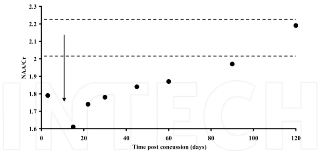
after the first concussion). Figures 1 and 2 illustrate the time course changes of NAA (reported in the Figures as the NAA/Cr ratio) in the two doubly concussed athletes (Patients 1 and 2) receiving the second head injury between the 1st and the 2nd1H-MRS, both
showing loss of consciousness < 2 min on field.
Table 1. Clinical features of doubly concussed athletes. The mean duration of symptom persistence lasted 5.8 ± 2.1 days after the 1st injury and 41.2 ± 13.0 days after the 2nd
concussion (p < 0.001 when compared to duration of symptoms observed after the first concussion). In both cases, symptoms disappeared much earlier than the time needed for complete NAA restoration.
At the time of the 1st resonance spectrum acquisition (3 days post-injury) both subjects
showed a consistent decrease in the NAA/Cr ratio. When effecting the 2nd MRS,
notwithstanding athletes were both initially advised to restrain from physical activity, they both declared to have started again their respective sport discipline because of symptom disappearance and to have received a second concussion few days later (mean value between repeat concussions = 9.5 ± 0.7). At this 2nd MRS analysis, the NAA/Cr ratio fell
slightly below 1.6 (-23.6% with respect to value in controls), a value very close to that observed in patients suffering from sTBI (Signoretti et al., 2010). In both these athletes, the second concussive episode produced a prolonged loss of consciousness (< 2 min). Both subjects admitted to have experienced, from the beginning up to clinical healing, much more severe and prolonged post concussive symptoms (mean value of symptom persistence = 55.5 days) than those lived following the first impact.
Figures 3, 4, 5 and 6 illustrate the NAA/Cr ratio recorded in four athletes receiving the second concussion between the 2nd and the 3rd MRS.
Patient 1 1.6 1.7 1.8 1.9 2 2.1 2.2 2.3 0 20 40 60 80 100 120
Time post concussion (days)
N
A
A
/C
r
Fig. 1. Change of NAA in doubly concussed athlete. NAA relative to Cr (NAA/Cr ratio) was measured by 1H-MRS in voxels properly positioned in the frontal lobes. The arrow indicates
the approximate time of occurrence of the 2nd concussive episode (see Table 1). Dotted lines
represent the range interval of the NAA/Cr ratio recorded in control healthy subjects. Notwithstanding prohibition to sustain physical activity, patient 1 restarted physical training immediately after symptom clearance (3 days after the 1st concussion), when
NAA/Cr was about 16% below the value recorded in controls.
Patient 2 1.6 1.7 1.8 1.9 2 2.1 2.2 2.3 0 20 40 60 80 100 120
Time post concussion (days)
N
A
A
/C
r
Fig. 2. Change of NAA in doubly concussed athlete. NAA relative to Cr (NAA/Cr ratio) was measured by 1H-MRS in voxels properly positioned in the frontal lobes. The arrow indicates
the approximate time of occurrence of the 2nd concussive episode (see Table 1). Dotted lines
represent the range interval of the NAA/Cr ratio recorded in control healthy subjects. Notwithstanding prohibition to sustain physical activity, patient 2 restarted physical training immediately after symptom clearance (4 days after the 1st concussion), when
Patient 3 1.6 1.7 1.8 1.9 2 2.1 2.2 2.3 0 15 30 45 60 75 90
Time post concussion (days)
N
A
A
/C
r
Fig. 3. Change of NAA in doubly concussed athlete. NAA relative to Cr (NAA/Cr ratio) was measured by 1H-MRS in voxels properly positioned in the frontal lobes. The arrow indicates
the approximate time of occurrence of the 2nd concussive episode (see Table 1). Dotted lines
represent the range interval of the NAA/Cr ratio recorded in control healthy subjects. Notwithstanding prohibition to sustain physical activity, patient 3 restarted physical training immediately after symptom clearance (8 days after the 1st concussion), when
NAA/Cr was about 13% below the value recorded in controls.
Patient 4 1.6 1.7 1.8 1.9 2 2.1 2.2 2.3 0 15 30 45 60 75 90
Time post concussion (days)
N
A
A
/C
r
Fig. 4. Change of NAA in doubly concussed athlete. NAA relative to Cr (NAA/Cr ratio) was measured by 1H-MRS in voxels properly positioned in the frontal lobes. The arrow indicates
the approximate time of occurrence of the 2nd concussive episode (see Table 1). Dotted lines
represent the range interval of the NAA/Cr ratio recorded in control healthy subjects. Notwithstanding prohibition to sustain physical activity, patient 4 restarted physical training immediately after symptom clearance (7 days after the 1st concussion), when
Patient 5 1.6 1.7 1.8 1.9 2 2.1 2.2 2.3 0 15 30 45 60
Time post concussion (days)
N
A
A
/C
r
Fig. 5. Change of NAA in doubly concussed athlete. NAA relative to Cr (NAA/Cr ratio) was measured by 1H-MRS in voxels properly positioned in the frontal lobes. The arrow indicates
the approximate time of occurrence of the 2nd concussive episode (see Table 1). Dotted lines
represent the range interval of the NAA/Cr ratio recorded in control healthy subjects. Notwithstanding prohibition to sustain physical activity, patient 5 restarted physical training immediately after symptom clearance (8 days after the 1st concussion), when
NAA/Cr was about 14% below the value recorded in controls.
Patient 6 1.6 1.7 1.8 1.9 2 2.1 2.2 2.3 0 15 30 45 60 75 90
Time post concussion (days)
N
A
A
/C
r
Fig. 6. Change of NAA in doubly concussed athlete. NAA relative to Cr (NAA/Cr ratio) was measured by 1H-MRS in voxels properly positioned in the frontal lobes. The arrow indicates
the approximate time of occurrence of the 2nd concussive episode (see Table 1). Dotted lines
represent the range interval of the NAA/Cr ratio recorded in control healthy subjects. Notwithstanding prohibition to sustain physical activity, patient 6 restarted physical training immediately after symptom clearance (5 days after the 1st concussion), when
At the time of the 2nd MRS, the four patients showed a recovery of NAA and affirmed to be
symptomless. Both these phenomena allowed athletes to violate the ban on sports so that they started practicing their respective disciplines before completion of brain metabolic recovery. At 22 days post-impact, we found in these subjects a significant further decline in the NAA/Cr ratio, the value of which was even lower than that recorded 3 days post-injury (Figures 3, 4, 5, 6 and 8). During the clinical consult, the four patients declared to have suffered from a second concussion (mean interval between the two concussions = 18.5 ± 2.1 days), interpreted on the field of minor relevance but being of surprisingly remarkable clinical severity and duration (mean value of symptom persistence = 34.0 days). It is worth underlining that in patient 5 completion of brain metabolic recovery was observed at the time of the 6th MRS, i.e. 39 days after the 2nd insult and 15 days after symptom
disappearance.
4. Discussion
According to our opinion, data reported in the present study strongly demonstrate that the occurrence of repeat concussion produced a significant increase in the time of recovery of brain metabolism (as evaluated in terms of NAA/Cr variations determined by 1H-MRS),
coupled to the appearance of clinical symptoms with increased severity and duration with those reported after a single concussive event (Vagnozzi et al., 2008, 2010). In sports medicine, this finding implies that it should be mandatory for concussed athletes to observe a period of restriction from physical activity until the process of normalization of brain metabolism is completed. Since also in these subjects the clearance of post-concussive clinical symptoms took place much before than the return of NAA to physiological values (Vagnozzi et al., 2008, 2010) it is our advise that monitoring alterations in the biochemistry of post-concussed neurons (NAA changes) by 1H-MRS should be considered a fundamental
tool to evaluate recovery of post concussed athletes for their safe return to play.
Supported by abundant literature, it is nowadays worldwide accepted that concussion triggers a cascade of molecular events that transiently alter the biochemistry of the post-concussed neurons, with particular involvement for mitochondrial-dependent energy metabolism. This condition prompted Hovda and coll. to hypothesize the insurgence of a period of brain vulnerability during which a second concussive event may have fatal consequences for the neuronal vitality (Giza & Hovda, 2001; Hovda et al., 1993). Our previous researches in rats undergoing repeat mTBI, using the closed-head weight-drop model of diffuse injury set up by Marmarou and coll. (Foda & Marmarou 1994; Marmarou et al., 1994), clearly demonstrated that, depending on the time interval between injuries, two repeat concussions may cause metabolic cerebral irreversible alterations typical of single sTBI (3 days between concussions) (Tavazzi et al., 2007; Vagnozzi et al., 2007), i.e. cumulative effect of the two concussions. If the two repeat injuries were viceversa spaced by 5 days the changes of brain metabolism are fully reversible and comparable to those recorded in single mTBI, i.e. the two concussions acted as separate events (Tavazzi et al., 2007; Vagnozzi et al., 2007). This strongly indicates the existence of a window of metabolic brain vulnerability during which neurons, when receiving a second insult of even very modest entity, can suffer from dramatic impairment of cell functions. This phenomenon, can be explained by hypothesizing that neurons, after the first mTBI, are deeply involved in the energy-consuming processes to restore cell homeostasis, therefore rendering cells more susceptible to injuries of even very modest entity. The duration for the completion of these
“repairing processes” corresponds to the window of brain vulnerability. In a pilot study in a restricted group of concussed athletes, we first monitored the time course of NAA decrease and recovery following concussion, thereby demonstrating the occurrence of the metabolic brain vulnerability status after an mTBI also in human beings (Vagnozzi et al., 2008). In the same study, we also described 2 cases of doubly concussed athletes who received a second impact during the period of energy metabolism recovery and who therefore underwent to a 15 days delay in complete NAA restoration (Vagnozzi et al., 2008). Recently, we provided unquestionable evidences indicating that the determination of NAA by 1H-MRS is a reliable
tool with which monitoring post-concussive periods in athletes. In this last study we demonstrated that results of the MRS analyses were independent on the MR apparatus (different MR suppliers), the field strength adopted (1.5 or 3.0 T) and the mode of spectra acquisition (Vagnozzi et al., 2010). Furthermore, the number of athletes enrolled (n = 40) and serially analyzed allowed to demonstrate that it is possible to determine the period of metabolic brain vulnerability for a safe return of athletes to play. Recently, different research groups confirmed our findings and successfully applied NAA evaluation by MRS to monitor the metabolic recovery of mildly-injured patients (Henry, 2010; Gasparovic et al., 2009; Sarmento et al., 2009; Yeo et al., 2011), thereby strongly corroborating the concept that methods capable of investigating at the molecular level are of great clinical relevance in the surveillance of post-concussed patients. On the other hand, the vast data in literature obtained in different models of mTBI (Barkhoudarian et al, 2011; Signoretti et al., 2010) clearly showed that post-concussive brain modifications are caused by a cascade of molecular events involving cerebral metabolism and, more in general, cerebral biochemistry. At present, in addition to the subjective indication of the patient, cognitive neuropsychological tests are widely used to assess the condition of mildly injured athletes. This type of monitoring has been considered one of the cornerstones for return to play after a concussion (Maroon et al, 2000; McClincy et al., 2006; McCrea et al., 2003; Schatz et al., 2006), even though concerns have been raised, including the question of when they should be used in the management and assessment of concussion (Collie et al., 2006; Gosselin et al., 2006; Randolph et al., 2005). Furthermore, none of the currently available diagnostic tests (Broglio et al., 2007; McCrea et al., 2003; Schatz et al., 2006; Register-Mihalik et al., 2008) are capable of measuring the unique, transient and potentially dangerous state of metabolic vulnerability experienced by the post-concussed brain tissue. Therefore, the need to find objective parameters to evaluate the extent of and recovery from concussion-induced cerebral damage has been stressed recently (Cantu, 2000). Our previous studies (Tavazzi et al., 2005; Vagnozzi et al., 2005, 2008, 2010) and the present research demonstrate that 1
H-MRS is capable of detecting significant neurochemical changes present in the injured brain despite the normal appearance of neuroimaging, absence of symptoms and normal neurological examination, i.e. we validated the use of a rapid, objective and sensitive diagnostic tool with which evaluating normalization of cerebral metabolism for a safe return of concussed athletes to play outside the window of metabolic brain vulnerability. Therefore, restraint from physical activity following concussion should be mandatory to avoid the risk of insurgence of SIS, with SIS being interpreted as an acute, fatal disease caused by uncontrolled brain swelling (Bowen, 2003; Cantu, 1998; Cobb & Battin, 2004; Logan et al., 2001; Mori et al., 2006; Saunders & Harbaugh, 2006). Results of the present study suggest that the concept of SIS might certainly be revised and could be broaden to any case of repeat concussion in which, after the second injury, a clear disproportion among the
entity of the concussive event, the post-concussive clinical symptoms and the cerebral metabolic recovery indeed exists. This restricted cohort of doubly concussed athletes could be included within the aforementioned definition of SIS. In fact, notwithstanding all athletes received two repeat concussions (for each athlete, both events were characterized by the same acute symptoms with no change in GCS and negative MRI), clinical symptoms after the 2nd impact lasted much longer than the 1st one, satisfying our first proposed criterium to
diagnose SIS. Moreover, in our previous studies we showed that the time to return the NAA/Cr to normal cerebral levels in singly concussed athletes is within 30 days post-impact. In the present cohort of doubly concussed athletes, the time required to measure values of the NAA/Cr ratio similar to those recorded in controls after the second concussion was of 81.2 ± 24.4 days. This much longer time for NAA recovery satisfy the second criterium we proposed to diagnose SIS. Independently on the inclusion in the SIS category, these athletes definitely showed a prolonged time of clearance of clinical symptoms and brain metabolism normalization. In our opinion, monitoring of brain metabolism in singly concussed athletes should drive the timetable of return to physical activity, especially for those practicing sports at risk of recurrent concussions (American football, boxe, ice hockey, rugby, alpine skiing, martial arts, soccer, etc.), according to this possible steps: 1) if upon first examination, NAA is below the value of healthy controls, i.e. altered energy metabolism, the athlete should rest with no physical activity (approximate post-concussion time interval of 1–15 days); 2) if, at the second examination, MRS suggests an initiation of the process of NAA recovery (i.e. quasi-normal energy metabolism), it is advisable that the athlete begin physical activity of increasing intensity (approximate post-concussion time interval of 16–22 days); 3) if, at the third MRS, progressive NAA replenishment is observed (i.e. normalized energy metabolism), then physical activity might be intensified to a ‘return to play’ level of conditions (approximate post-concussion time interval of 23–30 days); 4) if, at the fourth MRS, normal NAA, i.e. normal energy metabolism, has been determined, it is suitable that athletes be permitted to return to play (approximate post-concussion time interval 30 days). Such a timetable could be adapted to any post-concussed, non-athlete patient and translated into recommendations differing on personal lifestyle during the recovery of NAA post-concussion: 1) NAA below control values (prolonged altered energy metabolism) would recommend rest, with no physical activity and sedentary lifestyle (approximate post-concussion time interval of 1–15 days); 2) signs of initiation of NAA recovery (i.e. quasi-normal energy metabolism) would suggest normal working activity and moderate physical activity (approximate post-concussion time interval of 16–22 days); 3) normal NAA at MRS (i.e. normal energy metabolism re-established) would implicate return to full normal lifestyle (approximate post-concussion time interval of 23–30 days). Results of this and of previous studies (Vagnozzi et al., 2008, 2010) indicated that the kinetic of NAA recovery, following a single concussion, is non-linear, with a very slow phase of about 15-20 days and a second faster period of 10-15 days. We have recently demonstrated that this non-linear time-course of post-traumatic NAA recovery may be due to the cerebral energy imbalance, assessed by high-energy phosphate quantification (ATP, ADP, AMP, etc.), caused mainly by mitochondrial malfunctioning, as indicated by altered mitochondrial phosphorylating capacity (measured by the ATP/ADP ratio) (Signoretti et al., 2010; Tavazzi et al., 2005). Under these conditions, the remarkable decrease in cerebral NAA, which mirrors the changes in brain ATP, may possibly be attributed to the general energy depression consequent to impaired mitochondrial functions (Lifshitz et al., 2003; Robertson
et al., 2006). Incorporating the data obtained in preclinical studies on mTBI, demonstrating decreased ATP concentration for a given period of time post-injury (Tavazzi et al. 2007; Vagnozzi et al., 2005, 2007), it is conceivable that the process of NAA normalization is markedly hindered by an imbalance of neuronal energy metabolism induced by concussion. In fact, NAA synthesis necessarily requires the availability and the energy of hydrolysis of acetyl-CoA (∆G = -31.2 kJ/mol), working as the acetyl group and energy donor in the acetylation reaction of aspartate catalyzed by ANAT. It is fundamental to understand that when acetyl-CoA is used for NAA synthesis there is an indirect high energy cost to the cell. In fact, since in this case acetyl -CoA will not enter the citric acid cycle (Krebs’ cycle) there will be a decrease in the production of reducing equivalents (3 NADH and 1 FADH2) as the
fuel for the electron transport chain. Since the oxidative phosphorylation is stoichiometrically coupled to the amount of electron transferred to molecular oxygen by the electron transport chain, the final result will be a net loss of 11 ATP molecules for each NAA molecule newly synthesized. Experimental studies (Signoretti et al., 2010) have shown that spontaneous re-synthesis of NAA occurs only after recovery of mitochondrial dysfunction with consequential return to normal ATP levels; therefore, it appears possible that normalization of NAA concentrations may occur only after the cerebral energy state has fully recovered. The slow normalization of the cell energetic could also be attributed to the drastic decrement of the nicotinic coenzyme pool that was observed in rat models of graded injury. In fact, previous studies (Tavazzi et al., 2005; Vagnozzi et al., 2007) showed the net diminution of the nicotinic coenzyme pool (NAD+ + NADH and NADP+ + NADPH) that
certainly plays a pivotal role in the final result of general depression of cell energy metabolism. This depletion jeopardizes either the reducing equivalent supply to mitochondrial oxidative metabolism, or the catalytic activity of dehydrogenase-mediated oxidoreductive reactions. To date, possible mechanisms for this phenomenon are the hydroxyl radical-induced hydrolysis of the N-glycosidic bond of the reduced forms of the nicotinic coenzymes NADH and NADPH and the activation of the enzyme NAD-glycohydrolase (Lautier et al., 1994). Both mechanisms cause the hydrolysis of these coenzymes and give rise to the same end products, i.e., ADP-ribose(P) and nicotinamide. Independently of the predominant mechanism, the final result is certainly deleterious for the correct functioning of cell metabolism. Finally, the augmentation of poly-ADP ribosylation reactions through the activation of the enzyme poly-ADP ribose polymerase (Du et al., 2003; Nanavaty et al., 2002; Pacher et al., 2002), has been demonstrated to trigger the mechanisms of apoptotic induction (Yu et al., 2002). The overall result should be to significantly contribute to the decrease in the rate of NAA recovery during the time period close to the head insult, when cells are more “metabolically vulnerable” and physical restriction is mandatory to avoid catastrophic consequences.
5. Conclusion
This and previous data (Vagnozzi et al., 2008, 2010) demonstrated that this process can be non-invasively followed in vivo by 1H-MRS giving clinically relevant information
concerning the duration of the window of metabolic brain vulnerability. This time interval should be characterized by restriction of physical activity to avoid the occurrence of second concussion with unpredictable consequences, from the delay in cerebral metabolic normalization (such a delay being not yet defined in duration) to the onset of uncontrolled brain edema (i.e., the current definition of SIS). In our opinion, we again provided the
experimental evidence in the dramatic discrepancy between the time required for the clearance of post-concussive clinical symptoms and the time needed to restore concussion-perturbed brain metabolism. Since 1H-MRS is the analytical method of choice and NAA the
biochemical parameter indirectly representing the brain energy metabolism, it should be strongly suggest ed to determine healing of post-concussed athletes and patients using this potent diagnostic tool. In light of the consequences of a second concussive event during the window of brain vulnerability, potentially catastrophic, it should be strongly recommended that the restriction of physical activity is mandatory and that the removal of this restriction is submitted to the full recovery of the NAA physiological level. Due to the potential catastrophic consequences of repeat concussions and the need to have clear diagnostic tools and protocols to study recovery of the post-concussed brain it is fundamental to undertake further studies to better understand these topics.
6. Acknowledgement
This work has been supported in part by research funds of the three Universities involved (Catania, Rome “Tor Vergata”, Rome Catholic “Sacro Cuore”).
7. References
Alexander, M.P. (1995). Mild traumatic brain injury: pathophysiology, natural history, and clinical management. Neurology, Vol. 45, No.7, (July 1995), pp. 1253–1260, ISSN 0028-3878
Barker P.B.; Soher, B.J.; Blackband, S.J.; Chatham, J.C.; Mathews, V.P. & Bryan, R.N. (1993). Quantitation of proton NMR spectra of the human brain using tissue water as an internal concentration reference. NMR in Biomedicine, Vol.6, No.1, (January-February 1993), pp. 89–94, ISSN 0952-3480
Barkhoudarian, G.; Hovda, D.A. & Giza, C.C. (2011). The molecular pathophysiology of concussive brain injury. Clinics in sports medicine, Vol.30, No.1, (January 2011), pp. 33-39, ISSN 0278-5919
Barth, J.T.; Freeman, J.R.; Broshek, D.K. & Varney, R.N. (2001). Acceleration-deceleration sport-related concussion: the gravity of it all. Journal of Athletic Training, Vol.36, No.3, (September 2001), pp. 253–256, ISSN 1062-6050
Baslow, M.H. (2003a). Brain N-acetylaspartate as a molecular water pump and its role in the etiology of Canavan disease: A mechanistic explanation. Journal of Molecular
Neuroscience, Vol.21, No. 3, pp. 185–190, ISSN 0895-8696
Baslow, M.H. (2003b). N-acetylaspartate in the vertebrate brain: Metabolism and function.
Neurochemical Research, Vol.28, No.6, (June 2003), pp. 941–953, ISSN 0364-3190 Bergsneider, M.; Hovda,. D.A.; Lee, S.M.; Kelly, D.F.; McArthur, D.L.; Vespa, P.M.; Lee, J.H.;
Huang, S.C.; Martin, N.A.; Phelps, M.E. & Becker, D.P. (2000). Dissociation of cerebral glucose metabolism and level of consciousness during the period of metabolic depression following human traumatic brain injury. Journal of
Neurotrauma, Vol.17, No.5, (May 2000), pp. 389–401, ISSN 0897-7151
Bigler, E.D. (2003). Neurobiology and neuropathology underlie the neuropsychological deficits associated with traumatic brain injury. Archives of Clinical Neuropsychology, Vol.18, No.6, (August 2003), pp. 595–627, ISSN 0887-6177
Bowen, A.P. (2003). Second impact syndrome: A rare; catastrophic; preventable complication of concussion in young athletes. Journal of Emergency Nursery, Vol.29, No.3, (June 2003), pp. 287–289, ISSN 0099-1767
Broglio, S.P.; Ferrara, M.S., Macciocchi, S.N.; Baumgartner, T.A. & Elliott, R. (2007). Test-retest reliability of computerized concussion assessment programs. Journal of athletic
training, Vol.42, No.4, (October-December 2007), pp. 509-514, ISSN 1062-6050 Brooks, W.M.; Friedman, S.D. & Gasparovic, C. (2001). Magnetic resonance spectroscopy in
traumatic brain injury. Journal of Head Trauma Rehabilitation, Vol.16, No.2, (April 2001), pp. 149–164, ISSN 0885-9701
Bruns, J. Jr. & Hauser, W.A. (2003). The epidemiology of traumatic brain injury: a review.
Epilepsia, Vol.44, Suppl.10, (September 2003), pp. 2–10, ISSN 0013-9580
Cantu, R.C. (1998). Second-impact syndrome. Clinics in Sports Medicine, Vol.17, No.1, (January 1998), pp. 37–44, ISSN 0278-5919
Cantu, R.C. & Voy, R. (1995). Second impact syndrome: a risk in any contact sport. The
Physician and Sport Medicine, Vol.23, No.6, (June 1995), pp. 27-34, ISSN 0091-3847 Cantu, R.C. (2000). Malignant brain edema and Second Impact Syndrome. In: Neurologic
Athletic Head and Spine Injuries, R.C. Cantu, (Ed.), 132-137, ISBN 072-1683-39-8, WB Saunders Company, Michigan , USA
Cantu, R.C. (2003). Recurrent athletic head injury: risks and when to retire. Clinics in Sports
Medicine, Vol.22, No.3, (July 2003), pp. 593–603, ISSN 0278-5919
Cantu, R.C. (2007). Athletic concussion: current understanding as of 2007. Neurosurgery, Vol.60, No.6, (July 2007), pp. 963–964, ISSN 1524-4040
Cobb, S. & Battin, B. (2004). Second-impact syndrome. Journal of School Nursing, Vol.20, No.5, (October 2004), pp. 262–267, ISSN 1050-8405
Collie, A.; Makdissi, M.; Maruff, P.; Bennell, K. & McCrory, P. (2006). Cognition in the days following concussion: comparison of symptomatic versus asymptomatic athletes
Journal of Neurology, Neurosurgery, and Psychiatry, Vol.77, No.2, (February 2006), pp. 241-245, ISSN 0022-3050
Delaney, J.S.; Abuzeyad, F.; Correa, J.A. & Foxford, R. (2005). Recognition and characteristics of concussions in the emergency department population. The Journal of Emergency
Medicine, Vol.29, No.2, (August 2005), pp. 189–197, ISSN 0736-4679
Du, L.; Zhang, X.; Han, Y.Y.; Burke, N.A.; Kochanek, P.M.; Watkins, S.C.; Graham, S.H.; Carcillo, J.A.; Szabó, C. & Clark, R.S. (2003). Intra-mitochondrial poly(ADP-ribosylation) contributes to NAD+ depletion and cell death induced by oxidative stress. The Journal of Biological Chemistry, Vol.278, No.20, (May 2003), pp. 18426– 18433, ISSN 0021-9258
Fabbri, A.; Servadei, F.; Marchesini, G.; Negro, A. & Vandelli, A. (2010). The changing face of mild head injury: temporal trends and patterns in adolescents and adults from 1997 to 2008. Injury, Vol.41, No.9, (September 2010), pp. 913-917, ISSN 1879-0267
Foda, M.A. & Marmarou, A. (1994). A new model of diffuse brain injury in rats: part II. Morphological characterization. Journal of Neurosurgery, Vol.80, No.2, (February 1994), pp. 301-313, ISSN 0022-3085
Friedman, S.D.; Brooks, W.M.; Jung, R.E.; Chiulli, S.J.; Sloan, J.H.; Montoya, B.T.; Hart, B.L. & Yeo, R.A. (1999). Quantitative proton MRS predicts outcome after traumatic brain injury. Neurology, Vol.52, No.7, (April 1999), pp. 1384–1391, ISSN 0028-3878
Garnett, M.R.; Blamire, A.M.; Corkill, R.G.; Cadoux-Hudson, T.A.; Rajagopalan, B. & Styles, P. (2000). Early proton magnetic resonance spectroscopy in normal-appearing brain correlates with outcome in patients following traumatic brain injury. Brain, Vol.123, No.10, (October 2000), pp. 2046–2054, ISSN 0006-8950
Gasparovic, C.; Yeo, R.A.; Mannell, M.; Ling, J.; Elgie, R.; Phillips, J.; Doezema, D. & Mayer, A.R. (2009). Neurometabolite concentrations in gray and white matter in mild traumatic brain injury: an H-1-magnetic resonance spectroscopy study. Journal of
Neurotrauma, Vol.26, No.10, (October 2009), pp. 1635-1643, ISSN 0897-7151
Giza, C.C. & Hovda, D.A. (2001). The neurometabolic cascade of concussion. Journal of
Athletic Training, Vol.36, No.3, (September 2001), pp. 228-235, ISSN 1062-6050 Gosselin, N.; Lassonde, M.; Petit, D.; Leclerc, S.; Mongrain, V.; Collie, A. & Montplaisir, J.
(2009). Sleep following sport-related concussions. Sleep Medicine, Vol.10, No.1, (January 2009), pp. 35–46, ISSN 1389-9457
Gosselin, N.; Theriault, M.; Leclerc, S.; Montplaisir, J. & Lassonde, M. (2006). Neurophysiological anomalies in symptomatic and asymptomatic concussed athletes. Neurosurgery, Vol.58, No.6, (June 2006), pp. 1151-1161, ISSN 1524-4040 Gouvier, W.D.; Cubic, B.; Jones, G.; Brantley, P. & Cutlip, Q. (1992). Postconcussion
symptoms and daily stress in normal and head-injured college populations.
Archives of Clinical Neuropsychology, Vol.7, No.3, (March 1992), pp. 193–211, ISSN 0887-6177
Guskiewicz, K.M.; Bruce, S.L.; Cantu, R.C.; Ferrara, M.S.; Kelly, J.P.; McCrea, M. & National Athletic Trainers’ Association. (2006). Research based recommendations on management of sport related concussion: summary of the National Athletic Trainers’ Association position statement. British Journal of Sports Medicine, Vol.40, No.1, (January 2006) , pp. 6–10, ISSN 0306-3674
Guskiewicz, K.M.; McCrea, M.; Marshall, S.W.; Cantu, R.C.; Randolph, C.; Barr, W.; Onate, J.A. & Kelly, J.P. (2003). Cumulative effects associated with recurrent concussion in collegiate football players: the NCAA Concussion Study. JAMA, Vol.290, No.19, (November 2003), pp. 2549–2555, ISSN 0098-7484
Henry, L.C.; Tremblay, S.; Boulanger, Y.; Ellemberg, D. & Lassonde, M. (2010). Neurometabolic changes in the acute phase after sports concussions correlate with symptom severity. Journal of Neurotrauma, Vol.27, No.1, (January 2010), pp. 65-76, ISSN 0897-7151
Hovda, D.A.; Badie, H.; Karimi, S.; Thomas, S.; Yoshino, A. & Kawamata T. (1993). Concussive brain injury produces a state of vulnerability for intracranial pressure perturbation in the absence of morphological damage. In: Intracranial pressure VIII. C.J.J. Avezaat, J.H.M. van Eijndhoven, A.I.R. Maas & J.T. Tans, (Eds.), 469–472, ISBN 038-7559-46-9, Springer-Verlag, Berlin
Hunt, T. & Asplund, C. (2010). Concussion assessment and management. Clinics in Sports
Medicine, Vol.29, No.1, (January 2010), pp. 5–17, ISSN 0278-5919
Ingebrigtsen, T.; Romner, B. & Kock-Jensen, C. (2000). Scandinavian guidelines for initial management of minimal, mild, and moderate head injuries. The Journal of Trauma, Vol.48, No.4, (April 2000), pp. 760–766, ISSN 0022-5282
Kay, T.; Newman, B.; Cavallo, M.; Ezrachi, O. & Resnick, M. (1992). Toward a neuropsychological model of functional disability after mild traumatic brain injury.
Kissick, J. & Johnstone, K.M. (2005). Return to play after concussion: principles and practice.
Clinical Journal of Sport Medicine, Vol.15, No.6, (November 2005), pp. 426–431, ISSN 1536-3724
Kurca, E.; Siva, K.S. & Kucera, P. (2006). Impaired cognitive functions in mild traumatic brain injury patients with normal and pathologic magnetic resonance imaging.
Neuroradiology, Vol.48, No.9, (September 2006), pp. 661–669, ISSN 0028-3940 Lautier, D.; Hoflack, J.C.; Kirkland, J.B.; Poirier, D. & Poirier, G.G. (1994). The role of
poly(ADP-ribose) metabolism in response to active oxygen cytotoxicity. Biochimica
et biophysica acta, Vol.1221, No.3, (April 1994), pp. 215–220, ISSN 0006-3002
Lifshitz, J.; Sullivan, P.G.; Hovda, D.A.; Wieloch, T. & McIntosh, T.K. (2004). Mitochondrial damage and dysfunction in traumatic brain injury. Mitochondrion, Vol.4, No.5-6, (September 2004), pp. 705-713, ISSN 1567-7249
Livingston, D.H.; Lavery, R.F.; Passannante, M.R.; Skurnick, J.H.; Baker, S.; Fabian, T.C.; Fry, D.E. & Malangoni, M.A. (2000). Emergency department discharge of patients with a negative cranial computed tomography scan after minor head injury. Annals of
Surgery, Vol.232, No.1, (July 2000), pp. 126-132, ISSN 0003-4932
Lloyd, D.A.; Carty, H.; Patterson, M.; Butcher, C.K. & Roe, D. (1997). Predictive value of skull radiography for intracranial injury in children with blunt head injury. Lancet, Vol.349, No.9055, (March 1997), pp. 821-824, ISSN 0140-6736
Logan, S.M.; Bell, G.W. & Leonard, J.C. (2001). Acute subdural hematoma in a high school football player after 2 unreported episodes of head trauma: A case report. Journal of
Athletic Training, Vol.36, No.4, (December 2001), pp. 433–436, ISSN 1062-6050 Longhi, L.; Saatman, K.E.; Fujimoto, S.; Raghupathi, R.; Meaney, D.F.; Davis, J.; McMillan,
B.S.A.; Conte, V.; Laurer, H.L. Stein, S.; Stocchetti N. & McIntosh, T.K. (2005). Temporal window of vulnerability to repetitive experimental concussive brain injury. Neurosurgery, Vol.56, No.2, (February 2005), pp. : 364–374, ISSN 1524-4040 Lovell, M.; Collins, M. & Bradley, J. (2004). Return to play following sports-related
concussion. Clinics in Sports Medicine, Vol.23, No.3, (July 2004), pp. 421-441, ISSN 0278-5919
Marmarou, A.; Foda, M.A.; van den Brink, W.; Campbell, J.; Kita, H. & Demetriadou, K. (1994). A new model of diffuse brain injury in rats: part I. Pathophysiology and biomechanics. Journal of Neurosurgery, Vol.80, No.2, (February 1994), pp. 291–300, ISSN 0022-3085
Maroon, J.C.; Lovell, M.R.; Norwig, J.; Podelek, K.; Powell, J.W. & Hartl, R. (2000). Cerebral concussion in athletes: evaluation and neuropsychological testing. Neurosurgery, Vol.47, No.3, (September 2000), pp. 659-669, ISSN 1524-4040
McClincy, M.P.; Lovell, M.R.; Pardini, J.; Collins, M.W. & Spore, M.K. (2006). Recovery from sports concussion in high school and collegiate athletes. Brain Injury, Vol.20, No.1, (January 2006), pp. 33–39, ISSN 0269-9052
McCrea, M.; Guskiewicz, K.M.; Marshall, S.W.; Barr, W.; Randolph, C.; Cantu, R.C.; Onate, J.A.; Yang, J. & Kelly, J.P. (2003). Acute effects and recovery time following concussion in collegiate football players: the NCAA Concussion Study. JAMA, Vol.290, No.19, ( November 2003), pp. 2556-2563, ISSN 0098-7484
McCrea, M.; Hammeke, T.; Olsen, G.; Leo, P. & Guskiewicz, K. (2004). Unreported concussion in high school football players: implications for prevention. Clinical
McCrory, P.R.; Meeuwisse, W.; Johnston, K.; Dvorak, J.; Aubry, M.; Molloy, M. & Cantu, R.C. (2009). Consensus statement on Concussion in Sport 3rd International Conference on Concussion in Sport held in Zurich, November 2008. Clinical Journal
of Sport Medicine, Vol.19, No.3, (May 2009), pp. 185-200, ISSN 1536-3724
McCrory, P.R. & Berkovic, S.F. (2001). Concussion: The history of clinical and pathophysiological concepts and misconceptions. Neurology, Vol.12, No.12, (December 2001), pp. 2283–2289, ISSN 0028-3878
McCrory, P.R. (2001). Does second impact syndrome exist? Clinical Journal of Sport Medicine, Vol.11, No.3, (July 2001), pp. 144–149, ISSN 1536-3724
Meehan, W.P. 3rd & Bachur, R.G. (2009). Sport-related concussion. Pediatrics, Vol.123, No.1,
(January 2009), pp. 114–123, ISSN 0031-4005
Mitsumoto, H.; Ulug, A.M.; Pullman, S.L.; Gooch, C.L.; Chan, S.; Tang, M.X.; Man, X.; Hays, A.P.; Floyd, A.G.; Battista, V.; Montes, J.; Hayes, S.; Dashnaw, S.; Kaufmann, P.; Gordon, P.H.; Hirsch, J.; Levin, B.; Rowland L.P. & Shungu, D.C. (2007). Quantitative objective markers for upper and lower motor neuron dysfunction in ALS. Neurology, Vol.68, No.17, (April 2007), pp. 1402–1410, ISSN 0028-3878
Miyake, M.; Kakimoto, Y. & Sorimachi, M. (1981). A gas chromatographic method for the determination of N-acetyl-L-aspartic acid; N-acetyl-alpha-aspartylglutamic acid and beta-citryl-L-glutamic acid and their distributions in the brain and other organs of various species of animals. Journal of Neurochemistry, Vol.36, No.3, (March 1981), pp. 804–810, ISSN 0022-3042
Moffett, J.R.; Namboodiri, M.A.; Cangro, C.B. & Neale, J.H. (1991). Immunohistochemical localization of N-acetylaspartate in rat brain. Neuroreport, Vol.2, No.3, (March 1991), pp.131–134; ISSN 0959-4965
Mori, T.; Katayama, Y. & Kawamata, T. (2006). Acute hemispheric swelling associated with thin subdural hematomas: Pathophysiology of repetitive head injury in sports. Acta
Neurochirurgica Supplement, Vol.96, pp. 40–43; ISSN ISSN 0001-6268
Nanavaty, U.B.; Pawliczak, R.; Doniger, J.; Gladwin, M.T.; Cowan, M.J.; Logun, C. & Shelhamer, J.H. (2002). Oxidant-induced cell death in respiratory epithelial cells is due to DNA damage and loss of ATP. Experimental lung research, Vol.28, No.8, (December 2002), pp. 591–607, ISSN 0190-2148
Nugent, G.R. (2006). Reflections on 40 years as a sideline physician. Neurosurgical Focus, Vol.21, No.4, (October 2006), pp. E2, ISSN 1092-0684
Pacher, P.; Liaudet, L.; Mabley, J.; Komjáti, K. & Szabó, C. (2002). Pharmacologic inhibition of poly(adenosine diphosphate-ribose) polymerase may represent a novel therapeutic approach in chronic heart failure. Journal of the American College of
Cardiology, Vol.40, No.5, (September 2002), pp. 1006–1016, ISSN 0735-1097
Pellman; E.J.; Viano, D.C.; Casson, I.R.; Tucker, A.M.; Waeckerle, J.F.; Powell, J.W. & Feuer, H. (2004). Concussion in professional football. Repeat injuries—Part 4.
Neurosurgery, Vol.55, No.4, (October 2004), pp. 860–873, ISSN 1524-4040
Ponsford, J. (2005). Rehabilitation interventions after mild head injury. Current Opinion in
Neurology, Vol.18, No.6, (December 2005), pp. 692–697, ISSN 1350-7540
Praticò, D.; Reiss, P.; Tang, L.X.; Sung, S.; Rokach, J. & McIntosh, T.K. (2002). Local and systemic increase in lipid peroxidation after moderate experimental traumatic brain injury. Journal of Neurochemistry, Vol.80, No.5, (March 2002), pp. 894–898, ISSN 0022-3042
Randolph, C.; McCrea, M. & Barr, W.B. (2005). Is neuropsychological testing useful in the management of sport-related concussion? Journal of Athletic Training, Vol.40, No.3, (July-September 2005), pp. 139-152, ISSN 1062-6050
Randolph, C.; Millis, S.; Bar, W.B.; McCrea, M.; Guskiewicz, K.M.; Hammeke, T.A. & Kelly, J.P. (2009). Concussion symptom inventory: an empirically derived scale for monitoring resolution of symptoms following sport-related concussion. Archives of
Clinical Neuropsychology, Vol.24, No.3, (May 2009), pp. 219–229, ISSN 0887-6177 Register-Mihalik, J.K.; Mihalik, J.P. & Guskiewicz, K.M. (2008). Balance deficits after
sports-related concussion in individuals reporting posttraumatic headache. Neurosurgery, Vol.63, No.1, (July 2008), pp. 76-80, ISSN 1524-4040
Robertson, C.L.; Soane, L.; Siegel, Z.T. & Fiskum, G. (2006). The potential role of mitochondria in pediatric traumatic brain injury. Developmental Neuroscience, Vol.28, No.4-5, pp. 432–446, ISSN 0378-5866
Sarmento, E.; Moreira, P.; Brito, C.; Souza, J.; Jevoux, C. & Bigal, M. (2009). Proton spectroscopy in patients with post-traumatic headache attributed to mild head injury. Headache, Vol.49, No.9, (October 2009), pp. 1345-1352, ISSN 0017-8748 Saunders, R.L. & Harbaugh, R.E. (1984). The second impact in catastrophic contact-sports
head trauma. JAMA, Vol.252, No.4, (July 1984), pp. 538–539, ISSN 0098-7484 Schatz, P.; Pardini, J.E.; Lovell, M.R.; Collins, M.W. & Podell, K. (2006). Sensitivity and
specificity of the ImPACT Test Battery for concussion in athletes. Archives of Clinical
Neuropsychology, Vol.21, No.1, (January 2006), pp. 91-99, ISSN 0887-6177
Shackford, S.R.; Wald, S.L.; Ross, S.E.; Cogbill, T.H.; Hoyt, D.B.; Morris, J.A.; Mucha, P.A.; Pachter, H.L.; Sugerman, H.J.; O'Malley, K. et al. (1992). The clinical utility of computed tomographic scanning and neurologic examination in the management of patients with minor head injuries. The Journal of Trauma, Vol.33, No. 3, (September 1992), pp. 385-394, ISSN 0022-5282
Signoretti, S.; Di Pietro, V.; Vagnozzi, R.; Lazzarino, G.; Amorini, A.M.; Belli, A.; D'Urso, S. & Tavazzi, B. (2010). Transient alterations of creatine, creatine phosphate, N-acetylaspartate and high-energy phosphates after mild traumatic brain injury in the rat. Molecular and Cellular Biochemistry, Vol. 333, No.1-2, (January 2010), pp. 269-277, ISSN 0300-8177
Swann, I.J.; MacMillan, R. & Strong, I. (1981). Head injuries at an inner city accident and emergency department. Injury, Vol.12, No.4, (January 1981), pp. 274-278, ISSN 1879-0267
Tagliaferri, F.; Compagnone, C.; Korsic, M.; Servadei, F. & Kraus, J. (2006). A systematic review of brain injury epidemiology in Europe. Acta Neurochirurgica (Wien), Vol.148, No.3, (November 2005), pp. 255–268, ISSN 0001-6268
Taheri, P.A.; Karamanoukian, H.; Gibbons, K.; Waldman, N.; Doerr, R.J. & Hoover, E.L. (1993). Can patients with minor head injuries be safely discharged home? Archives
of Surgery, Vol.128, No.3, (March 1993), pp. 289-292, ISSN 0272-5533
Tavazzi, B.; Lazzarino, G.; Leone, P.; Amorini, A.M.; Bellia, F.; Janson, C.G.; Di Pietro, V.; Ceccarelli, L.; Donzelli, S.; Francis, J.S. & Giardina, B. (2005). Simultaneous high performance liquid chromatographic separation of purines; pyrimidines; N-acetylated amino acids; and dicarboxylic acids for the chemical diagnosis of inborn errors of metabolism. Clinical Biochemistry, Vol.38, No.11, (November 2005), pp. 997–1008, ISSN 0009-9120
Tavazzi, B.; Vagnozzi, R.; Signoretti, S.; Amorini, A.M.; Belli, A.; Cimatti, M.; Delfini, R., Di Pietro, V.; Finocchiaro, A. & Lazzarino, G. (2007). Temporal window of metabolic brain vulnerability to concussions: oxidative and nitrosative stresses-part II.
Neurosurgery, Vol.61, No.2, (August 2007), pp. 390–396, ISSN 1524-4040
Truckenmiller, M.E.; Namboodiri, M.A.; Brownstein, M.J. & Neale, J.H. (1985). N-Acetylation of L-aspartate in the nervous system: differential distribution of a specific enzyme. Journal of Neurochemistry, Vol.45, No.5, (November 1985), pp. 1658–1662, ISSN 0022-3042
Vagnozzi, R.; Marmarou, A.; Tavazzi, B.; Signoretti, S.; Di Pierro, D.; del Bolgia, F.; Amorini, A.M.; Fazzina, G.; Sherkat, S. & Lazzarino, G. (1999). Changes of cerebral energy metabolism and lipid peroxidation in rats leading to mitochondrial dysfunction after diffuse brain injury. Journal of Neurotrauma, Vol.16, No.10, (October 1999), pp. 903–913, ISSN 0897-7151
Vagnozzi, R.; Signoretti, S.; Tavazzi, B.; Cimatti, M.; Amorini, A.M.; Donzelli, S.; Delfini, R. & Lazzarino, G. (2005). Hypothesis of the post-concussive vulnerable brain: experimental evidence of its metabolic occurrence. Neurosurgery, Vol.57, No.1, (July 2005), pp. 164–171, ISSN 1524-4040
Vagnozzi, R.; Tavazzi, B.; Signoretti, S.; Amorini, A.M.; Belli, A.; Cimatti, M.; Delfini, R.; Di Pietro, V.; Finocchiaro, A. & Lazzarino, G. (2007). Temporal window of metabolic brain vulnerability to concussions: mitochondrial-related metabolic impairment-part I. Neurosurgery, Vol.61, No.2, (August 2007), pp. 379–389, ISSN 1524-4040 Vagnozzi, R.; Signoretti, S.; Tavazzi, B.; Floris, R.; Ludovici, A.; Marziali, S.; Tarascio, G.;
Amorini, A.M.; Di Pietro, V.; Delfini, R. & Lazzarino, G. (2008). Temporal window of metabolic brain vulnerability to concussion: a pilot 1H-MRS study in concussed
athletes-part III. Neurosurgery, Vol.62, No.6, (June 2008), pp. 1286-1295, ISSN 1524-4040
van der Naalt, J. (2001). Prediction of outcome in mild to moderate head injury: a review.
Journal of Clinical and Experimental Neuropsychology, Vol.23, No.6, (December 2001), pp. 837–851, ISSN 1380-3395
Vos, P.E.; Battistin, L.; Birbamer, G., Gerstenbrand, F.; Potapov, A.; Prevec, T.; Stepan, Ch.A.; Traubner, P.; Twijnstra, A.; Vecsei, L. & von Wild, K. (2002). EFNS guideline on mild traumatic brain injury: report of an EFNS task force. European Journal of
Neurology, Vol.9, No.3, (May 2002), pp. 207-219, ISSN 1468-1331
Yates, D.; Aktar, R.; Hill, J. & Guideline Development Group. (2007). Assessment, investigation, and early management of head injury: summary of NICE guidance.
British Medical Journal, Vol.335, No.7622, (October 2007), pp. 719-720, ISSN 0959-8146
Yeo, R.A.; Gasparovic, C.; Merideth, F.; Ruhl, D.; Doezema, D. & Mayer, A.R. (2011). A longitudinal proton magnetic resonance spectroscopy study of mild traumatic brain injury. Journal of Neurotrauma, Vol.28, No.1, (January 2011), pp. 1-11, ISSN 0897-7151
Yu, S.W.; Wang, H.; Poitras, M.F.; Coombs, C.; Bowers, W.J.; Federoff, H.J.; Poirier, G.G.; Dawson, T.M. & Dawson, V.L. (2002). Mediation of poly(ADP-ribose) polymerase-1-dependent cell death by apoptosis-inducing factor. Science, Vol.297, No.5579, (July 2002), pp. 259–263, ISSN 0036-8075