Jo
urnal o
f
Rad
io
lo
gy
Case Rep
orts
ww
w.Ra
diol
ogyC
ase
s.com
Mayer-Rokitansky-Kuster-Hauser Syndrome
diagnosed by Magnetic Resonance Imaging. Role of
Imaging to identify and evaluate the uncommon
variation in development of the female genital tract.
Valeria Fiaschetti
1*, Amedeo Taglieri
1, Vito Gisone1, Irene Coco
1, Giovanni Simonetti
11. Department of Diagnostic Imaging, Molecular Imaging, Interventional Radiology and Radiotherapy, Tor Vergata University Hospit al Foundation, University of Rome Tor Vergata, Italy
* Correspondence: Valeria Fiaschetti, MD, Department of Diagnostic Imaging, Molecular Imaging, Interventional Radiology and Radiotherapy, Tor Vergata University Hospital Foundation, University of Rome Tor Vergata, Italy
Radiology Case. 2012 Apr; 6(4):17-24 :: DOI: 10.3941/jrcr.v6i4.992
ABSTRACT
Mayer-Rokitansky-Kuster-Hauser (MRKH) syndrome is a spectrum of Müllerian duct anomalies characterized by congenital aplasia of the uterus and of the upper part (2/3) of the vagina, in young women presenting otherwise with normal endocrine status. The ovaries and fallopian tubes are present. It is one of the most common causes of primary amenorrhea and affects at least 1 out of 4500 women. Its penetrance varies, as does the involvement of other organ systems and itcan be isolated (type I) or associated with other malformations (type II). The MRKH syndrome usually remains undetected until the patient presents with primary amenorrhea despite normal development of secondary sexual characteristics, so imaging evaluation can demonstrate in one setting, non invasively, the anomalies in development of genital tract. We report a case of MRKH syndrome in a 16-year-old woman who presented with primary amenorrhea, stressing the role and benefit of imaging in the differential diagnosis.
C
ASE
R
EPORT
A 16 year old girl was sent by a general practitioner for suspected primary amenorrhea. Medical history documents the beginning of pubertal development at age of 9 years and early thelarche at age of 11 years. Patient did not suffer any pain and had a normal development for both height and weight, with well developed secondary sexual characters (normal pubic and axillary hair and breast development). Vaginal examination was deferred as patient was virgo intacta. A diagnosis of imperforate hymen could not beexcluded at this stage. A hormone profile laboratory test was arranged.
Chromosomal studies performed on patient was of a normal karyotype of 46, XX. Pelvic ultrasound imaging demonstrated agenesis of the uterus and the presence of a tubular structure. There was difficulty visualizing the ovaries.
Therefore, a pelvic MRI examination was conducted to integrate the US findings.
The MRI study confirmed uterus and cervix absence, associated with agenesis of the upper-middle third of the vagina (fig.1). The ovaries were morphological and sized in accordance with age of the patient (3.6ml),with multiple fluid millimetric cysts (fig.2) but located higher than average position.
Noteworthy absence of utero-ovarian ligament (proper ovarian ligament) and broad ligament. This findings were compatible with the MRKH syndrome. Diagnostic integration with abdomen ultrasound excluded aplasia of Müllerian duct, unilateral renal agenesis, and anomalies of the cervico-thoracic somites (MURCS). An echocardiogram showed normal heart functionality with no evidence of morphological anomalies. CASE REPORT
Radiology Case. 2012 Apr; 6(4):17-24
Jo
urnal o
f
Rad
io
lo
gy
Case Rep
orts
ww
w.Ra
diol
ogyC
ase
s.com
18 Thyroid ultrasonography showed no abnormalities .A surgeon specialized in reconstructive surgery and a consultant psychologist were involved and no surgery was planned for the patient at this stage. The findings and implications regarding potential fertility and childbearing was explained to the patient. An interdisciplinary management in a specialized center was planned.
After initial non-operative treatment, the patient underwent vaginoplasty.
Primary amenorrhea affects approximately 5% of amenorrheic women and it's commonly diagnosed in girls with normal pubertal development at the age of 16 years or as early as 14 years in girls with no pubertal development. A diagnosis is reached by a thorough history and physical examination focusing on pubertal development and following appropriate diagnostic algorithms. It may be due to a congenital Müllerian anomaly as the Mayer-Rokitansky-Kuster-Hauser syndrome. Although MRKH syndrome is a rare condition with a reported incidence of 1:4000, it represents the second most common cause of primary amenorrhea [1, 2]. The diagnosis is generally formulated at puberty, with development of secondary sexual characteristics in the absence of menarche and the diagnosis is of exclusion based on negative genetic and endocrine tests.
MRKH syndrome is a complex malformation comprising vaginal atresia with other variable Müllerian duct abnormalities namely absent or rudimentary uterus. It was first described in 1829 by Mayer, who demonstrated partial and complete duplications of the vagina in four stillborns as an anomaly among many, including cleft lip and limb, cardiac and urological defects. In 1838, Rokitansky went on to describe 19 adult autopsy cases of utero-vaginal agenesis including three in which unilateral renal agenesis was noted. Finally in 1961, Hauser and Schreiner emphasized the importance of distinguishing between this syndrome and testicular feminization, both of which feature defective vaginal development.
This syndrome is the less common finding of complete agenesis of any of the structures derived from the paramesonephric Müller's ducts (uterus, cervix, upper 2/3 of vagina) in females with a normal genotype, phenotype and endocrine status [3].
Apart from anatomically, histologically and functionally normal ovaries placed high on the side of the pelvis, which are therefore inaccessible for rectal examination, the syndrome includes several developmental anomalies dependent on the degree of Müllerian agenesis. Müllerian duct anomalies include the congenital absence or atresia of the upper 2/3 of vagina and uterine abnormalities, which range from total or partial absence to a double uterus or one without an opening to the introitus and renal anomalies. It may be seen in association with cloacal anomalies or occur in children with normal external genitalia [4].
The etiology is unknown, and it is usually a sporadic disorder, although a few familial cases have been described.
The urinary and genital system development is embryologically associated. The embryonic Müllerian ducts fuse, creating the uterus, cervix and upper two-thirds of the vagina. The fallopian tubes develop from the unfused ends of the Müllerian system.
In patients with MRKH syndrome, the failed development results in a variety of abnormalities as described above, but most commonly vaginal atresia and agenesis of the uterus and cervix. As the lower one-third of the vagina originates from ectodermal cells, it will develop and form a short, blind pouch or dimple in the perineum. The external genitalia develop normally. Ovarian development and function is preserved, as these organs arise from primitive mesoderm and migrate to the pelvis as the fetus grows [3].
MRKH syndrome Type II is associated in 40% of cases with kidney abnormalities (15% of these girls/women will be born with only one kidney), 10% will have hearing problems, and 10-12% will have skeletal abnormalities.
Duncan et al[5] in 1979, in a review of 30 cases proposed the designation of the entity consisting of anomalies of all 3 systems as "MURCS Association". The fourmost common malformations specifically described in the association are: 1. uterine hypoplasia or aplasia, 2. renal agenesis or ectopia, 3. vertebral anomalies and 4. adult stature less than 152 cms.
Strubbe et al. suggested that two separate subgroups existed - the typical group which included those with an isolated form of congenital agenesis of the vagina and uterus and the atypical group which included those with obligatory characteristic finding of agenesis of the vagina and uterus in addition to extra-genital features, including asymmetrical muscle buds and renal anomalies, amongst others. The term GRES (genital, renal, ear, skeletal) syndrome was suggested for this latter atypical group[6].
Pittock et al. describe cardiac anomalies, an anomaly not previously described, in 16% of their patients suggesting that a search for associated anomalies including cardiac defects is indicated in all such patients [7].
Urogenital dysplasia is part of the differential diagnosis associated with the MRKH syndrome. It is the association of Müllerian anomalies with unilateral or bilateral renal agenesis, which in some families shows autosomal dominant inheritance with reduced penetrance [8, 9]. The Müllerian anomalies include a unicornuate or bicornuate uterus, complete duplication of the uterus, or various types of vaginal abnormality such as a vaginal septum.
MRKH has been reported in association with vertebral anomalies, rib anomalies, hypoplastic or absent radii, abnormalities of the carpal bones and hypoplastic phalanges. The only lower limb abnormality reported has been bilateral femoral hypoplasia [6].
Jo
urnal o
f
Rad
io
lo
gy
Case Rep
orts
ww
w.Ra
diol
ogyC
ase
s.com
In the diagnostic workup, assessment of the sex hormones must be regarded as a basic component as ovarian function is usually preserved with normal hormonal levels. Chromosomal analysis should be performed to differentiate it from other syndromes presenting with primary amenorrhea and is an essential tool as diagnosis is based on exclusion. Laparoscopy is the gold standard for diagnosis of Müllerian agenesis.
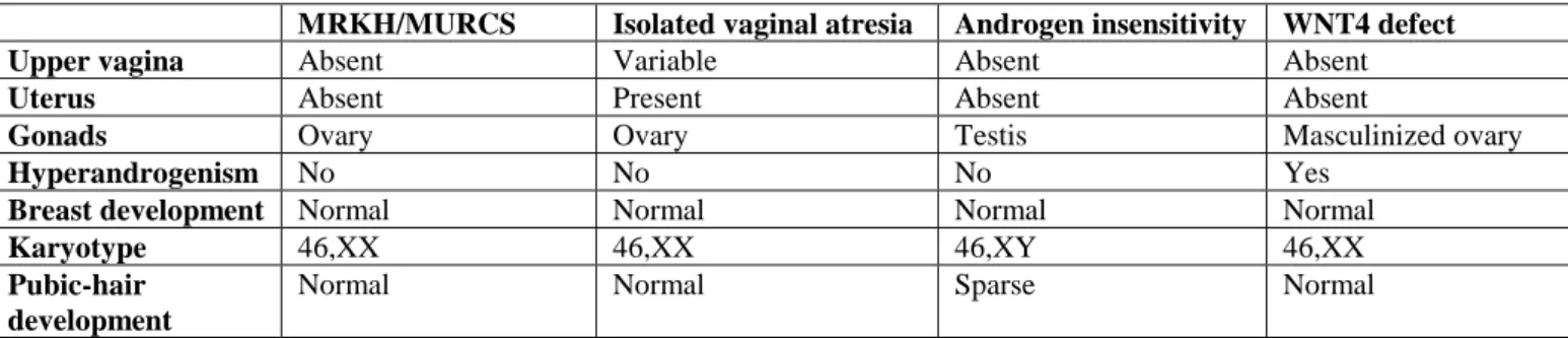
Differential diagnosis of Müllerian aplasia includes patients presenting with primary amenorrhea and with normal secondary sexual characteristics. This should first lead to exclusion of gonadal dysgenesis. The differential diagnosis includes congenital absence of uterus and vagina (aplasia or agenesis), isolated vaginal atresia, androgen insensitivity syndrome (AIS) and wingless-type MMTV integration site family, member 4 (WNT4) defect (Differential Table).Transverse vaginal septum and imperforate hymen, which can be initially misleading, are not included. Indeed, patients with these latter conditions have normal cervix and uterus, both of which are palpable on rectal examination.
AIS formerly known as testicular feminization, is an X-linked recessive condition resulting in a failure of normal masculinization of the external genitalia in chromosomally male individuals. This failure of virilization can be either complete androgen insensitivity syndrome (CAIS) or partial androgen insensitivity syndrome (PAIS), depending on the amount of residual receptor function. Both individuals with partial androgen insensitivity syndrome and individuals with complete androgen insensitivity syndrome have 46,XY karyotypes. Individuals with complete androgen insensitivity syndrome have female external genitalia with normal labia, clitoris, and vaginal introitus. The phenotype of individuals with partial androgen insensitivity syndrome may range from mildly virilized female external genitalia (clitorimegaly without other external anomalies) to mildly undervirilized male external genitalia (hypospadias and/or diminished penile size).
In either case, affected individuals have normal testes with normal production of testosterone and normal conversion to dihydrotestosterone (DHT), which differentiates this condition from 5-alpha reductase deficiency. Because the testes produce normal amounts of Müllerian-inhibiting factor (MIF), affected individuals do not have fallopian tubes, a uterus, or a proximal (upper) vagina.
The basic etiology of androgen insensitivity syndrome is a loss-of-function mutation in the androgen receptor (AR) gene. Loss of AR function means that, despite normal levels of androgen synthesis, the typical post-receptor events that mediate the effects of hormones on tissues do not occur. This results in the phenotype of prenatal undervirilization of external genitalia, absence of pubic and axillary hair, lack of acne, and absence of voice changes at puberty. All patients with androgen insensitivity syndrome are chromosomally and gonadally male. Most cases of AIS are identified in the newborn period by the presence of inguinal masses, which later are identified as testes during surgery. Some patients are first seen in the teenage years for evaluation of primary amenorrhea. Many of these patients have a history of surgery
for hernias and/or the presence of gonads in the inguinal canals, which were considered ovaries and returned to the abdomen.
WNT4 defects is a condition characterized by loss-of-function mutations in the WNT4 gene that appear to cause developmental abnormalities of sexual differentiation.
This condition is similar but distinct from MRKH syndrome (see Etiology section) and may therefore lead to confusion. It seems quite clear that other cases will soon be reported in the literature, making it important to include this new syndrome in the differential diagnosis of MRKH/MURCS. Evidence of hyperandrogenism in women presenting with normal female phenotype should then initially direct the clinicians to suspect WNT4 as a cause.
Ultrasonography can be used to define the Müllerian structures in infrequent cases where palpation is unrevealing [10].
Trans-abdominal ultrasonography is a simple and noninvasive method, and must be the first investigation in evaluating patients with suspected Müllerian aplasia. This technique reveals an absence of the uterine structure between the bladder and the rectum. However, a quadrangular retro-vescical structure may be wrongly identified as a hypoplastic or juvenile uterus: this fact corresponds to the vestigial lamina located underneath the peritoneal fold, itself situated transversally to the posterior side of the bladder, where utero-sacral ligaments attach. Since the vestigial lamina shows no cavity, there is no evidence of a hyperechogenic line, which normally corresponds to the uterine mucous membrane [11]. Finally, renal malformations must be systematically evaluated during this scan.
However, ultrasonography is an operator-dependent technique that can fail at identifying anatomical structures of the pelvis [12, 13]. MRI can accurately evaluate utero-vaginal anomalies thanks to multi-planar imaging and the use of differently weighted sequences, allowing the best visualization of all structures being examined [18].
MRI is a non-invasive technique that provides a more sensitive and more specific means of diagnosis than ultrasonography. It is increasingly becoming an essential diagnostic tool complementary to laparoscopy [14].MRI has a greater accuracy than ultrasonography and even laparoscopy in defining the exact anatomic characteristics of MRKH syndrome [15].
It should be performed when ultrasonographic findings are inconclusive or incomplete. MRI enables detailed road-mapping of the pelvic anatomy and morphology of the internal female genital organs and allows an accurate evaluation of the uterine aplasia, as well as a clear visualization of the rudimentary horns and ovaries [16, 17]. Determination of the length of agenetic segments and demonstration of the location of ectopic gonads is also offered by MRI.
Radiology Case. 2012 Apr; 6(4):17-24
Jo
urnal o
f
Rad
io
lo
gy
Case Rep
orts
ww
w.Ra
diol
ogyC
ase
s.com
20 The uterine aplasia is best characterized on sagittalimages, while vaginal aplasia is best evidenced on transverse images [18]. Moreover, MRI can be used at the same time to search for associated renal and skeletal malformations. Previous surgery makes interpretation more difficult and, if possible, MRI should be carried out prior to any surgery.
Consequently, MRI facilitates the surgical approach, e.g. vaginal reconstruction. Differentiating MRKH syndrome from other Müllerian anomalies and more specifically from malformations of the distal genital tract is a necessity dictated not only by the different surgical strategies required by each anomaly, but also by the different reproductive potential and psychological impact [14].
Mayer-Rokitansky-Kuster-Hauser syndrome is one of the most common causes of primary amenorrhea and the MRI demonstration of vaginal, cervical, and uterine morphology contributes significantly to treatment planning and patient management. Sonographic study is useful to evaluate the genitourinary tract for diagnosing any associated renal anomalies. Magnetic Resonance Imaging is more precise than sonography and less invasive and expensive than laparoscopy, contributing significantly to treatment planning and patient management. Although this condition has psychologically devastating consequences today anatomical defects can be surgically treated allowing a normal sexual function and reproduction thanks to the assisted techniques. Therefore correct evaluation of these patients and proper management is mandatory.
1. Oppelt P, Renner SP, Kellermann A, et al. Clinical aspects of MRKH syndrome: recommendations for clinical diagnosis and staging. Hum Reprod 2006; 21: 792. PMID: 16284062
2. Kaya H, Sezik M, Ozkaya O, et al. MRKH Syndrome associated with unilateral gonadal agenesis. A case report. J Reprod Med 2003; 48:902. PMID: 14686026
3. Li S, Qayyum A, Coakley FV, Hricak H. 2000. Association of renal agenesis and Müllerian duct anomalies. Journal of Computer Assisted Tomography 24:829 - 834. PMID: 11105695
4. Bykowski J. 1990. Magnetic resonance imaging in Mayer-Rokitansky-Kuster-Hauser syndrome. Obstetrics and Gynecology 76:593 - 596. PMID: 2145530
5. Duncan PA, Shapiro LR, Stangel JJ, Klein RM, Addonzizio JC. The MURCS association: Müllerian duct aplasia, renal aplasia, and cervicothoracic somite dysplasia. J Pediatr. 1979 Sep;95(3):399-402. PMID: 469663
6. Strubbe EH, Cremers CWRJ, Willemsen WNP, Rolland R, Thijn CJ. 1994. The Mayer - Rokitansky - Kuster - Hauser (MRKH) syndrome without and with associated features: two separate entities?. Clinical Dysmorphology 3:192 - 199. PMID: 7981853
7. Pittock ST, Babovic-Vuksanovic D, Lteif A. 2005. Mayer-Rokitansky-Kuster-Hauser anomaly and its associated malformations. American Journal of Medical Genetics 135: 314 - 316. PMID: 15887261
8. Schimke RN, King CP. 1980. Hereditary urogenital adysplasia. Clinical Genetics 18: 417 - 420. PMID: 7449179 9. Biedel CW, Pagon RA, Zapata JO. 1984. Müllerian anomalies and renal agenesis: autosomal dominant urogenital adysplasia. Journal of Pediatrics 104: 861 - 863. PMID: 6726517
10. Morcel K, Camborieux L; Programme de Recherches sur les Aplasies Müllériennes, Guerrier D. Mayer-Rokitansky-Kuster-Hauser (MRKH) syndrome. Orphanet J Rare Dis.2007 Mar 14; 2:13. PMID: 17359527
11. Paniel BJ, Haddad B, el Medjadji M, Vincent Y: Value of ultrasonography in utero-vaginal aplasia. J GynecolObstetBiolReprod - Paris1996, 25: 128-130. PMID: 8690860
12. Lang I, Babyn P, Oliver G. MR imaging of pediatric uterovaginal anomalies. PediatrRadiol 1999. 29:163-1. PMID: 10201032
13. Fedele L, Dorta M, Brioschi D et al. Magnetic resonance imaging in Mayer-Rokitansky-Kuster-Hauser syndrome. ObstetGynecol 1990. 76: 593-596. PMID: 2145530
14. Pompili G, Munari A, Franceschelli G, Flor N, Meroni R, Frontino G, Fedele L, Cornalba G. Magnetic resonance imaging in the preoperative assessment of Mayer-Rokitansky-Kuster-Hauser syndrome. Radiol Med. 2009 Aug; 114 (5): 811-26. PMID: 19484353
15. Janssens F, Verswijvel G, Mestdagh G. Mayer-Rokitansky-Küster-Hauser syndrome. JBR-BTR. 2004 May-Jun; 87 (3):140-1. PMID: 15293682
16. Maubon A, Ferru JM, Courtieu C, Mares P, Rouanet JP. Gynecological malformations. Classification and contribution of different imaging methods. J Radiol1996, 77: 465-475. PMID: 8760613
17. Carrington BM, Hricak H, Nuruddin RN, Secaf E, Laros RK Jr., Hill EC. Müllerian duct anomalies: MR imaging evaluation. Radiology 1990, 176: 715-720. PMID: 2202012 18. Troiano RN, McCarthy SM. Müllerian duct anomalies:
imaging and clinical issues. Radiology 2004, 233: 19-34. PMID: 15317956
REFERENCES TEACHING POINT
Jo
urnal o
f
Rad
io
lo
gy
Case Rep
orts
ww
w.Ra
diol
ogyC
ase
s.com
Figure 1 (top): 16 year old girl with
Mayer-Rokitansky-Kuster-Hauser syndrome. Transabdominal ultrasonography (Philips iU22 xMATRIX ultrasound system - Curved array probe 4-9 MHz) Axial scan showing dead-end inferior third of vagina (white arrow). The image confirms the absence of the uterus with interposition of meteoric bowel loops.
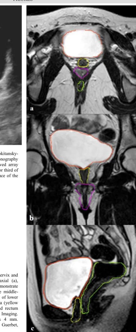
Figure 2 (right): 16 year old girl with absence of cervix and
uterus in MRKH syndrome. MRI T2-weighted axial (a), coronal (b) and sagittal (c) images. This scans demonstrate absence of uterus and cervix, with agenesis of the middle-upper third of the vagina. Noteworthy the presence of lower third of the vagina. Bladder (orange profile), urethra (yellow profile), lower portion of vagina (pink profile) and rectum (green profile) are outlined. (3T Magnetic Resonance Imaging. Protocol: TR 4,750 ms; TE 120 ms; thickness 4 mm. Gadoterate N-methylglucamine, Dotarem®, Guerbet, 0.5mL/Kg).
Radiology Case. 2012 Apr; 6(4):17-24
Jo
urnal o
f
Rad
io
lo
gy
Case Rep
orts
ww
w.Ra
diol
ogyC
ase
s.com
22Etiology The cause of MRKH is unknown. Several genes have been tested to study the possibility of the syndrome being genetic, but no single factor has yet been identified to be responsible for it.
Incidence Around 1 in 4500 female births.
Gender ratio The syndrome only occurs in females.
Age predilection Congenital malformation. Usually remains undetected until the patient present with primary amenorrhea despite normal female sexual development.
Risk factors Unknown. Most cases are sporadic, although familial cases have been reported.
Treatment After initial non-surgical treatment with use of successive dilators, the patient underwent vaginal reconstruction. The management also includes evaluation for associated congenital anomalies and psychological support.
Prognosis Surgery allows patients to have normal sexual function while fertility prognosis is unfortunately compromised.
Imaging finding PELVIC US IMAGING
Agenesis of the uterus or rudimentary one associated with presence of a tubular structure. The ovaries are morphological and sized.
MRI
-Axial plane: presence of bladder and rectum without interposition of uterus. The ovaries are morphological and sized.
-Sagittal plane: No uterus can be made out and only the lower vagina is seen.
-Coronal plane: uterus and cervix absence associated with agenesis of the upper-middle third of the vagina.
Table 1: Summary table for Mayer-Rokitansky-Kuster-Hauser (MRKH) syndrome
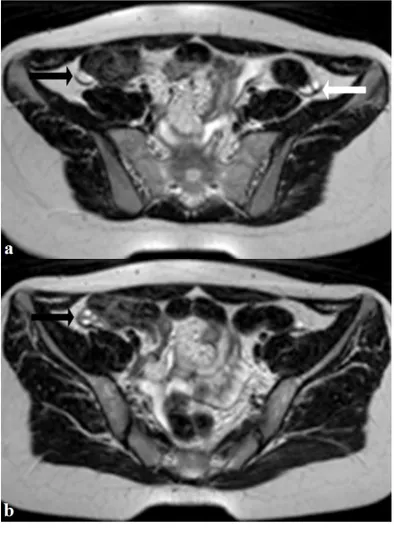
Figure 3 (left): 16 years old girl with
Mayer-Rokitansky-Kuster-Hauser syndrome. MRI T2-weighted axial images showing left (white arrow) and right (black arrows) ovaries with normal size and morphology but located higher than average position. Noteworthy absence of utero-ovarian ligament (proper ovarian ligament) and broad ligament. (3T MR imaging clinical unit Achieva, Philips Medical Systems, Eindhoven, the Netherlands. Protocol: TR 4,750 ms; TE 120 ms; thickness 4 mm. Gadoterate N-methylglucamine, Dotarem®, Guerbet, 0.5mL/Kg).
Jo
urnal o
f
Rad
io
lo
gy
Case Rep
orts
ww
w.Ra
diol
ogyC
ase
s.com
MRKH/MURCS Isolated vaginal atresia Androgen insensitivity WNT4 defect Upper vagina Absent Variable Absent Absent
Uterus Absent Present Absent Absent
Gonads Ovary Ovary Testis Masculinized ovary
Hyperandrogenism No No No Yes
Breast development Normal Normal Normal Normal
Karyotype 46,XX 46,XX 46,XY 46,XX
Pubic-hair development
Normal Normal Sparse Normal
Table 2: Differential diagnosis table for Mayer-Rokitansky-Kuster-Hauser (MRKH) syndrome
Radiology Case. 2012 Apr; 6(4):17-24
Jo
urnal o
f
Rad
io
lo
gy
Case Rep
orts
ww
w.Ra
diol
ogyC
ase
s.com
24 AIS: Androgen insensitivity syndromeAR: Androgen Receptor
CAIS: Complete androgen insensitivity syndrome DHT: DiHydroTestosterone
GRES: Genital, Renal, Ear, Skeletal syndrome MIF: Müllerian-inhibiting factor
MRI: Magnetic Resonance Imaging MRKH: Mayer-Rokitansky-Kuster-Hauser
MURCS: Müllerian hypoplasia/aplasia, renal agenesis and cervico-thoracic somite dysplasia association
PAIS: Partial androgen insensitivity syndrome
WTH 4: wingless-type MMTV integration site family, member 4
Mayer-Rokitansky-Kuster-Hauser; pelvic magnetic resonance; amenorrhea; vaginoplasty; Müllerian abnormalities; infertility
Brunetto Boscherini, MD; Elisabetta Del Duca, MD from Pediatric Unit, Tor Vergata University Hospital Foundation, University of Rome Tor Vergata, Italy and Giuseppe Sorrenti, MD from Section of Gynecology and Obstetrics, Department of Surgery, Tor Vergata University Hospital Foundation, University of Rome Tor Vergata, Italy.
Online access
This publication is online available at: www.radiologycases.com/index.php/radiologycases/article/view/992
Peer discussion
Discuss this manuscript in our protected discussion forum at: www.radiolopolis.com/forums/JRCR
Interactivity
This publication is available as an interactive article with scroll, window/level, magnify and more features.
Available online at www.RadiologyCases.com Published by EduRad
www.EduRad.org ABBREVIATIONS
KEYWORDS