Alma Mater Studiorum
Alma Mater Studiorum –– Università di Bologna
Università di Bologna
DOTTORATO DI RICERCA IN
Biologia Cellulare, Molecolare e Industriale
"Biologia Funzionale e Molecolare"
Ciclo XXV
Settore Concorsuale di afferenza: 05/I1 Genetica e Microbiologia
Settore Scientifico disciplinare: Bio19/ Microbiologia
Molecular and functional characterization of
the chemotactic genes in the PCBs-‐degrader
Pseudomonas pseudoalcaligenes KF707
PhD Student: Dott.ssa Tania Triscari Barberi
Tutor:
Chiar. mo Prof. Davide Zannoni
PhD Coordinator:
Chiar. mo Prof. Vincenzo Scarlato
Table of Contents
Preface
1Chapter A - General introduction
4A-1 Motility systems 4
A-2 The link between flagellar rotation and the bacterial swimming behaviour 6
A-3 Chemotaxis network in Escherichia coli 8
A-3.1 Signal transduction in response to a negative stimulus 9
A-3.2 Signal transduction in response to a positive stimulus 9
A-3.3 Signal transduction in response to multiple stimuli 10
A-4 Components of the “receptor-signalling complexes” 10
A-4.1 Chemoreceptors: structure and classification 10
A-4.2 The histidine kinase CheA and the adapter protein CheW 12
A-4.3 Organization of the receptors-signal complexes in clusters 13
A-5 Homologies among bacterial chemotactic pathways 13
A-6 Regulation of the signal termination 15
A-7 Memory in the chemotactic response 16
A-8 Role of motility and chemotaxis in biofilm formation 18
Chapter B - General Materials and Methods common to Chapters D, E, F
20B-1 Bacterial strains, media and growth conditions 20
B-2 Extraction of genomic DNA from Pseudomonas pseudoalcaligenes KF707 23
B-4 DNA sequencing and sequence analysis 25
B-5 Conjugation 25
B-6 Electroporation of Pseudomonas pseudoalcaligenes KF707 27
B-7 Construction of Pseudomonas pseudoalcaligenes KF707 mini-Tn5 transposon mutant library 28
Chapter C - The Genome Project of the polychlorinated-biphenyl degrader
Pseudomonas pseudoalcaligenes KF707
29C-1 Introduction 29
C-1.1 Prokaryotic genome projects pipeline 30
C-1.1.1 Second generation sequencing technologies 30
C-1.1.2 Overview of computational workflow for prokaryotic assembly and annotation of sequenced prokaryotic genomes 32
C-1.1.2.1 Reads quality control 33
C-1.1.2.2 Genome assembly 34
C-1.1.2.3. Genome scaffolding 35
C-2 Materials and Methods 37
C-2.1 Pseudomonas pseudoalcaligenes KF707 genome sequencing and preliminar analyses 37
C-2.2 Next generation sequencing data quality analysis 37
C-2.3 Genome assembly 37
C-2.4 Optical map and contigs scaffolding 38
C-2.5 Gene prediction 38
C-3.1 454 and Illumina reads datasets 39
C-3.2 Genome assembly 43
C-3.2.1 Assembly using reference genomes 43
C-3.2.2 de-novo assembly strategies 43
C-3.3 Contigs scaffolding 44
C-3.4 Genes prediction and annotation 46
C-4 Discussion 49
Chapter D - Bioinformatics analysis of genes involved in motility and
chemotaxis in Pseudomonas pseudoalcaligenes KF707 and contruction of
chemotactic mutants
51D-1 Introduction 51
D-2 Materials and Methods 52
D-2.1 Identification of genes involved in motility and chemotaxis 52
D-2.2 Bacterial strains and growth conditions 53
D-2.3 Amplifications of chemotactic target genes flanking regions and subsequent molecular fusion by using “Gene SOEing” method 53
D-2.4 Construction of recombinat plasmids containing fragments with deleted chemotactic genes and conjugation into Pseudomonas pseudolacaligenes KF707 wild type strain 56
D-3 Results 59
D-3.1 Motility and chemotaxis genes clusters in Pseudomonas pseudoalcaligenes KF707 genome 59 D-3.2 Amplification of cheA genes flanking regions and fusion
by Gene SOEing (splicing overlap extension) 65
D-3.3 Construction of recombinant conjugative plasmids carrying fragments with deleted target chemotactic genes and conjugation into Pseudomonas pseudolacaligenes KF707 wild type strain 67
D-4 Discussion 69
Chapter E - Role of chemotactic genes in Pseudomonas pseudoalcaligenes
KF707 motile behaviour and biofilm formation
73E-1 Introduction 73
E-2 Materials and Methods 74
E-2.1 Bacterial strains and growth conditions 74
E-2.2 Motility assays 74
E-2.2.1 Swimming 75
E-2.2.2 Swimming in presence of metals 75
E-2.2.3 Swimming chemotaxis assay 76
E-2.2.4 Plugs chemotaxis assay 76
E-2.2.5 Quantitative chemotaxis assays 76
E-2.2.6 Contrast phase microscopy 78
E-2.2.7 Swarming 78
E-2.2.8 Twitching 79
E-2.3 Evaluation of biofilm growth 79
E-2.3.1 Biofilm and planktonic growth curves 81
E-2.3.2 Confocal Laser Scanning microscopy (CLSM) 82
wild type and chemotactic mutant strains 84
E-3.2 Role of cheA genes in Pseudomonas pseudoalcaligenes KF707 biofilm formation and development 91
E-4 Discussion 95
Chapter F - Searching for a Quorum Sensing (QS) system
in Pseudomonas pseudoalcaligenes KF707
98F-1 Introduction 98
F-1.1 Bacterial Quorum Sensing: general features 98
F-1.2 Bacterial QS systems 99
F-1.2.1 Vibrio fischeri luxI/luxR system: the QS paradigm in gram negative bacteria 100
F-1.2.2 lux-like QS systems in Gram- Bacteria 101
F-1.2.3 Structure and function of the LuxR proteins family 103
F-1.3 Structural diversity in QS signal molecules 104
F-1.4 Synthesis and detection of AHLs signal molecules 105
F-1.5 Role of QS in swarming motility and biofilm development 106
F-2 Materials and Methods 107
F-2.1 Bacterial strains and growth conditions 107
F-2.2 T-streaks bioassays 108
F-2.3 Extraction of N-acyl-homoserine lactone 108
F-2.4 AHL reporter plate bioassays 109
F-2.5 TLC and detection of AHLs 110
F-2.7 Genome analysis for lux homologous searching 111
F-3 Results 112
F-3.1 Agar-Bioassays for the detection of QS molecules 112
F-3.2 TLC analyses on planktonic and biofilm organic extracts 114
F-3.3 Bioinformatics analysis on Pseudomonas pseudoalcaligenes KF707 genome for luxI/luxR homologues systems searching 116
F-4 Discussion 118
Conclusions
121Preface
Bacteria may encounter a large spectrum of different environments during their life cycles. Indeed, the capacity to adapt and survive in changing environments is a fundamental property of living cells and bacteria have developed effective mechanisms to regulate their behaviour accordingly. Chemotaxis, i.e. the migration of microorganisms under the influence of a chemical gradient, allows bacteria to approach chemically favorable niches for their growth and survival avoiding unfavourable ones. Since the most of microorganisms inhabiting heterogeneous environments are motile, the chemotactic behavior is achieved by integrating signals received from receptors that sense the environment. Apparently, the main reason for which environmental bacteria have retained during the “evolution” a large number of genes involved in motility and chemotaxis (Macnab, 1996), is because they provide a selective advantage and play a significant role in the dynamics of microbial populations (Pilgram and Williams, 1976; Freter et al., 1978; Kennedy and Lawless, 1985; Kennedy, 1987; Kelly et al., 1988; Lauffenburger, 1991).
Bacterial chemotaxis can be therefore considered the prerequisite for population survival, metabolism and interactions within ecological niches (Chet and Mitchell, 1979). In line with this, it has been reported that chemotaxis has important roles in colonization of plant roots by plant growth-promoting Pseudomonas fluorescens (De Weger et al.,1987; de Weer at al., 2002), infections of plants by Pseudomonas syringae and Ralstonia solanacearum (Yao and Allen, 2006), and animal infections by Pseudomonas aeruginosa (Drake and Montie, 1998 ). Notably, chemotaxis is also a selective advantage to degradative bacteria which colonize contaminated sites as microorganisms, with a
chemotactic ability toward xenobiotic compounds in polluted niches, have been isolated and characterized (Harwood et al., 1990; Grimm and Harwood, 1997; Bhushan et al., 2000a; Bhushan et al., 2000b; Parales and Harwood, 2002).
The soil bacterium Pseudomonas pseudoalcaligenes KF707 is know for its ability to degrade biphenyl and polychlorinated biphenyls (PCBs) (Furukawa et al, 1986), to which the strain is chemically attracted. PCBs are toxic compounds of great concern since they have been recognized as important harmful environmental contaminants in the EPA (Environment Protection Agency) priority list of pollutants.
The understanding of bacterial chemotaxis toward pollutants is a topic of particular interest, so that strategies for bioremediation by means of strains with degradative abilities, have been developed. However, the low bioavailability of organic contaminants is a limitation for the microbial remediation of contaminated sites, as toxic hydrophobic chemicals are often adsorbed to a non-aqueous-phase-liquid (NAPL) (Stelmack et al., 1999). In bioremediation processes, target compounds can be easily accessible to bacteria by dissolution in the aqueous phase; alternatively microorganisms might have access to a polluted surface through biofilm formation. In this respect, chemotaxis is a key factor in biofilm formation (Pratt and Kolter, 1998; O’Toole and Kolter, 1998; , Prigent-Combaret et al, 1999; Watnick and Kolter, 1999) and flagella are required for attachment to solid surfaces and the initiation of biofilm formation (Pratt and Kolter, 1998; Stelmack et al., 1999). In addition, motility and chemotaxis are required for biofilm growing bacteria to move along the surface, facilitating the spread of the biofilm (Stelmack et al., 1999).
Recent findings have shown that a Pseudomonas pseudoalcaligenes KF707 chemotactic mutant in a cheA gene (che stands for chemotaxis) is impaired in motility and chemotaxis as well as in biofilm development (Tremaroli et al, 2011). However,
recent studies on sequencing, assembly and annotation of Pseudomonas pseudoalcaligenes KF707 genome (Triscari et al., 2012; see also this Thesis work), have clearly demonstrated that the KF707 genome contains multiple putative operons encoding for different chemotaxis pathways and therefore multiple cheA genes are present. This finding was not surprising since genome analyses have revealed that a large number of environmental motile bacteria, such as Pseudomonas spp., Vibrio spp., Rhodobacter spp., own several gene clusters involved in chemosensing and chemotactic signal transduction, which may work in parallel or be expressed under different environmental conditions.
The goals of this present study were to investigate the role in motility, chemotaxis as well as in biofilm formation, of the various cheA genes we found by sequencing analysis of KF707 genome and to compare their functions with those previously attributed to a cheA gene in a KF707 mutant strain constructed by a mini-Tn5 transposon insertion (Tremaroli et al., 2011). Further, since it has been reported that communication via quorum sensing (QS) is involved in organizing group motility and biofilm formation, the ability to produce signal molecules by KF707 was also investigated.
CHAPTER A - General introduction
Motility and chemotaxis are peculiar traits common to many bacterial species. In the microbial world different kinds of motility can be observed. In addition, microorganisms can be sensitive to different stimuli and responde to them with variable taxis strategies.
A-1 Motility systems
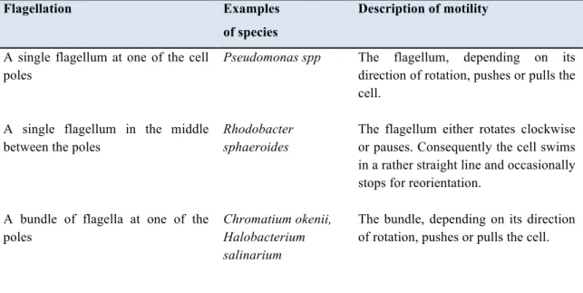
Swimming is the most common strategy for motility in fluid environments and is the outcome of the flagellar rotation, which exert a pushing force that drives bacteria at a speed up to 20–60 nm/sec. Several types of flagellar motility have been found and they depend on the number and the position of flagella and on the species. In Table A-1.1, various types of flagellar motility are listed.
Table A-1.1: Variety of flagellar motility in bacteria. This table was taken and modified from
Eisenbach (2001).
Flagellation Examples
of species
Description of motility
A single flagellum at one of the cell poles
Pseudomonas spp The flagellum, depending on its direction of rotation, pushes or pulls the cell.
A single flagellum in the middle between the poles
Rhodobacter sphaeroides
The flagellum either rotates clockwise or pauses. Consequently the cell swims in a rather straight line and occasionally stops for reorientation.
A bundle of flagella at one of the poles
Chromatium okenii, Halobacterium salinarium
The bundle, depending on its direction of rotation, pushes or pulls the cell.
Table A-1.1 continued
A bundle of flagella at each of the two poles
Some cells of
H. salinarium
The bundles, depending on their direction of rotation, push or pull the cell. Consequently, the cell goes back and forth or stops
5–10 flagella randomly distributed around the cell
Escherichia coli, Salmonella typhimurium, Bacillus subtilis
Most of the time the flagella rotate counterclockwise and the cell swims in a rather straight line (a run). Intermittently, the flagella rotate clockwise or pause, as a result of which the cell undergoes a vigorous angular motion (a tumble)
A polar tuft of 2 flagella + 2–4 lateral flagella
Agrobacterium tumefaciens
Flagella rotate clockwise or pause; consequently the cell swims in a rather straight line or turns
One flagellum at one end, one or more Flagella subterminally at each end. All the
flagella are contained within the periplasmic space
Spirochaetes The cells exhibit smooth swimming, reversals, flexing and pausing. When the flagellar bundles at both cell poles rotate in opposite directions (one pulls and one pushes), the cell swims in a rather straight line. When both bundles switch synchronously, the cell reverses. When both bundles rotate in the same direction, the cell flexes
Swarming is an organized translocation of differentiated cells on a solid surface generally due to type IV pili and cell-to-cell communication appears to be essential for this motile behaviour. Swarming bacteria located in the outer layer of a colony, expand outwardly and the evacuated space is filled with new growing cells. Irregular branches can appear at the periphery of the colony, forming a dendritic pattern on the surface.
Gliding is a particular kind of movement characterized by bacterial migration on a solid surface covered with a liquid film, without the formation of external structures and no cellular differentation.
Twitching motility is a form of translocation on solid surfaces which is dependent on pili-assisted motility (Henrichsen 1972;1983).
Propulsion by actin filaments is a peculiar mode of motility to pathogens such as Listeria, Shigella and Rickettsia in host eukaryotic cells. The bacteria assemble actin filaments for propulsion in the cytoplasm of the infected host cell.
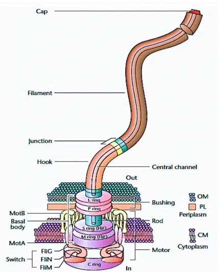
A-2 The link between flagellar rotation and the bacterial swimming behaviour Flagella are specialized structures (see Fig. A-2.1) which enable bacteria to swim in an aqueous solution.
Fig. A-2.1: Structural organization of a bacterial flagellum. It consists of three major parts:
a basal body, a hook and a filament. Bacterial flagella may vary between species and families, but the main structural aspects are common to all. This picture was taken and modified from Eisenbach (2001).
Bacteria such as E. coli have two main swimming patterns: smooth swimming in a straight direction (run) and an overturning motion (tumble). In absence of stimuli, when the concentration of nutritional compounds in the enviroment is uniform, cells run for about a second, then tumble for about a tenth of a second, changing orientation and, as a consequence, running in a new direction. Consequently, the bacterial cells walk randomly, with no net vectorial movement (Fig. A-2.2a). Specifically, the run is the consequence of a counterclockwise rotation while the tumble is the consequence of a clockwise rotation of the flagella.
When a cell detects increasing concentrations of attractants or decreasing concentrations of repellents, tumbles occur less frequently, and there is a net movement towards attractants and away from repellents (Fig. A-2.2b). Cells make temporal comparisons of chemo-effectors concentrations during a run and they decide, second by second, the movement direction, suppressing tumbles if the level of chemo-attractant increases. On the other hand, negative stimuli increase the probability of clockwise rotation (Tsang et al., 1973) and cells tumble more frequently.
Fig. A-2.1.: Bacterial biased random walk in absence of stimuli (a) and movement under
attractant gradient (b). This picture was taken and modified from Sourjik and Wingreen (2012).
A-3 Chemotaxis network in Escherichia coli
Prokaryotic chemosensory pathways are depending on two-component signal transduction system with conserved components regulating flagellar activity (Stock et al., 2000; Wolanin et al., 2002). Generally, a two-component system includes a histidine protein kinase (HPK) that catalyzes the transfer of a phosphoryl group from ATP to an aspartate residue on the response regulator (Borkovich et al., 1989).
The E. coli chemosensory network is considered as the simplest model to describe bacterial chemotaxis although much more complex as compared to a basic two-components system. Indeed, the six Che proteins, CheA, CheW, CheY, CheZ, CheR and CheB and the five chemoreceptors, Tsr, Tar, Tap, Trg and Aer, constitute the E. coli chemotaxis system.
The CheA protein, unlike orthodox membrane-bound histidine kinases, does not interact directly with chemo-effectors, because it lacks of a sensory domain. It is in fact connected to the transmembrane receptor proteins (chemoreceptors) via the ‘adapter’ protein CheW. Together, chemoreceptors - CheA - CheW, form large complexes that integrate enviromental informations to control CheA kinase activity in phosphorylating the response regulator CheY. CheR and CheB are, respectively, involved in methylation and demethylation of the chemoreceptors cytoplasmic domain (West et al., 1995; Djordjevic and Stock, 1997). CheR is an S-adenosyl-methionine-dependent methyl-transferase that methylates specific glutamate residues ; CheB is an esterase with an opposite function as it hydrolyzes the methyl esters formed by CheR. These antagonist activities play a critical role in adaptation (Okumura et al., 1998; Levit and Stock 2002; Sourjik and Berg, 2002), conferring
also a memory mechanism. The stochastic nature of these modifying activities ensures a variety of receptor sensitivity and capacity of response in the different cells of a bacterial population.
A-3.1 Signal transduction in response to a negative stimulus
When chemo-repellents bind to the receptors, they switch to an active form and together with CheW, stimulate CheA autophosphorylation. The histidine kinase, in turn phosphorylates CheY. Phosphorylated CheY (CheY∼P) diffuses to flagellar motors (Li et al., 1995; Sourjik and Berg, 2002), where it acts as an allosteric regulator on the flagellar proteins FliM, changing the sense of rotation from counterclockwise to clockwise and, consequently, tumble occurs (Alon et al., 1998). The response is termined by the CheZ phosphatase, by enhancing CheY~P dephosphorylation (Stock, A.M. and Stock, J.B., 1987; Wang and Matsumura, 1996).
A-3.2 Signal transduction in response to a positive stimulus
When chemo-attractants bind to the receptors, they do not undergo to conformational changes, so CheA autophosphorylation is inhibited, causing reduced levels of CheY~P and promoting smooth swimming as the probability of clockwise rotation decreases. The results are prolonged runs alterned to rare tumbles.
A-3.3 Signal transduction in response to multiple stimuli
Generally, cells are exposed to multiple positive and negative stimuli. Bacteria are able to integrate all these inputs and to show a unique behavioural response. Thus, movement towards attractants and away from repellents is determined by the efficiency of temporary response and the memory of past informations, properties that allow bacteria to make second-to-second decisions to continue swimming or tumbling and change direction (Berg, 2000; Bourret and Stock, 2002; Wadhams and Armitage, 2008).
A-4 Components of the “receptor-signalling complexes”
Receptors-signalling complexes can be viewed as ternary complexes resulting from the interactions between the membrane receptors and the chemotaxis proteins CheW and CheA. Receptors act anchoring the chemotactic proteins in the inner membrane and are necessary for signal transmission from the periplasmic domain (which binds the ligand), through the membrane, to the cytoplasmic complex.
In the following paragraphs the single components of receptors-signalling complexes are described.
A-4.1 Chemoreceptors: structure and classification
Chemoreceptors are transmembrane proteins with variable periplasmic sensing domains – which are able to bind specific ligands - and a conserved cytoplasmic domain – which acts as a scaffold for the anchoring of the histidine kinase CheA and the adpter CheW (Le Moual and Koshland, 1996; Zhulin, 2001). Binding of a ligand to the sensing domain causes a conformational change,
inducing a “piston-like movement" which in turn causes the transmission of signals across the cell membrane for the control of CheA kinase activity in the cytoplasm (Mowbray and Sandgren, 1998), (Otteman et al., 1998; 1999). The function of the chemoreceptors is strictly related to their structure. The cytoplasmic part of chemoreceptors can be divided into four subdomains: (i) the histidine kinase, adenylyl cyclase, methyl-binding proteins and phosphatases domain (HAMP); (ii) methylated helix 1 (MH1); (iii) signaling domain; (iv) methylated helix 2 (MH2). Together the methylated helixes (MH1 and MH2) contain four or more glutamate residues that are substrates for CheR and CheB modification (Terwilliger et al., 1983; 1984). Since chemoreceptors are substrates for methylation and demethylation, they are also known as methyl-accepting chemotaxis proteins (MCPs).
MCPs are classified on the basis of different properties: cellular localization (membrane-bound or cytoplasmatic), abundance, size (cluster I receptors with a ligand-binding region between 120 and 210 amino acids whereas cluster II receptors have larger binding regions of 220–299 amino acids), the ligand-binding region (extra-cellular space or cytosol). Notably, MCPs are different with respect to the sequence of their periplasmic part and the presence of the binding site for CheR in their cytoplasmic side. Thus, receptors possessing this CheR-binding site are known as “major receptors” and can function independently. The other MCPs, without this binding site, have an adaptation mechanism depending on the presence of the first type of receptors: this may explain a possible reason for receptors organization in clusters. Moreover, several MCPs are able to respond to different compounds at the same time: for example the E. coli Tar receptor, sense
aspartate and maltose. Aspartate binds directly to the periplasmic ligand binding domain (Yen et al., 1996) whereas maltose binds to the periplasmic maltose binding protein (MBP) associated to the MCP.
A-4.2 The histidine kinase CheA and the adapter protein CheW
CheA and CheW chemotactic proteins play an important role in the organization of clusters of receptors. On the other hand, MCPs represent anchors for the assembly of chemotactic proteins. Recent studies have shown that deleted mutants in cheA or cheW genes are impaired in receptor arrays formation. In order to understand the interaction between of both CheA and CheW, it is fundamental to know the tridimensional structures of these proteins and if particular conserved domains are involved in the interaction with the cytoplasmic receptor domain.
CheA is divided into five domains with specific and distint structure and function: the histidine phosphotransfer domain (P1), the response regulator binding domain (P2), the dimerization domain (P3), the histidine protein kinase catalytic domain (P4), and the regulatory domain (P5). The P1 domain belongs to the histidine phosphotransfer (HPT) family of proteins that transfer the phosphoryl groups between ATP and the phospho-accepting aspartate of the response regulators. The response regulator binding domain, P2, is flanked by two flexible linker sequences connecting it to P1 and P3 (Zhou et al., 1996). When P2 is in complex with CheY, the CheY active site undergoes a conformational change that increases the accessibility of the phospho-acceptor aspartate, Asp57. More importantly, P2 binds CheY in close proximity to the phospho-P1 domain and increases its effective concentration (Stewart, 1997; 2000). P3 and P4 domains
constitute the histidine protein kinase (HPK) catalytic core. P5 is homologous to CheW (Bilwes et al., 1999) and mediates binding to the chemoreceptor signaling domains (Levit et al., 2002).
CheW is a monomeric soluble protein, know as adpter and its role is anchoring the histidine Kinase CheA to the chemoreceptors arrays (Surette and Stock, 1996; Griswold and Dhalquist, 2002; Griswold et al., 2002).
A-4.3 Organization of the receptors-signal complexes in clusters
Generally, bacterial chemoreceptors are organized in clusters located at one or both the cell poles. The chemoreceptors are organized into units of ‘trimers of dimers’, which form ternary signalling complexes with the chemotaxis histidine protein kinase CheA and the linker protein CheW. In these clusters, receptors with different ligand specificities are uniformly mixed and arranged in hexagonal arrays. Receptor arrays are not perfectly regular structures: the hexagonal order appears to be distorted (Khursigara et al., 2008) with a variable stoichiometry of the receptors to CheW and CheA (Levit et al, 2002; Sourjik and Berg, 2004). All the other chemotaxis proteins localize to the clusters by interaction with either receptors or CheA and CheW. CheR and CheB both bind to the NWETF pentapeptide sequence at the C-terminus of the major receptors.
A-5 Homologies among bacterial chemotactic pathways
Unlike E. coli, the most of known bacterial species show more complex chemotactic pathways as they possess multiple chemotaxis proteins and cytoplasmic chemoreceptors, alternative adaptation and signal termination strategies (Rao et al., 2008; Schweinitzer and Josenhans, 2010; Silversmith, 2010)
(see Table A-5.1). Many species possess homologues of the CheA, CheB, CheR, CheW and CheY chemotaxis proteins. Studies on Rhodobacter sphaeroides (Porter et al., 2008) have provided proof for the existence of multiple signalling cascades. This bacterium has three major operons (cheOp1-3) encoding homologues of signalling proteins and two different flagellar systems, named fla1 and fla2 (del Campo et al., 2007). Experimental observations have shown that genes encoded by cheOp1 control the activity of the fla2 system whereas proteins of cheOp2 and cheOp3 regulate fla1 activity. The transmembrane chemoreceptors localized at the cell poles were found to interact with proteins encoded by cheOp2 whereas the cytoplasmic chemoreceptors cluster with proteins encoded by cheOp3 (Wadhams et al., 2003). Therefore, cytoplasmic and membrane chemoreceptors form two separate signalling complexes, enabling Rhodobacter sphaeroides to sense cytoplasmic and extracellular signals independently. However, it was observed that there are interactions between the two signalling pathways as the loss of either cheOp2 or cheOp3 signalling proteins causes lack of chemotaxis, hence both signalling pathways are necessary to generate a chemotactic response (Porter et al., 2002). As annotation of putative chemotaxis genes is based on nucleotidic sequence similarity, there is evidence that not all annotated chemotaxis gene clusters are involved in taxis. For example, Myxococcus xanthus was found to have eight gene clusters containing proteins typically associated with taxis. Some of these clusters are involved in taxis whereas others can be associated with developmental processes leading to the formation of fruiting bodies (Zusman et al., 2007).
Table A-5.1: Some example of homologues and alternative chemosensory-like pathways in
bacteria (reviewed in Porter et al., 2011).
E.coli R.sphaeroides P.aeruginosa M.xanthus
Number of MCP 5 13 26 21 Chemoreceptor types Transmembrane Transmembrane Cytoplasmic Transmembrane Cytoplasmic Transmembrane Cytoplasmic Chemotaxis pathways 1 3 4 8 Gene sets encoding flagella 1 2 1 0 Signal termination
cheZ cheA3 cheZ cheC
homologue Role of che-like
pathways
chemotaxis chemotaxis c-diGMP biofilm
EPS production
A-6. Regulation of the signal termination
The chemotactic signalling cascade is characterized by a specific lifetime, which guarantees an effective response and allows to recover the pre-stimulus steady-state. The control of the lifetime of the cellular response has a crucial role in signal transduction systems and is depending on CheY∼P dephosphorylation. The response regulator can catalyze its self-dephosphorylation, but this occurs slowly. Generally, CheY∼P dephosphorylation is due to other phosphatases. Some kinases are able to dephosphorylate their response regulator (Zhu et al., 2000; Gao and Stock, 2009; Kenney, 2010), but the most of the times dephosphorylation is catalyzed by the phosphatase CheZ. This enzyme consists of two symmetric monomers, each containing a binding site for CheY~P. It has been reported that the binding between CheZ and CheY~P shows a positive cooperativity (Blat et al., 1998; Silversmith et al., 2008). Therefore, CheZ activity is suppressed at low CheY~P concentration, thus ensuring that CheY~P levels do not get too low and maintaining a steady-state condition.
Many species have multiple homologues of the E. coli chemosensory system (Silversmith et al., 2005) and some of them are involved in CheY~P dephosphorylation mechanism. For example, Sinorhizobium melioti owns two CheY homologues (Guhaniyogi et al., 2008), one of which is able to interact with the flagellar motor and the other one is involved in signal termination as it acts as a phosphate sink (Lukat et al., 1991). Another example is that in B. subtilis, where in lack of CheZ, CheY~P dephosphorylation is due to FliY enzyme, homologue with the CheX-like phosphatase proteins found in other species (Park et al., 2004).
A-7. Memory in the chemotactic response
The chemosensory pathway in E. coli is maintained at a steady-state of the histidine kinase CheA activity and, as a consequence, of the CheY~P levels. The system is set up for an optimal response to both positive and negative stimuli. Adaptation, due to different mechanisms of feedback, works in order to guarantee this balanced state, enabling a bacterial population to sense a temporal gradient of attractant and/or repellent. One example of feedback mechanism is the modification, by methylation/demethylation, of the MCPs cytoplasmic signalling and adaptation domain, containing the NWETF peptide and specific glutamate residues. The chemotactic protein CheR shows high affinity towards this pentapeptide and, constitutively, adds methyl groups to glutamate. This works antagonistically to CheB, a methylesterase that - when phosphorylated by CheA - removes methyl groups from glutamate residues. The methylation/demethylation of MCPs is depending on their own state, associated to the binding of the ligand. Active MCPs (bound to the ligand) are demethylated by CheB and inactive MCPs
(without ligand) are methylated by CheR (Alon et al., 1999; Boldog et al., 2006). Moreover, even though the CheB methylesterase and the response regulator CheY are both activated by the histidine kinase CheA, CheB is phosphorylated with a slight delay as compared to CheY: this mechanism ensures that the switch of the flagellar motor can occur before adaptation.
This simple system is common to many bacterial species (Marchant et al., 2002). Other species have CheV, CheC and CheD enzymes, as additional proteins involved in adaptation feedback loops (Szurmant and Ordal, 2004; Rao et al., 2008). As it has been reported in Bacillus subtilis, CheV acts by modulating CheA activity, via a CheW-like domain. CheC acts as a CheY phosphatase. CheD is a MCPs deamidase, which converts glutamine residues in glutamate, which can be modified by CheB or CheR. All these mechanisms have the important role to reset the chemosensory system at the pre-stimuls steady-state condition.
The time-lag between the chemotactic response and the adaptation is known as “memory” lenght (Macnab and Koshland, 1972), and it depends on the stimulus strength, the gradient stepness and also on the bacterial lifestyle. An optimal memory lenght must allow bacteria to “remember” still relevant past conditions in order to compare them to present ones and choose the swimming direction (Macnab and Koshland, 1972).
It has been reported that bacteria in a population show variability in adaptation time, memory lenght (Vladimivor et al., 2008; Meir et al., 2010), number of CheR and CheB units (Li and Hazelbauer, 2004) and MCPs abundance and distribution in the membrane (Thiem and Sourjik, 2008; Greenfield et al., 2009). The fact that all the cells in a population, do not have the same behaviour in
unpredictable and variable environmental conditions, can be seen as an evolutionary advantage which guarantees the survival of the polulation as a whole.
A-8. Role of both motility and chemotaxis in biofilm formation
Commonly, in natural environments, bacteria grow as biofilms, i.e. organized mixed cells communities adhering to biotic or abiotic surface and packaged in an extracellular polysaccharide matrix known as exopolysaccharide (EPS) (Costerton et al., 1995). Interestingly, bacteria can switch between the planktonic and the biofilm lifestyles in response to nutritional cues.
Biofilm formation occurs accordingly to a gradual and well regulated process, namely:
• the adhesion to a surface via the cells pole, this step being known as “reversible attachment” (O’Toole and Kolter 1998a-b; Hinsa et al., 2003);
• “irreversible attachment” via the long cell axis (Marshall et al., 1971; Fletcher 1996);
• micro-colonies formation via the recruitment of planktonic cells from the medium or migration of attached cells on the surface by twitching motility (O’Toole et al., 2000a; O’Toole and Kolter, 1998b);
• cells maturation with the formation of the EPS matrix (Danese et al., 2000; Hellmann et al., 1996; Watnick and Kolter 1999; Yildiz and Schoolnik, 1999); • dispersal of cells due to starvation (Gjermansen et al., 2005).
Klausen et al., (2003), have reported that flagella and type IV pili are important factor in P. aeruginosa biofilm development, as they mediate attachment
to solid surfaces. Other studies have suggested that swarming motility - depending on quorum sensing (QS, see § F), rhamnolipid production, type IV pili and the presence of flagellum - can also contribute to early stages of P. aeruginosa biofilm formation (Köhler et al., 2000). Moreover, it is likely that motility and chemotaxis are required to swim towards nutrients associated with a surface.
CHAPTER B - General Materials and Methods Common to Chapters
D, E, F.
B-1 Bacterial strains, media and growth conditions
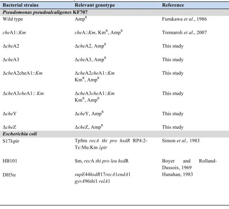
All strains and plasmids used in this study are listed in Table B-1.1.
Table B-1.1: Bacterial strains and plasmids.
Bacterial strains Relevant genotype Reference
Pseudomonas pseudoalcaligenes KF707
Wild type AmpR Furukawa et al., 1986
cheA1::Km cheA::Km, KmR, AmpR Tremaroli et al., 2007
ΔcheA2 ΔcheA2, AmpR This study
ΔcheA3 ΔcheA3, AmpR
This study ΔcheA2cheA1::Km ΔcheA2cheA1::Km KmR, AmpR This study ΔcheA3cheA1:: Km ΔcheA3cheA1::Km KmR, AmpR This study
ΔcheY ΔcheY, AmpR This study
ΔcheZ ΔcheZ, AmpR This study
Escherichia coli
S17λpir TpSm recA thi pro hsdR
RP4:2-Tc:Mu:Km λpir
Simon et al., 1983
HB101 Sm, recA thi pro leu hsdR Boyer and
Rolland-Dussoix, 1969
DH5α supE44hsdR17recA1endA1
gyrA96thi1 relA1
Table B-1.1: continued
Top10F’ F´{lacIq, Tn10(TetR)} mcrA
Δ(mrr-hsdRMS-mcrBC) Φ80lacZΔM15ΔlacX74 recA1
araD139 Δ(ara leu) 7697 galU galK rpsL (StrR) endA1 nupG
InvitrogenTM
pSB401 harbouring the luxCDABE plasmid
construct Winson et al., 1998 JM109 endA1,glnV44,thi-1,relA1 gyrA96,recA1,mcrB+,Δ(lacproAB) e14-[F'traD36proAB+lacIq lacZΔM15] hsdR17(rK-mK+) Yanish-Perron et al., 1985 Agrobacterium tumefaciens NTL4 / Farrand et al., 2002 WCF47 / Zhu et al., 1998 Chromobacterium violaceum / CV026 KmR McClean et al., 1997
Plasmids Relevant genotype or
characteristics Reference
pUC19 AmpR, cloning vector Sambrook et al., 1989
pUT mini-Tn5 Km AmpR KmR, delivery plasmid for
mini -Tn5 Km
de Lorenzo et al., 1990
pRK2013 KmR ori ColE1
RK2-Mob+ RK2-Tra+
Figursky et al., 1979
pG19II GmR sacB lacZ, cloning vector
conjugative plasmid
Maseda et al., 2003
pSB401 luxCDABE reporter fusion Winson et al., 1998
pZLR4 lacZ reporter fusion Farrand et al., 2002
pCF218 codifing for traR Zhu et al., 1998
pCF372 traI promoter-lacZ phusion Zhu et al., 1998
Liquid cultures of all bacterial strains were grown in agitation at 150 rpm at the optimal temperature (Escherichia coli at 37°C; Pseudomonas pseudoalcaligenes,
Agrobacterium tumefaciens and Chromobacterium violaceum at 30°C). The compositions of the media used in this study are reported in Table B-1.2.
Table B-1.2: Media composition. Rich media Luria-Bertani (LB) pH 7 Trypton Yeast extract NaCl 10g/l 5g/l 10g/l Defined media
Sucrose-Asparagine (SA) pH 7 Sucrose
Asparagine K2HPO4 MgSO4 10% (w/v)* 20g/l 2g/l 1g/l 5ml/l
Minimal salt medium (MSM) pH 7 K2HPO4
KH2PO4 (NH4)2SO4 MgSO4* CaSO4* MnSO4* FeSO4* 4,4g/l 1,7g/l 2,6g/l 0,4g/l 0,0031g/l 0,05g/l 0,01g/l
Succinate*or byphenil crystals 5mM
AB glucose pH 7 20X buffer solution*:
K2HPO4 NaH2PO4xH2O 20X salts solution*: NH4Cl MgSO4 KCl CaCl2 FeSO4x7H2O
Carbon source: glucose
60g/l 23g/l 20g/l 2,9g/l 3g/l 0,2g/l 0,05g/l 5g/l
The asterix (*) indicates medium components which were prepared as concentrated stock solutions, autoclaved separately and added at the medium at the final concentration of 1X.
For growth on solid media, agar was added at the final concentration of 15 g/l. X-Gal stock solution was prepared at a concentration of 50 mg/ml and stored in 1 ml aliquotes, protected from light, at -20°C. Antibiotics stock solutions were prepared as reported in Table B-1.3 and stored at -20°C in 1 ml aliquotes until use.
Table B-1.3: Antibiotics stock solutions and concentrations used for selective growth.
Stock solution Final Concentration in µg/ml
P.ps.alcaligenes - E.coli - A.tumefaciens – CV026
Ampicillin, 100 mg/ml, water solution Kanamycin, 100 mg/ml, water solution Gentamycin, 30 mg/ml, water solution Tetracycline, 20 mg/ml, 70% ethanol solution Spectinomycin, 50 mg/ml, water solution
100 50 / / 50 50 / 50 20 20 30 / 20 20 20 / / / 50 /
B-2 Extraction of genomic DNA from Pseudomonas pseudoalcaligenes KF707 Genomic DNA from Pseudomonas pseudoalcaligenes KF707 was extracted with the following protocol. A 10 ml of an over-night grown culture was centrifuged at 5000 rpm at 4°C for 15 minutes and washed with 10 ml of TES solution ( 50 mM TrisHCl, 20 mM EDTA, 50 mM NaCl pH 8.0). The cell pellet was resuspended in 5 ml of TE buffer (50 mM TrisHCl, 20 mM EDTA pH 8.0). Lysozyme solution, prepared in the same buffer, was added at the final concentration of 20 mg/ml; the solution was incubated at 37°C for 30 minutes and mixed by inversion every 10 minutes. At the end of incubation, 500 µl of a 10%
SDS solution and Proteinase K at the final concentration of 10 mg/ml were added, followed by 1 h incubation at 37°C; the reaction was stopped by adding a solution of 10 mM EDTA and 3 mM sodium acetate. The lysate was incubated with RNase at 37°C for 1h after which an iso-volume of a phenol-chlorophorm-isoamyl alcohol 25:24:1 v/v mixture was added and the sample was mixed by inversion at room temperature for 15 minutes. The water phase containing the genomic DNA was separeted from the organic phase and cell debris by centrifugation at 5000 rpm at 4°C for 15 minutes. The extraction was repeated three times and phenol traces were removed by adding an iso-volume of a 24:1 v/v mixture of chlorophorm-isoamyl alcohol. The water phase was recovered after centrigugation at 5000 rpm at 4°C for 15 minutes in a clean beacker. 1.5 volumes of cold absolute ethanol were added to the extracted water phase and the genomic DNA was collected using a clean glass stick. The DNA was washed by immersing the glass stick in a cold 70% ethanol solution and then air dried. After this, the stick with DNA was immersed in a small volume of sterile nuclease free water and left at 4°C over-night to allow the DNA to suspend. The resuspended genomic DNA preparation was stored at the temperature of -20°C.
B-3 DNA manipulations and genetic techniques
All restriction digests, ligations, cloning and DNA electrophoresis, were performed using standard techniques (Sambrook et al, 1989). Taq polymerase, the Klenow fragment of DNA polymerase, alkaline phosphatase, restriction endonucleases and T4 DNA ligase were used as specified by the vendors (Roche, Fermentas, Invitrogen, Sigma-Aldrich, NEB Biolabs). The plasmid pUC19 was
routinely used as the cloning vector and recombinant plasmids were introduced into E. coli host by transformation of chemically competent cells, prepared according to the CaCl2 method (Sambrook et al, 1989). To detect the presence of insert DNA, X-Gal was added to agar media at a final concentration of 50 µg/ml. X-Gal stock solutions were prepared at a final concentration of 50mg/ml in N-N-dimethylformamide and stored as 1 ml aliquots at - 20 °C protected from light. Kits for plasmid mini- midi- and maxi-preps, PCR purification and DNA gel extraction were obtained from QIAGEN (Milan, Italy) and used according to the manifacturer’s instructions.
B-4 DNA sequencing and sequence analysis
Genomic DNA fragments of interest were cloned in the pUC19 cloning vector and positive plasmids were sent for sequencing to the BMR-genomics service of the University of Padova (Padova, Italy). Samples were prepared according to the recommended procedures (www.bmr-genomics.it). M13 Forward and Reverse primers were used for sequencing the extremities of DNA fragments cloned into the pUC19 vector from the M13 promoter. Sequence identities were determined by DNA homology searches using the BLAST program to search both NCBI and TIGR databases.
B-5 Conjugation
Day I. Donor, receiver and helper strains were streaked out on LB agar plates with the appropriate antibiotics. LB plates were incubated over-night at 37°C and 30°C for E. coli and P. pseudoalcaligenes KF707 optimal growth temperature
respectively. E. coli HB101 strain carrying the mobilization plasmid pRK2013 was commonly used as helper strain for tri-parental mating (see Table 1 for strain and plasmid features).
Day II. Donor, receiver and helper strains were inoculated in LB broth from single colonies grown on the agar plates. The appropriate antibiotics were added to LB medium in order to maintain selection. LB liquid cultures were grown over-night at the appropriate temperature under agitation at 150 rpm.
Day III. Donor, receiver and helper strains were inoculated with a 1% inoculum in liquid LB medium without antibiotics from over-night grown liquid cultures. Cells were grown at the appropriate temperature and under shaking for 2 – 3 h, in order to obtain early exponential phase cultures (OD660 ~ 0.2 – 0.3). 1 ml aliquot from each culture was collected in a sterile tube, spun down at room temperature and washed twice with 1 ml LB medium. Cells were suspended in 1 ml of fresh LB and then used for the preparation of conjugation mix by adding equal volumes (100 µl) of donor, receiver and helper suspensions to a sterile tube. The conjugation mix was incubated at 30 °C for 30 min and spots were plated onto well dry LB agar plates without selection. Controls for each conjugation were carried out with 100 µl of the receiver, donor or helper cell suspensions alone added to sterile tubes and processed in the same way as conjugation mix. LB plates were incubated for 24 h at 30°C.
Day IV. The bacterial biomass was collected from each plate with a sterile loop and suspended in 1 ml of fresh LB with 20 % glycerol. 10 fold serial dilutions of cell suspensions of conjugation mix and controls were carried out in 0.9 % saline. The remaining part of the conjugation mix suspended in LB with 20 %
glycerol was stored at – 80 °C. Appropriate dilutions were plated on agar plates containing the antibiotics for transconjugants selection. For the selection of KF707 transconjugants, cells were plated on SA or AB glucose medium in the presence of appropriate antibiotics. The two media were used to counter-select E. coli donor and helper strains, given that these medium do not support E. coli growth, thus resulting selective for P. pseudoalcaligenes KF707. Plates were incubated at the appropriate temperature until transconjugants growth was clearly visible (i.e. 24 h for E. coli transconjugants growing on LB and at least 36 h for KF707 growing on SA or AB glucose).
Day V. Transconjugants were streaked out on the appropriate agar media in the presence of antibiotic selection and incubated at the optimal temperature until growth was clearly visible. The selection was repeated at least twice, in order to obtain a pure culture and remove both donor and helper strain backgrounds.
B-6 Electroporation of Pseudomonas pseudoalcaligenes KF707
Pseudomonas pseudoalcaligenes KF707 was inoculated over-night in 10 ml of Luria-Bertani broth without NaCl. 1 mL of the overnight culture was transferred to 100 ml of the same media in a 500 ml flask. Cells were grown at the appropriate temperature (30°C) and under shaking (150 rpm) until the culture reached the exponential phase (OD600 ∼ 0.5-0.6). Cells were collected by centrifugation at 5000 rpm at 4°C for 15 minutes and then washed three times with ice-cold 300 mM sucrose solution (the first two times with 100 ml and the last one with 50 ml of the washing solution). The cells were harvested by spin at 5000 rpm for 15 minutes at 4ºC and after discarding the supernatant, they were resuspended in 1 ml of sucrose
300mM. 100 µl aliquots from the suspension were transferred into microcentrifuge tubes on ice and immediately used for the electroporation. 1 µg of DNA of interest was added to the 100 µl aliquot to be electroporated. The mix was incubated on ice 5-10 minutes before being transferred in 0.2 cm-cuvettes (Biorad) and being subjected to electroporation with the following parameters: 2.5 kV, 25 µF and 400 Ω. After incubation on ice for 1 minute, 500 µl of SOC recovery medium was added to each electroporated suspension. The cells were then recovered for 2 hours under shaking at 150 rpm at 30ºC before being spread onto LB plates supplemented with the appropriate antibiotic.
B-7 Construction of Pseudomonas pseudoalcaligenes KF707 miniTn5 transposon mutant library
Random mutagenesis was performed by inserting miniTn5 Km transposon into the chromosome of P. pseudoalcaligenes KF707 using bi-parental conjugation with E. coli S17-λpir/mini-Tn5 Km (donor strain) and P. pseudoalcaligenes KF707 (receiver strain) as previously described (de Lorenzo et al., 1990). Kanamycin resistant exconjugants were selected on SA plates supplemented with Km (50 mg/ml.
CHAPTER C
The Genome Project of the polychlorinated-biphenyl degrader
Pseudomonas pseudoalcaligenes KF707
C-1 Introduction
Pseudomonas pseudoalcaligenes KF707 is a soil biphenyl and PCBs (polychlorinated biphenyls) degrader (Furukawa et al., 1986), able to grow both planktonically as well as biofilm (Tremaroli et al., 2008) even in the presence of various toxic metals and metalloids (Di Tomaso et al., 2002; Zanaroli et al., 2002, Tremaroli et al., 2007). KF707 shows also chemotactic response towards biphenyl and PCBs (Tremaroli et al., 2010), physiolgical traits that enable KF707 to survive in hostile environments and also to be employed in bioremediation procedures in polluted sites.
In order to obtain more information about the genetic bases of the peculiar physiological aspects and environmental behaviour of KF707 strain, such as chemotaxis, biofilm formation and metabolic degradation properties, we recently started the ”Genome project of Pseudomonas pseudoalcaligenes KF707” in collaboration with Prof. R.J.Turner (University of Calgary, Calgary, Ca) and Prof. M.Attimonelli (University of Bari, Bari I).
Next - generation - sequencing (NGS) technologies as 454 Life Sciences pyrosequencing (Genome Sequencer FLX System, Roche Applied Science) and Illumina (HiSeq2000, Solexa), were performed. Output data were statistically analyzed, validated and subsequently assembled using the Newbler software based
on the OLC (overlap-layout-consensus) approach and the AbySS software based on the Brujin-graph approach (Pevzner et al., 2001). Optical Mapping technology (Samad et al., 1995) was also performed with the aim to complete the sequence assembly of the whole genome. The RAST (Rapid Annotations using Subsystems Technology) Prokaryotic Genome Annotation server (Aziz RK et al., 2008) was used for genes annotation.
C-1.1 Prokaryotic genome projects pipeline
C-1.1.1 Second generation sequencing technologies
Next generation sequencing technologies had have a big impact on genomics. They are know as massively parallel systems, since they ground on the use of plataforms which deliver several Gbp (Giga base pair) of DNA sequences per week, with a dramatic drop in cost as compared to shotgun sequencing based on the Sanger method (www.genome.gov/sequencingcosts). Moreover, they allow to bypass library construction and to avoid bias generated during the sub-cloning process.
Four second generation platforms are available (the Roche/454 FLX, the Illumina/Solexa Genome Analyzer, the Applied Biosystems (ABI) SOLiD Analyzer and the Polonator G.007), although, currently, they have already been supplanted by third generation sequencing technologies. P. pseudoalcaligenes KF707 genome sequencing have been performed by means of 454 FLX and Illumina platforms.
The sequencing via the GS FLX (454 – pyrosequencing) involves four main steps, from purified DNA to analyzed results (Margulies et al., 2005). The
first step consists in the library preparation: a low amount of DNA (few µg) is fragmented by nebulization into 300-800 bp fragments, purified, blunted and phosphorylated. Adapters (A and B) are added to each end and used for both amplification and sequencing. The B adapters contain 5' biotin tags, which allow the fragments to remain immobilized on streptavidin-coated magnetic beads during the denaturation, whereas not-biotinylated strands are released. In the second step amplification starts after the beads are dropped off into independent microreactors and emulsified with a mixture containing PCR reaction components. In the third step pyrosequencing is performed: nucleotides are flowed across a Pico-Titer-Plate device in a fixed order. During the extension step by means of a DNA polymerase, released pyrophosphate (PPi) is converted by the sulfurylase enzyme in ATP, which is subsequently used by luciferase enzyme to emit photons (pyrosequencing). This chemioluminescent signal is recorded by a CCD camera. The combination of signal intensity and positional information generated across the Pico-Titer-Plate device, allows the software to determine the sequence of more than 1.000.000 individual reads of about 500 bp in length. The output is provided in a *.sff (standard flowgram format) file, which contains the sequences and the corresponding quality scores for all the high-quality reads (filtered reads).
Illumina technology is a platform based on a sequencing-by-synthesis (SBS) approach and gives as output paired-end reads of about 150 bp in lenght. Genomic DNA is randomly fragmented, adapters are ligated to both ends of the fragments, which subsequently are immibilized on the surface of a flow ell channels. Unlabeled nucleotides and enzyme are added to initiate solid-phase
bridge amplification. The enzyme incorporates nucleotides to build stranded bridges on the solid-phase substrate and million of clusters of double-stranded DNA are generated: this represents the library used for the subsequent sequencing. The first sequencing cycle begins by adding a PCR reaction mixture with labeled dNTPs. Indeed, these modified dNTPs have their 3’-OH chemically inactivated, ensuring the incorporation of only one base per cycle. When the first dNTP is incorporated, emission of fluorescence occurs, the signal is captured and the first base is identified. The sequencing cycles are repeated to determine the sequence of all the fragments in the library, one base at a time (Mardis, 2008). The standard sequencing output files of the HiSeq 2000 consist of a ∗.bcl (base call) files, containing the “bases calls” and quality scores relative to each cycle. Subsequently they can be converted into ∗qseq.txt files by BCL Converter (www.illumina.com).
C-1.1.2 Overview of computational workflow for prokaryotic assembly and annotation of sequenced prokaryotic genomes
C-1.1.2.1 Reads quality control
Although new generation technologies have reduced the time and the cost of whole-genome sequencing, reads are more error-prone than those obtained by performing Sanger sequencing approach. Moreover, NGS data need to be clipped to remove low-quality regions and adapter sequences. Therefore, a quality check is necessary before starting the assembly. Several softwares have been developed to overcome these problems. In order to remove contaminations (low quality regions and adapters), all sequences must be collected and
processed by using open source softwares such as FastQC, FastX, Trimmomatic and HtSeq. FastQC is usually used for quality check; FastX and Trimmomatic are employed for Illumina paired-ends clipping. Furthermore, assembly softwares (Newbler and AbySS), provided by the sequencing companies, include alghorithms for quality assessment and clipping.
C-1.1.2.2 Genome assembly
Assembly is a hierarchical procedure which allows the contruction of the original DNA sequence by align and joining groups of reads into contigs and contigs into scaffolds. The scaffolds, also called supercontigs or metacontigs, define the contig order and orientation and the sizes of the gaps between them (Miller et al., 2010). Two assembly strategies can be adopted: assembly using reference genomes or de novo assembly. Several softwares have been developed for both assembly approaches.
The Newbler software (Margulies et al, 2005), based on an “overlap–layout– consensus” (OLC) approach, allows to obtain a consensus alignment of all the reads, genereting step by step longer contigs (Pevzner et al., 2001; Miller et al., 2010).
The AbySS software (Simpson et al., 2009), is based on the “de Brujin graph approach” (DBG) and it works by breaking up the reads in oligomers of k length. The de Bruijn graph is constructed on the resulting k-mers groups. The graph contains nodes of (k−1) in length (Pop, 2009); two nodes are linked by an edge if the adjacent (k−1)-mers have an exact overlap of length (k−2). Euler, Velvet, AllPaths, SOAP-denovo are assembly softwares all based on the “de Brujin
graph” approach. (reviewed by Miller et al., 2010). MOSAIK assembler ( see McKernan et al., 2009) is suggested for short-reads data and for cross-species comparison and can be used in assemblies using reference genomes (§ C-2.3).
C-1.1.2.3 Genome scaffolding
Newbler and AbySS assembly algorithms, as well as others based on both OLC and DBG, increase reads length, however they do not give as outup the complete closed genome. Consequently, the assembled genome is only a draft version (Nielsen et al., 2009). Generally, the complete map of a genome, could be closed by re-sequencing the genome and with high probability the assembly of new data may give in output contigs that overlap with those of the previous assembly; eventually, gaps may be closed by performing chromososme walking by PCR. In addition, the latter strategy is quite expensive and represents also a waste of time. Therefore, other computational approaches for scaffolding are suggested.
Physical and genetic maps may be helpful for scaffolding (Beyer et al., 2007). Physical maps are obtained by means of genome restriction and electrophoretic separation of the fragments; moreover, the migration pattern is useful for clones overlapping (Nathans and Smith, 1975). Long Read DNA Extension Methods were developed on the basis of restriction mapping.
Optical Mapping System (Samad et al., 1995) is the most common “Long Read DNA Extension Methods” technology which gives whole genome analysis (Lin JY, 1999). Maps are constructed by restriction analysis (~ 500 Kb in size fragments are obtained) and directly visualized by
fluorescence microscopy. Resultig restriction maps are used as scaffolds to assemble contigs and orienting them in the right directions. Moreover, they give informations about the size of the gaps, the size of the genome and reveal assembly errors. OpGen (www.opgen.com) has developed an advanced technology to construct optical maps. The first step consists in the genome extraction by Adapted Agencourt Genfind V2 bead or agarose plug extractions, procedures that both allow to obtain an as much as possible intact genome. Sample is then electrostatically fixed on the surface of the MapCard and processed by adding a mixture containing restriction enzyme, reaction buffer, stain. After processing, the instrument scans the lanes of the MapCard surface, measuring each fragments and collecting data to assemble the genome. The MapSolver software has been developed to manipulate data from Optical Mapping: it is useful to perform comparison with other optical solved genomes; contigs can be aligned and correctly oriented on the Optical Map covering up to 80-90% of the genome (Nagarajan et al., 2008), allowing to validate assemblies and identify probes to close gaps for whole genome finishing.
C-1.1.2.4 Genome annotation
The genome annotation (structural and functional) consists on the identification of elements on the genome and assigning to them a biological information. Structural annotation identifies ORFs and their localization, gene structure, coding regions and location of regulatory motifs. Functional
annotation consists in the assignment of biological information to genomic elements such as biochemical and biological functions, regulation and expression. Several algorithms have been developed for gene prediction and annotation.
GeneMark (http://exon.gatech.edu/) supplies a group of gene prediction softwares GeneMark-P, GeneMark.hmm-P, GeneMarkS) for prokaryotic gene annotation (Borodovsky and McIninch, 1993). They allow online access and sequences in multiple formats (FASTA, EMBL, GenBank, PIR, or Phylip) can be processed. The sequences are analyzed by carrying on the genetic code in one of six possible frames (including three frames in complementary DNA strand). In addition to the basic GeneMark, the GeneMark.hmm algorithm allows to find exact gene starts.
RAST (Rapid Annotations using Subsystems Technology) is an automated annotation service for gene prediction and metabolic reconstruction (Aziz RK et al., 2008). The prokaryotic genome of interest, in the form of a set of contigs in FASTA format, is uploaded to start the computational process. Contigs are scanned and genes are identified and assigned to subsystems of FGIfam protein families collection. To identify the tRNA, tRNAscan-Se is used (Lowe and Eddy, 1997) while rRNA encoding genes are identified by the "search-for-RNAs" (Overbeek et al., 2005) tool.
C-2 Materials and Methods
C-2.1 Pseudomonas pseudoalcaligenes KF707 genome sequencing and preliminary analyses
Next–generation sequencing technology 454 Life Sciences pyrosequencing (§ C-1.1.1) was performed at the NRC Plant Biotechnology Institute (Saskatoon, Canada), using the Genome Sequencer FLX System (Roche Applied Science), in a quarter of a PicoTiterPlate.
Illumina (Solexa) sequencing (§ C-1.1.1) was performed at the IGA (Institute of Applied Genomics, Udine, Italy) on 1/3 of an Illumina HiSeq2000 platform.
C-2.2 Next generation sequencing data quality analysis
454 reads were filtered by the GS FLX platform and checked by the in-built tools of the Newbler assembly software (§ 1.1.2.1). FASTQC software (§ C-1.1.2.1) was used to perform quality control of the Illumina reads dataset, whereas Trimmomatic software (§ C-1.1.2.1) was employed for clipping. Bases at the extremities of each Illumina read -i.e. the adpter oligomers used during the sequencing run - were cut.
C-2.3 Genome assembly
Assembly was performed adopting two different approaches: use of reference genomes of two Pseudomonas strains (P. mendocina ymp and P. aeruginosa PAO1, phylogenetically related to KF707) and de-novo assembly (§ C-1.1.2.2).
MosaikAssembler was employed for assembly with reference genomes. The software consists of four modular programs: Build, Aligner, Sort and Assembler.
Mosaik Build translates external read formats to a format that the aligner can use. In addition, to processing reads, the program also converts reference sequences from a FASTA file to an efficient binary format. Mosaic Aligner performs pairwise alignment between reads of the read dataset and the set of reference sequences. The maximum mismatch percent threshold was set at a value of 0.2. In this way all sequences with a mismatch equal or bigger than 20% were excluded. MosaikSort takes the alignment output and prepares it for multiple sequence alignment. MosaikAssembler takes the sorted alignment file and produces a multiple sequence alignment which is saved in an assembly file format.
With regard to the de novo assembly, 454 reads dataset was assembled with the Newbler software (v.2.3), with default parameters for single-end libraries. Illumina paired end reads were processed with the AbySS software, only after trimming was performed to improve the reads quality assessed by FastQC.
C-2.4 Optical map and contigs scaffolding
The P. pseudoalcaligenes KF707 optical map was constructed at the Canadian Food Inspection Agency, following the protocol supplied by OpGen (http://www.opgen.com). Genome was extracted following the Adapted Agencourt Genfind V2 bead or agarose plug extraction protocols and digested withBamHI. Optical map data were provided in a *.xml file, compatible with the MapSolver software (§ C-1.1.2.2).
C-2.5 Gene prediction
GeneMark software and Rast server were used for gene prediction and annotation (§ C-1.1.2.4).