A R T I C L E
O p e n A c c e s s
A four-year survey (2011
–2014) of West
Nile virus infection in humans, mosquitoes
and birds, including the 2012
meningoencephalitis outbreak in Tunisia
Abir Monastiri
1, Badereddine Mechri
1, Ana Vázquez-González
2, Meriadeg Ar Gouilh
3, Mohamed Chakroun
4,
Chawki Loussaief
4, Maha Mastouri
5, Najet Dimassi
6, Lamjed Boughzala
7, Mahjoub Aouni
1and Jordi Serra-Cobo
8Abstract
A West Nile virus (WNV) outbreak occurred in Tunisia between mid-July and December 2012. To assess the epidemiological features of the WNV transmission cycle, human cerebrospinalfluid samples from patients with suspected cases (n = 79), Culex pipiens mosquitoes (n = 583) and serum specimens from domestic and migratory birds (n = 70) were collected for 4 years (2011–2014) in the Tunisian Sahel region. Viral testing was performed by polymerase chain reaction (PCR). The WNV genome was detected in 7 patients (8.8%), 4 Culex pipiens pools, and a domestic mallard (Anas platyrhynchos). All PCR-positive samples were from the Monastir region. Phylogenetic analysis revealed that two different WNV strain groups circulated, and isolates from the reservoir (bird), vector (Culex pipiens), and dead-end hosts (humans) were closely related. The Monastir region is a hot-spot for WNV infection, and the reiterative presence of WNV over the years has increased the risk of viral reemergence in Tunisia, which highlights the need for more enhanced and effective WNV surveillance in humans with public awareness campaigns strengthened by monitoring mosquitoes and maintaining avian surveillance for early detection of WNV circulation.
Introduction
The West Nile virus (WNV) is an arthropod-borne virus of the Flaviviridae family, genus Flavivirus, belonging to the Japanese encephalitis serocomplex and has a complex life cycle, involving several bird species as primary hosts, mosquitoes as primary vectors and humans, and horses as incidental or dead-end hosts1,2. WNV is maintained in nature via native birds who serve as the local amplifying hosts, as well as infected migratory birds who spread the virus intercontinentally3. Most human WNV infections are subclinical or asymptomatic;
however, symptomatic persons may experience a mild influenza-like illness that induces fever, headache, myal-gia, malaise, vomiting, and diarrhea. Less than 1% of infected patients develop neuroinvasive diseases, includ-ing meninclud-ingitis, encephalitis, or acute flaccid paralysis, with some fatalities occurring in elderly and immuno-compromised people4–6.
In recent decades, WNV has expanded geographically and is now endemic in Southern and Eastern Europe, Africa, North America, West Asia, the Middle East and Australia7. WNV circulation in the Mediterranean basin has been confirmed, and large human WNV infection outbreaks continue to be reported8,9. In Tunisia, WNV caused three major human epidemics with fatalities and severe central nervous system diseases, such as meningitis and encephalitis, which occurred in 199710,11, 20035,12, and more recently in 20126, with sporadic cases recorded © The Author(s) 2018
Open Access This article is licensed under a Creative Commons Attribution 4.0 International License, which permits use, sharing, adaptation, distribution and reproduction in any medium or format, as long as you give appropriate credit to the original author(s) and the source, provide a link to the Creative Commons license, and indicate if changes were made. The images or other third party material in this article are included in the article’s Creative Commons license, unless indicated otherwise in a credit line to the material. If material is not included in the article’s Creative Commons license and your intended use is not permitted by statutory regulation or exceeds the permitted use, you will need to obtain permission directly from the copyright holder. To view a copy of this license, visithttp://creativecommons.org/licenses/by/4.0/.
Correspondence: Abir Monastiri ([email protected])
1
Laboratory of Contagious Diseases and Biologically Active Substances, LR99ES27, Faculty of Pharmacy, Avicenne Street, 5000 Monastir, Tunisia
2
Laboratory of Arbovirus and Imported Viral Diseases, National Microbiology Center, Instituto de Salud Carlos III, 28220 Madrid, Spain
Full list of author information is available at the end of the article
1234567890()
:,;
1234567890(
in 2007, 2010, 2011, and 20169,13,14. The WNV strains identified during 1997 and 2003 human outbreaks belonged to lineage 1a and were closely related to the Israeli-American cluster5,11,15. WNV is thought to be endemic/enzootic in Tunisia, as shown by several ser-ological studies performed in humans16–18, wild birds19, and other mammals20–23. Furthermore, WNV has been detected in mosquitoes from Central Tunisia24.
The Tunisian Sahel is a coastal region of Eastern Tunisia near Malta and the Sicilian Islands. It stretches along the eastern shore of three governorates (Sousse, Monastir, and Mahdia). The Sahel region was affected by the three WNV epidemics, and its governorates are considered hotspots22 based on avifauna biodiversity and the regional abundance of wetlands and mosquito populations, primarily Culex species, which are potential WNV vectors in Tunisia25,26. Moreover, the Sahel region is the resting and wintering place of many migratory and native bird species, which could be a risk factor for WNV infection25,27. Thus, monitoring
Flavi-virus infections in this coastal region is important for public health.
In the present study, we report outcomes of the first molecular WNV characterization from clinical samples from patients who had suspected WNV infections or neurological symptoms during the 2012 outbreak in the Tunisian Sahel region. The study also provides a survey of mosquitoes and birds conducted to compare the WNV strains circulating in wildlife and humans and to obtain epidemiological information about the WNV transmis-sion cycle in the Tunisian Sahel region.
Results
Patient clinical characteristics and WNV detection
Seventy-nine patients (48 males and 31 females) from 22 localities in the Tunisian Sahel region were admitted to Fattouma Bourguiba University Hospital of Monastir (Tunisia) and distributed as follows: 19 infants (24%), 18 children (23%), 36 adults (45%), and 6 elderly patients (8%) (Table 1). The mean patient age was 21.42 years (range: 1 month–76 years).
Among the 79 cerebrospinal fluid (CSF) samples, 7 (8.8%) were positive for WNV RNA (5 females and 2 males) originating from 7 localities (Fig.1): 4 and 1 were obtained in November and December 2012, respectively, and 2 in January 2013 (Table 2). Four patients had neu-roinvasive diseases: three had meningitis and one meningoencephalitis with a past medical history of hypertension, one patient experienced febrile seizures and two patients died. All patients received supportive treat-ment, and the brain computed tomography scan of the meningoencephalitis patient was normal.
WNV detection in mosquitoes
A total of 583 Culex (Cx.) pipiens female mosquitoes were collected and grouped into 25 pools (Table 3). Among the 25 mosquito pools tested for WNV RNA, 4 pools sampled in 4 localities (Khniss, Agba, Moôtmar-Sahline, Oued Khniss) in the Monastir region were posi-tive by PCR (Fig. 1). Mosquitoes from 1 and 3 of the WNV-positive pools were captured in October 2011 and November 2012, respectively.
WNV detection in migratory and domestic birds
Among the 70 bird serum samples, only one bird tested positive for WNV RNA: a domestic mallard (Anas pla-tyrhynchos) sampled in November 2013 from a livestock farm in Ouardanine-Oued El Guelta in the Monastir region (Fig.1; Table4).
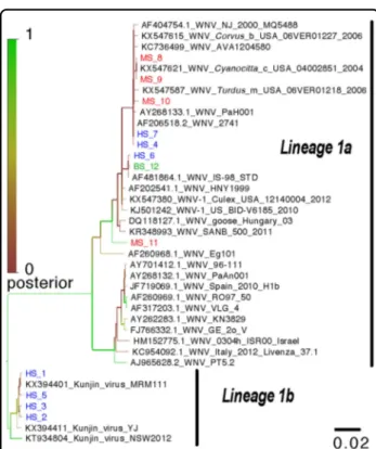
Phylogenetic analysis
A phylogenetic tree was constructed from a 283-bp section of the NS5 gene region amplified by the WNV primers (WNV2+/WNV2−). The tree including the 12 WNV isolates from this study and their relationship with other WNV strains is presented in Fig.2. As shown in the figure, only strains from lineage 1 were detected. The WNV-positive PCR amplicons obtained from three patients, the bird and the 4 mosquito pools formed a monophyletic cluster and were closely related to a Tuni-sian WNV strain isolated in 1997 (PAH001) as well as the Israeli strain, IS-98. The 4 remaining human sequences
Table 1 Distribution of patients and confirmed West Nile virus cases by year of sampling in the Monastir region, Tunisia
2012 2013 2014
A B C D A B C D A B C D
Women 11 4 (36%) 12 1 (8%) 8 0
Men 21 1 (5%) 16 1 (6%) 11 0
Total 32 5 (16%) 15 5 (34%) 28 2 (7%) 13 2 (15%) 19 0 11 0
formed a second distinct cluster and belonged to the Kunjin strains group (Lineage 1b).
Sequences obtained were submitted in GenBank under the accession number, MF371349-60.
Discussion
During the summer-autumn of 2012, a WNV infection outbreak occurred in Tunisia after an apparent silent period following the 2003 epidemic, although
Table 2 Clinical and demographic data and virologicalfindings of the seven West Nile virus-positive patients in the Monastir region, Tunisia
Patient Sample Year Virus Sex Age (years) Fever (°C) Symptoms Locality Geographic coordinates
1 CSF 2012 HS_1 Ma 65.00 39.00 C, F, Nr, M Bembla 35°40′55.10″N/10°45′58.19″E 2 CSF 2012 HS_5 F 50.00 38.60 F, Nr, Gp, ME Khniss 35°43′5.28″N/10°48′50.79″E 3 CSF 2012 HS_7 F 31.00 38.50 C, F, Nr, V, M Monastir 35°45′51.31″N/10°48′40.64″E 4 CSF 2012 HS_3 F 0.90 38.70 Sh, D, F, V Moôtmar 35°45′4.29″N/10°41′27.23″E 5 CSF 2012 HS_6 F 1.00 38.00 F, S Ouardanine 35°42′31.36″N/10°40′43.66″E 6 CSF 2013 HS_2 Ma 66.00 — A, C Sayada 35°39′40.80″N/10°54′15.94″E
7 CSF 2013 HS_4 F 36.00 39.00 C, F, Nr, V, M Ksar Helal 35°38′42.94″N/10°52′24.14″E
CSF cerebrospinal fluid, HS human sample, Ma male, F female, A asthenia, C headache, Sh shock, S seizures, D diarrhea, F fever, Gp general physical deterioration, Nr nuchal rigidity, V vomiting, M meningitis, ME meningoencephalitis
Fig. 1 Geographic distribution of PCR-positive human, avian and pooled mosquito samples in the Monastir governorate, Sahel region, Tunisia. 1, Bembla; 2, 8, Kniss; 3, Monastir; 4, Moôtmar; 5, Ouardanine; 6, Sayada; 7, Ksar Helal; 9, Agba; 10, Moôtmar- Sahline; 11, Oued khniss; 12, Ouardanine Oued el Guelta. Red circles: positive human samples. Black circles: negative human samples. White triangles: positive mosquito pools. Black triangles: negative mosquito pools. Blue square: positive bird sample. Black squares: negative bird samples
epidemiological and clinical surveys have demonstrated WNV circulation in humans5,10–12, as well as in mosqui-toes24, and seropositivity has been reported in equids20–22, dromedaries23, and wild birds19in the years since thefirst WNV epidemic in 1997. However, to date, no information on the epidemiological characteristics of the WNV transmission cycle in the Tunisian Sahel region has been available. This coastal region of Tunisia is interesting to study from an epidemiological viewpoint as (i) the first WNV human cases during 1997, 2003, and 2012 out-breaks have been reported in the Tunisian Sahel region; (ii) it is located on the northern coast of Africa across the
Mediterranean Sea from Italy and could therefore serve as a gateway for birds migrating between Africa and Europe; and (iii) wetlands identified in the Tunisian Sahel region are overwintering and resting areas for several bird species.
On the basis of our WNV RNA detection results from human samples, the WNV prevalence (8.8%) was low and is consistent with a previous study that reported WNV genome detection in clinical specimens by nested PCR (8.8%) during the 2003 meningitis/meningoencephalitis epidemic in the Monastir region5.
Table 3 Features ofCulex pipiens mosquitoes (n = 583) sampled for West Nile virus testing in the Sahel region, Tunisia
No. of females analyzed Capture day Locality Geographic coordinates nRT-PCR results
45 2011/10/03 Khniss 35°43′13.08″N/10°49′11.11″E MS_8
7 2011/10/25 Khniss 35°43′4.06″N/10°48′50.91″E Negative
15 2011/10/31 Khniss 35°43′21.0″N/10°49′12.84″E Negative
Total 2011 = 67
25 2012/09/07 Oued khniss 35°43′21.45″N/10°48′33.95″E Negative
46 2012/09/15 Oued khniss Negative
19 2012/11/01 Agba 35°45′19.31″N/10°49′39.04″E MS_9
12 2012/11/08 Sahline aéroport 35°44′39.29″N/10°44′15.86″E Negative
9 2012/11/12 Moôtmar- Sahline 35°45′10.70″N/10°42′16.47″E MS_10
19 2012/11/17 Oued khniss 35°43′18.44″N/10°48′50.28″E Negative
7 2012/11/18 Oued khniss Negative
12 2012/11/21 Oued khniss MS_11
7 2012/11/22 Oued khniss Negative
9 2012/11/24 Oued khniss Negative
Total 2012 = 165
24 2013/09/02 Oued khniss 35°43′18.44″N/10°48′50.28″E Negative
20 2013/09/15 ONAS Ouardanine 35°43′12.38″N/10°40′25.19″E Negative
29 2013/09/27 Agba 35°45′3.98″N/10°49′38.54″E Negative
34 2013/09/01 Oued Hamdoun 35°46′45.17″N/10°41′0.28″E Negative
33 2013/09/03 Oued Hamdoun Negative
45 2013/09/14 Oued Hamdoun Negative
30 2013/09/15 Sahline aéroport 35°44′39.29″N/10°44′15.86″E Negative
18 2013/10/01 Jemmel 35°37′30.96″N/10°46′0.70″E Negative
Total 2013 = 233
25 2014/09/01 Mahdia 35°25′5.35″N/11° 0′49.28″E Negative
20 2014/09/15 Mahdia Negative
34 2014/09/30 Mahdia Negative
39 2014/11/01 Frina 35°43′36.54″N/10°48′59.62″E Negative
Total 2014 = 118
Phylogenetic analysis revealed that our WNV isolates belonged to two distinct clusters: human, avian and mosquito isolates closely related to the virulent WNV strains isolated in Tunisia (1997) and in Israel (1998), suggesting the presence of an active and local WNV transmission cycle in the Monastir area. The second cluster comprised four isolates from human samples possibly related to the Kunjin virus. These findings revealed the emergence of new pathogenic WNV strains different from those circulating in the Monastir area during the 2003 outbreak5. Two of the four patients whose CSF samples tested positive for Kunjin virus RNA presented with neuromeningeal diseases: the elderly and adult patient had meningitis and meningoencephalitis with a past medical history of hypertension, respectively. Kunjin virus detection in patients with neurological manifestations raises the question of whether risk factors
have a role in the disease severity since the Kunjin virus is considered less virulent than other WNV types28. The contributing factors of age and chronic diseases may relate to a decreased blood–brain barrier integrity, which facilitates access of such a neurotropic virus to the central nervous system, and therefore, predisposes infected patients to neurological complications29.
Per the National Observatory of New and Emerging Diseases (Tunisia), the 2012 WNV epidemic occurred earlier in Tunisia from mid-July and persisted beyond the usual occurrence period to December with a wider geo-graphical spread in addition to an increasing number of reported neuroinvasive disease cases and deaths com-pared to the two previous epidemics of 1997 and 2003, which likely indicates a stronger WNV dynamic in Tunisia in 201213,30. This epidemic coincided with WNV emergence in several other European countries31–34and
Table 4 West Nile virus surveillancefindings on domestic (n = 23) and migratory birds (n = 47) sampled in the Sahel region, Tunisia
Species D/W A/B Capture day Locality Geographic coordinates nRT-PCR results
Cormorant W 0/6 2012/10-11 Sahline-Sabkha- Airport 35°45′27.67″N/10°42′59.54″E Negative
Egret W 0/3 2012/10-11 Sahline-Sabkha- Airport Negative
Pintail W 0/3 2012/11/22 Sahline-Sabkha- Airport Negative
Snipe W 0/3 2012/11/30 Sahline-Sabkha- Airport Negative
Spoonbill W 0/3 2012/11/30 Sahline-Sabkha- Airport Negative
European herring gull W 0/5 2013/09/13 Sahline-Sabkha- Airport Negative European herring gull W 0/2 2012/12/12 Ouardanine 35°42′34.74″N/10°40′32.82″E Negative European starling W 0/3 2012/12/12 Mesjed Aïssa 35°43′36.38″N/10°43′5.67″E Negative
European starling W 0/3 2013/01/16 Mesjed Aïssa Negative
European herring gull W 0/5 2013/10/28 Saline-Sahline 35°45′47.34″N/10°42′25.72″E Negative White duck D 0/4 2013/11/01 Mahdia 35°30′32.19″N/10°57′49.17″E Negative Barbary duck D 0/3 2013/11/12 Irrigation area of Sahline 35°44′6.95″N/10°42′12.08″E Negative
Chickens D 0/3 2013/11/12 Irrigation area of Sahline Negative
Gray goose D 0/2 2013/11/12 Sahline-Sabkha- Airport 35°44′38.72″N/10°44′16.01″E Negative
Hen D 0/3 2013/11/12 Sahline-Sabkha- Airport Negative
European starling W 0/4 2013/11/22 Oued Hamdoun 35°46′27.35″N/10°41′18.63″E Negative Wild duck D 1/2 2013/11/25 Ouardanine Oued el Guelta 35°43′35.18″N/10°40′25.48″E BS_12
Gray goose D 0/2 2013/11/25 Ouardanine Oued el Guelta Negative
European herring gull W 0/1 2013/11/28 Mahdia 35°24′50.80″N/10°34′3.45″E Negative
European herring gull W 0/1 2013/11/29 Mahdia Negative
Seagull W 0/4 2013/11/29 Mahdia Negative
Egret W 0/1 2013/11/30 Mahdia Negative
Wild duck D 0/2 2013/12/07 Hergla Foukaïa 36° 1′11.21″N/10°30′5.51″E Negative
Gray goose D 0/2 2013/12/07 Hergla Foukaïa Negative
the United States35, who also reported earlier occurrences of an increased number of human WNV infection cases. In our study, among the seven WNV-positive cases, four were obtained in November 2012, one in December 2012 and two in January 2013, while WNV was undetected in patients during the transmission seasons of 2013 and 2014. Fewer WNV human cases and small-scale epidemics were reported in 2014 in Southern and Central Europe, as well as in the Middle East, compared to the exceptional transmission seasons of 2012 and 2013, which could be linked to the summer weather conditions in 2014 (temperature and precipitation), which were unfavorable for viral maintenance and amplification36,37
.
The seven WNV-positive cases originated from seven localities (Monastir, Bembla, Khniss, Ouardanine, Moôt-mar, Ksar Helal, Sayada) in the Monastir governorate. Notably, the Monastir area is a former hotbed of WNV infection and was the first governorate affected by the 2012 epidemic13. The presence of a local and active WNV transmission cycle in this region suggests that Monastir is
a high-risk area for WNV infection as demonstrated by previous epidemiological studies22,38.
In the current study, WNV-positive cases included three patients with non-specific non-neurological clinical manifestations and four patients with laboratory-confirmed central nervous system infections. However, even without neurological manifestations, WNV infection can cause significant public health problems, as patients such as children and workers who sought medical atten-tion could not attend school or work because of the ill-ness39. Only hospitalized patients were included in this study. Thus, WNV can cause mild self-limited febrile manifestations that may not require hospitalization; therefore, the overall disease incidence may be underestimated.
The roles of migratory birds as introductory hosts, as well as indigenous wild and domestic birds as potential WNV-circulating reservoirs, have previously been dis-cussed40,41, and several attempts to isolate the virus from birds have been documented42–44. A domestic mallard
(Anas platyrhynchos) sampled in November 2013 from a livestock farm in Ouardanine-Oued El Guelta in the Monastir region, tested positive for WNV RNA. This sampling site was chosen based primarily on a serological survey conducted at the same farm that showed evidence of WNV antibodies in domestic ducks and highlighted WNV circulation in this area; however, no WNV genomic RNA was detected45. Asian studies showed the receptivity and susceptibility of ducks to WNV infection as well as the birds’ involvement in WNV transmission46,47
. Our results suggest that this WNV-positive domestic duck was viremic at the time of sampling, and could therefore act as a WNV reservoir; thus, it may have been involved in the local WNV transmission cycle. However, per Hofmeister et al.48, it is unlikely that ducks are amplifying hosts or that they play significant roles in WNV transmission. The homology between the sequences obtained from this bird and the mosquitoes suggests that the virus is overwintered locally or reintroduced seasonally, and the environment favors viral amplification. In a similar study conducted in Italy, WNV persistence was also confirmed through viral circulation in birds and mosquitoes43. Furthermore, the studied area is situated near wadi El Guelta with approximately 5 km of wetlands, namely Sahline-Sabkha-Airport and Saline-Sahline, with high concentrations of migratory birds, some of which were sampled and inclu-ded in this study. Although WNV was undetected in the 47 migratory bird samples, the likely contribution of viremic migratory birds in repeatedly introducing WNV in Tunisia, followed by local viral transmission via amplifying avian hosts, has been clearly assessed in many Tunisian studies10,12,19.
Cx. pipiensmosquitoes are considered the most impor-tant vectors in transmitting WNV43,49,50. Entomological
Fig. 2 Phylogenetic analysis of identified West Nile virus sequences from humans, pooled mosquitoes and birds in the Monastir governorate, Sahel region, Tunisia. Phylogenetic analysis was performed using Bayesian analysis based on the TN93 evolution model, a gamma distribution and invariable sites. The tree includes 12 strains isolated in this study and 27 homologous nucleotide sequences from the West Nile virus NS5 gene obtained from the GenBank library. HS human sample, MS mosquito sample, BS bird sample
surveys conducted after WNV outbreaks in Tunisia showed the abundance and role of Cx. pipiens as potential WNV vectors in Tunisia11,24,26,51,52. WNV-positive Cx. pipiens pools sampled in 4 localities (Khniss, Agba, Moôtmar-Sahline, and Oued-Khniss) in the Monastir governorate indicated viral circulation during the 2011 and 2012 seasons. Despite no human cases having been reported in the Tunisian Sahel region in 201153, our find-ings strongly suggest a silent WNV circulation during the 2011 transmission season and that the onset of the 2012 WNV outbreak was favored by ecological factors and environmental conditions. The epidemic occurred during a very hot summer season with a high rainfall, which pro-vided optimal conditions for mosquito proliferation. Interestingly, such exceptionally warm climate conditions were observed during the two previous epidemics in Tunisia25, as well as in 2012 during the WNV outbreaks in Europe31,33,34and the United States54. Although WNV was undetected in Cx. pipiens mosquitoes collected in 2013 and 2014, a study conducted in central Tunisia reported viral detection in Cx. pipiens during the 2014 transmission season suggesting WNV persistence in the country24. More studies are needed regarding vector competence and overwintering cycles in mosquitoes, local birds and potential non-avian reservoirs23 as an endemization mechanism in Tunisia.
Isolate clustering from human and mosquito WNV-positive samples in this study confirms the potential role of Cx. pipiens as a bridge species between birds and mammals, and its involvement in the WNV transmission cycle in Tunisia has been described in several Tunisian entomological studies24,26,52. WNV detection in both human and Cx. pipiens specimens sampled in the Mon-astir region during the 2012 outbreak suggests that after WNV amplification via an enzootic transmission cycle, human exposure to infected mosquitoes and the viral shift to humans may have occurred under ecological and environmental conditions including the close proximity of human habitats to wetlands that host migratory bird settlements and are suitable areas for mosquitoes55. All patient and Cx. pipiens mosquito specimens in this study sampled during the 2013 and 2014 transmission seasons were WNV-negative. However, WNV detection in birds in 2013 (this study) and in mosquitoes in 201424suggest that the risk of WNV reemergence in Tunisia should be considered high.
As no vaccine or specific treatment exists for WNV infections, only preventive measures, such as wearing long-sleeve shirts and long pants, applying skin repellents and avoiding being outdoors at dusk could reduce the risk of WNV infection by half56. Although vector control measures including adulticidal and larvicidal treatments were implemented early in Tunisia in spring 2012, it appears that the population’s low background immunity
to WNV and the emergence of new pathogenic strains may partly explain the reoccurrence of WNV human cases and the spread of the virus to new areas13,16. Thus, human surveillance should be enhanced by updating risk area maps and public awareness campaigns among the population and health care professionals throughout the country. WNV persistence may lead to future outbreaks in Tunisia and along bird migratory routes between Africa and Europe, highlighting the need for entomological studies on persistence mechanisms and identification of mosquito species acting as competent bridge vectors in WNV transmission24. Setting up avian surveillance based on serological surveys of domestic birds cohabiting with humans and/or sentinel chickens to assess WNV enzootic transmission might provide useful epidemiological infor-mation such as low-noise WNV circulation, which can therefore be used as a warning system to detect viral circulation early and implement prevention and control measures57,58.
Materials and methods
Ethics statement
The study protocol was approved by the Ethics and Research Committee of the Fattouma Bourguiba Uni-versity Hospital (Monastir, Tunisia, Committee’s advice on 20 September 2013), and written informed consent was obtained from the 79 patients.
Patients and clinical sample collections
Seventy-nine CSF samples were obtained from patients admitted to different departments of the Fattouma Bourguiba University Hospital (Monastir, Tunisia) from November 2012 to December 2014. Per routine clinical care, lumbar punctures were performed by the examining physician on patients showing signs of suspected acute meningitis upon admission at the emergency department or within 24 h of hospitalization. CSF specimens were processed for microbiological testing by routine hospital procedures and were bacterial pathogen-free. CSF cyto-biochemical examinations provided pleocytosis as well as protein and glucose concentrations.
Based on the examining physician’s clinical diagnosis and medical records, patients with febrile symptoms and/ or exhibiting suspicious signs of neuromeningeal infec-tion, such as aseptic meningitis, encephalitis, and meningoencephalitis, were included in the study. Com-plete demographic characteristics and clinical findings were obtained for all patients.
Study sites and mosquito collections
Mosquito sampling was performed at different sites in the Sahel region’s 3 coastal governorates, Monastir, Sousse, and Mahdia, mostly from September to Novem-ber for 4 years (2011–2014). Two battery-powered CDC
light-traps were placed at 14 sampling sites and operated approximately from sunset to sunrise. Epidemiological characteristics were considered for the sampling site selection including occurrence of WNV human cases, proximity to wetlands and migratory bird settlement flight. Mosquito traps were placed in urban (houses and gardens) and rural (riding stables and livestock farms) environments.
Field-collected specimens were transported to the laboratory, and female Cx. pipiens were morphologically identified using identification keys59. Female Cx. pipiens were then pooled by date and collection site by handling mosquitoes individually with sterile stainless steel twee-zers with a maximum of 50 individuals per pool and stored in cryotubes at−70 °C until being assayed for viral detection.
Prior to nucleic acid extraction, mosquito pools were homogenized following a procedure described previously60.
Bird collections
Seventy serum samples from 23 domestic and 47 migratory birds were obtained from 2012 to 2014 during the autumn season and active mosquito circulation peri-ods. Migratory bird blood sampling was conducted at several bird wintering and resting sites. Domestic bird blood was collected from livestock farms located on wild bird flyways and/or close to wetlands in the Tunisian Sahel region. For each bird, 5 ml of blood was obtained and centrifuged at 12 000 rpm for 10 min. Serum samples were stored at−20 °C until tested.
Detection of WNV RNA
Total RNA was extracted from 200-µl samples using the Trizol® LS Reagent (Sigma-Aldrich, Madrid, Spain) method for CSF samples and mosquito pool supernatants. Sera from domestic and migratory birds were processed using Trizol®BD Reagent (Sigma-Aldrich, Madrid, Spain). RNA was then purified with chloroform and precipitated with isopropanol (Sigma-Aldrich, Madrid, Spain).
After washing with 70% ethanol, the pellet was dried and eluted in 30µl of RNase-free water (Qiagen, Barce-lona, Spain). A negative control consisting of RNase-free water was included in this step.
Generic RT-PCR was performed using degenerated primers, Flavi1+ and Flavi1−, whereas generic nested PCR was performed using degenerated primers, Flavi2+ and Flavi2−, as previously described61
. These two primer sets were designed to amplify a conserved region of theflavivirus genome located in the NS5 gene encoding for polymerase. A second generic nested PCR was per-formed using degenerated primers designed to specifically target the NS5 gene of the WNV genome (WNV2+:
5′8485AARCCYCTNCTYAAYTCWGAYAC3′8507/WNV2
−: 5′8813TCRTTSARNACNWRYTTIRCWCC3′8791).
The RT-PCR step was performed using the One Step RT-PCR kit (Qiagen, Barcelona, Spain) per the manufacturer’s instructions using 10 µl of RNA and 100 pmol of each primer (Flavi1+/Flavi1−) in a 50-µl total reaction volume. Samples underwent an initial cycle at 50 °C for 30 min and 95 °C for 15 min, followed by 40 PCR cycles at 94 °C for 30 s, 40 °C for 4 min, 72 °C for 1 min, and afinal elongation step at 72 °C for 10 min.
The nested-PCR reaction was performed using the Taq PCR Core kit (Qiagen, Barcelona, Spain) per the manu-facturer’s instructions. One microliter of the first ampli-fication product was added to 100 pmol of the second primer set (Flavi2+/Flavi2−) in a final volume of 50 µl. The reaction mixture was then subjected to the following amplification conditions: an initial denaturation step at 94 °C for 2 min followed by 40 PCR cycles at 94 °C for 30 s, 50 °C for 1 min, 72 °C for 30 s, and a final elongation step at 72 °C for 10 min. The expected PCR product size was 143 bp.
For the second nested WNV-specific PCR reaction, 1 µl of the RT-PCR product was added to 50µl of the total reaction mixture containing 100 pmol of each primer (WNV2+/WNV2−) with the following cycling condi-tions: 94 °C for 3 min and 40 PCR cycles at 94 °C for 1 min, 45 °C for 3 min, 72 °C for 1 min, and a final elonga-tion step at 72 °C for 10 min. The expected amplicon size was 328 bp.
After the amplifications, 10 µl of nested-PCR product was loaded into a 2% GelRed (Biotium) stained agarose electrophoresis gel in TBE buffer and visualized under ultraviolet light. Amplicon sizes were determined by comparing them with a 100-bp DNA ladder (Qiagen, Barcelona, Spain).
Sequencing and phylogenetic analysis
PCR products were purified using the QIAquick PCR purification kit (Qiagen, Barcelona, Spain) and bi-directionally sequenced (Macrogen Inc., Amsterdam, the Netherlands). The phylogenetic analysis (Bayesian analysis) was conducted using the beast package62, with the TN93 evolution model, a gamma distribution and invariable sites. The clock parameters were set to uncorrelated lognormal, using a coalescent constant size model. The chain length was set at 10 million iterations to produce an ESS (effective sampling size) superior to 200. The maximum credibility tree with branch length in number of substitutions was defined from ten thousand trees after discarding 10% and was edited using FigTree (http://tree.bio.ed.ac.uk/software/
Acknowledgements
We gratefully thank all hospital physicians of the Neurological, Pediatric, Emergency and Neurosurgery Departments and the laboratory personnel in the Microbiology Department at the Fattouma Bourguiba University Hospital of Monastir (Tunisia) for their help in obtaining patient clinical samples and laboratory data; the Ministry of Agriculture (Tunisia) and the Regional Office of Agriculture Development (Monastir, Tunisia) for providingfield study permissions, mosquito traps and avian samples within the framework of national passive epidemiological surveillance of avian influenza and West Nile virus in wild birds in Tunisia; and all technicians and staff at the Genomics Unit, Centros Cientificos y Tecnologicos UB (CCiTUB) at the Parc Cientific de Barcelona (PCB), University of Barcelona, for technical support. Abir Monastiri received a fellowship grant from the Ministry of Higher Education and Scientific Research (Tunisia). Funding for this work was provided by the Ministry of Higher Education and Scientific Research (Tunisia). Author details
1Laboratory of Contagious Diseases and Biologically Active Substances,
LR99ES27, Faculty of Pharmacy, Avicenne Street, 5000 Monastir, Tunisia.
2Laboratory of Arbovirus and Imported Viral Diseases, National Microbiology
Center, Instituto de Salud Carlos III, 28220 Madrid, Spain.3Department of
Infection and Epidemiology, Institut Pasteur, 75015 Paris, France.4Infectious
Diseases Department, Fattouma Bourguiba University Hospital, 5000 Monastir, Tunisia.5Laboratory of Microbiology, Fattouma Bourguiba University Hospital, 5000 Monastir, Tunisia.6Unit of Genetic Research, Biodiversity and Valorization
of Bioresources, UR03ES09, Higher Institute of Biotechnology, Taher Hadded Avenue, 5000 Monastir, Tunisia.7Ministry of Agriculture-Tunis, CRDA, 5000
Monastir, Tunisia.8IRBIO and Department of Animal Biology, Faculty of Biology, University of Barcelona, Av. Diagonal, 08028 Barcelona, Spain
Conflict of interest
The authors declare that they have no conflict of interest.
Received: 23 September 2017 Revised: 5 December 2017 Accepted: 1 January 2018
References
1. Thiel, H. J, Collett, M. S. & Gould, E. A. et al. in Virus Taxonomy. VIIIth Report of the International Committee on Taxonomy of Viruses (ICTV) (eds Fauquet, C. M., Mayo, M. A., Maniloff, J., Desselberger, U. & Ball, L. A.) 979–996 (Elsevier/ Academic Press, London, 2005).
2. Blitvich, B. J. Transmission dynamics and changing epidemiology of West Nile virus. Anim. Health Res. Rev. 9, 71–86 (2008).
3. Rappole, J. H. & Hubálek, Z. Migratory birds and West Nile virus. J. Appl. Microbiol. 94, 47–58 (2003).
4. Kramer, L. D., Li, J. & Shi, P. Y. West Nile virus. Lancet Neurol. 6, 171–181 (2007). 5. Riabi, S., Gaaloul, I. & Mastouri, M. et al. An outbreak of West Nile Virus infection in the region of Monastir, Tunisia, 2003. Pathog. Glob. Health 108, 148–157 (2014).
6. Bouatef, S., Hogga, N. & Ben Dhifallah, I. et al. Monitoring and current situation of meningitis and meningoencephalitis to West Nile virus in Tunisia. Tun. Rev. Infect. 6, 181–182 (2012).
7. World Health Organization. West Nile Virus. Media Centre. Fact sheet No. 354. (WHO, Geneva, 2011).http://www.who.int/mediacentre/factsheets/fs354/en/. Accessed July 2011.
8. Di Sabatino, D., Bruno R. & Sauro, F. et al. Epidemiology of West Nile disease in Europe and in the Mediterranean Basin from 2009 to 2013. Biomed. Res. Int. 2014, 907852 (2014).
9. European Centre for Disease Prevention and Control. Epidemiological Update: West Nile Virus Transmission Season in Europe (ECDC, Solna, 2016).https://ecdc. europa.eu/en/news-events/epidemiological-update-west-nile-virus-transmission-season-europe-2016.Accessed 15 December 2016.
10. Triki, H., Murri, S. & Le Guenno, B. et al. West Nile viral meningoencephalitis in Tunisia. Med Trop. 61, 487–490 (2001).
11. Feki, I., Marrakchi, C. & Ben Hmida, M. et al. Epidemic West Nile virus ence-phalitis in Tunisia. Neuroepidemiology 24, 1–7 (2005).
12. Hachfi, W., Bougmiza, I. & Bellazreg, F. et al. Second epidemic of West Nile virus meningoencephalitis in Tunisia. Med Mal. Infect. 40, 456–461 (2010). 13. Observatoire National des Maladies Nouvelles et Emergentes. Bilan de la
surveillance des Infections à Virus West Nile en Tunisie, Année 2012 (ONMNE, Tunis, 2013).http://www.onmne.tn/fr/images/Bulletin2WN.pdf.Accessed April 2013.
14. Benjelloun, A., El Harrak, M. & Belkadi, B. West Nile disease epidemiology in North-West Africa: bibliographical review. Transbound. Emerg. Dis. 63, 153–159 (2016).
15. Charrel, R. N., Brault, A. C. & Gallian, P. et al. Evolutionary relationship between Old World West Nile virus strains. Evidence for viral geneflow between Africa, the Middle East, and Europe. Virology 315, 381–388 (2003).
16. Riabi, S., Gallian, P. & Gaaloul, I. et al. Prevalence of IgG antibodies against West Nile virus in blood donors during the 2003 outbreak in Tunisia. Trans. R. Soc. Trop. Med. Hyg. 104, 507–509 (2010).
17. Bahri, O., Dhifallah, I. & Ben Alaya-Bouafif, N. et al. Sero-epidemiological study of West Nile virus circulation in human in Tunisia. Bull. Soc. Pathol. Exot. 104, 272–276 (2011).
18. Kooli, I., Loussaif, C. & Ben Brahim, H. et al. West Nile virus (WNV) presenting as acute cerebellar ataxia in an immunocompetent patient. Rev. Neurol. 173, 238–240 (2017).
19. Hammouda, A., Lecollinet, S. & Hamza, F. et al. Exposure of resident sparrows to West Nile virus evidenced in South Tunisia. Epidemiol. Infect. 143, 3546–3549 (2015).
20. Ben Hassine, T., Hammami, S. & Elghoul, H. et al. Detection of circulation of West Nile virus in equine in the north-west of Tunisia. Bull. Soc. Pathol. Exot. 104, 266–271 (2011).
21. Ben Hassine, T., De Massis, F. & Calistri, P. et al. First detection of co-circulation of West Nile and Usutu viruses in equids in the south-west of Tunisia. Transbound. Emerg. Dis. 61, 385–389 (2014).
22. Bargaoui, R., Lecollinet, S. & Lancelot, R. Mapping the serological prevalence rate of West Nile fever in equids, Tunisia. Transbound. Emerg. Dis. 62, 55–66 (2015).
23. Hassine, T. B., Amdouni, J. & Monaco, F. et al. Emerging vector-borne diseases in dromedaries in Tunisia: West Nile, bluetongue, epizootic haemorrhagic disease and Rift Valley fever. Onderstepoort J. Vet. Res 84, 1–3 (2017). 24. Wasfi, F., Dachraoui, K. & Cherni, S. et al. West Nile virus in Tunisia, 2014: first
isolation from mosquitoes. Acta Trop. 159, 106–110 (2016).
25. El Ghoul H. Renforcement de la surveillance et des systèmes d’alerte pour la fièvre catarrhale ovine, la fièvre du Nil Occidental et la rage au Maroc, en Algérie et en Tunisie– Fièvre du Nil Occidental: historique et situation épidémiologique en Tunisie. Projet GCP/RAB/002/FRA, FAO. 24 p. (2009).
26. Krida, G., Rhim, A. & Daaboub, J. et al. New evidence for the potential role of Culex pipiens mosquitoes in the transmission cycle of West Nile virus in Tunisia. Med Vet. Entomol. 29, 124–128 (2015).
27. Hamdi, N. & Chard-Cheikhrouha, F. Estimation du nombre total des oiseaux aquatiques hivernant en Tunisie: période 2001/2002 à 2006/2007. Rev. Ecol. 66, 231–253 (2011).
28. Garcia-Sastre, A. & Endy, T. P. in Encyclopedia of Microbiology (ed. Moselio, S.) 3rd edn, 313–320 (Elsevier/Academic Press, Oxford, 2009).
29. Hall, R. A., Broom, A. K. & Smith, D. W. et al. The ecology and epidemiology of Kunjin virus. Curr. Top. Microbiol Immunol. 267, 253–267 (2002).
30. EpiSouth. Weekly Epi Bulletin—N°239 10th October—17th October 2012. http://www.episouthnetwork.org/sites/default/files/bulletin_file/
eweb_239_18_10_12.pdf.
31. Barzon, L., Pacenti, M. & Franchin, E. et al. Large human outbreak of West Nile virus infection in north-eastern Italy in 2012. Viruses 5, 2825–2839 (2013). 32. Popović, N., Milošević, B. & Urošević, A. et al. Outbreak of West Nile virus
infection among humans in Serbia, August to October 2012. Eur. Surveill. 18, 206–213 (2013).
33. Pem-Novosel, I., Vilibic-Cavlek, T. & Gjenero-Margan, I. et al. First outbreak of West Nile virus neuroinvasive disease in humans, Croatia, 2012. Vector Borne Zoonotic Dis. 14, 82–84 (2014).
34. Pervanidou, D., Detsis, M. & Danis, K. et al. West Nile virus outbreak in humans, Greece, 2012: third consecutive year of local transmission. Eur. Surveill. 19, 2075–2078 (2014).
35. Centers for Disease Control and Prevention. West Nile Virus (WNV) Human Infections Reported to ArboNET, by State, United States, 2012 (CDC, Atlanta, 2012). http://www.cdc.gov/westnilestatsMaps/finalMapsData/index.html. Accessed 20 February 2013.
36. European Centre for Disease Prevention and Control. Cumulative number of West Nile fever cases by affected area in 2014 (ECDC, Solna, 2014).http://ecdc. europa.eu/en/healthtopics/west_nile_fever/West-Nile-fever-maps/Pages/ 2014-table.aspx. Accessed 30 November 2014.
37. Bahuon, C. & Lecollinet, S. Des saisons de transmission du virus West Nile contrastées enEurope. Bull. Epid. St. Anim. Alim. 67, 19–22 (2015).
38. Ben Hassine, T., Conte, A. & Calistri, P. et al. Identification of suitable areas for West Nile virus circulation in Tunisia. Transbound. Emerg. Dis. 64, 449–458 (2017).
39. Hayes, E. B., Sejvar, J. J. & Zaki, S. R. et al. Virology, pathology, and clinical manifestations of West Nile virus disease. Emerg. Infect. Dis. 11, 1174–1179 (2005).
40. Rappole, J. H., Derrickson, S. R. & Hubálek, Z. Migratory birds and spread of West Nile virus in the Western Hemisphere. Emerg. Infect. Dis. 6, 319–328 (2000).
41. Jourdain, E., Zeller, H. G. & Sabatier, P. et al. Prevalence of West Nile virus neutralizing antibodies in wild birds from the Camargue area, southern France. J. Wildl. Dis. 44, 766–771 (2008).
42. Malkinson, M., Banet, C. & Weisman, Y. et al. Introduction of West Nile virus in the Middle East by migrating white storks. Emerg. Infect. Dis. 8, 392–397 (2002). 43. Calzolari, M., Gaibani, P. & Bellini, R. et al. Mosquito, bird and human surveil-lance of West Nile virus and Usutu viruses in Emilia-Romagna Region (Italy) in 2010. PLoS ONE 7, 3805–3808 (2012).
44. Maquart, M., Boyer, S. & Rakotoharinome, V. M. et al. High prevalence of West Nile virus in domestic birds and detection in 2 new mosquito species in Madagascar. PLoS One 11, 01475–01489 (2016).
45. Bargaoui R. Epidémiologie de lafièvre West Nile en Tunisie. Thèse Pour obtenir le grade de Docteur de l’Université de Montpellier II. Virologie. Uni-versité de Montpellier II, Sciences et Techniques du Languedoc (2012). 46. Shirafuji, H., Kanehira, K. & Kubo, M. et al. Experimental West Nile virus infection
in aigamo ducks, a cross between wild ducks (Anas platyrhynchos) and domestic ducks (Anas platyrhynchos var. domesticus). Avian Dis. 53, 239–244 (2016).
47. Kalaiyarasu, S., Mishra, N. & Khetan, R. K. et al. Serological evidence of wide-spread West Nile virus and Japanese encephalitis virus infection in native domestic ducks (Anas platyrhynchos var domesticus) in Kuttanad region, Kerala, India. Comp. Immunol. Microbiol. Infect. Dis. 48, 61–68 (2016). 48. Hofmeister, E., Porter, R. E. & Franson, J. C. Experimental susceptibility of wood
ducks (Aix sponsa) for West Nile virus. J. Wildl. Dis. 51, 411–418 (2015).
49. Bagheri, M., Terenius, O. & Oshaghi, M. A. et al. West Nile virus in mosquitoes of Iranian wetlands. Vector Borne Zoonotic Dis. 15, 750–754 (2015).
50. López, R. H., Soto, S. U. & Gallego-Gómez, J. C. Evolutionary relationships of West Nile virus detected in mosquitoes from a migratory bird zone of Colombian Caribbean. Virol. J. 12, 80 (2015).
51. Bouattour, A., Rhaiem, A. & Ghrammam, M. et al. Rapport d’enquête ento-mologique suite à l’apparition des cas de West Nile. Rapport technique, Insti-tut Pasteur Tunis. Laboratoire d'entomologie médicale. 5 p. (1998). 52. Amraoui, F., Krida, G. & Bouattour, A. et al. Culex pipiens, an experimental
efficient vector of West Nile and Rift Valley fever viruses in the Maghreb region. PLoS ONE 7, 3675–3677 (2012).
53. European Centre for Disease Prevention and Control. Cumulative number of West Nile fever cases by affected area in 2011 (ECDC, Solna, 2011).http://ecdc. europa.eu/en/healthtopics/west_nile_fever/West-Nile-fever-maps/Pages/ 2011table.aspx. Accessed 30 November 2011.
54. Murray, K. O., Ruktanonchai, D. & Hesalroad, D. et al. West Nile virus, Texas, USA, 2012. Emerg. Infect. Dis. 19, 1836–1838 (2003).
55. Ben Hassine, T., Calistri, P. & Ippoliti, C. et al. Analysis of biotic and abiotic factors influencing the occurrence of West Nile virus infection in Tunisia. Arch. Inst. Pasteur Tunis. 91, 43–50 (2014).
56. Loeb, M., Elliott, S. J. & Gibson, B. et al. Protective behavior and West Nile virus risk. Emerg. Infect. Dis. 11, 1433–1436 (2005).
57. Rizzoli, A., Rosà, R. & Rosso, F. et al. West Nile virus circulation detected in northern Italy in sentinel chickens. Vector Borne Zoonotic Dis. 7, 411–417 (2007). 58. Chaintoutis, S. C., Dovas, C. I. & Danis, K. et al. Surveillance and early warning of West Nile virus lineage 2 using backyard chickens and correlation to human neuroinvasive cases. Zoonoses Public Health 62, 344–355 (2015).
59. Brunhes, J., Rhaim, A. & Geoffroy, B. et al. Les Culicidae d’Afrique Méditerra-néenne. (Institute for Research Development, Montpellier, France, 2000). 60. González-Reiche, A. S., Monzón-Pineda Mde, L. & Johnson, B. W. et al.
Detection of West Nile viral RNA fromfield-collected mosquitoes in tropical regions by conventional and real-time RT-PCR. Methods Mol. Biol. 630, 109–124 (2010).
61. Sánchez-Seco, M. P., Rosario, D. & Domingo, C. et al. Generic RT-nested-PCR for detection offlaviviruses using degenerated primers and internal control fol-lowed by sequencing for specific identification. J. Virol. Methods 126, 101–109 (2005).
62. Drummond, A. J. & Rambaut, A. BEAST: Bayesian evolutionary analysis by sampling trees. BMC Evol. Biol. 7, 214–214 (2007).