Radiology and histopathology of the bent limbs
in campomelic dysplasia: implications in the aetiology of the disease
and review of theories
U. E. Pazzaglia I and G. Beluffi 2
1 Orthopedic Clinic, University of Pavia and
z Department of Radiology, Pediatric Radiology Section, IRCCS Polictinico San Matteo, Pavia, Italy
Abstract.
The study of the bent bones in a case of
campomelic dysplasia and a review of the literature
suggest a repair process at the site of angulation. A
theory that can explain most of the features of the
disease is presented; it supposes a damage of the car-
tilage model at the midshaft, followed by a fracture
of the thin bone collar in the earlier stage of ossifica-
tion of the diaphysis.
Following the description by Maroteaux et al. [32]
and Bianchine et al. [6] of campomelic dysplasia,
many cases have been reported of this usually lethal
entity, in which multiple osseous defects (bending of
tubular bones is the most striking) and malforma-
tions of other organs are associated [24, 31, 40, 44, 46,
48, 49].
The genesis of this condition remains uncertain
and both an exogenous factor acting in the mother in
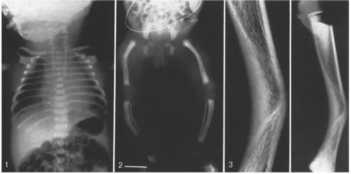
Fig. 1. The chest is bell-shaped, with slender, horizontal ribs and hypoplastic scapulae
Fig.2. Bowing of the tubular bones of lower limbs; hypoplastic pelvic girdle bones and dislocation of the hips
Fig.3. X-ray of the tibia after dissection: angulation of the bone between the proximal ¾ and the distal %. On the concave side the cortex has been replaced by dense bone in which the rayed pattern is evident. Metaphyseal trabeculae are normal
U. E. Pazzaglia and G. Beluffi: Campomelic dysplasia 51
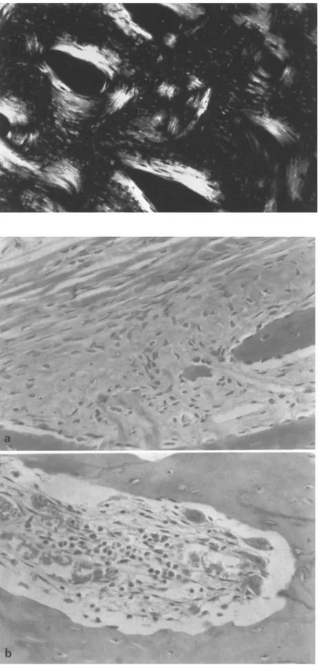
Fig.4.
Haematoxylin-eosin (100x). Polarized light; remodelling of the dense bone on the concave side of angulation, with Haversian canals running in the mass of woven bonethe first period of gestation and a genetic factor in
the baby have been proposed. In our presentation
the histopathological findings of the bent bones of a
girl who died at 11 days of age are reported: the
study of them led to formulation of ideas about the
pathogenesis of the disease.
Case report
This 3700-g female infant was born at week 41 of gestation of a 32-year-old mother and a 40-year-old father.
There is no consanguinity in the kindred; the mother is affect- ed by hyperthyroidism, but there is no record of drug use during gestation. A previous male child was normal. There is no record of abortion. Delivery was spontaneous and natural; the placenta, weighing 500 g was normal. At birth the infant was dyspneic and cyanotic, requiring mechanical ventilation, and markedly hypo- tonic. After an initial improvement with spontaneous ventilation,
the infant had to be maintained with mechanical ventilation until she died at 11 days of age.
The head circumference was 39 cm, chest circumference 34 cm and crown-rump length 48 cm. There was hypertelodsm, a flat na- sal bridge, micrognathia and cleft of the soft palate, low set ears and redundant skin around the neck. The lower limbs were slight- ly shorter than normal (more so on the right than the left) with an- terior bowing of the femora, tibiae and fibulae; a skin dimple over the right tibia; and bilateral talipes equinovarus. External genital- ia were normal. The abdomen was swollen; the liver and spleen enlarged.
X-rays showed an enlarged skull and micrognathia. The chest was bell shaped, with slender, horizontal ribs (Fig. 1) and absence of the sternal ossification nuclei. The scapulae were hypoplastic and clavicles had lateral hooks.
The iliac wings were hypoplastic, and the hip joints were dislo- cated (Fig. 2). The lower limb tubular bones are described in detail in the next section.
Chromosomes were 46, XX.Autopsy, carried out 3 h after death revealed: atelectasia of both lungs with a small infarction area in the left lower lobe; absence of olfactory bulbs; thymic hy- poplasia; enlarged liver and spleen.
Fig. 5. Haematoxylin-eosin (240 x). Polarized light; transverse section of the Haversian ca- nals in the mass of woven bone on the con- cave side
Fig.6A and B. Haematoxylin-eosin
(250x). A t h e origin of Haversian canals be- neath the periosteum; B osteoclasts and ves- sels at the apex of a new Haversian canal
U. E. Pazzaglia and G. Beluffi: Campomelic dysplasia 53
Material and methods
The right femur, tibia and fibula were dissected from the soft tis- sues. A specimen of the tibial, proximal growth plate was immedi- ately fixed in glutaraldehyde and embedded in methacrylate. Un- decalcified sections were stained with haematoxylin-eosin and Von Kossa method. Radiographs of the remaining bone speci- mens were taken and then they were decalcified in a 10% E D T A solution. After inclusion in paraffin, serial sections in the plane of the bending were prepared and stained with haematoxylin eosin.
Observations
Radiographs.
In the femur there was an angle of 157 o between the axes of the proximal % and the distal % of the bone.Angulation in the tibia and fibula were, respectively, 151 ° and 153 °, between the proximal % and the distal ~/3 of the bones. The metaphyseal bone trabecutae, as well as the cortex on the convex side (excepting the site of angulation) were unremarkable. On the concave side the normal cortex had been replaced by broad dense bone in which were thin radiotransparent lines in fan-like arrange- ment converging toward the apex of the bend (Fig. 3).
Histology. The
histopathology of the femur, tibia and fibula was the same, and only the tibia will be described in detail.The epiphysis and growth plate cartilage (and its ossification) were normal. The primary trabeculae, easily recognized because of the central core of cartilage, were normal in their architecture and structure and were undergoing the expected, remodelling.
On the concave side, instead of the normal cortical bone, a less organized woven bone was observed lying between periosteum and the metaphyseal trabeculae. Straight parallel canals, contain- ing blood vessels, ran through it (primary osteons) from the peri- osteum toward the apex of the cone (Figs. 4 and 6). The surfaces of these canals were the site of remodelling, producing osteonal structures (Fig. 5). This organization was responsible for the rayed pattern observed in X-rays of the specimen.
Periosteal features were unremarkable and no signs of en- hanced appositional activity were actually evident.
Discussion
The histological findings in the bent bones of cam-
pomelic dysplasia suggest a repair process with an-
gulation and with new periosteal bone laid down on
the concave side and remodelling of the mass of
woven bone so formed.
As observed by Roth et al. [39], this process is
identical to the healing of malaligned fractures and it
is not specific to campomelic dysplasia, since the
same thickening of the concave side is observed in
other congenital angulation of tubular bones [9, t l ,
14, 23, 28, 35]. The same histological features can be
observed in lower limb experimental bowing ob-
tained in chicken embryos by injecting insulin at the
5th day [15, 16].
A further confirmation of the former statement is
given by the only two cases of campomelic dysplasia,
who surived [8, 32], and by all the other non-lethal
cases of congenital bowing of the legs, where a spon-
taneous correction of the angulation occurs as the
child grows up.
Moreover the growth plate cartilage and the
metaphyses look normal and abnormalities de-
scribed in previous articles [4, 26, 29, 31] do not stand
up to critical review, as Austin et at. [3] have already
observed.
A theory of the aetiology and pathogenesis of
campomelic dysplasia should take account of these
observations, but must also satisfactorily explain the
features presented constantly in the dysplasia. These
may be summarized as follows: level of angulation
always between the proximal % and the distal 2A in
the femur the opposite in the tibia and fibula; a de-
gree of shortening of the bone, independent from an-
gulation (the so-called short limb variety of campo-
melic dysplasia is not considered here, because in
these cases there are radiographic signs of meta-
physeal involvement [18, 21, 22, 27, 51]; symmetry of
lower limb lesions; upper limb tubular bones unaf-
fected or showing only a minor degree of dysplasia;
talipes equino-varus; scapula and pelvic girdle bones
hypoplastic, with dislocation of the hips; other bone
defects (skull, spine, ribs); other organ defect (CNS,
respiratory tract, heart, urinary tract, gonads); spo-
radic cases, but also reported in brothers [34, 42, 45,
47].
The cartilage model of each skeletal element is
formed before the end of the embryonic period (8th
postovulatory week). The model of tubular bones is
sharply defined by the perichondrium; the youngest
cells are at the ends, where most of the growth oc-
curs, while the oldest are in the mid-section of the
shaft, where, before ossification starts (primary ossi-
fication center) they enlarge, their cytoplasm be-
comes vescicular and the amount of intercellular ma-
trix increases. A bone collar is laid down by the
perichondrium (now periosteum) around the mid-
shaft before the matrix calcifies and vascular inva-
sion starts.
The development of the lower limb buds is a little
later than the upper and this is observed at all stages,
namely, mesenchymal differentiation, chondrifica-
tion and beginning of ossification [37].
Hypothetically, possible damage to the cells of
the midshaft leaving healthy those of the ends of the
model) may explain the features observed in cam-
pomelic dypslasia. The lesion of the central part o f
the model is followed by the fracture of the thin bone
collar. Since the embryo is surrounded by the amni-
otic liquid and pressure is transmitted uniformily
through liquids, the displacement of the cartilagine-
ous model fragments may be conditioned by the in-
trinsic tension generated by muscles in the limb or by
the postion of the limbs relatively to the body of the
angulation is always anterior in the tibia and antero-
lateral in the femur.
The absence of bending of the upper limb bones
may be explained by the more advanced develop-
mental stage, with a thicker (and so more resistant to
fracture) bone collar, while in an earlier stage the me-
chanical forces are not strong enough to bend the
cartilage model.
In the cartilage of the pelvic girdle and scapula
the lesion results in a loss of cells and a hypoplastic
mature bone. The same factor may be responsible of
the spine, skull and upper respiratory tract cartilage
alterations and of other organs malformations.
In the tubular bones the lesion affects the mid-
shaft, while the distal ends of the model grow nor-
mally. Since the distal cartilage plate of the femur
grows faster than the proximal and vice versa in the
tibia, then at birth, when the dysplasia is observed,
the apex of the bending is between the proximal ~A
and the distal % in the femur and the opposite is seen
in the tibia. The loss in length of the bone is therefore
due exclusively to the midshaft lesion of the cartilage
model.
Two mechanical theories of congenital bowing of
the long bones have been presented, one by Middle-
ton [33], the other by Chapple and Davidson [101 and
Caffey [9] and by Angle [21. Both of them presuppose
a mechanical force constantly applied to the devel-
oping bone. Two points are against these theories: (1)
there is a real shortening of the bone; (2) metaphy-
seal trabeculae are straight and parallel to the axis of
the bone and one would expect to find them bent if a
constant load were to be applied to the extremities of
the bone during growth. The main argument raised
by Middleton to support his theory is the constant
association of talipes equino-varus to bending; how-
ever, there is no evidence that muscular imbalance
could be secondary to the bone malformation.
With regard to aetiology, in the first reports atten-
tion was addressed to an exogenous factor [7, 19, 20,
29, 30, 38, 43], while later it shifted to a genetic defect
[1, 5, 6, 12, 13, 17, 25, 26, 34, 36, 47].
The pathogensis of the disease and the normal
metaphyseal growth plate cartilage favour a factor
acting in a limited span of time during the develop-
ment of the embryo.
Moreover it is interesting to observe that before
1970 few cases which fit in with campomelic dyspla-
sia have been reported [4, 8, 50], but since then their
number increased to over 100 cases. The question of
an increasing frequence of the dysplasia has been
raised by Segr6 et al. [411 and by Spranger et al. [43];
the question of an increasing frequency deserves a
careful consideration.
Acknowledgement. The
authors wish to thank Dr. E Tenti, who made available the pathological material for this study, and Dr. E D. Byers for the useful discussion of the case.References
1. Angle CR (1954) Congenital bowing and angulation of the long bones. Pediatrics 13:257
2. Ameri MR, Alebouyeh M, Amirfeyz M, Ziai M, Rafii MR, Gandjour A (1978) Das kampomele Syndrom. Monatsschr Kinderheilkd 126:687
3. Austin GE, Gold RH, Mirra JM, Perry S, Moedjono S (1980) Longlimbed campomelic dwarfism. A radiologic and patho- logic study. Am J Dis Child 134:1035
4. Bain AD, Barrett HS (1959) Congenital bowing of the long bones: report of a case. Arch Dis Child 34:524
5. Beluffi G, Fraccaro M (1982) Genetical and clinical aspects of campomelic dysplasia. In: Skeletal dysplasias, 53. Alan R Liss, New York
6. Bianchine JW, Risenberg HM, Kanderian SS, Harrison HE (1971) Camptomelic dwarfism. Lancet 1 : 1017
7. Blessinger GM (1970) Syndrome of multiple osseous deformi- ties. Lancet 2:982
8. Bound JP, Finlay HVL, Rose FC (1952) Congenital anterior angulation of the tibia. Arch Dis Child 27:179
9. Caffey J (1947) Prenatal bowing and thickening of tubular bones with multiple cutaneous dimples in arms and legs. Am J Dis Child 74:543
10. Chapple CC, Davidson DT (194I) A study of the relationship between fetal position and certain congenital deformities. J Pediatr 18:483
11. Conway TJ (1958) Prenatal bowing and angulation of long bones. Am J Dis Child 95: 305
12. Cremin BJ, Orsmond G, Beighton P (1973) Autosomal reces- sive inheritance in campomelic dwarfism. Lancet 2: 488 13. Dagna Bricarelli F, Fraccaro M, Lindsten J, Mtiller U, Baggio
P, Doria Lambda Carbone L, Hjerpe A, Lindgren F, Mayero- v/t A, Ringertz H, Ritzen EM, Rovetta DC, Sicchero C, Wolf U (1981) Sex-reversed XY females with campomelic dysplasia are H-Y negative. Hum Genet 57: 15
t4. Dunn AW, Aponte GE (1962) Congenital bowing of the tibia and femur. J Bone Joint Surg 44-A: 737
15. Duraiswami PK (1950) Insulin-induced skeletal abnormalities in developing chickens. Br Med J 2:384
16. Duraiswami PK (1952) Experimental causation of congenital skeletal defects and its significance in orthopaedic surgery. J Bone Joint Surg 34-B: 646
17. Fontaine G, Walbaum R, Farriaux JP, Tilmont P, Peuzin F, Delecour M (1980) Le conseil grnrtique dans la dysplasie campomrlique. A propos de deux observations, J Genet Hum 28: 267
18. Fryns JP, Annicq P, Ulrix M, Van Den Berghe H (1983) Con- genital bowing of the tong bone. Acta Paediatr Scand 72: 789 19. Gardner LI, Assemany SR, Neu RL (1970) 46, XY female: an-
ti-androgenic effect of oral contraceptive ? Lancet 2:667 20. Gardner LI, Assemany SR, Neu RL (1971) Syndrome of multi-
U. E. Pazzaglia and G. Beluffi: Campomelic dysplasia 55 21. Hall BD, Spranger JW (1979) Familiar congenital bowing with
short bones. Radiology 132:611
22. Hall BD, Spranger JW (1980) Congenital bowing of the long bones. A review and phenotype analysis of 13 undiagnosed cases. Eur J Pediatr 133: 131
23. Heyman CH, Herndon CH (1949) Congenital posterior angu- lation of the tibia. J Bone Joint Surg 31-A: 571
24. Hoefnagel D, Wurster D, Carey D, Harris GJ, Pilliod J (1972) Camptometic dwarfism, Lancet 1 : 260
25. Houston CS, Opitz JM, Spranger JW, MacPherson RI, Reed MH, Gilbert EF, Herrmann
J,
Schinzel A (1983) The campo- melic syndrome: review, report of 17 cases, and follow-up on the currently 17-year-old boy first reported by Maroteaux et al. in 1971. Am J Med Genet 15:326. Hovm611er ML, Osuna A, Ekl6f O, Fredga K, Hjerpe A, Lind- sten J, Ritzrn M, Stanescu V, Svenningsen N (1977) Camp- tomelic dwarfism. A genetically determined mesenchymal dis- order combined with sex reversal. Hereditas 86:51
27. Khajavi A, Lachman R, Rimoin D, Schimke RN, Dorst J, Handmaker S, Ebbin A, Perreault G (1976) Heterogeneity in the campomelic syndromes: long and short-bones varieties. Radiology 120:641
28. Kozlowski K, Bfitzler HO, Galatius-Jensen F, Tulloch A (1978) Syndromes of congenital bowing of the long bones. Pediatr Radiol 7:40
29. Krous HF, Turbeville DF, Altshuler GP (1979) Campomelic syndrome. Possible role of intrauterine viral infection. Terato- logy 19:9
30. Kucera J (1972) Syndrome of multiple osseous defects. Lancet 1 : 260
31. Lee FA, Isaacs H Jr, Strauss J (1972) The "campomelic" syn- drome: short life-span dwarfism with respiratory distress, hy- potonia, peculiar facies, and multiple skeletal and cartilagi- nous deformities. Am J Dis Child 124:485
32. Maroteaux P, Spranger J, Opitz JM, K u ~ r a J, Lowry RB, Schimke RN, Kagan SM (1971) Le syndrome campomrlique. Presse Med 25:1157
33. Middleton DS (1934) Studies on prenatal lesions of striated muscles as a cause of congenital deformity. Edinburgh Med J 41 : 401
34. Mojedono S J, Crandal BF, Sparkes RS, Feldman GM, Austin GE, Perry S (1980) The campomelic syndrome in a singleton and monozygotic twins. Clin Genet 18:397
35. Newell RLM, Durbin FC (1976) The aetiology of congenital angulation of tubular bones with constriction of the medullary canal and its relationship to congenital pseudoarthrosis. J Bone Joint Surg 58-B: 444
36. Noyal P, Vermeulin C, Hibon D, Meck JM (1982) La dysplasie campomblique. Un cas de survie au-del/t de 4 ans. Arch Fr Pe- diatr 39:621
37. O'Rahilly R, Gardner E (1976) The embriology of bone and bones. In: Ackerman LV, Spjnt H J, Abell MR (eds) Bones and joints. Williams &Wilkins, Baltimore, p 1
38. Papp Z, Gardo S (1971) Effect of exogenous hormons on the fetus. Lancet 1 : 753
39. Roth SI, Jimenez JF, Husted S, Seibert JJ, Haynes DW (1982) The histopathology of camptomelia (bent limbs). A dyscon- drogenesis. Clin Orthop 167:152
40. Schmickel RD, Heidelbergere KP, Poznanski AK (1973) The campomelique syndrome. J Pediatr 82: 299
41. Segr6 A, Beluffi G, Peretfi G (1978) Camptomelic syndrome. A rare type of congenital dwarfism associated with skeletal and other abnormalities. Ital J Orthop Traumatol 4:237
42. Shafai T, Schwartz L (1976) Camptomelic syndrome in sib- lings. J Pediatr 89:512
43. Spranger J, Langer LO, Maroteaux P (1970) Increasing fre- quency of a syndrome osseous defects? Lancet 2:716 44. Storer J, Grossman H (1974) The campomelic syndrome. Ra-
diology 111:673
45. Strive A, Wiedemann H-R (1971) Congenital bowing of the long bones in two sisters. Lancet 2:495
46. Tokita N, Chandra-Sekhar HK, Daly JF, Becker MH, Aleksic S (1979) The campomelic syndrome. Temporal bone histo- pathologic features and otolaryngologic manifestations. Arch Otolaryngol 105:449
47. Thurmon TF, De Fraites EB, Anderson EE (1973) Familiar camptomelic dwarfism. J Pediatr 83: 841
48. Wagner EJ, Hawey PJ (1983) Case report 230. Skeletal Radiol 9:283
49. Weiner DS, Benfield G, Robinson H (1976) Camptomelic dwarfism. Report of a case and review of the salient features. Clin Orthop 116:29
50. Williams ER (1943) Two congenital deformities of the tibia: congenital angulatione and congenital pseudoarthrosis. Br J Radiol 16:371
51. Winter R, Rosenkranz W, Hofmann H, Ziefler H, Becker H, Borkenstein M (1985) Prenatal diagnosis of campomelic dys- plasia by ultrasonography. Prenat Diagn 5: I
Received: 18 March 1986; accepted: 10 May 1986
Dr. U. E. Pazzaglia Clinica Ortopedica dell' University di Pavia Via Taramelli
1-27100 Pavia Italy