Repetitive Fragmentation Products of Albumin and
␣1-Antitrypsin in Glomerular Diseases Associated with
Nephrotic Syndrome
Giovanni Candiano,* Luca Musante,*
†Maurizio Bruschi,*
†Andrea Petretto,
‡Laura Santucci,*
†Piero Del Boccio,
§储¶Barbara Pavone,
§储¶Francesco Perfumo,**
Andrea Urbani,
§储¶Francesco Scolari,
††and Gian Marco Ghiggeri*
*Laboratory on Pathophysiology of Uremia and **Department of Nephrology,
‡Mass Spectrometry Core Facility, G.
Gaslini Children Hospital, and
†Renal Child Foundation, Genoa,
§Department of Biomedical Science, Universita` degli
Studi di Chieti e Pescara,
储Centro Studi sull’Invecchiamento, Fondazione Universita` “G. D’Annunzio,” Chieti,
††Department of Nephrology, University of Brescia, Brescia, and
¶IRCCS-Fondazione Santa Lucia, Rome, Italy
Even if nephrotic syndrome is characterized by massive urinary loss of major plasma proteins, a clear structural character-ization based on proteomics has never been reported. Urine and plasma of 23 patients with different idiopathic nephrotic syndromes (10 steroid-sensitive minimal-change nephropathy, seven steroid-resistant FSGS, and six membranous glomeru-lonephritis) were analyzed with two-dimensional electrophoresis in soft gel, Western blot, and matrix-assisted laser desorp-tion/ionization time of flight mass spectrometry; 72 urinary components corresponded to fragments of albumin and/or of
␣1-antitrypsin. Several repetitive fragmentation motives and a few differences among different pathologies were found.
Several (21 of 72) urinary albumin fragments also were detected in plasma, although in lower concentration, suggesting a preferential excretion. The bulk of components with low molecular weight were detected only in urine, suggesting an in situ formation; zymograms with albumin as substrate showed the presence in urine of specific proteases. A final but not secondary point was the characterization of albumin adducts that harbor both the COOH and NH2 terminal parts of the protein, suggesting the formation of new covalent chemical groups. Altogether, these new findings reveal unexpected structural and functional aspects of proteinuria that may play a key role in pathogenesis. Characterization of urinary fragmentation patterns should be extended to other renal diseases.
J Am Soc Nephrol 17: 3139 –3148, 2006. doi: 10.1681/ASN.2006050486
N
ephrotic syndrome is a clinical condition that is char-acterized by severe hypoproteinemia that is pro-duced by protracted losses in urine of major plasma proteins that are not counterbalanced by resynthesis. The large spectrum of renal diseases that cause proteinuria and nephrotic syndrome ranges from primary diseases such as FSGS, mini-mal-change nephropathy (MCN), and membranous glomeru-lonephritis (MGN) to secondary conditions such as diabetes and amyloidosis (1). Although each renal disease that leads to proteinuria has specific pathogenic mechanisms (immunologicversus metabolic versus plasma factors), there is a common
background that includes structural glomerular alterations, in particular of podocytes, with failure in control of protein traf-ficking (2– 4). The identification of specific biomarkers in urine should greatly improve the diagnostic potential and also direct
the clinical management. The great accessibility of urine makes this fluid an ideal target for the search of disease markers (5,6). The definition of the mechanisms of glomerular filtration of proteins represented a historic research advance in the early 1980s, when it became clear that, in the presence of a normal slit diaphragm, sieving properties mainly are a function of both dimension and charge of any given molecule and that also conformation plays a role (7). It also was postulated that vari-ations in protein structure could influence proteinuria in patho-logic conditions, such as FSGS, but a clear demonstration of this hypothesis has never been reported so far (8 –10). Few data are available on structural aspects of plasma proteins, in particular of albumin in human renal diseases that are associated with nephrotic syndrome. Advances in the field of proteomics allow today a definite structural approach, including a fine structure definition, and this new technology potential should prompt us to revise our knowledge on the structural aspects of albumin and of other major plasma proteins in nephrotic syndrome. Within the frame work of a collaborative program on proteom-ics of biologic fluids, we performed studies on the composition of plasma and urine in different pathologic conditions that cause proteinuria, such as primary nephrotic syndrome in chil-dren (MCN and FSGS) and MGN in adults. We describe here an
Received May 17, 2006. Accepted August 7, 2006.
Published online ahead of print. Publication date available at www.jasn.org.
Address correspondence to:Dr. Gian Marco Ghiggeri, Laboratory on Pathophys-iology of Uremia, G. Gaslini Children Hospital, Largo G. Gaslini, 5, 16148 Genova, Italy. Phone:⫹39-010-380742; Fax: ⫹39-010-395214; E-mail: [email protected]
unexpected finding: The presence of fragmentation products of two major plasma proteins, albumin and ␣1-antitrypsin (␣1AT). The finding of both repetitive fragmentation motives and specific patterns in different renal diseases suggests com-mon fragmentation pathways as well as specific mechanisms. In the case of albumin, we also could describe the formation of adducts that suggest stable changes of fine protein structure. As a whole, these data suggest new, unexplored mechanisms un-derlying kidney damage in primary renal diseases.
Materials and Methods
Patients
Three groups of patients with nephrotic syndrome and different pathologic backgrounds were enrolled in the study (see Table 1); the diagnosis was based on clinical data and on renal histology. The general criteria for enrollment were (1) presence of nephrotic syndrome (proteinuria⬎40 mg/h per m2) with onset at⬍16 yr of age and, in cases of steroid dependence and/or resistance, availability of a renal biopsy; (2) absence of familial traits of nephrotic syndrome and/or relevant mutations of slit-diaphragm genes (NPHS2, CD2AP, and ACTN4) (11– 14); and (3) presence of nephrotic syndrome at any age with MGN. Molecular screening was done as already described (CD2 and ACTN) (12). Blood samples that were collected in EDTA and morning fresh urine that was collected in sterile conditions were immediately centri-fuged at 3°C for 10 min, then stored at ⫺80°C for ⬍4 mo before processing. In all cases, plasma and urine were collected at the begin-ning of proteinuria (i.e., before the starting of any therapy). Clinical parameters included blood cell counts, serum creatinine and urea lev-els, cholesterol, triglycerides, liver function tests, antinuclear antibod-ies, antineutrophil and antibodies against native DNA, and antibodies against hepatitis virus B and C. All evaluations were performed ac-cording to international standardized procedures. Renal biopsies were processed by standard procedures that included hematoxylin-eosin, periodic acid-Schiff, and green Masson staining; immunofluorescence of frozen samples was carried out with a panel of anti-serum protein antibodies against IgA, IgM, IgG, C3, and C4. Biopsy samples were evaluated by a pathologist who discussed all relevant aspects with clinicians. Adult patients and parents (for patients who were younger than 18 yr) were requested to give their informed consent to DNA analysis. The following groups of patients were studied.
Steroid-Sensitive or Steroid-Dependent Primary Nephrotic Syn-drome (MCN). Ten children with nephrotic syndrome had been treated with prednisolone (2 mg/kg starting dosages followed by ta-pering) (15,16) and presented a good initial response. Six patients were
in stable remission at the end of the therapeutic cycle; another four presented steroid dependence and received the minimum amount of prednisolone required to achieve remission. Renal morphology was analyzed to define renal lesions before starting therapy with major immune suppressors. Renal histology showed MCN on the basis of absence of immune deposits in glomeruli and of histologic lesions. Electron microscopy was not required in these cases.
Steroid-Resistant Primary Nephrotic Syndrome (FSGS). Seven children had strict resistance to prednisolone given at the dosage above and received cyclosporine as associated therapy. Cyclosporine was given in all cases at 5 mg/kg starting dosage, followed by tapering to reach the minimum dosage required for maintaining cyclosporine se-rum levels between 50 and 100 ng/ml (17). All of these patients had a histologic diagnosis of FSGS on the basis of the presence of at least one segmental area of glomerulosclerosis in a histologic sample that con-tained at least six glomeruli.
MGN. Six patients had a histologic diagnosis of MGN performed at the onset of proteinuria; they were adults with the exception of a young girl aged 12 yr. After the diagnosis, all patients with MGN received steroids as single drug (1 to 2 mg/kg) or in combination with immune suppressors (18) or angiotensin-converting enzyme inhibitors (ramipril 5 to 8 mg/m2). MGN was considered a primary form on the basis of negativity of relevant immunologic parameters (antinuclear antibody, nDNA, C3, and ANCA) and viral tests
Normal Control Subjects. The control group consisted in 10 nor-mal donors of the laboratory staff (five women and five men; mean age 28 yr; range 22 to 34 yr) and their children (n⫽ 6; four girls and two boys; mean age 4 yr; range 2 to 6 yr). Plasma and morning fresh urine were collected in sterile conditions and immediately frozen at⫺80°C as above.
Two-Dimensional Electrophoresis
Two-dimensional (2D) electrophoresis was performed in soft gels as described previously (19). Sample delipidation was achieved using a solution that consisted of tri-n-butyl-phosphate:acetone:methanol (1:12: 1), cooled in ice. Fourteen milliliters of this mixture was added to each sample to reach a final acetone concentration of 80% (vol/vol) and incubated at 4°C for 90 min. The precipitate was pelleted by centrifu-gation at 2800⫻ g for 20 min at 4°C. After washing with the same dilapidating solution, it was centrifuged again and then air-dried. Finally, samples were dissolved in the focusing solution (7 M urea, 2 M thiourea, 4% [wt/vol] 3-[3-(cholamidopropyl)-dimethylammonio]-1-propanesulphonate, 5 mM tributyl-phosphina, 20 mM iodoacetamide, 40 mM Tris, 0.1 mM EDTA [pH 8.5], and a 1% [vol/vol] carrier
Table 1. Clinical and biochemical parameters in 23 patients with idiopathic nephrotic syndrome according to renal
pathology (10 steroid-sensitive MCN, seven steroid-resistant FSGS, and six MGN)
an Gender(M/F) Age(yr) Age atOnset (yr) Proteinuria (g/24 h) Serum Creatinine (mg/dl) Serum Cholesterol (mg/dl) Serum Albumin (g/dl) Therapy (St/CsA/ACEi) Steroid-sensitive MCN 10 7/3 8.2 (3 to 16) 3.0 (1.5 to 6) 6.5 (2 to 7) 0.5 (0.3 to 0.8) 348 (230 to 670) 1.2 (0.9 to 1.4) 10/0/4 Steroid-resistant FSGS 7 5/2 8 (5 to 16) 3 (1 to 7) 4.8 (2 to 7) 0.6 (0.4 to 1.0) 300 (200 to 460) 1.6 (1.1 to 2.2) 6/6/4 MGN 6 4/2 39 (12 to 55) 36 (10 to 53) 6 (1 to 8) 1.1 (0.5 to 1.4) 280 (220 to 500) 1.5 (1.1 to 2) 6/0/6
aIn all cases, plasma and urine were collected before any therapy was started and before a correct diagnosis was made on the basis of clinical and pathologic findings. All patients who were classified as steroid-sensitive MCN presented a good sensitivity to steroids, although with some relapses of proteinuria. In these cases, MCN was diagnosed on the basis of the absence of Ig deposition in glomeruli and of any histologic alteration. Electron microscopy was not required in these cases. Results are given as mean range of variation. NS, nephrotic syndrome; MCN, minimal-change nephropathy; St, steroids; CsA, cyclosporine; ACEi, angiotensin-converting enzyme inhibitors.
ampholyte cocktail) that contained 60% of the pH 3.5 to 10 and 40% of the pH 4 to 8 intervals. Before isoelectric focusing, samples were incubated in this solution for 3 h to allow proper reduction and alky-lation. For prevention of overalkylation during the isoelectric focusing step, excess of iodoacetamide was destroyed by adding a molar amount of dithiothreitol. The first dimension strips that were used for 2D maps were 18-cm-long, soft, homemade, immobilized-pH-gradient gels. In the second dimension, proteins were separated on the basis of their size in 6 to 16% T gradient polyacrylamide gel slabs that were 180⫻ 160 ⫻ 1.5 mm.
For Western blot, proteins were transblotted to nitrocellulose mem-branes Protean BA (Schleicher & Schuell, Dassel, Germany) with a Novablot semidry system using a continuous buffer system with 2-amino 2-idroxymethyl 1,3-propanediol tris 38 mM, glycine 39 mM, SDS 0.035%, and methanol 20%. The transfer was achieved at 1.55 mA/cm2for 1.5 h.
Gel/Membrane Staining and Image Analysis
After separation in SDS-PAGE gels, the proteins were visualized by a double-staining procedure: First the methyl-trichloroacetate negative staining (20) followed by the silver staining or Blue silver colloidal Coomassie (21) for preparative mass spectrometry analysis. Images of stained gels were digitized using a GS800 photometer and were ac-quired using a Versa DOC 400. All images were analyzed with the PD Quest software (Bio-Rad, Hercules, CA). Chemiluminescence was used detect proteins after Western blot. Hybridization was preceded by an overnight incubation at room temperature with a blocking solution of 3% BSA in TBS Tween 0.15% (vol/vol; TBST). Incubation with primary anti-human albumin and anti-human␣1AT antibodies (see the Valida-tion of Antibody Specificity secValida-tion) diluted 1:40,000 (vol/vol) was performed in 3% (wt/vol) BSA in TBST for 2 h at room temperature. The membrane then was washed with TBST four times, 15 min each, before the incubation with peroxidase-conjugated goat anti-rabbit IgG diluted 1:40,000 (vol/vol) in 3% (wt/vol) BSA in TBST for 2 h at room temperature. The membrane then was washed four times, 15 min each, before developing the immune blot with the Super-Signal West Pico chemiluminescent Substrate (Pierce, Rockford, IL). The detection of fluorescence signals was acquired with Versa Doc 4000 (Bio-Rad).
Validation of Antibody Specificity
For Western blot, the following two antibodies were used: (1) Poly-clonal rabbit anti-human albumin (A0001; Dako, Glostrup, Denmark) and (2) polyclonal rabbit anti-human␣1AT (A0012; Dako). The second antibody was a peroxidase-conjugated goat anti-rabbit IgG from Vector Laboratories (Burlingame, CA). Anti-actin antibodies from the same source were used to exclude cross-reactions. The specificity of anti-albumin antibodies also was tested by repeating Western blot after pull-down of the protein, in which case no reactivity was observed. Lack of cross-immunoreactivity also can be defined on the basis of comparison between Western blot for albumin and for␣1AT.
Tryptic Digestion and Matrix-Assisted Laser Desorption/
Ionization Time of Flight Protein Identifications
Protein spots were excised from 2D PAGE gel, and cysteines were reduced with dithiothreitol and alkylated with iodoacetamide. After derivatization, the samples were incubated at 37°C for 16 to 18 h with 20l of a buffer solution, 50 mM ammonium bicarbonate, that con-tained 10 ng/l sequence-grade trypsin (Sigma, St. Louis, MO). The reaction was stopped by adding a final concentration of 0.1% trifluoro-acetic acid and by freezing gel plugs at⫺20°C. After digestion, samples
were used for protein identification by mass fingerprint matrix-assisted laser desorption/ionization time of flight (MALDI-TOF) analysis.
For the mass spectrometric analysis, the tryptic digests were ex-tracted by ZipTip C18 reverse phase according to the manufacturer (Millipore, Bedford, CA). Elution and spotting were obtained with a 50% acetonitrile/water solution saturated with ␣-cyano-4-hydroxycin-namic acid (Sigma) and deposited onto individual spots on the target MTP Ground Steel 384 (Bruker Daltonics, Billerica, CA). MALDI-TOF spectra were obtained by Reflex IV time-of-flight instrument (Bruker Daltonics) equipped with a 337-nm nitrogen laser. Mass spectra were acquired in reflectron-positive ion mode at a voltage of 20.00, 16.80, and 8.90 kV for ion source 1, 2, and lens, respectively, and 23.00 kV for Reflectron in the 700- to 4500-m/z range. A 50-pmol/l peptide-mix solution of angiotensin I (1296.68 Da), ACTH 18-39 (2465.19 Da), bra-dykinin (1060.57 Da), renin substrate (1758.93 Da), and Glu-FibrinoPep (1570.67 Da) was used as external calibration standard. Monoisotopic peaks were selected for peptide fingerprinting using the FlexAnalysis 2.0 software (Bruker Daltonics), and BioTools Version 2.2 was used to submit monoisotopic peptide masses for protein identification. Mass spectrometry (MS) data inference was compared with the National Center for Biotechnology Information nonredundant database data by the MASCOT software (Matrix Science, http://www.matrixscience-.com). Protein identification was considered significant at P⬍ 0.05 with respect to a random matching event. The query was restricted to human proteins, the maximum tolerance for masses was 100 ppm after an internal calibration using autolytic trypsin product, at most one miss-cleavage for tryptic peptides was allowed, and the modifications accepted were carbamidomethylation of cysteines and possible oxida-tion of methionines. In accordance with the Proposed Publicaoxida-tion Guidelines for the Analysis and Documentation of Peptide and Protein Identifications (Paris consensus), we included in Table 2 the complete figures of merit regarding the MS data. In particular, these include (1) matched peptides (the number of tryptic peptides actually aligned on the identified protein sequence), (2) sequence coverage (the percentage of the identified protein sequence covered by the MS data), and (3) identification score (a probabilistic measure of the goodness of the MS protein identification). All of the included protein identifications were significant at P⬍ 0.05.
Zymograms
Human serum albumin (Baxter) was reduced in 8 M urea, 65 mM dithioerythritol, 100 mM Tris(hydroxymethyl)aminomethane (TRIS), and 0.1 mM EDTA for 1.5 h at room temperature; 260 mM iodoacet-amide then was added for 1 h at room temperature. Overalkylation was stopped by addition of 260 mM of dithioerythritol. Albumin finally was dialyzed three times against 10 L of 10 mM TRIS-HCl (pH 8.8).
Albumin zymography was performed on 8 to 16% polyacrylamide gels (160⫻ 140 ⫻ 0.75 mm) that contained 2 mg/ml carboxymethylated albumin under Laemmli conditions (22). After electrophoresis (12 mA, constant current at 12°C), gels were washed twice in 250 ml of 2.5% Triton X-100 (30 min each at 4°C) and incubated in 50 mM TRIS-HCl and 5 mM CaCl2(pH 7.8; 48 h, 37°C). Gels were fixed in 40% ethanol and 10% acetic acid and stained with 0.2% Coomassie brilliant blue G-250 (40% ethanol, 10% acetic acid) and destained appropriately (40% ethanol, 10% acetic acid). Gelatinase activity was evident as cleared regions.
Results
Fresh urine was obtained from normal children and adults and from patients with nephrotic syndrome at the start of proteinuria. The study cohorts consisted of two groups of children with
pri-mary nephrotic syndrome and different response to steroids and an additional group that consisted mainly of adults with MGN. After the therapeutic approach, nephrotic children were subdi-vided in responsive and resistant to steroids, and in case of de-pendence or resistance, renal histology was evaluated to exclude other primary and secondary pathologies.
Normal urine from adults and children was analyzed re-petitively by a combination of 2D electrophoresis and MS, and the same pattern was always observed. Owing to the very low protein level in normal urine, gels were overloaded
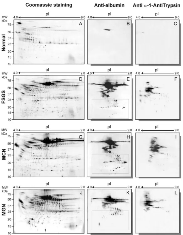
with 2 ml of normal urine (containing 200 g of total pro-tein); a major band of uromodulin (acid protein with 80 kD molecular weight) was evident, together with a few other acid proteins between 50 and 20 kD and a low amount of intact albumin (Figure 1, A and B). Therefore, the constant pattern of normal urine in both children and adults was characterized by the absence of albumin fragments. The com-position of nephrotic urine was remarkably different. Nu-merous fragments of albumin and of ␣1AT in fact were detected in urine of all patients with a few patterns common
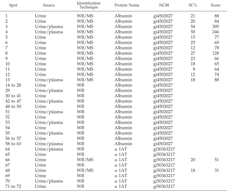
Table 2. Urinary and plasma proteins from patients with nephrotic syndrome were characterized by
two-dimensional electrophoresis-Western blot and by MALDI-TOF
aSpot Source IdentificationTechnique Protein Name NCBI SC% Score
1
Urine
WB/MS
Albumin
gi4502027
21
88
2
Urine
WB/MS
Albumin
gi4502027
20
84
3
Urine/plasma
WB/MS
Albumin
gi4502027
54
305
4
Urine/plasma
WB/MS
Albumin
gi4502027
50
246
5
Urine
WB/MS
Albumin
gi4502027
15
77
6
Urine
WB/MS
Albumin
gi4502027
25
69
7
Urine
WB/MS
Albumin
gi4502027
12
78
8
Urine
WB/MS
Albumin
gi4502027
27
128
9
Urine
WB/MS
Albumin
gi4502027
23
66
10
Urine
WB/MS
Albumin
gi4502027
18
65
11
Urine
WB/MS
Albumin
gi4502027
8
64
12
Urine
WB/MS
Albumin
gi4502027
12
74
13
Urine/plasma
WB/MS
Albumin
gi4502027
18
88
14 to 28
Urine
WB
Albumin
gi4502027
29
Urine/plasma
WB
Albumin
gi4502027
30 to 41
Urine
WB
Albumin
gi4502027
42 to 47
Urine/plasma
WB
Albumin
gi4502027
48 to 50
Urine
WB
Albumin
gi4502027
51
Urine/plasma
WB
Albumin
gi4502027
52
Urine
WB
Albumin
gi4502027
53
Urine/plasma
WB
Albumin
gi4502027
54
Urine
WB
Albumin
gi4502027
55
Urine/plasma
WB
Albumin
gi4502027
56 to 57
Urine
WB
Albumin
gi4502027
58 to 63
Urine/plasma
WB
Albumin
gi4502027
64
Urine/plasma
WB
␣ 1AT
gi50363217
65
Urine
WB
␣ 1AT
gi50363217
66
Urine
WB/MS
␣ 1AT
gi50363217
20
51
67
Urine
WB
␣ 1AT
gi50363217
68
Urine
WB/MS
␣ 1AT
gi50363217
18
31
69
Urine
WB
␣ 1AT
gi50363217
70
Urine/plasma
WB
␣ 1AT
gi50363217
71 to 72
Urine
WB
␣ 1AT
gi50363217
aA total of 72 spots were identified as fragments of albumin and/or of␣1-antitrypsin (␣1AT). Most (n ⫽ 72) were recovered from urine, and only some (n⫽ 21) were also detected in plasma. According to the Proposed Publication Guidelines for the Analysis and Documentation of Peptide and Protein Identifications (Paris consensus), we included in this table the complete figures of merit regarding the mass spectrometry (MS) data. In particular, these include sequence coverage (SC%), indicating the percentage of the identified protein sequence covered by the MS data, and identification score (Score), indicating a probabilistic measure of the goodness of the MS protein identification. All included protein identifications were significant at
P⬍ 0.05. MALDI-TOF, matrix-assisted laser desorption/ionization time of flight; NCBI, National Center for Biotechnology
to different diseases and several differences that are de-scribed separately next.
Albumin
Intact albumin was the major protein in nephrotic urine. In parallel, numerous albumin fragments that covered the entire
range of molecular weight from 64 to 20 kD were detected. An example is given in Figure 1E (Western blot with anti-albumin antibodies) and shows the pattern of a morning fresh urine sample that was obtained from a child with steroid resistance and FSGS; by comparison, a urine sample that was obtained from a child with steroid-sensitive nephrotic syndrome is
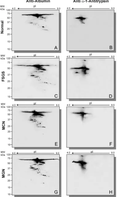
Figure 1. Two-dimensional (2D) electrophoresis and Coomassie staining of a single spot of fresh urine obtained from a normal volunteer
(A) and from patients with nephrotic syndrome caused by different renal pathologies such as FSGS (D), minimal-change nephropathy (MCN; G), and membranous glomerulonephritis (MGN; L). Western blot with anti-albumin and anti–␣1-antitrypsin (anti-␣1AT) polyclonal antibodies of the same urinary sample as above: normal-albumin (B), normal-␣1AT (C), FSGS-albumin (E), FSGS-␣1AT (F), MCN-albumin (H), MCN-␣1AT (I), MGN-albumin (M), and MGN-␣1AT (N). 2D electrophoresis was performed in soft gels according to the technique described by Bruschi et al. (19). In case of normal urine, 2 ml of urine (containing 200g of total protein) was loaded onto the gel. In case of heavy proteinuria, 1l of urine (containing 200 g of protein) was loaded.
shown in Figure 1, G and H. In both cases, the same amount of protein analyzed in normal urine (1l, containing 200 g of protein) was loaded on gels. The presence of albumin frag-ments was confirmed by Western blot analysis of several urine samples from patients both with steroid-resistant FSGS (Fig-ures 1, D through F, and 2, tracks 10 through 16) and with steroid-sensitive MCN (Figures 1, G and H, and 2, tracks 4 through 9 and tracks 17 through 20). Figure 2 shows the pattern of urinary albumin in both reduced (⫹ mercaptoethanol) and nonreduced (⫺ mercaptoethanol) conditions, indicating that some albumin fragments react with each other and form aggre-gates through disulphide bridges that are destroyed by mer-captoethanol. Finally, albumin fragments also were detected with some differences in patients with MGN (Figure 1, L and M). These patients lacked, in fact, the isoforms with the lowest
molecular weight. A comprehensive characterization of the differences between FSGS/MCN and MGN by dual labeling is in progress and will be presented in the near future.
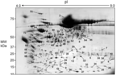
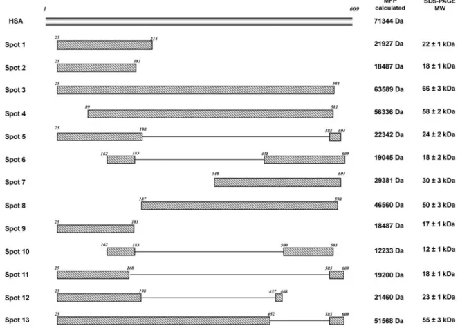
Overall, 63 fragments of albumin were detected clearly in nephrotic urine (Figure 3); they were recovered from the gel and analyzed by MS that unequivocally confirmed their iden-tity. The molecular weight of albumin fragments was calculated both from the human serum albumin (HSA) sequence that was obtained from the mass fingerprint experiments and from the SDS-PAGE. Both molecular weight values showed a good cor-relation, demonstrating that most of the HSA sequence was investigated in the MALDI-TOF-MS experiments. Moreover, few protein spots (Figure 4) showed consistently a peculiar distribution of the fingerprint peptide pattern that contained both the N-terminal region and the C-terminal domain with a complete lack of any MS signals for the internal sequence region. The reconstructed joint domains were calculated from mass fingerprint data that defined the HSA sequence regions that consistently were matched by experimentally measured tryptic peptides. In addition, the molecular weights of albumin spots estimated from 2D PAGE were not compatible with an albumin isoform that spanned the complete continuous se-quence. Such tryptic peptide distribution could not be ex-plained directly by the presence of the 16 disulfide bridges that were reported for the HSA structure given the strong reducing conditions that were used in sample preparation and separa-tion. Therefore, we might speculate on the presence of covalent bound intramolecular cross-links that would be resistant to the strong denaturing conditions that were used in protein separa-tion.
To exclude that the pattern of albumin fragmentation that was observed in nephrotic urine was due, in fact, to proteases in the tubule and/or in urine, we repeated the same analysis with Western blot in the companion plasma, where a large part of fragmentation products were recognized. The results are shown in Figure 5 (a 2D analysis and Western blot of plasma
Figure 2. Gradient electrophoresis in the presence and in the
absence of-mercaptoethanol and Western blot with anti-albu-min antibodies of urine from normal donors (2 ml of urine; tracks 1 through 3, patients with steroid-sensitive MCN (tracks 4 through 9 and tracks 17 through 20), and patients with steroid-resistant FSGS (tracks 10 through 16). In the presence of -mercaptoethanol, several low molecular isoforms of albumin were detected. In its absence, a few low molecular weight isoforms still were present with several dimers of albumin indicating the formation of disulphide bridges. Normal urine did not contain low or high molecular weight isoforms.
Figure 3. 2D electrophoresis of the sample of urine of Figure 1D,
in which all spots that were characterized by mass spectrome-try (MS) and/or Western blot are numbered and described in Table 2.
albumin in one patient per group) and are summarized in Table 2. Twenty-one of the spots that were characterized as albumin/ ␣1AT in urine also were present in plasma even at a lower concentration, suggesting preferential excretion of low molec-ular components. However, a lot of low molecmolec-ular weight isoforms that were observed in urine were not present in plasma (see Table 2), suggesting an in situ fragmentation pro-cess that implies the presence of proteases in urine. In general, the components that were detected only in urine presented a low molecular weight and migrated to an area of gels indicated by a square in gels of Figure 1. On this basis, the presence of urinary proteases was postulated and experiments were planned to demonstrate their existence (see the Urinary Pro-teases Specific for Albumin section).
␣1AT
␣1AT is practically absent in normal urine (Figure 1, A and C). Intact and fragmented␣1AT instead is well evident in urine of all different cohorts of patients (Figure 1, F, I, and N). MS confirmed identity of␣1AT (Table 2). As for albumin also for ␣1AT, experiments with Western blot were carried out in plasma, where the presence of a part of␣1AT fragments could be confirmed (Figure 5). Again, comparison of plasma and
urine␣1AT composition showed that a few isoforms with low molecular weight are produced in situ as an effect of proteases.
Urinary Proteases Specific for Albumin
To explain the formation of albumin fragments in urine, we performed a zymogram with albumin as substrate that indi-cated the presence of specific proteases. Comparison of normal and nephrotic urine demonstrated the presence of two major bands with protease activity that were upregulated in nephrotic patients (Figure 6). This finding indirectly supports the exis-tence of a specific mechanism for in situ digestion of albumin in urine.
Discussion
This study was aimed at defining protein composition of urine and plasma in nephrotic patients and at revaluating relevant structural aspects of major proteins such as albumin. For this purpose, we took advantage of the progress that has been made in the vast area of proteomics. Different clinical and pathologic conditions that are determined by different mecha-nisms such as primary nephrotic syndrome in children (perme-ability factors?) and MGN (immune complex) (23,24) were con-sidered. The following relevant findings reveal unexpected
Figure 4. Schematic representation of urinary albumin peptide composition as assigned by matrix-assisted laser desorption/
ionization time of flight MS to spots that were recovered from 2D gels. A few spots (1, 2, 3, 4, 5, and 9) represented fragments of albumin. Several other spots harbored both the N-terminal region and in the C-terminal domain with a complete lack of any MS signals of the internal sequence region.
elements that suggest pathogenic implications and warrant wide discussion. The first regards the composition of nephrotic urine that practically contains only intact and fragmentation products of albumin and of␣1AT. With the exception of a few low molecular weight urinary isoforms, some albumin and ␣1AT fragments also were detected in plasma, suggesting that fragmentation takes place at this site. Few isoforms instead are produced in urine, and we could demonstrate the presence in urine of specific proteases. A parallel aspect is how fragmen-tation influences renal handling of both proteins, which is the reason that nephrotic urine is enriched in fragments. The final point is about albumin adducts and their significance.
It is relevant that most of the proteins that are detected in
urine of children with nephrotic syndrome correspond to intact albumin and␣1AT or their fragments as it clearly emerges from the comparison of the 2D gels of the same urinary sample stained by Coomassie and Western blot reported in Figure 1. MS confirmed this conclusion.
To our knowledge, the finding of protein fragments in serum and urine of patients with renal diseases is new and suggests different fragmentation mechanisms. The high concentration of albumin fragments in nephrotic urine suggests a preferential urinary excretion and reflects the partial maintenance of size selectivity properties of the kidney. Albumin fragments ac-count, in fact, for 25 to 30% of total albumin in urine of ne-phrotic patients, whereas their proportion in serum does not exceed 5%. Considering that in nephrotic syndrome at least 2 to 3 g of albumin is lost every day in urine and that up to 1 g (30%) is represented by fragments, it can be concluded that almost 30 g of fragments is cleared monthly from plasma, accounting for 30% of the albumin pool in a patient who weighs 30 kg. Our conclusion is that preferential urinary excretion of albumin fragments in nephrotic patients represents an efficient depura-tive mechanism that restores normal albumin composition in plasma, and the same considerations apply to␣1AT. It is critical to this conclusion that also normal plasma contains albumin fragments, which suggests that in nephrotic syndrome the frag-mentation process is amplified. This seems even more interest-ing for␣1AT, which acts as a major circulating defense against exogenous proteolysis enzymes of viral or bacterial derivation and also controls the proteolysis response that is triggered by normal immunologic process. It is a suicide protein in the sense that it attracts proteases at a bait region and is degraded after binding by the proteosome (25,26). To our knowledge, nothing like this has been described before in human pathology, and
Figure 5. 2D electrophoresis and Western blot with
anti-albu-min (A, C, E, and G) and anti-␣1AT (B, D, F, and H) polyclonal antibodies of plasma from normal volunteers (A and B) and from patients with nephrotic syndrome caused by different renal pathologies such as FSGS (C and D), MCN (E and F), and MGN (G and H).
Figure 6. Zymograms with albumin as substrate of normal urine
(tracks 1 and 2) and urine from patients with nephrotic syn-drome caused by various types of renal diseases: Steroid-resis-tant FSGS (tracks 3 through 8), steroid-sensitive MCN (tracks 9 through 13), and MGN (tracks 14 through 16). Two major bands common to both normal and nephrotic urine are present at different concentrations. Arrow indicates a band with protease activity that is present only in urine of patients with FSGS. ST indicates molecular weight standards (Bio-Rad).
experimental and in vitro models are required to investigate this aspect further. It seems relevant that inactivation of␣1AT by leukocytes has been reported extensively in the literature, this mechanism involving the production of free radicals from ac-tivated neutrophils and activation of specific proteases (27–29). This observation reinforces the hypothesis of a role of free radicals in nephrotic syndrome that has been proposed in the past but never received experimental support.
A final but not secondary point is the description of albumin adducts. They were characterized by MS as peptides that har-bor both the COOH and NH2 terminal parts of the protein. Such evidence cannot be explained simply by the presence of disulfide bridges or the adventitious lack of MS signals given the 2D electrophoresis separation conditions and the calculated molecular weight. These data might be suggestive of the pres-ence of a covalent chemical adduct that links the two otherwise separated parts of albumin (30). Such cross-links have been described as end products of Amadori compounds that are generated from the glycation of lysine residues. Nevertheless, the large number of chemical structures that are formed during nonezymatic glycation does not allow us to draw any conclu-sion on the new molecules described here (31). The presence of albumin adducts in FSGS is new and obviously will receive further attention in the near future for a definite chemical characterization.
Conclusion
Albumin and␣1AT fragments represent the major proteins in urine of patients with nephrotic syndrome, where at least 50 isoforms of these proteins were characterized by proteomics analysis. Most of these isoforms derive from plasma, but a few were formed in situ by specific proteolysis. Albumin adducts that harbor both the COOH and NH2 terminal parts of the protein also were detected, suggesting the formation of cova-lent chemical adducts. These new findings reveal unexpected structural and functional aspects of proteinuria that may play a key role in pathogenesis.
Acknowledgments
This work was done with the financial support of the Italian Ministry of Health and a grant from the Renal Child Foundation. We also acknowledge Fondazione Mara Wilma e Bianca Querci for the financial support to the project “Nuove evoluzioni sulla multifattorialita` della sindrome nefrosica.”
Data were critically discussed with Prof. R. Gusmano, and we ac-knowledge her role. The manuscript was revised by Ms. Capurro.
References
1. Bonilla-Felix M, Parra C, Dajani T, Ferris M, Swinford RD, Portman RJ, Verani R: Changing patterns in the histopa-thology of idiopathic nephrotic syndrome in children.
Kid-ney Int 55: 1885–1890, 1999
2. Koop K, Eikmans M, Baelde HJ, Kawachi H, De Heer E, Paul LC, Bruijn JA: Expression of podocyte-associated mol-ecules in acquired human kidney diseases. J Am Soc
Neph-rol 14: 2063–2071, 2003
3. Schmid H, Henger A, Cohen CD, Frach K, Grone HJ,
Schlondorff D, Kretzler M: Gene expression profiles of podocyte-associated molecules as diagnostic markers in acquired proteinuric diseases. J Am Soc Nephrol 14: 2958 – 2966, 2003
4. Benigni A, Gagliardini E, Tomasoni S, Abbate M, Rugge-nenti P, Kalluri R, Remuzzi G: Selective impairment of gene expression and assembly of nephrin in human dia-betic nephropathy. Kidney Int 65: 2193–2200, 2004
5. Haubitz M, Wittke S, Weissinger EM, Walden M, Rupprecht HD, Floege J, Haller H, Mischak H: Urine pro-tein patterns can serve as diagnostic tools in patients with IgA nephropathy. Kidney Int 67: 2313–2320, 2005
6. Branten AJ, du Buf-Vereijken PW, Klasen IS, Bosch FH, Feith GW, Hollander DA, Wetzels JF: Urinary excretion of beta2-microglobulin and IgG predict prognosis in idio-pathic membranous nephropathy: A validation study.
J Am Soc Nephrol 16: 169 –174, 2005
7. Rennke HG, Venkatachalam MA: Glomerular permeability of macromolecules. Effect of molecular configuration on the fractional clearance of uncharged dextran and neutral horseradish peroxidase in the rat. J Clin Invest 63: 713–717, 1979
8. Ghiggeri GM, Ginevri F, Candiano G, Oleggini R, Perfumo F, Queirolo C, Gusmano R: Characterization of cationic albumin in minimal change nephropathy. Kidney Int 32: 547–553, 1987
9. Ghiggeri GM, Candiano G, Ginevri F, Gusmano R, Ciardi MR, Perfumo F, Delfino G, Cuniberti C, Queirolo C: Renal selectivity properties towards endogenous albumin in minimal change nephropathy. Kidney Int 32: 69 –77, 1987 10. Ginevri F, Ghiggeri GM, Candiano G, Oleggini R, Bertelli
R, Perfumo F, Queirolo C, Gusmano R: Endogenous albu-min as a marker of renal selectivity in steroid-unresponsive nephrotic syndrome. Nephron 52: 133–138, 1989
11. Boute N, Gribouval O, Roselli S, Benessy F, Lee H, Fuchs-huber A, Dahan K, Gubler MC, Niaudet P, Antignac C: NPHS2, encoding the glomerular protein podocin, is mu-tated in autosomal recessive steroid-resistant nephrotic syndrome. Nat Genet 24: 349 –354, 2000
12. Caridi G, Bertelli R, Di Duca M, Dagnino M, Emma F, Onetti Muda A, Scolari F, Miglietti N, Mazzucco G, Murer L, Carrea A, Massella L, Rizzoni G, Perfumo F, Ghiggeri GM: Broadening the spectrum of diseases related to podo-cin mutations. J Am Soc Nephrol 14: 1278 –1286, 2003 13. Shih NY, Li J, Karpitskii V, Nguyen A, Dustin ML,
Kana-gawa O, Miner JH, Shaw AS: Congenital nephrotic syn-drome in mice lacking CD2-associated protein. Science 286: 312–315, 1999
14. Kaplan JM, Kim SH, North KN, Rennke H, Correia LA, Tong HQ, Mathis BJ, Rodriguez-Perez JC, Allen PG, Beggs AH, Pollak MR: Mutations in ACTN4, encoding alpha-actinin-4, cause familial focal segmental glomerulosclero-sis. Nat Genet 24: 251–256, 2000
15. ISKDC: Prospective, controlled trial of cyclophosphamide therapy in children with nephrotic syndrome. Report of the International study of Kidney Disease in Children.
Lancet 2: 423– 427, 1974
16. ISKDC: Primary nephrotic syndrome in children: Clinical significance of histopathologic variants of minimal change and of diffuse mesangial hypercellularity. A Report of the International Study of Kidney Disease in Children. Kidney
17. Ponticelli C, Rizzoni G, Edefonti A, Altieri P, Rivolta E, Rinaldi S, Ghio L, Lusvarghi E, Gusmano R, Locatelli F, et
al.: A randomized trial of cyclosporine in steroid-resistant
idiopathic nephrotic syndrome. Kidney Int 43: 1377–1384, 1993
18. Ponticelli C, Zucchelli P, Passerini P, Cesana B: Methyl-prednisolone plus chlorambucil as compared with meth-ylprednisolone alone for the treatment of idiopathic mem-branous nephropathy. The Italian Idiopathic Memmem-branous Nephropathy Treatment Study Group. N Engl J Med 327: 599 – 603, 1992
19. Bruschi M, Musante L, Candiano G, Ghiggeri GM, Herbert B, Antonucci F, Righetti PG: Soft immobilized pH gradient gels in proteome analysis: A follow-up. Proteomics 3: 821– 825, 2003
20. Candiano G, Porotto M, Lanciotti M, Ghiggeri GM: Nega-tive staining of proteins in polyacrylamide gels with methyl trichloroacetate. Anal Biochem 243: 245–248, 1996 21. Candiano G, Bruschi M, Musante L, Santucci L, Ghiggeri
GM, Carnemolla B, Orecchia P, Zardi L, Righetti PG: Blue silver: A very sensitive colloidal Coomassie G-250 staining for proteome analysis. Electrophoresis 25: 1327–1333, 2004 22. Laemmli UK: Cleavage of structural proteins during the
assembly of the head of bacteriophage T4. Nature 227: 680 – 685, 1970
23. Vincenti F, Ghiggeri GM: New insights into the pathogen-esis and the therapy of recurrent focal glomerulosclerosis.
Am J Transplant 5: 1179 –1185, 2005
24. Salant DJ, Darby C, Couser WG: Experimental membra-nous glomerulonephritis in rats. Quantitative studies of
glomerular immune deposit formation in isolated glomer-uli and whole animals. J Clin Invest 66: 71– 81, 1980 25. Miyamoto Y, Akaike T, Maeda H: S-nitrosylated human
alpha(1)-protease inhibitor. Biochim Biophys Acta 1477: 90 – 97, 2000
26. Cho Y, Jang YY, Han ES, Lee CS: The inhibitory effect of ambroxol on hypochlorous acid-induced tissue damage and respiratory burst of phagocytic cells. Eur J Pharmacol 383: 83–91, 1999
27. Dean RT, Nick HP, Schnebli HP: Free radicals inactivate human neutrophil elastase and its inhibitors with compa-rable efficiency. Biochem Biophys Res Commun 159: 821– 827, 1989
28. Wallaert B, Aerts C, Gressier B, Gosset P, Voisin C: Oxida-tive inactivation of alpha 1-proteinase inhibitor by alveolar epithelial type II cells. J Appl Physiol 75: 2376 –2382, 1993 29. Ottonello L, Dapino P, Scirocco M, Dallegri F, Sacchetti C:
Proteolytic inactivation of alpha-1-antitrypsin by human neutrophils: Involvement of multiple and interlinked cell responses to phagocytosable targets. Eur J Clin Invest 24: 42– 49, 1994
30. Ahmed N, Dobler D, Dean M, Thornalley PJ: Peptide map-ping identifies hotspot site of modification in human se-rum albumin by methylglyoxal involved in ligand binding and esterase activity. J Biol Chem 280: 5724 –5732, 2005 31. Ahmed N, Thornalley PJ: Quantitative screening of protein
biomarkers of early glycation, advanced glycation, oxida-tion and nitrosaoxida-tion in cellular and extracellular proteins by tandem mass spectrometry multiple reaction monitor-ing. Biochem Soc Trans 31: 1417–1422, 2003