Research Article
The ‘‘Gender Factor’’ in Wearing-Off among Patients with
Parkinson’s Disease: A Post Hoc Analysis of DEEP Study
Delia Colombo,
1Giovanni Abbruzzese,
2Angelo Antonini,
3Paolo Barone,
4Gilberto Bellia,
1Flavia Franconi,
5Lucia Simoni,
6Mahmood Attar,
1Emanuela Zagni,
1Shalom Haggiag,
7and Fabrizio Stocchi
81Novartis Farma S.p.A., Origgio, Varese 21040, Italy
2Department of Neurosciences, University of Genoa, Genoa 16132, Italy 3Department of Parkinson’s Disease, IRCCS San Camillo, Venice 30126, Italy 4Scuola Medica Salernitana, Universit`a degli Studi di Salerno, Salerno 84100, Italy 5Department of Biomedical Science, University of Sassari, Sassari 07100, Italy 6MediData srl, Modena 41123, Italy
7Department of Neurology, San Camillo-Forlanini Hospital, Rome 00151, Italy
8Department of Neurology, Institute of Research and Medical Care, IRCCS San Raffaele, Rome 00163, Italy
Correspondence should be addressed to Gilberto Bellia; [email protected]
Received 11 October 2014; Revised 13 December 2014; Accepted 21 December 2014
Academic Editor: Lawrence W. Svenson
Copyright © 2015 Delia Colombo et al. This is an open access article distributed under the Creative Commons Attribution License, which permits unrestricted use, distribution, and reproduction in any medium, provided the original work is properly cited.
Background. The early detection of wearing-off in Parkinson disease (DEEP) observational study demonstrated that women with
Parkinson’s disease (PD) carry an increased risk (80.1%) for wearing-off (WO). This post hoc analysis of DEEP study evaluates gender differences on WO and associated phenomena. Methods. Patients on dopaminergic treatment for≥1 year were included in this multicenter observational cross-sectional study. In a single visit, WO was diagnosed based on neurologist assessment as well as the use of the 19-item wearing-off questionnaire (WOQ-19); WO was defined for scores≥2. Post hoc analyses were conducted to investigate gender difference for demographic and clinical features with respect to WO. Results. Of 617 patients enrolled, 236 were women and 381 were men. Prevalence of WO was higher among women, according to both neurologists’ judgment (61.9% versus 53.8%,𝑃 = 0.045) and the WOQ-19 analysis (72.5% versus 64.0%, 𝑃 = 0.034). In patients with WO (WOQ-19), women experienced ≥1 motor symptom in 72.5% versus 64.0% in men and ≥1 nonmotor symptom in 44.5% versus 36.7%, in men. Conclusions. Our results suggest WO as more common among women, for both motor and nonmotor symptoms. Prospective studies are warranted to investigate this potential gender-effect.
1. Background
Parkinson’s disease (PD) is one of the most common age-related progressive neurodegenerative disorders, with no identifiable cause. PD is slightly more common in men than in women in most studies, usually ranging from a 1.2 : 1 ratio up to a 1.5 : 1 ratio [1], and men seem to be at higher risk for PD [2–4]. The reasons for the increased risk in men are not known; probably “male lifestyle” could account for some of the excess incidence in men [5]. Alternatively, there is increasing evidence from in vitro as well as clinical studies in
humans that estrogen may be neuroprotective [6]. Sex-related differences have been reported in the onset of symptoms and type of motor symptoms as well as in medication use [7, 8]. Notably, normal human basal ganglia, specifically within the dopamine system, are sexually dimorphic and that may influence the onset and progression of PD [8].
Levodopa is the most efficacious treatment in the man-agement of PD. Unfortunately, chronic use of traditional levodopa/dopa decarboxylase inhibitor formulations is asso-ciated with the development of motor complications, such as wearing-off (WO) and dyskinesia that occur in the majority
Volume 2015, Article ID 787451, 10 pages http://dx.doi.org/10.1155/2015/787451
of PD patients. The WO effect, or end-of-dose failure, refers to a decrease in the length of time that each dose of levodopa controls symptoms. “Off ” states that result in motor and nonmotor symptoms, freezing of gait (FOG), and falling are disabling for many patients. Considered to be the major source of disability in PD patients, recognition of these complications is critical in order to develop different strategies designed not only to treat these problems when they develop, but also to prevent troublesome complications associated with potential risk factors.
We previously conducted an observational, cross-sectional, multicenter study called Early DEtection of
wEaring off in Parkinson disease (DEEP Study) [9, 10]. The primary goal was to look at the frequency of WO phenomena among a wide population of Italian patients with PD and secondly to assess associated phenomena, such as FOG and nonmotor symptoms, as well as assessing the impact on patient’s QoL. In our sample WO occurred since the early years of the disease; furthermore younger age, unified Parkinson’s disease rating scale (UPDRS) part II score, duration of anti-Parkinson (APD) treatment, and female gender were found significantly associated with WO. Our data showed women having an 80.1% higher risk of experiencing WO than men [9]. This exploratory study aimed to further examine what role the “gender factor” in WO would play, that is, to characterize disparities between women and men in frequency and features of WO symptoms. For this purpose, we conducted a post hoc analysis of the DEEP study database. This study is also part of the gender-medicine project (METAGEM), being carried out with the aim to describe clinical outcomes and therapeutic approach by gender, through the analysis of observational studies conducted in Italy, among different therapeutic areas [11].
2. Methods
Patients prospectively recruited were men and women aged 18 years or older, with PD (Hoehn and Yahr Stages 1– 5), nondemented, under levodopa (LD), and/or dopamine
agonists (DAs) therapy for≥1 year before the study screening.
The full DEEP study design, including complete inclu-sion/exclusion criteria, has been published [9]. The study was conducted in accordance with Good Clinical Practice and the Declaration of Helsinki. The study protocol and amendments were approved by each local Ethics Committees or Institutional Review Boards of all 37 participating centers (both academic and hospital based) across Italy (Additional file 1). All patients provided written, informed consent before study participation.
During a single visit, neurologists experienced in move-ment disorders and previously subjected to targeted training acquired standard demographic and detailed clinical infor-mation on all participants who met the criteria, via a struc-tured interview and by examination. Therapy was expressed in terms of Levodopa equivalent daily dose (LEDD) [12]. Subjects were assessed with the UPDRS and the Hoehn and Yahr scale (H&Y). The diagnosis of WO was made on both
the neurologist evaluation and the basis of the patient self-assessment using the Italian version of the 19-item
Wearing-Off Questionnaire (WOQ-19) [10]. The WOQ-19 consists of 9 items assessing fluctuations of motor symptoms, including tremor, difficulty in speech, weakness, problems with bal-ance, slowness, reduced dexterity, general stiffness, muscle cramps, and difficulty getting out of the chair and 10 items assessing fluctuations of nonmotor symptoms, including anxiety, sweating, mood changes, numbness, panic attacks, cloudy mind/dullness of thinking, abdominal discomfort, experience hot and cold, pain, and aching [10]. For each item, patients were asked to tick whether symptoms were present and whether they improved after the following dose of
anti-Parkinson treatment: a cutoff of≥2 improved symptoms has
been previously established to make diagnosis of WO [10].
3. Statistical Analysis
Descriptive analyses were mean and standard deviation (SD) or median and interquartile range (IQR). Only the fully completed scales were considered evaluable for the statistical analysis. Gender differences analyses were performed on all patients who responded to inclusion-exclusion criteria. Dif-ferences in demographics/baseline characteristics between patients with WO and patients with no WO (as assessed using WOQ-19 and the neurologist assessment) were estimated
using𝑡-test for continuous data and the Chi-square test for
categorical data. As post hoc analyses, all P values presented are exploratory. Patients with missing data in selected param-eters were not evaluated for those paramparam-eters. All analyses were performed with SAS v. 9.2 and Enterprise Guide 4.3.
4. Results
Of the 634 patients screened, 617 (97.3%) met the inclusion criteria: 236 (38.2%) were women and 381 (61.8%) were men (Table 1). The excluded patients did not differ significantly on demographic or clinical parameters with respect to the study population [9]. Baseline demographic data are
pro-vided in Tables1and2. Evaluation of gender differences in
the study sample indicated no difference in age, history of concomitant diseases, caregiver support, body mass index,
or coffee consumption. As shown in Table 1, women were
less likely to be married/cohabiting (𝑃 = 0.0001) and have less education (𝑃 = 0.0002). In almost the third of cases women were housewives and were less likely to be employed (𝑃 = 0.0046). Men were more likely to be past (𝑃 < 0.0001) or current smokers (𝑃 = 0.0059) and to be frequent alcohol consumers (𝑃 < 0.0001), although more often engaged in physical activity on a regular basis (𝑃 = 0.0005).
A shown inTable 2, disease duration, H&Y staging, and
UPDRS and MMSE scores were statistically indistinguishable between both groups. Postural instability/gait difficulties (PIGD) phenotype was found in 53.0% of the whole sample,
although more prevalent in women (57.0% versus 50.5%,𝑃 =
0.005), while the tremor dominant (TD) phenotype was more
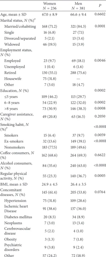
Table 1: Demographic and lifestyle data of DEEP study population: women versus men.
Women 𝑁 = 236 𝑁 = 381Men 𝑃 Age, mean± SD 67.0± 8.9 66.6± 9.4 0.6602 Marital status,𝑁 (%)# Married/cohabiting 168 (71.2) 321 (84.3) 0.0001 Single 16 (6.8) 27 (7.1) Divorced/separated 5 (2.1) 13 (3.4) Widowed 46 (19.5) 15 (3.9) Employment status, 𝑁 (%) Employed 23 (9.7) 69 (18.1) 0.0046 Unemployed 1 (0.4) 6 (1.6) Retired 130 (55.1) 288 (75.6) Housewife 75 (31.8) — Other 7 (3.0) 18 (4.7) Education,𝑁 (%) 0.0002 ≤5 years 109 (46.2) 113 (29.7) 6–8 years 54 (22.9) 122 (32.0) 0.0002 >8 years 73 (30.9) 146 (38.3) 0.0008 Caregiver assistance, 𝑁 (%) 49 (20.8) 63 (16.5) 0.2050 Smoking habit,𝑁 (%)# <0.0001 Smokers 15 (6.4) 37 (9.7) 0.0059 Ex-smokers 32 (13.6) 149 (39.1) <0.0001 Nonsmokers 183 (77.5) 189 (49.6) Coffee consumers,𝑁 (%) 162 (68.6) 264 (69.3) 0.6622 Alcohol consumers, 𝑁 (%) 84 (35.6) 240 (63.0) <0.0001 Regular physical activity,𝑁 (%) 55 (23.3) 140 (36.7) 0.0005 BMI, mean± SD 24.9± 4.5 26.4± 3.5 Concomitant diseases,𝑁 (%) 145 (61.4) 205 (53.8) 0.0764 Hypertension 75 (31.8) 109 (28.6) Ischemic heart Disease 91 (38.6) 137 (36.0) Diabetes mellitus 20 (8.5) 34 (8.9) Neoplasms 7 (3.0) 13 (3.4) Cerebrovascular disease 5 (2.1) 4 (1.0) Obesity 3 (1.3) 7 (1.8) Psychiatric disorders 9 (3.8) 9 (2.4) Other 57 (24.2) 72 (18.9)
#Missing data for<10 patients/variable.
Prevalence of WO was higher among women, accord-ing to both neurologists’ judgment (61.9% versus 53.8%, 𝑃 = 0.0495) and WOQ-19 analysis (72.5% versus 64.0%,
81.3 76.6 56.1 72.5 79.8 64.3 47.9 64 0 10 20 30 40 50 60 70 80 90 Total
Rates of WO diagnosis (WOQ-19)
Dis eas e d ura tio n Men Women >10 y 5–10 y <5 y
Figure 1: Rates of WO diagnosis according to WOQ-19 stratified by disease duration: women versus men.
𝑃 = 0.034) (Figure 1). Taking into account the symptoms reported as usually improving after the following dose of anti-Parkinson agents (APD), otherwise defined as WO
symptoms, 3.3± 2.5 versus 3.0 ± 2.6 were motor symptoms
(𝑃 = 0.222) and 1.2 ± 1.8 versus 0.9 ± 1.5 were nonmotor symptoms (𝑃 = 0.0125), respectively, in women and men. The frequency of symptoms reported by patients through the WOQ-19, stratified by gender and disease duration, is listed
inTable 3. Among patients with a diagnosis of WO according
to WOQ-19, women experienced≥1 motor symptom in 72.5%
versus 64.0% in men and≥1 nonmotor symptom in 44.5%
versus 36.7%, respectively, in men (Figure 2).
With regard to APD therapy, no differences were observed in the use of the different classes of APDs (𝑃 = 0.4457) or for LEDD values (844.2 ± 679.0 versus 874.4 ±
749.1,𝑃 = 0.622).
Finally, women reported significantly higher PDQ-8
scores than men (31.3± 18.4 versus 27.7 ± 19.1, 𝑃 = 0.023).
5. Discussion
WO is an important feature of PD, often marking the end of the “honeymoon period.” The WO manifestations can be extremely heterogeneous from subject to subject, and early recognition allows timely optimization of treatment that may impact patient care and long-term clinical outcomes. The reasons underlying these complications are not fully under-stood and well-recognized risk factors for the development of WO and dyskinesias include young age at onset, low body weight, severity of disease higher levodopa dose, association of levodopa with entacapone, once daily intake of levodopa, duration of levodopa therapy, more severe UPDRS Part II,
and female gender [9, 13–15]. This paper was dedicated to
assess gender differences in WO and is based on the post hoc analysis of the DEEP study, a large epidemiological survey that enrolled more than 600 patients with PD across Italy. This study specifically investigated for WO and shows that women are more likely to suffer from WO symptoms
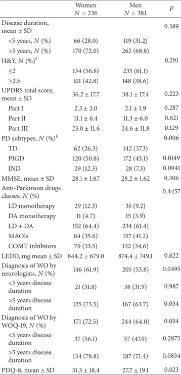
Table 2: Gender differences on clinical aspects of DEEP population: women versus men.
Women 𝑁 = 236 𝑁 = 381Men 𝑃 Disease duration, mean± SD 0.389 <5 years, 𝑁 (%) 66 (28.0) 119 (31.2) >5 years, 𝑁 (%) 170 (72.0) 262 (68.8) H&Y,𝑁 (%)# 0.291 ≤2 134 (56.8) 233 (61.1) ≥2.5 101 (42.8) 148 (38.6)
UPDRS total score,
mean± SD 36.2± 17.7 38.1± 17.4 0.223 Part I 2.3± 2.0 2.1± 1.9 0.287 Part II 11.1± 6.4 11.3± 6.0 0.621 Part III 23.0± 11.6 24.6± 11.8 0.129 PD subtypes,𝑁 (%)§ 0.006 TD 62 (26.3) 142 (37.3) PIGD 120 (50.8) 172 (45.1) 0.0149 IND 29 (12.3) 28 (7.3) 0.0041 MMSE, mean± SD 28.1± 1.67 28.2± 1.62 0.306 Anti-Parkinson drugs classes,𝑁 (%) 0.4457 LD monotherapy 29 (12.3) 35 (9.2) DA monotherapy 11 (4.7) 15 (3.9) LD + DA 152 (64.4) 234 (61.4) MAOIs 84 (35.6) 157 (41.2) COMT inhibitors 79 (33.5) 132 (34.6) LEDD, mg mean± SD 844.2 ± 679.0 874.4± 749.1 0.622 Diagnosis of WO by neurologists,𝑁 (%) 146 (61.9) 205 (53.8) 0.0495 <5 years disease duration 21 (31.8) 38 (31.9) 0.987 >5 years disease duration 125 (73.5) 167 (63.7) 0.034 Diagnosis of WO by WOQ-19,𝑁 (%) 171 (72.5) 244 (64.0) 0.034 <5 years disease duration 37 (56.1) 57 (47.9) 0.2875 >5 years disease duration 134 (78.8) 187 (71.4) 0.0834 PDQ-8, mean± SD 31.3± 18.4 27.7± 19.1 0.023 SD: standard deviation; H&Y: Hoehn and Yahr staging; UPDRS: unified Parkinson’s disease rating scale; PD: Parkinson’s disease; TD: tremor dom-inant; PIGD: postural instability and gait difficulties; IND: intermediate; MMSE: mini-mental state examination; LD: levodopa; DA: dopamine-agonist; MAOIs: monoamine oxidase B inhibitors; COMT: catechol-O-methyltransferase; LEDD: levodopa equivalent daily dose; WO: wearing-off; WOQ-19: 19-item wearing-off questionnaire; PDQ-8: 8-item Parkinson’s disease questionnaire.
§PD subtypes could not be calculated for 39 males and 25 females. Paired
post hoc Chi-squares take TD as reference.
#
Missing data for<10 patients/variable.
as compared to men and that female gender confers an increased risk for WO equal to 80.1% [9]. This finding was
66.7% 85.9% 80.5% 34.8% 49.4% 45.3% 55.5% 79.4% 71.9% 23.5% 44.7% 38.1% 0 20 40 60 80 100 Total Total R at e o f W O sym p to m s Disease duration Women Men <5 y >5 y <5 y >5 y
Nonmotor symptoms Motor symptoms
Figure 2: Rates of motor and nonmotor WO symptoms: women versus men.
confirmed both by the neurologist assessment (61.9% versus
53.8%,𝑃 = 0.049) and by patients themselves thorough the
WOQ-19 (72.5% versus 64.0%,𝑃 = 0.034). Among patients
with WO (WOQ-19), women more frequently complained
of≥1 motor (72.5% versus 64.0%) and ≥1 nonmotor (44.5%
versus 36.7%) symptoms of WO. Our findings have been supported in a previous clinic-based sample of patients with PD, where WO symptoms have been shown to occur more
frequently in women (46% versus 29%, 𝑃 = 0.02) [16].
Furthermore, durations to develop WO and dyskinesia were shown to be shorter in women compared to men, as well as disease progression being slightly faster for women [17]. A similar “gender effect” has already been shown for the occurrence of levodopa-induced dyskinesias, as it has been claimed that dyskinesias occur more frequently in women
if disease duration is >5 years [18, 19] and with a shorter
time latency than men [20]. Otherwise, in a large British community-based study, men and women did not differ in the occurrence of motor fluctuations or dyskinesias [13]. In a recent post hoc analysis of a large prospective trial, female gender was identified as a specific predicting factor for dyskinesias and WO as well, and the authors hypothesized that it would reflect increased levodopa concentrations in women because of lower body weight [15]. This interesting hypothesis, in addition to providing an explanation of the association with female gender, would place the emphasis on a parameter, such as body weight, which in clinical practice is generally overlooked for therapy decisions. Data available from our observational study do not allow analyzing this aspect, as patients were undergoing very heterogeneous treatments and were poorly comparable to one another.
In DEEP population women and men were substantially homogeneous for age, disease duration, H&Y staging, and neurological disability (UPDRS; MMSE); otherwise smoking habits, alcohol consumption, and physical activity differed, and this reflects what is observed in the general population [21]. To our knowledge no data indicate a direct association of these social factors with WO; nevertheless, it is plausible that they might influence PD treatment and vice versa, and we cannot exclude possible association with the development of
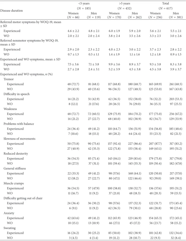
Table 3: Frequency of experienced symptoms and WO symptoms, as reported by DEEP population, through the WOQ-19, stratified by disease duration: women versus men.
Disease duration
<5 years >5 years Total
(𝑁 = 185) (𝑁 = 432) (𝑁 = 617)
Women
(𝑁 = 66) (𝑁 = 119)Men (𝑁 = 170)Women (𝑁 = 262)Men (𝑁 = 236)Women (𝑁 = 381)Men Referred motor symptoms by WOQ-19, mean
± SD
Experienced 4.6± 2.2 4.8± 2.1 6.0± 1.9 5.9± 2.0 5.6± 2.1 5.5± 2.1
WO 2.0± 2.1 2.0± 2.4 3.8± 2.4 3.5± 2.6 3.3± 2.5 3.0± 2.6
Referred nonmotor symptoms by WOQ-19, mean± SD
Experienced 2.9± 2.0 2.3± 2.2 4.0± 2.3 3.0± 2.2 3.7± 2.3 2.8± 2.2
WO 0.7± 1.3 0.5± 1.1 1.4± 1.9 1.1± 1.6 1.2± 1.8 0.9± 1.5
Experienced and WO symptoms, mean± SD
Experienced 7.5± 3.6 7.1± 3.8 9.9± 3.6 8.9± 3.7 9.3± 3.8 8.3± 3.8
WO 2.7± 2.8 2.4± 3.1 5.2± 3.9 4.5± 3.8 4.5± 3.8 3.9± 3.7
Experienced and WO symptoms,𝑛 (%) Tremor Experienced 48 (72.7) 81 (68.1) 117 (68.8) 180 (68.7) 165 (69.9) 261 (68.5) WO 29 (43.9) 40 (33.6) 96 (56.5) 127 (48.5) 125 (53.0) 167 (43.8) Difficulty in speech Experienced 14 (21.2) 51 (42.9) 62 (36.5) 152 (58.0) 76 (32.2) 203 (53.3) WO 8 (12.1) 21 (17.6) 28 (16.5) 76 (29.0) 36 (15.3) 97 (25.5) Weakness Experienced 48 (72.7) 72 (60.5) 129 (75.9) 184 (70.2) 177 (75.0) 256 (67.2) WO 14 (21.2) 27 (22.7) 68 (40.0) 102 (38.9) 82 (34.7) 129 (33.9)
Problems with balance
Experienced 24 (36.4) 49 (41.2) 110 (64.7) 136 (51.9) 134 (56.8) 185 (48.6) WO 7 (10.6) 18 (15.1) 48 (28.2) 64 (24.4) 55 (23.3) 82 (21.5) Slowness of movements Experienced 50 (75.8) 90 (75.6) 157 (92.4) 227 (86.6) 207 (87.7) 317 (83.2) WO 27 (40.9) 42 (35.3) 122 (71.8) 153 (58.4) 149 (63.1) 195 (51.2) Reduced dexterity Experienced 36 (54.5) 85 (71.4) 143 (84.1) 219 (83.6) 179 (75.8) 317 (79.8) WO 18 (27.3) 37 (31.1) 101 (59.4) 145 (55.3) 119 (50.4) 182 (47.8) General stiffness Experienced 22 (33.3) 49 (41.2) 98 (57.6) 168 (64.1) 120 (50.8) 217 (57.0) WO 12 (18.2) 27 (22.7) 80 (47.1) 122 (46.6) 92 (39.0) 149 (39.1) Muscle cramps Experienced 36 (54.5) 57 (47.9) 100 (58.8) 138 (52.7) 136 (57.6) 195 (51.2) WO 11 (16.7) 11 (9.2) 37 (21.8) 48 (18.3) 48 (20.3) 59 (15.5)
Difficulty getting out of chair
Experienced 24 (36.4) 36 (30.2) 98 (57.6) 137 (52.3) 122 (51.7) 173 (45.4) WO 6 (9.1) 11 (9.2) 62 (36.5) 79 (30.1) 68 (28.8) 90 (23.6) Anxiety Experienced 42 (63.6) 49 (41.2) 112 (65.9) 123 (46.9) 154 (65.3) 172 (45.1) WO 10 (15.1) 13 (10.9) 46 (27.1) 45 (17.2) 56 (23.7) 58 (15.2) Sweating Experienced 16 (24.2) 30 (25.2) 85 (50.0) 102 (38.9) 101 (42.8) 132 (34.6) WO 3 (4.5) 4 (3.4) 19 (11.2) 28 (10.7) 22 (9.3) 32 (8.4)
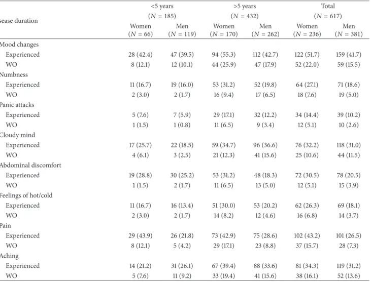
Table 3: Continued.
Disease duration
<5 years >5 years Total
(𝑁 = 185) (𝑁 = 432) (𝑁 = 617)
Women
(𝑁 = 66) (𝑁 = 119)Men (𝑁 = 170)Women (𝑁 = 262)Men (𝑁 = 236)Women (𝑁 = 381)Men Mood changes Experienced 28 (42.4) 47 (39.5) 94 (55.3) 112 (42.7) 122 (51.7) 159 (41.7) WO 8 (12.1) 12 (10.1) 44 (25.9) 47 (17.9) 52 (22.0) 59 (15.5) Numbness Experienced 11 (16.7) 19 (16.0) 53 (31.2) 52 (19.8) 64 (27.1) 71 (18.6) WO 2 (3.0) 2 (1.7) 16 (9.4) 17 (6.5) 18 (7.6) 19 (5.0) Panic attacks Experienced 5 (7.6) 7 (5.9) 29 (17.1) 32 (12.2) 34 (14.4) 39 (10.2) WO 1 (1.5) 1 (0.8) 11 (6.5) 9 (3.4) 12 (5.1) 10 (2.6) Cloudy mind Experienced 17 (25.7) 22 (18.5) 59 (34.7) 96 (36.6) 76 (32.2) 118 (31.0) WO 4 (6.1) 3 (2.5) 21 (12.3) 41 (15.6) 25 (10.6) 44 (11.5) Abdominal discomfort Experienced 19 (28.8) 30 (25.2) 53 (31.2) 48 (18.3) 72 (30.5) 78 (20.5) WO 1 (1.5) 2 (1.7) 11 (6.5) 13 (5.0) 12 (5.1) 15 (3.9) Feelings of hot/cold Experienced 11 (16.7) 16 (13.4) 51 (30.0) 53 (20.2) 62 (26.3) 69 (18.1) WO 2 (3.0) 2 (1.7) 14 (8.2) 12 (4.6) 16 (6.8) 14 (3.7) Pain Experienced 29 (43.9) 26 (21.8) 73 (42.9) 75 (28.6) 102 (43.2) 101 (26.5) WO 8 (12.1) 5 (4.2) 29 (17.1) 23 (8.8) 37 (15.7) 28 (7.3) Aching Experienced 14 (21.2) 31 (26.1) 67 (39.4) 88 (33.6) 81 (34.3) 119 (31.2) WO 5 (7.6) 11 (9.2) 33 (19.4) 41 (15.6) 38 (16.1) 52 (13.6)
WO and contribution for the differences between women and men. Large cohort studies would address these hypotheses.
DEEP women reported poorer QoL than men (𝑃 = 0.023), probably due to WO, as we have previously reported
that the number of motor symptoms (0.34008;𝑃 < 0.0001)
and nonmotor symptoms of WO (0.33595; 𝑃 < 0.0001)
correlates with PDQ-8 score and that by linear regression analysis the presence of each additional WO symptom, as identified by WOQ-19, corresponds to an increase of 1.15 points of the PDQ-8 score (𝑃 < 0.0001) [9].
According to treatment guidelines in PD, the symp-tomatic control of WO should be obtained by increasing and fractionating the dose of levodopa or by the addition of other drugs, such as DAs, catechol-O-methyl transferase (COMT) inhibitors, or monoamine oxidase inhibitors [22– 24]. Surprisingly, in front of the higher prevalence of WO, DEEP women were indistinctly treated as men, both in terms of classes of APD and by LEDD analysis. This could have several explanations. First, women and men may exhibit different pharmacological response to APD, making a direct comparison between women and men difficult. Although gender differences are generally not acknowledged in pub-lished clinical guidelines of PD management, evidence of
gender disparities in medication response, pharmacokinetics, and tolerability suggests the need for paying more attention to differences between women and men in clinical management. Levodopa treatment has been found to significantly improve motor function in women more than in men [25]. Studies suggest that women have greater levodopa bioavailability
[26, 27] and different clearance of dopaminergic agents,
which may play a role in gender-specific dosing of PD medications and may explain why women are more likely
to have levodopa-related dyskinesias [18,19,28]. The COMT
inhibitors have also been reported to have different tolerabil-ity profiles based on gender, which may result from disparities
in optimal levodopa dosage [29,30].
Second, the higher frequency of WO among women may reflect a state of undertreatment. If this is the case, we may interpret it as a more “gentle” treatment approach adopted for women with PD, compared to men, perhaps due to safety and tolerability concerns. This hypothesis is in line with the findings of a large population-based study including more than 25,000 patients with PD (Thomson Reuter’s Marketscan), as men were found to be more likely to be treated with an APD in the first year after diagnosis (72.9%
Table 4: Members of the DEEP study group.
First name Last name Affiliation City
Fabrizio Stocchi Dipartimento di Neurologia IRCCS San Raffaele Pisana di Roma Roma
Laura Vacca Dipartimento di Neurologia IRCCS San Raffaele Pisana di Roma Roma
Peter P. Pramstaller Neurologia Ospedale di Bolzano Bolzano
Maurizio Facheris Neurologia Ospedale di Bolzano Bolzano
Mario Guidotti Neurologia Ospedale Valduce Como
Elisabetta Corengia Neurologia Ospedale Valduce Como
Giulio Riboldazzi Neurologia Ospedale di Circolo e Fondazione Macchi Varese
Serena Leva Neurologia Ospedale di Circolo e Fondazione Macchi Varese
Alberto Priori Neurologia Fondazione Ospedale Maggiore Policlinico Milano
Filippo Cogiamanian Neurologia Fondazione Ospedale Maggiore Policlinico Milano
Gianni Pezzoli Centro Parkinson Istituti Clinici di Perfezionamento Milano
Canesi Margherita Centro Parkinson Istituti Clinici di Perfezionamento Milano
Alberto Albanese Neurologia I Fondazione IRCCS Istituto Nazionale Neurologico C.
Besta; Universit`a Cattolica del Sacro Cuore Milano
Paola Soliveri Neurologia I Fondazione IRCCS Istituto Nazionale Neurologico C.
Besta Milano
Daniele Picco Neurologia Riabilitativa Fondazione Salvatore Maugeri IRCCS
Veruno Veruno
Fabrizio Pisano Neurologia Riabilitativa Fondazione Salvatore Maugeri IRCCS
Veruno Veruno
Leonardo Scarzella Neurologia Ospedale Evangelico Valdese Torino
Alessia Tavella Neurologia Ospedale Evangelico Valdese Torino
Leonardo Lopiano Neurologia 4 A.S.O. Molinette Torino
Maurizio Zibetti Neurologia 4 A.S.O. Molinette Torino
Michele Tinazzi U.O. Neurologia Ospedale Civile Maggiore-Borgo Trento Verona
Sarah Ottaviani U.O. Neurologia Ospedale Civile Maggiore-Borgo Trento Verona
Franco Valzan`ıa Clinica Neurologica Nuovo Ospedale Sant’Agostino-Estense Modena
Sara Contardi Clinica Neurologica Nuovo Ospedale Sant’Agostino-Estense Modena
Rocco Quatrale UO Neurologia Arcispedale Sant’Anna Ferrara
Mariachiara Sensi UO Neurologia Arcispedale Sant’Anna Ferrara
Roberto Ceravolo U.O. Neurologia Ospedale Santa Chiara Pisa
Carlo Rossi U.O. Neurologia Ospedale Santa Chiara Pisa
Massimo Cincotta Neurologia Azienda Sanitaria Firenze-S Giovanni di Dio (SGDD) Firenze
Paola Vanni Neurologia Azienda Sanitaria Firenze-S Giovanni di Dio (SGDD) Firenze
Ubaldo Bonuccelli U.O. Neurologia Ospedale Versilia Camaiore
Paolo Del Dotto U.O. Neurologia Ospedale Versilia Camaiore
Maria Gabriella Ceravolo Clinica Neuroriabilitazione Az. Ospedali Riuniti Ancona
Marianna Capecci Clinica Neuroriabilitazione Az. Ospedali Riuniti Ancona
Roberta Marchese Centro Parkinson-Dipartimento Neuroscienze Universit`a degli Studi
di Genova Genova
Tiziano Tamburini Centro Parkinson-Dipartimento Neuroscienze Universit`a degli Studi
di Genova Genova
Astrid Thomas CeSI-Centro Studi Invecchiamento Fondazione Universit`a Gabriele
D’Annunzio Chieti
Iole Borrelli CeSI-Centro Studi Invecchiamento Fondazione Universit`a Gabriele
D’Annunzio Chieti
Roberto Marconi Neurologia Ospedale della Misericordia Grosseto
Simone Gallerini Neurologia Ospedale della Misericordia Grosseto
Table 4: Continued.
First name Last name Affiliation City
Valerio Pisani Clinica Neurologica Policlinico Tor Vergata Roma
Anna Rita Bentivoglio Neurologia Universit`a Cattolica S. Cuore Policlinico Gemelli Roma
Giovanna Lor`ıa Neurologia Universit`a Cattolica S. Cuore Policlinico Gemelli Roma
Maria Francesca De Pandis U.O. Riabilitazione Parkinson Ospedale San Raffaele Cassino Cassino
Giovanna Federici U.O. Riabilitazione Parkinson Ospedale San Raffaele Cassino Cassino
Valentino Manzo Neurologia, Padiglione F,Amb UVA AORN A. Cardarelli Napoli
Alfonso Mauro Struttura Semplice Malattia di Parkinson AORN San Giovanni di Dio
e Ruggi d’Aragona Salerno
Paolo Barone Centro Parkinson Dipartimento Scienze Neurologiche Universit`a
Federico II Napoli Napoli
Marina Picillo Centro Parkinson Dipartimento Scienze Neurologiche Universit`a
Federico II Napoli Napoli
Marcello Moccia Centro Parkinson Dipartimento Scienze Neurologiche Universit`a
Federico II Napoli Napoli
Stefano Ruggieri Neurologia Istituto Mediterraneo Neuromed Pozzilli
Nicola Modugno Neurologia Istituto Mediterraneo Neuromed Pozzilli
Paolo Lamberti Neurologia “Amaducci” Az. Osp. Univ. Policlinico Consorziale Bari
Claudia Dell’Aquila Neurologia “Amaducci” Az. Osp. Univ. Policlinico Consorziale Bari
Giulio Cicarelli Neurologia A.O.R.N. San Giuseppe Moscati Avellino
Aldo Quattrone Clinica Neurologica Universit`a Magna Grecia Catanzaro
Giuseppe Nicoletti Clinica Neurologica Universit`a Magna Grecia Catanzaro
Antonino Cannas Neurologia Policlinico Universitario di Monserrato Monserrato
Paolo Solla Neurologia Policlinico Universitario di Monserrato Monserrato
Mario Z`appia Clinica Neurologica I Policlinico Universitario Catania
Alessandra Nicoletti Clinica Neurologica I Policlinico Universitario Catania
Letterio Morgante Clinica Neurologica Policlinico G. Martino Messina
Francesca Morgante Clinica Neurologica Policlinico G. Martino Messina
Marco D’Amelio Dipartimento di Biomedicina Sperimentale e Neuroscienze Cliniche,
Universit`a di Palermo Palermo
Valeria Terruso Neurologia Az. Osp. Univ. Policlinico “P. Giaccone” Palermo
Roberto Eleopra SOC Neurologia Az. Osp. Univ. S.Maria della Misericordia Udine
Marco Mucchiut SOC Neurologia Az. Osp. Univ. S.Maria della Misericordia Udine
Manuela Pilleri U.O. Malattia di Parkinson IRCCS Ospedale San Camillo Venezia
Roberta Biundo U.O. Malattia di Parkinson IRCCS Ospedale San Camillo Venezia
Stefania Nassetti U.O.C. Neurologia Ospedale Bellaria Bologna
Roberto Michelucci U.O.C. Neurologia Ospedale Bellaria Bologna
APD (23.9% versus 21.3%,𝑃 < 0.0001), compared to women
[31]. Third, since nonmotor symptoms appeared to more heavily affect WO phenomena in women, clinicians could be oriented to prescribe other nondopaminergic therapies, rather than APD. Among DEEP women, the analysis of individual WO symptoms shows the greater prevalence of nonmotor symptoms such as “anxiety” (23.7% versus 15.2%), “mood changes” (22.0% versus 15.5%), and “pain” (15.7% versus 7.3%). This supports previous observations, where women were found more likely than man to be prescribed
an antidepressant (53.1% versus 38.5%, 𝑃 < 0.0001), an
antipsychotic (19.2% versus 14.4%, 𝑃 < 0.0001), and an
anxiolytic (19.6% versus 14.7%,𝑃 < 0.0001) [31].
Given the complexity of PD, as well as the potential for patient characteristics to affect WO symptoms and their management, our study allows an initial assessment of the “gender effect” in the WO manifestations and management in the real-world setting. Nevertheless, our study has a number of limitations: (a) as a post hoc analysis it was not originally designed to assess gender differences of WO symptoms; (b)
statistical analysis was mainly descriptive and𝑃 values are
only explorative; (c) there are some limitations of the original study, such as patients selection bias and no analysis of non-APD medications; furthermore only symptoms included in WOQ-19 were assessed, which precludes making assump-tions about the contribution of other symptoms.
In conclusion, sex differences in WO among subjects with PD appear to exist and may have implications for the optimal utilization of APD therapy. Our results further put in perspec-tive current clinical management of WO symptoms, raising concern about the appropriateness of treatment approach and also the risk of generalizing data derived from trials in which women are often underrepresented. Further research into the long-term implications of these disparities is needed, and studies such as ours, although limited, suggest that “gender effect” should be carefully considered in designing clinical studies in PD.
Conflict of Interests
D. Colombo is a part-time employee of Novartis Farma Italy and received grants from Allergan and Aventis. G. Abbruzzese received honoraria for speaking engagements from Abbvie, Lundbeck, and GlaxoSmithKline. A. Antonini received honoraria for consulting services and symposia from Abbott, Boheringer Ingelheim, GSK, Lundbeck, UCB, Novartis, and Merck Serono. Paolo Barone received hon-oraria as Consultant and Advisory Board Memberships for Novartis, Schwarz Pharma/UCB, Merck Serono, Eisai, Solvay, General Electric, and Lundbeck and received research support from Boehringer Ingelheim, Novartis, Schwarz Pharma/UCB, Merck Serono, Solvay, and Lundbeck. G. Bellia is an employee of Novartis Farma Italy. F. Franconi has no conflict of interests or financial disclosures. L. Simoni is an employee of Medidata srl in charge of the study management designated by the sponsor Novartis Farma Italy. M. Attar is an employee of Novartis Farma Italy. E. Zagni is an employee of Novartis Farma Italy. S. Haggiag has received grants from Novartis Farma Italy, Lundbeck, and UCB Pharma. F. Stocchi received consulting fees from GSK, Teva, Boehringer Ingel-heim, Newron, Merck Serono, Novartis, Lundbeck, Impax, Schering Plough, MSD, and UCB.
Acknowledgments
This study was supported by an unrestricted educational grant issued by Novartis, Italy. The authors are grateful to Medidata for data collection and analysis as well as the
members of the DEEP study group listed inTable 4.
References
[1] L. W. Svenson, G. H. Platt, and S. E. Woodhead, “Geographic variations in the prevalence rates of Parkinson’s disease in Alberta,” Canadian Journal of Neurological Sciences, vol. 20, no. 4, pp. 307–311, 1993.
[2] M. Baldereschi, A. Di Carlo, W. A. Rocca et al., “Parkinson’s disease and parkinsonism in a longitudinal study: two-fold higher incidence in men,” Neurology, vol. 55, no. 9, pp. 1358– 1363, 2000.
[3] R. Caslake, K. Taylor, N. Scott et al., “Age-, gender-, and socioeconomic status-specific incidence of Parkinson’s disease and parkinsonism in North East Scotland: the PINE study,”
Parkinsonism and Related Disorders, vol. 19, no. 5, pp. 515–521,
2013.
[4] L. M. L. de Lau, P. C. L. M. Giesbergen, M. C. de Rijk, A. Hofman, P. J. Koudstaal, and M. M. B. Breteler, “Incidence of parkinsonism and Parkinson disease in a general population: the Rotterdam Study,” Neurology, vol. 63, no. 7, pp. 1240–1244, 2004.
[5] G. F. Wooten, L. J. Currie, V. E. Bovbjerg, J. K. Lee, and J. Patrie, “Are men at greater risk for Parkinson’s disease than women?”
Journal of Neurology, Neurosurgery & Psychiatry, vol. 75, no. 4,
pp. 637–639, 2004.
[6] N. C. Inestrosa, M.-P. Marzolo, and A. B. Bonnefont, “Cellular and molecular basis of estrogen’s neuroprotection: potential relevance for Alzheimer’s disease,” Molecular Neurobiology, vol. 17, no. 1–3, pp. 73–86, 1998.
[7] M. Lubomski, R. L. Rushworth, W. Lee, K. L. Bertram, and D. R. Williams, “Sex differences in Parkinson’s disease,” Journal of
Clinical Neuroscience, vol. 21, pp. 1503–1506, 2014.
[8] K. M. Smith and N. Dahodwala, “Sex differences in Parkinson’s disease and other movement disorders,” Experimental
Neurol-ogy, vol. 259, pp. 44–56, 2014.
[9] F. Stocchi, A. Antonini, P. Barone et al., “Early DEtection of wEaring off in Parkinson disease: the DEEP study,”
Parkinson-ism & Related Disorders, vol. 20, no. 2, pp. 204–211, 2014.
[10] G. Abbruzzese, A. Antonini, P. Barone et al., “Linguistic, psychometric validation and diagnostic ability assessment of an Italian version of a 19-item wearing-off questionnaire for wearing-off detection in Parkinson’s disease,” Neurological
Sci-ences, vol. 33, no. 6, pp. 1319–1327, 2012.
[11] D. Colombo, G. Bellia, D. Vassellatti, E. Zagni, S. Sgarbi, and S. Rizzoli, “A gender-medicine post hoc analysis (MetaGeM) project to test sex differences in previous observational studies in different diseases: methodology,” Open Access Journal of
Clinical Trials, vol. 6, pp. 111–116, 2014.
[12] C. L. Tomlinson, R. Stowe, S. Patel, C. Rick, R. Gray, and C. E. Clarke, “Systematic review of levodopa dose equivalency reporting in Parkinson’s disease,” Movement Disorders, vol. 25, no. 15, pp. 2649–2653, 2010.
[13] A. Schrag and N. Quinn, “Dyskinesias and motor fluctuations in Parkinson’s disease: a community-based study,” Brain, vol. 123, no. 11, pp. 2297–2305, 2000.
[14] J. A. Obeso, M. C. Rodriguez-Oroz, P. Chana, G. Lera, M. Rodriguez, and C. W. Olanow, “The evolution and origin of motor complications in Parkinson’s disease,” Neurology, vol. 55, supplement 4, pp. S13–S20, 2000.
[15] C. Warren Olanow, K. Kieburtz, O. Rascol et al., “Factors predictive of the development of Levodopa-induced dyskinesia and wearing-off in Parkinson’s disease,” Movement Disorders, vol. 28, no. 8, pp. 1064–1071, 2013.
[16] M. A. Stacy, H. Murck, and K. Kroenke, “Responsiveness of motor and nonmotor symptoms of Parkinson disease to dopaminergic therapy,” Progress in Neuro-Psychopharmacology
and Biological Psychiatry, vol. 34, no. 1, pp. 57–61, 2010.
[17] K. Sato, T. Hatano, K. Yamashiro et al., “Prognosis of Parkinson’s disease: time to stage III, IV, V and to motor fluctuations,”
Movement Disorders, vol. 21, no. 9, pp. 1384–1395, 2006.
[18] “Impact of deprenyl and tocopherol treatment on Parkinson’s disease in DATATOP patients requiring levodopa. Parkinson Study Group,” Annals of Neurology, vol. 39, no. 1, pp. 37–45, 1996. [19] K. E. Lyons, J. P. Hubble, A. I. Tr¨oster, R. Pahwa, and W. C. Koller, “Gender differences in Parkinson’s disease,” Clinical
[20] S. Hassin-Baer, I. Molchadski, O. S. Cohen et al., “Gender effect on time to levodopa-induced dyskinesias,” Journal of Neurology, vol. 258, no. 11, pp. 2048–2053, 2011.
[21] K. T. Knoops, L. C. de Groot, D. Kromhout et al., “Mediter-ranean diet, lifestyle factors, and 10-year mortality in elderly European men and women: the HALE project,” The Journal of
the American Medical Association, vol. 292, no. 12, pp. 1433–1439,
2004.
[22] S. H. Fox, R. Katzenschlager, S.-Y. Lim et al., “The
move-ment disorder society evidence-based medicine review update:
treatments for the motor symptoms of Parkinson’s disease,”
Movement Disorders, vol. 26, no. 3, pp. S2–S41, 2011.
[23] R. Pahwa, S. A. Factor, K. E. Lyons et al., “Practice parameter: treatment of Parkinson disease with motor fluctuations and dyskinesia (an evidence-based review): report of the quality standards subcommittee of the American Academy of Neurol-ogy,” Neurology, vol. 66, no. 7, pp. 983–995, 2006.
[24] K. Seppi, D. Weintraub, M. Coelho et al., “The movement disor-der society evidence-based medicine review update: treatments for the non-motor symptoms of Parkinson’s disease,” Movement
Disorders, vol. 26, no. 3, pp. S42–S80, 2011.
[25] J. H. Growdon, K. Kieburtz, M. P. McDermott, M. Panisset, and J. H. Friedman, “Levodopa improves motor function without impairing cognition in mild non-demented Parkinson’s disease patients,” Neurology, vol. 50, no. 5, pp. 1327–1331, 1998. [26] K. Kompoliti, C. H. Adler, R. Raman et al., “Gender and
pramipexole effects on levodopa pharmacokinetics and phar-macodynamics,” Neurology, vol. 58, no. 9, pp. 1418–1422, 2002. [27] C. E. Wright, T. L. Sisson, A. K. Ichhpurani, and G. R. Peters,
“Steady-state pharmacokinetic properties of pramipexole in healthy volunteers,” The Journal of Clinical Pharmacology, vol. 37, no. 6, pp. 520–525, 1997.
[28] P. Martinelli, M. Contin, C. Scaglione, R. Riva, F. Albani, and A. Baruzzi, “Levodopa pharmacokinetics and dyskinesia: are there sex-related differences?” Neurological Sciences, vol. 24, no. 3, pp. 192–193, 2003.
[29] K. M. Jorga, “Pharmacokinetics, pharmacodynamics, and tol-erability of tolcapone: a review of early studies in volunteers,”
Neurology, vol. 50, no. 5, supplement 5, pp. S31–S38, 1998.
[30] S. A. Parashos, C. L. Wielinski, and J. A. Kern, “Frequency, reasons, and risk factors of entacapone discontinuation in Parkinson disease,” Clinical Neuropharmacology, vol. 27, no. 3, pp. 119–123, 2004.
[31] J. Castelli-Haley, M. J. Lage, and M. Tarrants, “Differences in Parkinson’s disease treatment by patient sex,” in Proceedings
of the American Academy of Neurology’s (AAN) 63rd Annual Meeting, abstract, Honolulu, Hawaii, USA, April 2011.
Submit your manuscripts at
http://www.hindawi.com
Neurology
Research International Hindawi Publishing Corporation
http://www.hindawi.com Volume 2014
Alzheimer’s Disease
Hindawi Publishing Corporationhttp://www.hindawi.com Volume 2014
Scientifica
Hindawi Publishing Corporation
http://www.hindawi.com Volume 2014
Hindawi Publishing Corporation
http://www.hindawi.com Volume 2014
BioMed
Research International
Hindawi Publishing Corporation
http://www.hindawi.com Volume 2014
Research and Treatment
The Scientific
World Journal
Hindawi Publishing Corporation
http://www.hindawi.com Volume 2014
Hindawi Publishing Corporation
http://www.hindawi.com Volume 2014
Neural Plasticity
Hindawi Publishing Corporation
http://www.hindawi.com Volume 2014
Parkinson’s
Disease
Hindawi Publishing Corporationhttp://www.hindawi.com Volume 2014
Research and Treatment
Autism
Sleep Disorders
Hindawi Publishing Corporationhttp://www.hindawi.com Volume 2014
Hindawi Publishing Corporation
http://www.hindawi.com Volume 2014
Neuroscience
Journal
Epilepsy Research and Treatment
Hindawi Publishing Corporation
http://www.hindawi.com Volume 2014
Hindawi Publishing Corporation
http://www.hindawi.com Volume 2014
Psychiatry
Journal
Hindawi Publishing Corporationhttp://www.hindawi.com Volume 2014
Computational and Mathematical Methods in Medicine
and Treatment
Hindawi Publishing Corporation
http://www.hindawi.com Volume 2014
Hindawi Publishing Corporation
http://www.hindawi.com Volume 2014
Brain Science
International Journal ofStroke
Research and TreatmentHindawi Publishing Corporation
http://www.hindawi.com Volume 2014
Neurodegenerative
Diseases
Hindawi Publishing Corporation
http://www.hindawi.com Volume 2014 Journal of
Cardiovascular Psychiatry and Neurology
Hindawi Publishing Corporation