UNIVERSITY OF CATANIA
DEPARTMENT OF PHARMACEUTICAL SCIENCES
INTERNATIONAL DOCTORATE IN PHARMACEUTICAL SCIENCES XXV Cycle
SEMMELWEIS UNIVERSITY - BUDAPEST
FACULTY OF MEDICINE
DEPARTMENT OF PHARMACOLOGY AND PHARMACOTHERAPY
Dr. Serena Brancati
Study of the role of substance P in the regulation of
gastric motility and gastric mucosal integrity in rats
_________________________________________________
DOCTORATE THESIS
_________________________________________________
Coordinator:
Prof. Giuseppe Ronsisvalle Supervisor:
Prof. Giovanna Maria Scoto Co-supervisor:
Prof. Klara Gyires
1
TABLE OF CONTENTS
1. INTRODUCTION...4
1.1. Overview of factors involved in gastric mucosal integrity………….4
1.1.1. Local control of gastric functions: the ENS………..4
1.1.2. Central control of gastric functions: the "brain-gut axis"...7
1.2. Gastric motor activity and its regulation by intrinsic and extrinsic pathways………...15
1.2.1. Motility patterns of the stomach...16
1.2.2. Control of gastric motility patterns...18
1.3. Regulation of gastric mucosal integrity...21
1.3.1. Structural and functional elements of the gastric mucosal defense....22
1.3.2. Mediators of gastric mucosal defense...25
1.3.3. CNS and gastric mucosal integrity...29
1.3.4. Gastric motility - gastric mucosal integrity...34
1.4. Substance P and regulation of gastric functions...38
1.4.1. Molecular biology of substance P and tachykinin receptors...40
1.4.2. Distribution of substance P and tachykinin receptors in the stomach and in the central nervous areas involved in the regulation of gastric functions………...44
1.4.3. Role of substance P in the regulation of gastric motility...51
2 1.4.5. Interaction between substance P and endogenous opioid system in the
maintenance of gastric mucosal integrity...61
2. AIMS OF THE STUDY...64
3. MATERIALS AND METHODS...66
3.1. Animals ...66
3.2. In vivo measurement of gastric motor activity...66
3.3. Gastric mucosal damage induced by acidified ethanol...68
3.4. Bilateral cervical vagotomy...69
3.5. Radioimmunoassay determination...70
3.6. Materials...71
3.7. Statistical analysis ...74
4. RESULTS………....75
4.1. GASTRIC MOTILITY - Experiments on urethane-anesthetized rats by using intragastric balloon...75
4.1.1. Effect of centrally administered substance P on basal gastric motor activity………..75
4.1.2. Effect of peripherally administered substance P on basal gastric motor activity...77
3 4.1.3. Analysis of the peripheral factors involved in the gastric motor effects
induced by intravenous administration of substance P ...80
4.2. GASTROPROTECTION - Experiments on acidified ethanol-induced gastric ulcer...86
4.2.1. Effects of centrally and peripherally administered substance P on the ethanol-induced lesion formation...86
4.2.2. Effect of centrally administered tachykinin receptor antagonists on the supraspinal gastric mucosal protective action of substance P ...88
4.2.3. Analysis of the role of endogenous opioid system in the gastroprotective effect of substance P………90
4.2.4. Analysis of the peripheral factors mediating the central gastroprotective effect of substance P...93
5. DISCUSSION...97
6. CONCLUSIONS...111
4
1. INTRODUCTION
1.1. Overview of the factors involved in the control of gastric functions
Gastric functions are regulated through a complex interacting network comprising several gut regulatory peptides, hormones, extrinsic afferent innervation, sympathetic and parasympathetic nerves and the enteric nervous system (ENS).
1.1.1. Local control of gastric functions: the ENS
The ENS is a part of the autonomic nervous systems (ANS) and its components form an integrated circuitry that controls and coordinates motility, blood flow and secretions in the gastrointestinal tract. The ENS is organized into an interconnected network of neurons and glial cells that are grouped into ganglia located in two major plexuses: the myenteric (Auerbach's) plexus and the submucosal (Meissner's) plexus 1. The myenteric plexus is positioned
between the longitudinal and circular muscle layers throughout the digestive tract, from the esophagus to the rectum. The submucosal plexus is positioned in the submucosa, being prominent only in the intestine. Indeed, the stomach almost completely lacks a ganglionated submucosal plexus and the myenteric plexus represents the only source of the intrinsic innervation of the muscle and the mucosa. ENS neurons can be classified according to their morphological, neurochemical, or functional properties 2. Depending on t heir morphology,
5 neurons are distinguished into Dogiel type I t o type VII and giant neurons. The function of neurons of the ENS depends on their chemical coding, which shows pronounced plasticity under pathophysiological conditions, as demonstrated in the margin of gastric ulcers and in atrophy following intestinal inactivity 3. More than 30 neurotransmitters have been identified in
the ENS, including either small molecules (e.g. norepinephrine and 5-HT), larger molecules (peptides) or gases (e.g. nitric oxide). Acetylcholine (Ach) represents the major excitatory transmitter of the ENS, whereas nitric oxide (NO) and vasoactive intestinal polypeptide (VIP) are the main inhibitory transmitters. According to their functions, enteric neurons are classified in sensory neurons, interneurons, motor neurons and vasomotor/secretomotor neurons. Intrinsic primary afferent sensory neurons (IPANs) are Dogiel Type II neurons innervating both mucosal and muscular layers of the stomach. They also synapse with each other forming self-reinforcing networks that issue outputs to interneurons, motor neurons, secretomotor neurons and vasodilator neurons 4. IPANs include mucosal chemosensors, mucosal mechanosensor and muscular tension receptors, thus providing the ENS with the kind of sensory information that it requires for the autonomic control of gastric functions. Myenteric interneurons are usually Dogiel type II neurons characterized by a single axon and a cell body with short lamellar of filamentous dendrites. They receive inputs from IPANs, extrinsic neurons, and from other interneurons and send synaptic outputs to other classes of enteric neurons. Interneurons
6 involved in motor reflexes project orally or anally and are designated as ascending or descending, respectively. The ascending interneurons are mainly cholinergic (therefore excitatory); they make synaptic contacts with other ascending interneurons, with excitatory motor neurons and with other classes of enteric neurons. The descending interneurons have a complex chemical coding including acetylcholine, NO, VIP, 5-HT and somatostatin.
Enteric motor neurons, which are S/Dogiel type 1 cells, comprise muscle motor neurons, secretomotor neurons and neurons innervating entero-endocrine cells, such as gastrin secreting entero-endocrine cells of the stomach. Muscle motor neurons innervate the longitudinal and circular muscle and the muscularis mucosae throughout the stomach. They can be either excitatory or inhibitory and release transmitters that provoke muscle contractions or relaxation. The major mediator of the contractile response at the neuroeffector junction is acetylcholine acting at muscarinic receptors. Acetylcholine is probably released from more than one population of cholinergic myenteric neurons along with other transmitters including substance P (SP). For the inhibitory neurons, which account for much of the descending accommodating inhibitory reflexes, the transmitters are NO, VIP, ATP, and possibly pituitary adenylate cyclase-activating polypeptide (PACAP), gamma aminobutyric acid (GABA), neuropeptide Y and carbon monoxide. They constitute the non-adrenergic non-cholinergic (NANC) inhibitory gastric transmission. As for interneurons, the excitatory and inhibitory motor pathways within the
7 myenteric plexus are polarized into ascending and descending projections, respectively, in all gastric regions 5-7. Sequential activation of polarized
circuits in the stomach is responsible for aboral transport of luminal content and mediates relaxation below and contraction above a stimulus. Myenteric secretomotor and vasomotor neurons control secretory activity of epithelia cells and blood flow, respectively. IPANs (but also extrinsic afferent neurons) exert a direct control of secretomotor and vasomotor neurons by releasing several neurotransmitters 8.
1.1.2. Central control of gastric functions: the "brain-gut axis"
The ENS is connected to the central nervous system (CNS) by both extrinsic afferent and extrinsic efferent nerve fibers, which constitute the two-way communication pathtwo-way between the gut and the brain (the so called "brain-gut axis") 9.
The afferent component of brain-gut communication system convey to the CNS the information about processes and condition in the gut and participate in the organization of autonomic and neuroendocrine reflex circuits and in the maintenance of mucosal homeostasis. The extrinsic afferent innervation of the stomach is constituted by vagal and spinal primary sensory fibers originating from somata in the nodose and dorsal root ganglia, respectively 10-13.
Associated mostly with non-myelinated and some thinly myelinated axons (C and Aδ fibers), the extrinsic sensory nerve fibers supply gastric mucosa,
8 submucosa (particularly arterioles), muscle, myenteric plexus and serosa. With these projections and their sensory modalities, they can respond to changes of the chemical environment in the gastric lumen, interstitial space and vasculature and to mechanical distortion of the stomach wall (typically distension, but also contraction or relaxation of the muscle). Although the intrinsic and extrinsic afferent innervation of the stomach are distinct in terms of origin and functional implications, they share a number of characteristics. Both group of sensory fibers have a similar innervation territories in mucosa and muscle, are responsive to both chemical and mechanical stimuli and share neurochemical traits (see above). In contrast, only extrinsic afferents are sensitive to capsaicin, the pungent ingredient of red pepper, because of the expression of the transient receptor potential cation channel of vanilloid type 1 (TRPV1), on which capsaicin acts specifically 14, 15. Opening of the
non-selective cation channel in response to capsaicin leads to an influx of Na+ and
Ca2+; as end effect, the membrane of the nerve ending is depolarized and gives
rise to afferent signals 16, 17.
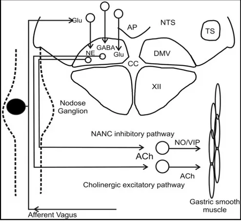
Vagal afferent fibers carry a large volume of information about the physiological status of the stomach directly to brainstem circuits regulating gastric functions. The central terminals of vagal afferent neurons enter the brainstem via the tractus solitarius and terminate within the nucleus tractus solitarii (NTS), using mainly glutamate as their neurotransmitter 18, 19. NTS is
9 postrema (AP) and dorsal motor nucleus of the vagus (DMV), constitutes the dorsal vagal complex (DVC). In the NTS, second order neurons integrate the sensory information from vagal afferents with inputs from other CNS regions involved in the regulation of autonomic functions 20-22 and project the
elaborated afferent information to the adjacent DMV, where are located parasympathetic preganglionic neurons that supply the vagal output to the stomach 19, 23. The connection to the bodies of the DMV completes the
so-called "vago-vagal reflex circuit", of primary importance for the regulation and coordination of several gastric functions. Second order NTS neurons project also to "higher" regions of the brain involved in the coordination of autonomic functions, including parabrachial nucleus, hypothalamic paraventricular nucleus (PVN), central nucleus of amygdala (CeA), bed nucleus of stria terminalis (BNST), ventral thalamus and insular cortex 24, 25.
Although NTS neurons have different biophysical and neurochemical properties, functional studies have determined that they primarily control the DMV through glutamatergic 18, 26, catecholaminergic 27, 28 and GABAergic 26 inputs. In general, GABA, acting on GABA-A receptors, mediates the inhibitory effects of the NTS on DMV neurons; conversely most of the excitation delivered to the DMV by the NTS is mediated by glutamate interacting with both NMDA and non-NMDA receptors 29, 30. Catecholamines
seem to be involved in both excitatory and inhibitory control of the DMV. The spinal afferent fibers are involved in the neural regulation of gastric
10 reflexes and sensations (in particular in the communication of pain associated with visceral organs). These fibers reach the stomach mainly via splanchnic and mesenteric nerves passing through prevertebral ganglia and forming collateral synapses with sympathetic ganglion cells 31. At central level, they
terminate predominantly in distinct laminae of the dorsal spinal cord where they are organized in a segmental manner and distributed over several spinal segments 32, 33. Typically, spinal afferents contain a variety of bioactive
peptides, including calcitonin gene-related peptide (CGRP) and the tachykinins SP and neurokinin A (NKA) 34, 35. The coexpression of CGRP and SP is characteristic of extrinsic afferent neurons, whereas intrinsic enteric neurons in the rat stomach do not coexpress these peptides. Although vagal afferent neurons also express CGRP and SP, spinal afferents represent the main extrinsic source of these neuropeptide in the rat stomach. Within the rat gastric wall, it is particularly the arterial and arteriolar system that receives a dense supply by spinal afferents expressing CGRP and SP 34-37. In addition,
some peptide-containing afferent fibers supply the myenteric plexus, the circular muscle layer and the gastric mucosa. These peptide-containing spinal afferents can release transmitters from their peripheral endings in response to a variety of stimuli; with this "efferent-like activity", they can regulate several gastric functions (including gastric mucosal blood flow, vascular permeability, acid secretion and motility) leading to an increased resistance of the gastric mucosa to injury and a facilitated repair of damaged tissue 10, 38. From this
11 point of view, spinal afferent fibers represent a local neural emergency system that is not tonically active, but is called into operation in the face of pending injury to the stomach (see below).
The efferent fibers of the brain-gut signalling system provide the parasympathetic and sympathetic innervation that helps control and coordinate the different gut functions including secretions, motility patterns and circulation.
Sympathetic control of the stomach stems from cholinergic preganglionic neurons located in the intermediolateral column of the thoracic spinal cord (T5-T9 segments), which, running in the thoracic splanchnic nerves (in particular in the greater splanchnic nerves), impinge on catecholaminergic postganglionic neurons in the coeliac ganglia that provide the stomach with most of its sympathetic supply 39, 40. Sympathetic preganglionic neurons give a
tonic drive to prevertebral postganglionic neurons of the coeliac ganglia, which in turn exert a permanent control of the stomach. These ganglia also receive synaptic inputs of central and peripheral origin so that they function as a constitutive part of a more complex system of nervous regulation 39, 40.
Postganglionic sympathetic fibers from coeliac ganglia run through the coeliac plexus - which also receives some fibers from the vagus 41 - along the vascular
supply of the stomach to innervate mainly myenteric neurons and blood vessels. The corresponding functions associated with these targets are regulation of motility and blood flow (particularly through the mucosa) 42.
12 Sympathetic regulation of gastric motility primarily involves inhibitory presynaptic modulation, via α2 adrenoreceptors, of postganglionic cholinergic
neurons in the myenteric plexus and of vagal cholinergic inputs to these neurons 43, 44. In this way sympathetic outflow inhibits both local excitatory
motor reflexes and extrinsic excitatory parasympathetic nervous activity. Norepinephrine from sympathetic fibers, acting on α1 adrenoreceptors, can
also have excitatory effects on enteric neurons. The stimulation of sympathetic nerves to the stomach elicits a characteristic blood flow response: a pronounced vasoconstriction that subsides within a few minutes to reach a steady state level of blood flow.
The stomach is highly dependent upon extrinsic parasympathetic (vagal) innervation and the gastric myenteric plexus essentially serves as a follower of vagal efferent inputs. As mentioned above, parasympathetic innervation of the stomach arises from vagal preganglionic neurons of the DMV 19, 45. The vast
majority of DMV neurons is cholinergic and activates cholinergic nicotinic receptors on postganglionic neurons within the stomach wall. Some DMV neurons also express immunoreactivity for nitric oxide synthase (NOS) and seem to have an inhibitory effect on gastric motility 46. Parasympathetic
preganglionic neurons innervating the stomach are site-specifically organized in the DMV: preganglionic neurons innervating the ventral corpus and antrum are chiefly located in the medial part of the left DMV, whereas those projecting to the dorsal corpus and antrum are predominantly observed in the
13 lateral and the medial part of the right DMV, respectively 47. DMV neurons
projecting to the stomach are remarkable in that they exhibit slow (1-2 Hz) spontaneous pacemaker-like activity (in vivo as well in vitro), the rate of which can be modulated by synaptic inputs 26, 48. Gastric-projecting DMV
neurons receive mainly glutamatergic, GABAergic and catecholaminergic inputs from the NTS. Several data have suggested that the firing rate of gastric-projecting DMV neurons is regulated by a tonic inhibitory GABAergic input arising from the NTS 49. Therefore, factors modulating GABAergic
inputs from the NTS to the DMV may have a significant impact on vagal control of the stomach. It has been documented that the ability of neurotransmitters and neuromodulators to affect tonic GABAergic connection between the NTS and the DMV depends upon the level of cAMP in the presynaptic GABAergic nerve terminals of the NTS. Under basal or resting conditions, glutamate released from vagal afferents dampens, by activating group II mGluRs, the level of cAMP in the NTS GABAergic terminals. As a consequence, receptors negatively coupled to adenylate cyclase are confined inside the synaptic terminal and neurotransmitters and neuromodulators cannot bind to them and affect GABA transmission 23. When cAMP levels
increases or the cAMP-PKA pathway is activated in the nerve terminal, the internalized receptors move rapidly and transiently to the membrane of the terminal, permitting other transmitters to bind to their receptors and modulate GABA release. Modulation of the NTS-DMV GABA transmission by
14 endogenous opioids acting on μ-opioid receptors is subjected to this type of regulation 50.
In the stomach, vagal efferents from DMV primarily innervate gastric myenteric neurons - that thus represents the parasympathetic postganglionic neurons - giving few branches to the submucosa or mucosa. Stimulation of vagal efferent fibers causes both inhibitory and excitatory effects in the stomach. Therefore, it is clear that both excitatory as well as inhibitory postganglionic neuroeffectors are released from enteric neurons in response to vagal inputs. The main excitatory postganglionic neurotransmitter is acetylcholine acting on muscarinic receptors in gastric smooth muscles, interstitial cells of Cajal (ICC) and parietal cells. Activation of this excitatory pathway enhances gastric motor activity and increases gastric acid secretion. The inhibitory postganglionic neurotransmitters are NO and vasoactive intestinal polypeptide (VIP) released by NANC myenteric neurons innervating gastric smooth muscles and ICC. Activation of this vagal NANC pathway produces a profound relaxation of the proximal stomach and depresses motility in the antrum 51, 52.
15
Fig. 1. Afferent and efferent vagal connection between the CNS and the stomach.
1.2. Gastric motor activity and its regulation by intrinsic and extrinsic pathways
The gastric wall comprises two layers of smooth muscles. An outer thin layer of cells arranged along the length of the stomach forms the longitudinal smooth muscle layer. A perpendicular, thicker, layer of cells immediately under the longitudinal muscle forms the circular smooth muscle layer. The smooth muscle cells of the stomach are connected by gap junctions and form an electrical syncytium. Therefore, electrical stimuli can spread between the cells through the gap junctions, causing parts of the muscle layer to act as one
16 single unit. The basic electrical rhythm in the stomach (as in the other parts of the gut) is fairly constant and characterized by slow waves, consisting of cyclic changes in the membrane potential due to activation and inactivation of different ion channels or pumps. These rhythmic electrical events may develop independently of neuronal activity and are responsible for rhythmic contractions of the muscles, either directly or indirectly by increasing the probability of an action potential. Electrical slow waves are initiated by interstitial cells of Cajal (ICCs) and spread passively to the smooth muscle cells. ICCs are mesenchymal cells typically situated between muscle cells or between myenteric neurons and muscle cells. They are coupled to each other and to muscle cells by gap junctions and act as pacemaker cells in the stomach walls 53.
1.2.1. Motility patterns of the stomach
Anatomically, the stomach is divided into fundus, corpus and antrum region, but with regard to its motor activity two parts can be distinguished: the proximal stomach, consisting of the fundus and the proximal part of the corpus, and the distal stomach, consisting of the distal part of the corpus and the antrum. The proximal stomach is characterized by tonic contractions but not by slow wave activity. The distal stomach, in contrast, exhibits slow wave activity, originating in a pacemaker region in the middle of corpus, and peristaltic contractions propagating towards the pylorus. Two different motor
17 patterns can be distinguished in the stomach: an interdigestive and a postprandial motor pattern. During the interdigestive phase, the proximal stomach muscle tone is high whereas the distal stomach is engaged in a recurrent contraction pattern known as the migrating myoelectrical complex (MMC). After food intake, the proximal stomach initially relaxes in response to swallowing to hold large amounts of food with limited increases in intraluminal pressure (receptive relaxation). When the food bolus reaches the stomach, gastric relaxation is maintained by another reflex triggered by the distension of the gastric wall. This second mechanism has been named "adaptive relaxation" or "gastric accommodation" and allows the stomach to serve as a reservoir of the ingested food. Then, a tonic contraction of the proximal stomach pushes the gastric content distally, whereas the distal stomach mixes and grinds the food by regular peristaltic contractions 54. The
coordinated tonic and peristaltic motor activity of the stomach generate a controlled flow of the gastric content to the duodenum. The subsequent gastric emptying depends on the coordination between the gastric motor activity and the contractile state of the pylorus sphincter and the proximal intestine.
18
1.2.2. Control of gastric motility patterns
The gastric motor activity is controlled by both the intrinsic and extrinsic innervation of the stomach. The ENS generates and propagates highly coordinated motor events such as peristalsis and triggers intrinsic ("short" or "intramural") motor reflexes, in which sensory information is transmitted within the ENS from IPANs to interneurons and then to effector neurons. Extrinsic nerves (sympathetic and parasympathetic) cooperate with ENS in modulating gastric motor programs and provide pathways for "long" or "extramural reflexes", i.e. reflex circuits involving neurons of the CNS 55 [Fig. 2].
Fig. 2. Schematic diagram illustrating the cooperation between central and enteric nervous system in the control and coordination of gastric motor activity.
The high basal muscle tone of the proximal stomach during the interdigestive phase is partially due to the myoelectrical properties of the fundus: the resting
19 membrane potential in the fundic muscles is near or above the mechanical threshold. In addition, muscle tone in the proximal stomach is sustained by constant cholinergic input mediated by the vagal efferent fibers from the DMV
26. The MMCs, which occur spontaneously during the interdigestive phase, are
modulated and coordinated by both ENS and extrinsic innervation. The ENS is necessary to coordinate the propagation of MMCs, whereas extrinsic nerves modulate the frequency and the regularity of the MMC cycles.
The gastric receptive relaxation of the proximal stomach after food intake is a "vago-vagal reflex". The distension of the esophagus by ingested food is detected by low threshold mechanoreceptors on vagal afferents that, in contrast to the relatively high threshold for the activation of spinal afferents, respond to more physiological stimuli. Sensory pathways from the esophagus run to the central subnucleus of the NTS, which, in turn, connects to the efferent vagal pathways from the DMV 56. The final element in the efferent
pathway is represented by myenteric inhibitory motor neurons releasing NO and VIP 51, 57. It seems that these neurotransmitters are co-released from the inhibitory motor neurons and are responsible for the different features of the NANC relaxation. NO would be responsible for the rapid beginning and the initial rapid development of the relaxation and VIP for the long duration of the relaxation. The effect is enforced by a simultaneous reflex that inhibits the excitatory cholinergic motor pathways to the stomach 56.
20 both vagal and intramural (local) reflex pathways with NO as common final inhibitory transmitter 57, 58. It has been demonstrated that stretch of the gastric
wall activates not only vagal afferents but also capsaicin-sensitive (spinal) afferent sensory fibers. These latter release CGRP that, in turn, induce the release of NO from myenteric neurons (either directly or indirectly by acting on myenteric interneurons), which causes relaxation of circular muscle and hence of the fundus 59.
The peristaltic motor activity of the stomach is a combination of oral contractions and anal relaxations that allows the progression of the gastric content to the intestine. This motor activity is induced by the simultaneous activation of ascending excitatory motor pathways, which use acetylcholine as main neurotransmitter, and descending inhibitory motor pathways, using principally NO and VIP 55. The intrinsic enteric innervation of the stomach is
essential for the initiation and propagation of gastric peristaltic contractions, but extrinsic nerves coordinate these contractions via parasympathetic and sympathetic pathways. Preganglionic parasympathetic neurons in excitatory and inhibitory vagal pathways connect to enteric neurons acting on smooth muscle and sympathetic neurons exert an inhibitory effect by a direct action on enteric neurons or by inhibiting transmitter release from preganglionic parasympathetic fibers 60. The reflex control of peristaltic activity of the
stomach relay on feedback from intrinsic and extrinsic afferent innervation of the gastric wall, which monitors the prevailing conditions and triggers the
21 appropriate motor response in the gastric smooth muscle. Both excitatory and inhibitory reflexes occur, inducing an enhancement and a reduction of peristaltic activity, respectively.
1.3. Regulation of gastric mucosal integrity
The maintenance of gastric mucosal integrity depends on the fine balance between aggressive factors (e.g. pepsin, gastric acid, proteases, different chemicals and bacterial invasion) and defensive mechanisms (including the layer of mucus, the bicarbonate secretion, the mucosal microcirculation and the cell renewal). Therefore, gastric mucosal damage may occur when noxious factors “overwhelm” an intact mucosal defense or when the mucosal defensive mechanisms are impaired. From a therapeutic point of view, this means that besides the classical approach that involves the inhibition of gastric acid secretion by H2 receptor antagonists and proton pump
inhibitors as well as eradication of Helicobacter pylori by antibiotics, augmentation of endogenous defensive mechanisms may represents another possible approach of anti-ulcer therapy.
The surveillance system of the gastric mucosa involves barriers (mucus gel layer, epithelial cells) and different mechanisms that are coordinated by the ENS and the CNS, the endocrine system and the immune system. Moreover these systems are cooperating with each other in the regulation of defensive processes 61.
22
1.3.1. Structural and functional elements of the gastric mucosal defense
"Mucosal defense" is a term used to describe the various factors and components that permit the mucosa to resist to injury. In a healthy organism, mucosal defense is a dynamic process and is enhanced when irritants are present in the stomach. The various levels of mucosal defense can be viewed in a structural sense, starting at the lumen and moving into deeper levels of the tissue.
The mucus gel layer covering the mucosa constitutes the first line of gastric mucosal defense. The physiological functions of this mucus barrier are to impede the diffusion of gastric acid, bacteria and different macromolecules such as bacterial toxins to the epithelial cells 62, 63. The mucus gel is secreted
by apical expulsion from surface epithelial cells and contains HCO3-, which
buffers gastric acidity and maintains a nearly neutral pH at the epithelial surface 64, and surfactant phospholipids with strong hydrophobic properties
that make the surface impermeable to the luminal acid 65.
The next level of gastric mucosal defense is formed by a continuous layer of tightly connected surface epithelial cells that prevent back diffusion of acid and pepsin, secrete mucus and bicarbonate and generate prostaglandins (PGs)
66, heat shock proteins 67 and other protective substances. The epithelium has
the ability to renovate itself continuously maintaining the structural integrity of the mucosa. A well-coordinated and controlled proliferation of progenitor
23 cells enables replacement of damaged or aged surface epithelial cells that are extruded into the lumen 68.
Gastric mucosal microcirculation is another essential element for maintaining gastric mucosal integrity, but also for healing of damaged mucosa 69. Mucosal
microcirculation is constituted by a dense network of capillaries underlying the surface epithelium that, at the base of surface epithelial cells, converge into collecting venules 70. In addition to supplying nutrients and oxygen to the
epithelium, gastric microcirculation also removes, dilutes and neutralizes noxious chemicals that diffuse into the mucosa from the lumen. It also plays a critical role in the disposal of H+ ions back-diffused from the lumen or parietal cells to the mucosa 71 and facilitates the delivery of bicarbonate to the
epithelium. When the epithelium is damaged, the microcirculation also contributes to create a microenvironment over the site of injury conducive for repair.
Mucosal blood flow is modulated by several endogenous substances. The endothelial cells lining the microvessels generate potent vasodilators such as NO and prostacyclin (PGI2) 68, which maintain viability of vessels and prevent
platelet and leukocyte adherence to the microvascular endothelium, thus preventing compromise of the microcirculation. Mucosal blood flow is also regulated by extrinsic innervation of the stomach. Vagal efferents from DMV are able to stimulate the release of NO by a cholinergic mechanism 72; they
also stimulate the gastric production of PGs 73, 74 - such as PGE
24 contribute to increase and maintain gastric mucosal blood flow (GMBF)75.
Capsaicin-sensitive primary afferent fibers innervating gastric mucosa and submucosal vessels release, in response to luminal aggressive factors or to acid back-diffusion, CGRP that, in turn, induces a hyperemic response mainly by stimulation of NO production, but also by a direct action on vascular smooth muscle cells 76, 77.
The mucosal immune system is a further component of the gastrointestinal surveillance system. The gut possesses a highly specialized immune system that contains organized and non-organized cellular elements 78, including antigen-sampling M cells, lymphocytes and immune-associated cells such as macrophages, eosinophils, neutrophils and mast cells. In addition, many epithelial cells are able to secrete chemokines (e.g., interleukin-8) and thus to recruit immune cells 79. The GI immune system is called into operation
whenever the mucosa is affected by microbial infection, allergen exposure, inflammation or other types of injury. The activation of immune cells induces the release of cytokines, PGs, leukotrienes, bradykinin, histamine, 5-HT and proteases that can either acutely excite sensory nerve fibres or alter their sensitivity in the long term 80.
25
1.3.2. Mediators of gastric mucosal defense
Several local mediators are involved in the regulation of the physiological defensive mechanisms mediating the resistance of the gastric mucosa to injury. These include PGs, gaseous mediators (NO and hydrogen sulfide) and neuropeptides (CGRP).
Continuous generation of PGE2 and PGI2 by the gastric mucosa is crucial for
the maintenance of mucosal integrity and protection against ulcerogenic and necrotizing agents 81. Almost all of the mucosal defense mechanisms are
stimulated and/or facilitated by PGs. They inhibit gastric acid secretion; stimulate mucus, bicarbonate and surfactant phospholipids production (increasing the mucosal hydrophobicity); enhance the mucosal content of sulfhydryl compounds (reduced glutathione) that are able of binding reactive free oxygen radicals; maintain and increase GMBF; inhibit platelet and leukocyte adhesion to vascular epithelium; accelerate epithelial restitution and mucosal healing. In addition, PGs inhibit mast cell activation reducing the release of inflammatory mediators (such as histamine, tumor necrosis factor-α, and platelet-activating factor) that have been suggested to contribute to the generation of mucosal injury in certain situations 81, 82.
NO is considered to be another essential mediator of the mucosal defensive mechanisms 83. As mentioned above, NO has been shown to participate in the
regulation of gastric mucosal microcirculation 84 and to mediate the
26 participate in gastric mucosal defense also by stimulation of mucus and bicarbonate secretion, inhibition of gastric acid secretion, modulation of the activity of mucosal immunocytes (e.g., mast cells and macrophages), reduction of leukocyte-endothelial adhesive interactions, and acceleration of mucosal damage healing 85. NO has proven to be the primary NANC
neurotransmitter in the GI tract 86. Not surprisingly, therefore, inhibition of
NOS results in disturbance of gastric blood flow, motility and secretion. NO also contributes to mucosal protection through its cytotoxic properties, a primary defense against ingested bacteria and parasites 87. In the stomach, suppression of NO synthesis renders the mucosa more susceptible to injury 83, whereas administration of NO donors can protect the stomach from injury 88.
However, when NO donors have been given in higher doses, extensive mucosal injury has been observed, suggesting that while physiological formation of NO plays a role in maintaining mucosal integrity, inappropriate release of NO can lead to mucosal injury 89. This could be due to a direct
cytotoxic action of NO or to the formation of oxidant metabolites like peroxynitrite 90.
NO seems to be involved in the mucosal protective effect of several anti-ulcer agents, like carbenoxolone 91, sucralfate 92 and aluminum-containing antacids 93, and in the mucosal protective process of experimental gastroprotective
agents, including capsaicin 94, opioids 95, pentagastrin and cholecystokinin-8 96.
27 Interestingly, the actions of NO overlap considerably with those of PGs. It has been observed that simultaneous suppression of both PGs and NO synthesis leads to a synergistic increase in mucosal susceptibility to injury and the gastric injury that can be induced by suppression of gastric PGs synthesis is prevented by administration of NO donors. These data suggest a close interaction between NO and PGs in the maintenance of mucosal integrity, that seems to be confirmed by the finding that these mediators can regulate the synthesis of each other 83, 97, 98.
Hydrogen sulfide is a further endogenously generated compound that exerts a strong mucosal protective action similar to NO; it reduces tumor necrosis factor α (TNF-α) expression, decreases leukocyte adherence to vascular endothelium, and inhibits NSAID-induced gastric mucosal injury 99, 100.
CGRP from capsaicin-sensitive primary afferent fibers innervating mucosa and submucosal vessels is one of the most important mediators of gastroduodenal defense. This neuropeptide plays its gastroprotective effect primarily through an increase of GMBF 101, 102. As mentioned above, the hyperemic effect of CGRP is mediated by NO, although the peptide can also acts directly on vascular smooth muscle 76. CGRP - through activation of
CGRP1 receptors - is also able to inhibit gastric acid secretion 103.
This antisecretory effect involves the release of somatostatin and inhibition of gastrin and acetylcholine release 104 and may contribute to its mucosal
28 only in the prevention of gastric mucosal damage, but also in facilitation of gastric ulcer healing 101, 105. This action may be due to the ability of the peptide
to enhance angiogenesis in vivo 101.
The pivotal role of CGRP released from peripheral terminals of visceral afferent fibers in gastric mucosal defense has been confirmed by several pharmacological studies. Low doses of capsaicin or TRPV1 agonists have clearly shown to exert gastroprotection via the stimulation of sensory nerves and the local release of CGRP 16, 106. In addition, close arterial infusion of
CGRP to the stomach - a route of administration that closely resembles the local release of the peptide in response to the stimulation of capsaicin-sensitive afferent fibers - significantly reduces gross mucosal damage caused by ethanol and aspirin 107. The gastroprotective effect of intragastric capsaicin
is abolished by systemic administration of the human C fragment of CGRP (hCGRP 8-37) 108, a CGRP antagonist, and of monoclonal antibodies to CGRP 109. The same agents have been also reported to enhance gastric mucosal
lesions induced, in rat, by ethanol 109, 110. Recent studies, performed using CGRP-knockout mice, have strongly confirmed these findings: the protective action of capsaicin against ethanol-induced lesions is completely abolished in CGRP -/- mice 101.
Different reports have suggested that CGRP is involved not only in the prevention of gastric mucosal damage, but also in facilitation of gastric ulcer
29 healing 101, 105. This action may be due to the ability of the peptide to enhance
angiogenesis in vivo 101.
CGRP-mediated protective action may be regulated by endogenous PGs. It is widely known that pain sensation is enhanced by PGE2 and PGI2 by
sensitizing the sensory nerves 111, with PGI
2 being more potent that PGE2 in
this sensitizing action 112. It has been reported that inhibition of PG synthesis
results in a reduction of the mucosal protective effect of capsaicin 113 and that
endogenous PGI2 facilitate the release of CGRP from capsaicin-sensitive
afferent fibers and gastric mucosal protection against ethanol 114, 115.
1.3.3. CNS and gastric mucosal integrity
Besides the structural and functional elements of gastric mucosal defense and the local release of protective mediators, the CNS also plays an important role in the maintenance of gastric mucosal integrity.
As mentioned previously, DVC and vagus nerve have a pivotal role in the regulation of gastric functions, including acid secretion, motor activity and mucosal defense 19. Within the DVC, NTS receives afferent sensory
information from the stomach, carried by vagal afferent fibers originating from nodose ganglion, and DMV sends preganglionic vagal efferent fibers innervating myenteric neurons in the gastric wall.
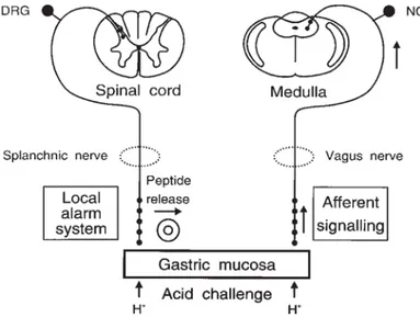
Disruption of gastric mucosal barrier by aggressive chemicals such as ethanol induces the back-diffusion of gastric acid from the lumen into the mucosa. The
30 surge of acid intruding the lamina propria stimulates spinal afferent fibers innervating the stomach (through activation of proton-sensitive TRPV1 ion channels), which induce a rapid increase of GMBF and initiate other defensive mechanisms by local release of CGRP 38. In parallel, acid challenge of the
mucosa activates vagal afferent fibers that communicate the pending injury to the NTS (Fig. 3), as demonstrated by expression in this area of messenger RNA (mRNA) for the immediate early gene c-fos 116 - which reflects neuronal
excitation and is hence widely used to visualize central neurons that receive a message from the periphery 117. Glutamate (via NMDA and AMPA receptors), but also SP and NKA (via NK1 and NK2 receptors) are involved in the communication of afferent information to NTS neurons 118.
Fig. 3. Neural circuits activated by acid back-diffusion into the gastric mucosa.
The vagal afferent input from the acid-threatened stomach is further relayed to hypothalamic and limbic areas of the rat brain (lateral parabrachial nucleus,
31 thalamic and hypothalamic paraventricular nuclei, supraoptic nucleus, subfornical organ, CeA and mediolateral habenula), but not to insular cortex (the major cerebral representation area of visceral input) 25; therefore, vagal afferent signalling of gastric challenge does not give rise to perception of pain, but evokes autonomic, endocrine, affective and behavioral reactions.
After its central processing, the afferent information is sent to the DMV leading to a modification of the activity of vagal efferent pathways to the stomach. Biochemical and pharmacological studies have shown that activation of these pathways stimulates, in the stomach, the release of PGs and NO 72, 119,
120 and the effector function of capsaicin-sensitive afferent fibers containing
CGRP 121. The exact mechanism by which central vagal activation stimulates
spinal afferents is still not well defined. PGs are probably involved in the sensitization of these fibers 115. Alternatively, acetylcholine from vagal
efferents could activate directly spinal afferents (an ability already demonstrated in the rat skin) 122; furthermore vagal activation is able to
induce, through activation of muscarinic receptors, the release of histamine and serotonin that are known to evoke sensory C-fiber excitation 123.
Several neuropeptides (e.g. TRH, neuropeptide Y, adrenomedullin and opioid peptides) have been demonstrated to induce a gastroprotective effect, after central administration, through the activation of this vagal cholinergic pathway and then through this NO/PGs/CGRP mechanism 121, 124-126.
32 regulation of gastric mucosal integrity mainly through descending neuronal projection to the DVC. They include two hypothalamic formations - the lateral hypothalamus (LH) and the PVN - but also the CeA and caudal raphe nuclei, namely the raphe pallidus (Rpa) and the raphe obscurus (Rob).
Anatomical data revealed that LH projects directly to NTS and DMV and receives ascending information from the NTS. LH is able to modulate the activity of DVC neurons involved in the regulation of gastric functions; the stimulation of LH induces predominantly inhibitory effects on NTS neurons and excitatory effects on DMV neurons 127. LH lesions have been reported to induce gastric mucosal erosion through a mechanism involving a decrease of mucosal barrier function, an increase of gastric acid secretion and gastric hypermotility 128, 129. Vagotomy or anticholinergic agents protect against
LH-induced gastric erosions suggesting the involvement of vagus nerve 130.
Also PVN has direct connections with the DVC and exert an inhibitory influence on NTS neurons, regulating their responsiveness to incoming vagal information, and an excitatory influence on DMV neurons, modulating the vagal output to the stomach 20. Electrical stimulation of PVN causes gastric
mucosal damage 131 and exacerbates stress-induced ulceration 20. A reduction
of GMBF and an increase of gastric motor activity are supposed to be involved in the detrimental effects of PVN stimulation, which are reduced by vagotomy and electrolytic lesion of PVN.
33 The caudal raphe nuclei - namely Rpa and Rob - represent a significant source of inputs to NTS and DMV through direct projections 132. Electrical or
chemical stimulation of Rpa or Rob enhances gastric motility 133, 134 and secretion of acid, pepsin and PGs 135, 136 by a vagal cholinergic pathway, but is
also able to induce an alteration of gastric mucosal resistance to lesion formation. For example, microinjection of kainic acid into the Rob or Rpa causes gastric erosions in fasted rats 137. On the other hand, kainic acid
microinjected into the Rpa at a subthreshold acid secretory dose induces gastric cytoprotection against ethanol injury 138.
Anterograde tract-tracing studies have revealed that efferent fibers from CeA terminate in both the NTS and the DMV, in regions that are involved in the regulation of gastric functions 139. Electrical stimulation of the CeA modifies
the activity of NTS and DMV neurons and enhances c-Fos expression in the NTS 21, 140 inducing vagal-dependent changes in gastric acid secretion, gastric
motility and gastric mucosal resistance. In particular, stimulation of some areas of the CeA increases gastric acid secretion and motility and produces gastric erosion, whereas stimulation of other areas inhibits gastric motility and acid secretion 141. Both excitatory and inhibitory changes were blocked by
34
1.3.4. Gastric motility - gastric mucosal integrity
Several experimental data have suggested the existence of a close relationship between gastric motility and maintenance of gastric mucosal integrity. All the most important peripheral mediators of gastric mucosal defense - namely NO, CGRP and PGs - are capable of modifying gastric motor activity, an ability that has been supposed to participate in their protective action.
As mentioned, NO represents, together with VIP, the major mediator of the NANC inhibitory motor transmission in the rat stomach. Acid challenge of gastric mucosa causes the activation of a subpopulation of myenteric nitrergic inhibitory motor neurons through a mechanism involving extrinsic capsaicin-sensitive afferent fibers and nicotinic cholinergic transmission 142, 143. The
activation of these nitrergic neurons may represent a physiologic response aimed at protecting the mucosa towards luminal noxae by inhibition of motor activity.
PGs have been reported to influence the gastric smooth muscle contractility and to have a complex effect on gastric motility 144, 145. In the distal stomach
endogenous PGs decrease the amplitude of contractions and the ability of the muscles to respond to excitatory stimuli. In the proximal stomach PGs have an opposite role: they promote tonic contraction 144. Mucosal endogenous PG
deficiency induced by indomethacin has been shown to be associated with gastric hypermotility, which seems to be an important factor in the
35 pathogenesis of gastric mucosal lesions produced by this non-steroidal anti-inflammatory drug (see below) 146. However, local deficiency of PGs cannot
induce, by itself, the formation of severe gastric damage in the absence of other risk factors such as intraluminal acid, chemical ablation of capsaicin-sensitive afferent fibers or stimulated gastric motility 147, 148.
Endogenous CGRP from spinal afferent fibers seems to have an inhibitory effect on gastric motility, which can contribute to the protective effect of intragastric capsaicin against ethanol-induced gastric mucosal damage 149. In
addition, it has been demonstrated that exogenously administered CGRP can inhibit gastric motility and emptying by a direct action on gastric smooth muscle through receptors linked with cAMP 150.
However, in spite of the intensive research, it is still not clear how alterations in gastric motor activity correlate with gastric ulcer formation. Both increased and decreased motility have been proposed to contribute to gastric mucosal damage.
Gastric contractions characterized by high amplitudes may induce microvascular disturbances in specific sites of the mucosa probably by abnormal compression of the gastric wall, thereby leading to insufficient mucosal blood flow, increased vascular permeability and cellular damage 151, 152. Furthermore, it is known that contraction of gastric circular smooth muscle
leads to the appearance of mucosal folds that has been implicated in the pathogenesis of several ulcer models, including ethanol-induced lesions 153.
36 Therefore, inhibition of gastric motility may lead to an attenuation of microvascular disturbances due to gastric hypermotility and to flattening of the mucosal folds, resulting in a reduction of mucosal vulnerability to irritants and of severity of damage.
As mentioned above, stimulated gastric motor activity, due to the gastric mucosal deficiency of PGs, seems to be an important factor in the pathogenesis of indomethacin-induced gastric mucosal damage 154. The ability
of different substances - such as amylin - to prevent mucosal lesions induced by indomethacin has been attributed to their inhibitory effect on enhanced gastric motility response observed in this ulcer model 155. Furthermore, under PG-deficient conditions induced by pretreatment with indomethacin, prokinetic drugs (that alone have no effects on gastric mucosal integrity) are able to induce gastric mucosal damage in the rat at doses that enhance gastric motility and emptying but not at doses that expedite gastric emptying only 147.
2-deoxy-D-glucose (2-DG), which enhances the gastric motility by a central vagal stimulation, causes by itself non-hemorrhagic lesions on gastric mucosa, but these lesions become hemorrhagic under PG-deficient conditions induced by a low dose of indomethacin 148.
An increased gastric motility seems to be associated also with gastric mucosal lesions induced by cold-restraint stress 151, 156; suppression of this gastric
hypermotility inhibits the stress-induced lesion formation.
37 canine stomach, ethanol induces an inhibition of antral phasic motor activity
157, which can account for the inhibition of gastric emptying observed in
several experimental studies, and a stimulation of corpus tonic motor activity
158. In the guinea pig stomach, ethanol induces only a contractile response in
both longitudinal and circular muscle 159, which may contribute to mucosal
necrosis and subsequent ulceration.
Exogenously administered PGs (in particular those acting on EP1 receptors)
inhibit gastric motor activity at the doses that significantly reduce the severity of gastric mucosal injury caused by ethanol 160, 161, suggesting a close relationship between the inhibited gastric motility and the cytoprotective action of PGs in this ulcer model.
An inhibition of gastric motility seems to be also involved in the protective action of intragastric capsaicin against ethanol 149. The inhibition of gastric
motility induced by capsaicin at gastroprotective doses is impaired by indomethacin pretreatment, desensitization of capsaicin-sensitive afferent neurons or CGRP antagonists 149, 162, suggesting the involvement of endogenous PGs and CGRP released from spinal afferents innervating the stomach.
In contrast with these reports, other experiments have suggested that also inhibition of gastric motility and delaying of gastric emptying may play an important role in the pathogenic mechanism of gastric ulcer formation, probably through a prolongation of the contact between ulcerogenic substance
38 and gastric walls. For example, the protective effect of the prokinetic drug metoclopramide against aspirin-induced gastric mucosal damage is thought to be mediated, at least in part, by acceleration of gastric emptying 163. Furthermore, the delay of gastric emptying caused by large doses of morphine has been proposed to aggravate the ethanol-induced gastric lesions 164.
However, the involvement of altered gastric motility in the pathogenesis of gastric mucosal damage has been questioned by some experimental data. Gutierrez-Cabano 165 has found that gastric contractile activity is unlikely to
play a major role in the development or prevention of gastric lesions induced by necrotizing agents such as 96% ethanol. Likewise, hypermotility is unlikely to serve as a major factor in stress ulceration, and the smooth muscle relaxing effect of atropine and verapamil may contribute only partly to their anti-ulcer effect 166. Moreover, inhibition of gastric motor activity seems to not
contribute to the protective effect of clonidine against ethanol-induced gastric mucosal damage 167.
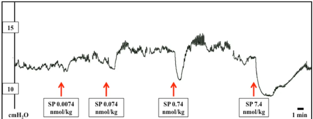
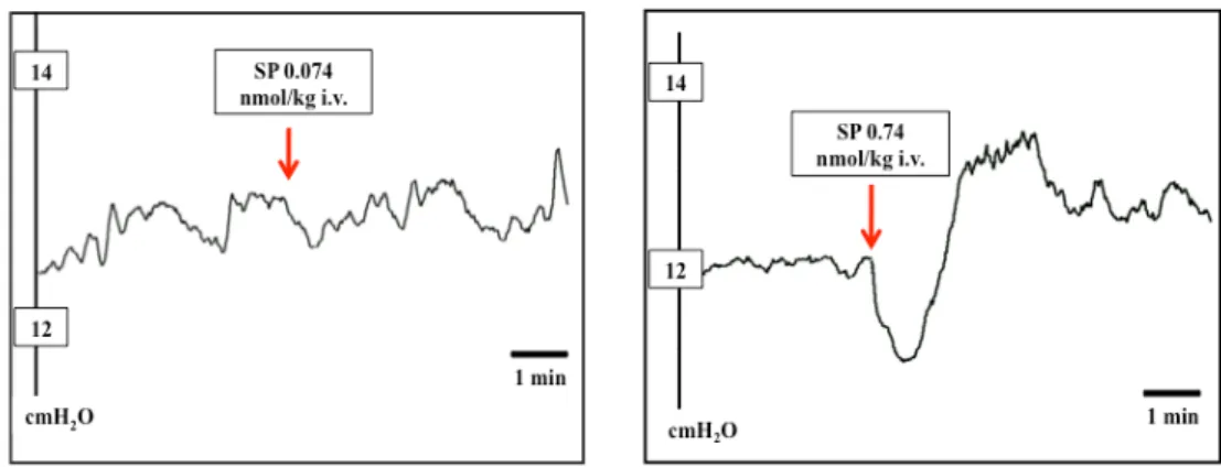
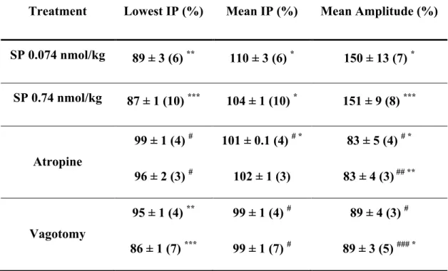
1.4. Substance P and regulation of gastric functions
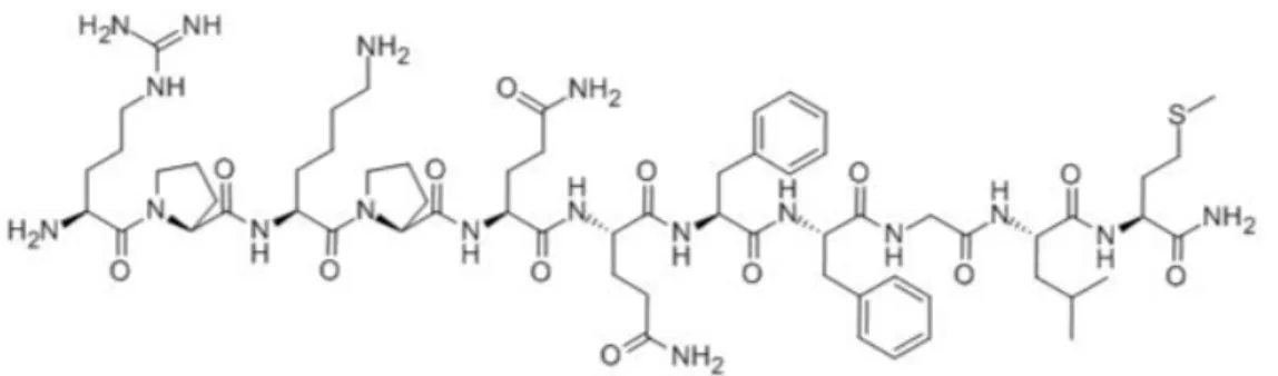
The undecapeptide SP (Fig. 4) is the most widely known representative of a family of small biologically active peptides, the tachykinins, which consist of a large number of mammalian and non-mammalian members characterized by a common and strongly evolutionarily conserved
carboxy-39 terminal amidated amino acid region, Phe-X-Gly-Leu-Met-NH2 (where X is
an aromatic or hydrophobic residue) 168.
Fig 4. Primary structure of substance P. The C-terminal sequence common to all the tachykinins is showed in red.
Besides SP, mammalian tachykinins include NKA, neurokinin B (NKB) and two elongated forms of NKA, neuropeptide K (NPK) and neuropeptide γ (NPγ) 169, 170. Recently (17 years after the isolation of NKA and NKB), other
tachykinin peptides, hemokinin-1 (HK-1), endokinin-1 (EK-1), endokinin A (EKA) and endokinin B (EKB), were identified in rodents and humans 171, 172.
Being considered of potential importance for understanding and therapy of human disease, tachykinins have become one of the largest investigated groups of neuropeptides and, despite the long period over which these substances have been studied, new informations on their functions continue to emerge. Ever since SP has been discovered to occur in the intestine and to contract gastrointestinal smooth muscle 173, the implication of tachykinins in
40 the regulation of gastrointestinal functions has been one of the most extensively studied areas of tachykinin research.
1.4.1. Molecular biology of substance P and tachykinin receptors
SP is encoded by the tachykinin precursor 1 (TAC1) gene (originally known as preprotachykinin (PPT)-A or PPT-I gene) that codifies also for NKA and its two elongated forms, NPK and NPγ. Transcription of TAC1 gene generates a pre-mRNA that could be spliced giving rise to four different mRNA isoforms (α, β, γ, and δ) that differ in their exon combinations 174. SP can be produced from all the mRNA isoforms, whereas NKA production is confined to the α and γ TAC1 mRNA (Fig. 5). This means that SP can be expressed alone, but NKA is always produced along with SP.
Translation of the mature mRNA from TAC1 gene generates a large polypeptide, designated as prepropeptide, that consists of a signal peptide, one or several copies of the neuropeptide and one or more spacer parts. The signal peptide is located at the N-terminal and allows the forming peptide to attach to and pass into the endoplasmic reticulum during synthesis; it is then rapidly cleaved off, after polypeptide synthesis, to allow the formation of the propeptide. This one is transported to the Golgi apparatus where the spacer parts are split off by proteases called convertases. After the cleavage of the propeptide, the final peptide is amidated at C-terminal by peptidyl-Gly-α-amidating monoxygenase, that use glycine as amide donor. SP is then packed
41 into storage vesicles budding off from the Golgi apparatus and is axonally transported from the perikaryon (were its synthesis is confined) to the nerve terminals for final enzymatic processing. From the nerve terminal the peptide, stored in large dense core vesicles, is released by a Ca2+-dependent exocytosis
process 175.
Fig. 5. Splices variants of TAC1 gene. Exons are showed as boxes. The positions of the predicted tachykinin peptides (SP, NKA, NPK, and NPγ) are indicated by underlining.
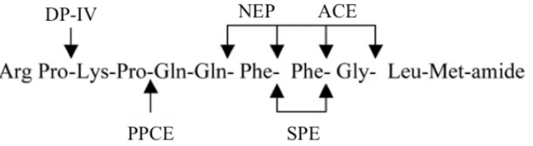
As with other peptide transmitters, the biological effects of synaptically released SP are terminated by enzymatic degradation, since neurons lack an active uptake mechanism for intact SP. Although several enzymes capable of hydrolyzing the peptide have been described, the cleavage of SP is carried out
42 mainly by angiotensin-converting enzyme (ACE), neutral endopeptidase (endopeptidase 24.11) (NEP) and substance P endopeptidase (SPE) 176. ACE is
a membrane-bound zinc metallopeptidase cleaving SP at Phe8-Gly9 and Gly9 -Leu10 as the major cleaving sites. ACE also acts as a peptidyl dipeptidase,
cleaving dipeptides from the remaining N-terminal fragment, and is thus able to generate the fragment (1-7) of SP 177. NEP primarily hydrolyzes SP at the
Gln6-Phe7, Phe7-Phe8 and Gly9-Leu10 bonds 178, 179. SPE is a metalloenzyme
highly selective for SP. This endopeptidase hydrolyzes SP mainly within and at the carboxylic side of the double-Phe bond thus releasing SP (1-7) and SP (1-8) fragments from the parent peptide. Other tachykinins lacking the double - Phe residues, such as NKA and NKB, are almost unaffected by this enzyme. SP is also cleaved, between the Pro4-Gln5 residues, by the post-proline
cleaving enzyme (PPCE) or prolyl endopeptidase 180. Post proline dipeptidyl
aminopeptidase (DPP-IV) successively removes the dipeptides Arg1-Pro2 and
Lys3-Pro4 from the undecapeptide 181. This enzyme is probably responsible for
the degradation of SP in the blood circulation 182.
43 The biological actions of SP are mediated by tachykinin (neurokinin: NK) receptors that belong to family 1 (rhodopsin-like) of G protein-coupled receptors. Tachykinin receptors have been first shown to be coupled to a Gq-protein and then to induce the activation of phospholipase Cβ (PLCβ) -
followed by production of 1,4,5-inositol triphosphate (IP3) and elevation of intracellular Ca2+ as second messengers - and of phospholipase A2 - followed
by an increase in arachidonic acid mobilization. Afterward, it has also been revealed that production of another second messenger, cAMP, was stimulated by tachykinin receptors coupled to Gs-protein.
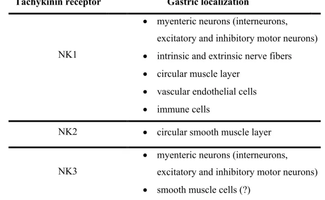
Currently three different tachykinin receptors, termed NK1, NK2 and NK3, have been identified. They are codified by three distinct genes: TACR1, encoding for NK1 receptor, TACR2, encoding for NK2 receptor, and TACR3, encoding for NK3 receptor. SP exhibits preferential binding to NK1 receptor, whereas NKA and NKB bind preferentially to NK2 and NK3 receptors, respectively. The rank order of potency for the NK1 receptor is SP > NKA > NKB, while it is NKA > NKB > SP for the NK2 receptor and NKB > NKA > SP for the NK3 receptor 183 (Table 1).
44 However, endogenous tachykinins are not highly selective for any given receptor, and all can act as full agonists on all three receptors under certain conditions such as receptor availability or at high peptide concentrations. For this reason SP activates not only NK1 receptors, but also NK2 and NK3 receptors in a number of tissues 183. The participation of NK2 and NK3
receptors, together with NK1 receptor, to the effects of SP has been confirmed in different experimental studies. For example, it has been observed that both NK1 and NK2 receptors are involved in the excitatory effect of SP on DMV neurons projecting to the stomach 184 and in the cardiovascular and behavioral effects induced by intracerebroventricular (i.c.v.) injection of the peptide 185, whereas all the three tachykinin receptors seem to be involved in the postsynaptic action of SP on PAG neurons 186.
1.4.2. Distribution of substance P and tachykinin receptors in the stomach and in the central nervous areas involved in the regulation of gastric functions
SP is highly abundant in the gastrointestinal tract, in which represents a neurotransmitter and a neuromodulator of primary importance. The main source of SP in the stomach is represented by intrinsic enteric neurons of the myenteric plexus, in which SP is extensively colocalized with choline acetyltransferase (ChAT) 2, 4. In contrast, SP does not coexist with VIP and
45 shown that SP is expressed by several classes of myenteric neurons including excitatory motor neurons and interneurons. Myenteric neurons expressing SP have been found to innervate the longitudinal muscle, circular muscle and muscularis mucosae of the murine, rat, guinea-pig and canine stomach 188-190.
In the enteric nervous system of the rat GI tract, γ isoform of TAC1 gene accounts for as much as 80-90% of the tachykinin encoding mRNA. This fact implies that in most (if not all) myenteric neurons of the stomach SP coexist with NKA. Extrinsic spinal afferents (capsaicin-sensitive) also significantly contribute to the SP content of the stomach. Characteristically many spinal afferents containing SP co-express CGRP, a combination of peptides that is not found in the ENS. The main targets of these spinal afferents are gastric blood vessels and mucosa. In contrast, less than 10% of extrinsic vagal afferents innervating the rat, mouse and guinea pig stomach express SP, making a relatively small contribution to the SP content of the stomach 34, 191.
The rest of SP content is contributed by enterochromaffin and immune cells of the gastric mucosa 192.
In the stomach, tachykinin NK1, NK2 and NK3 receptors are widely distributed. NK1 receptors have been detected on myenteric neurons, including interneurons, NOS-immunoreactive (IR) inhibitory motor neurons and ChAT-IR excitatory motor neurons. Furthermore, NK1 receptors have been found on intrinsic and extrinsic nerve fibers throughout the stomach, smooth muscle cells of the circular muscle layer and vascular endothelial cells
46
193. Likewise, immune cells involved in mucosal defense, such as enterocytes,
eosinophils, mucosal mononuclear cells (e.g., lymphocytes) and mast cells, can express NK1 receptors. These locations are congruent with a role of gastric NK1 receptors in regulating neuronal excitability, release of neurotransmitters, motility, vascular permeability, blood flow and inflammatory processes. In addition, the presence of NK1 receptors has been also demonstrated on gastric chief cells, where they seem to be involved in pepsinogen secretion 194. Double-staining experiments have demonstrated that
a majority of NK1 receptor expressing nerve fibers in the circular and longitudinal muscle layers and a minority of NK1 receptor expressing nerve fibers in the myenteric plexus contain SP. The observation that SP is co-localized with NK1 receptors raises the possibility that an autocrine regulatory feedback mechanism exists to control the release of SP in the stomach. It is worthy of note that in Wistar rat stomach a high percentage of SP-NK1 co-expressing fibers also express CGRP, suggesting an interaction between SP and the regulation of extrinsic spinal afferent fibers innervating the stomach
193.
NK2 receptors are typically expressed by the circular muscle layer of the stomach 195, 196, while NK3 receptors are largely confined to myenteric
neurons 197. In addition, NK3 receptors have been localized on muscle cells of
the stomach, although there is little pharmacological evidence that these receptors play a functional role (Table 2). However, the density of NK3