Department of Biomedical and Biotechnological Sciences University of Catania School of Medicine
International PhD Program in Neuroscience
XXXI Cycle
___________________________________________________________
Walter Gulisano
A renewed vision for Amyloid-β and tau in
Alzheimer’s disease pathophysiology
Ph.D. THESIS
Coordinator: Prof. Salvatore Salomone
Tutor: Prof. Agostino Palmeri
Co-Tutor: Prof. Daniela Puzzo
___________________________________________________________
Academic Year 2017-18
Index
Synopsis ... 3
General introduction ... 5
Preface ... 6
Amyloid-β peptide and Alzheimer’s disease: more than one century of research .... 7
A revaluated player in Alzheimer’s disease pathogenesis: Tau protein ... 11
Aβ and tau oligomers: a game at the synapse resulting in memory impairment ... 15
Aβ and tau activity dependent secretion, neuronal uptake, and spreading of the disease ... 17
References ... 23
Chapter 1: The effect of Amyloid-β peptide on synaptic plasticity and memory is influenced by different isoforms, concentrations, and aggregation status ... 31
Abstract ... 32
Introduction ... 33
Results... 34
Discussion ... 43
Conclusion ... 47
Materials and methods ... 48
References ... 51
Chapter 2: Amyloid-β peptide is needed for cGMP-induced long-term potentiation and memory ... 53
Abstract ... 54
Introduction ... 55
Results... 56
Discussion ... 67
Materials and methods ... 72
References ... 75
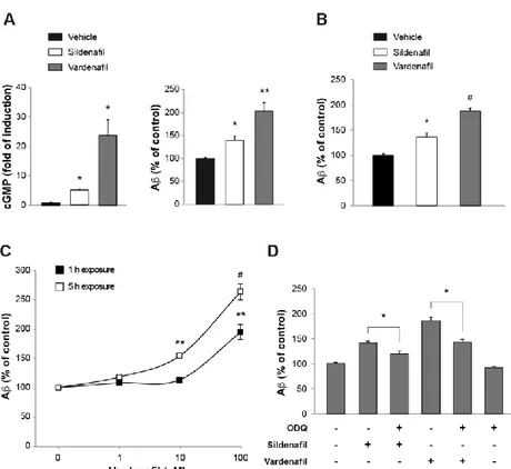
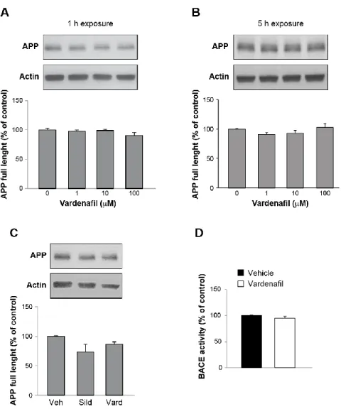
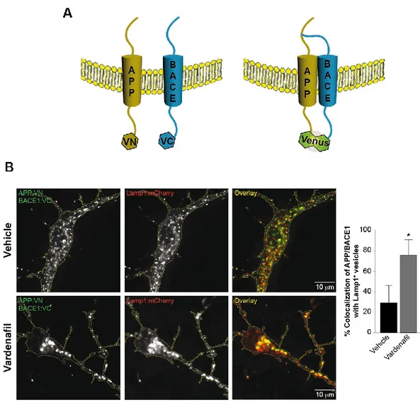
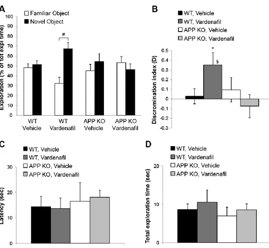
Chapter 3: Sub-efficacious doses of phosphodiesterase 4 and 5 inhibitors improve memory in a mouse model of Alzheimer’s disease ... 78
Introduction ... 80
Results... 81
Discussion ... 89
Materials and methods ... 92
References ... 96
Chapter 4: Extracellular tau oligomers produce an immediate impairment of LTP and memory ... 98
Abstract ... 99
Introduction ... 100
Results... 101
Discussion ... 113
Materials and methods ... 122
Supplementary Information ... 127
References ... 136
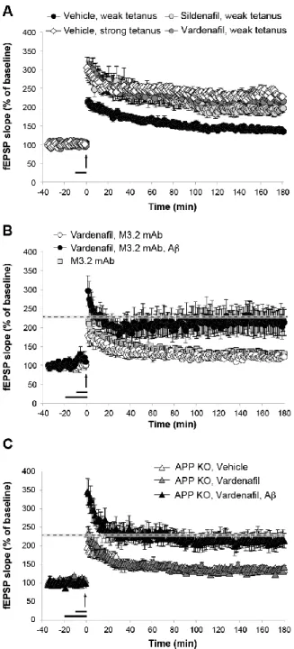
Chapter 5: LTP and memory impairment caused by extracellular Aβ and tau oligomers is APP-dependent ... 140
Abstract ... 141
Introduction ... 142
Results... 143
Discussion ... 154
Materials and methods ... 158
References ... 164
Concluding remarks ... 167
Synopsis
The aim of this thesis was to study the pathogenetic mechanisms underlying Alzheimer’s disease (AD), a neurodegenerative disorder affecting the elderly and characterized by memory loss, personality changes and cognitive dysfunction leading to dementia.
I will discuss the main projects in which I participated aimed at understanding the role of the main molecular interactors involved in AD pathogenesis, i.e. Amyloid-beta peptide (Aβ) and tau protein, on hippocampal synaptic plasticity and memory in animal models. After reviewing the pathophysiological models that have been developed so far, our general purpose was to study novel aspects of Aβ and tau involvement in physiological and pathological conditions to give a different interpretation of the disease.
The present thesis is divided into different sections, as follows:
1. The General Introduction provides a comprehensive overview on the role of Aβ and tau in both physiology and pathology. The purpose is to summarize the state of the art, reporting the main discoveries that have driven and inspired the research projects included in this thesis (Gulisano et al, J Alzh Dis, 2018).
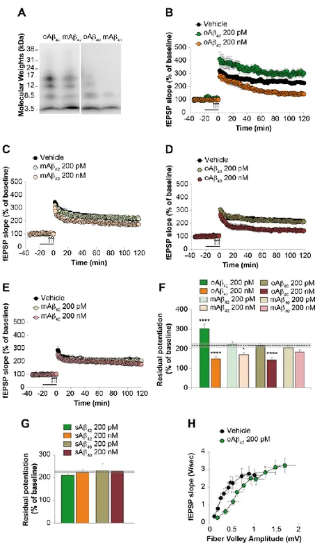
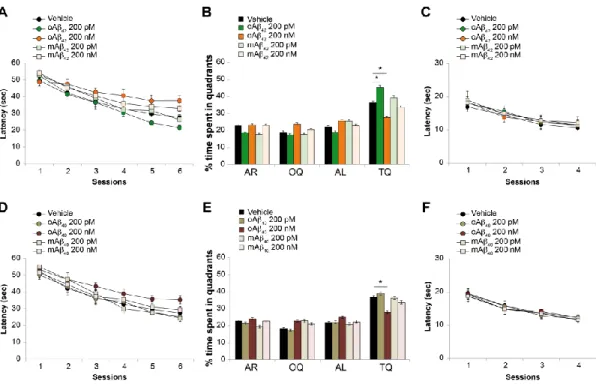
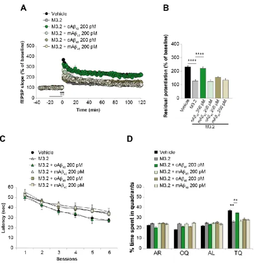
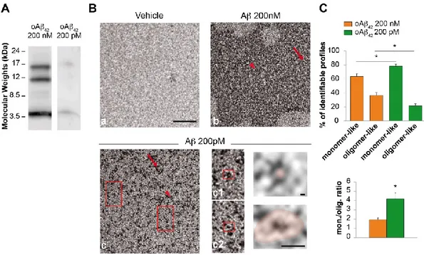
2. Chapters 1-3 focus on the physiological and pathological role of Aβ. In particular: • Chapter 1 reports a study performed to understand whether different
aggregation status (monomers vs. oligomers), concentrations (200 nM vs. 200 pM) and isoforms (Aβ40 vs Aβ42) of the peptide exert a beneficial or detrimental effect on hippocampal synaptic plasticity and memory (Gulisano et al, Neurobiol Aging, 2018).
• Chapter 2 reports a study aimed at understanding the relationship between Aβ and cGMP and the underlying molecular mechanisms during hippocampal
synaptic plasticity and memory in physiological conditions (Palmeri et al., J Neurosci, 2018).
• Chapter 3 reports a pre-clinical study in which the increase of cGMP and cAMP obtained by a treatment with sub-efficacious doses of phosphodiesterase inhibitors was able to revert the AD phenotype in animal models of AD (Gulisano et al., Neuropharm, 2018).
3. Chapter 4 and 5 focus on the role of tau and its interaction with Aβ. In particular: • Chapter 4 reports a study showing that an acute exposure to extracellular tau
oligomers is able to impair synaptic plasticity and memory. Here, the interaction between Aβ and tau was also studied by evaluating the effect of a combination of subthreshold doses of Aβ and tau oligomers (Fa’ et al., Sci Rep, 2016). • Chapter 5 reports a study focusing on the role of Amyloid precursor protein
(APP) as a common target mediating the detrimental effects of Aβ and tau on synaptic plasticity and memory (Puzzo et al., eLife, 2017).
4. A General Discussion will summarize the relevance of these findings giving new insights into possible future directions.
General introduction
“The saddest aspect of life right now is that science gathers knowledge faster than society gathers wisdom.”
Preface
Alzheimer’s disease (AD) is a neurodegenerative disorder clinically characterized by dementia, defined as a deficit of memory function and at least one other cognitive domain (language, praxis, gnosis, executive function, judgment, and abstract thought) as well as functional impairment, without alteration of the state of consciousness. In the last decades, AD has gained rising attention for its growing prevalence in aging populations, with 46.8 million people affected by the pathology worldwide, a number expected to increase up to 74.7 million in 2030 and 131.5 million in 2050. Besides representing a serious health and social problem, the disease causes exorbitant costs for the healthcare system estimated as 604 billion dollars in 2010 that represented a 35.4% increase in only 5 years1,2. Despite the numerous efforts to counteract the
disease, no therapies have so far proven to prevent AD onset or progression. To date, data from thousands of basic, pre-clinical, and clinical studies have identified amyloid-β peptide (Aamyloid-β) and tau protein as the key actors in the pathophysiology of AD, mainly because of their deposition in the characteristic histopathological brain lesions, the senile plaques for Aβ and the neurofibrillary tangles (NFTs) for tau, and the increase of their soluble forms in the brain of AD patients. However, therapeutic approaches aimed to decrease Aβ levels that have been attempted so far, have failed. Similarly, tau-based clinical trials have not yet produced positive findings.
The overall goal of this chapter is to provide a critical assessment of the literature on mechanisms underlying disease occurrence and progression. Specifically, we will revisit studies on Aβ and tau, as well as on their interaction, discussing the amyloid hypothesis - that has dominated the AD field in the last 25 years - and the role of tau protein.
Amyloid-β peptide and Alzheimer’s disease: more than one
century of research
Aβ derives from a complex cleavage of APP, a type I single-pass transmembrane protein constituted by 639–770 amino acids in humans, and highly expressed in the central nervous system where it exerts a variety of physiological functions3. APP is
initially cleaved by α-secretase or β-secretase, generating soluble and carboxyterminal fragments (CTF). α-secretase activity leads to the formation of sAPPα and CTF83, whereas β-secretase generates sAPPβ and CTF99. Then, γ-secretase intervenes, further cleaving CTF83 and CTF99, generating the intracellular peptide AICD/AID (amyloid intracellular domain) and a small p3 peptide from CTF83, and AICD/AID and Aβ from CTF99. Based on this biochemical processing, the cascade initiated by α-secretase has been considered neuroprotective when compared with the β-secretase cleavage, leading to the amyloidogenic cascade and the formation of Aβ4. Based on the γ-secretase site
of cutting, different isoforms of Aβ can be generated, composed of 38–43 amino acids. Aβ40 is the predominant species, whereas Aβ42 is present at lower concentrations but has received more attention in the AD field due to its high propensity to form aggregates. However, in the brain of AD patients, Aβ38 and truncated forms at N-terminal region, i.e., Aβ15, Aβ16, and Aβ17, have been also detected5. Aβ is
undoubtedly the most studied protein in AD and its putative role in the pathogenesis of the disease has oriented drug development and clinical trials for several decades. But how and why did the AD amyloidogenic theory emerge? From a historical perspective, it was at the beginning of the last century when Alois Alzheimer and other European neuropsychiatrists, e.g., Gaetano Perusini, attributed a nosographic identity to a form of “mental” disorder characterized by memory loss, hallucinations, and disorientation. At that time, the most influent personalities in psychiatry, Sigmund Freud and Emilin Kraeplin, fervently disputed on the origin of psychiatric illness, respectively emphasizing the role of the psyche or of organic and genetic factors. The mind/brain diatribe led several scientists to seek for the “material” causes of mental diseases. In this context, Alzheimer and Perusini, strongly supported by Kraeplin,
observed that the psychiatric symptoms of dementia could be correlated to peculiar histological lesions in postmortem brains. In the autopsy of the first described AD patient, Auguste Deter, cortical atrophy, neurons filled with neurofibrils, and extracellular miliary foci of an unknown substance were observed. After Alzheimer’s death, research studies on the disease were few until the 1980s, when epidemiological studies revealed an increase of patients affected by primary dementia.
It was during these years that key discoveries were made, fated to influence research in the field until today. Based on Alzheimer’s histological descriptions, Aβ and tau were recognized as the main components of extracellular foci (senile plaques) and intracellular neurofibrils (NFTs), respectively6–8. In the same period, the first genetic
mutation linked to dementia was identified on chromosome 21 coding for the APP9.
This autosomal dominant disease was responsible for early onset AD (EOAD) characterized by high levels of Aβ. Other genetic mutations were identified in Familiar Alzheimer’s disease (FAD), involving genes responsible for Aβ production such as presenilin 1 (PS1) on chromosome 14, which mutation is the most common cause of EOAD, and presenilin 2 (PS2) on chromosome 1. Consistent with these findings, the presence of AD-like pathology in patients affected by Down’s syndrome, due to a trisomy of chromosome 21, reinforced the idea that the increase of Aβ played a major role in AD pathogenesis. Based on these data, in 1995 the first mouse model of AD carrying an APP mutation was engineered10 and, over time, different models for
pre-clinical studies have been generated based on the most common mutations observed in FAD11. These findings contributed to the excitement around the “Amyloid Cascade
Hypothesis”12–14, recognized as the pathogenic mechanism underlying AD. Because
insoluble fibrils of Aβ were present in AD plaques, and could be formed in vitro from synthetic Aβ, they have dominated the scene until a fundamental breakthrough confirmed by several in vitro and in vivo studies indicated that soluble forms of Aβ were also present in the brain15,16. Aβ soluble aggregates range from monomers to
oligomers (molecular aggregates consisting of a few monomer units) and pre-clinical studies confirmed that dimers, trimers, tetramers, dodecamers, and high molecular weight oligomers were all able to induce neurotoxic effects as well as to induce an
immediate impairment of synaptic plasticity, and in particular of hippocampal long-term potentiation (LTP), thought to be the electrophysiological correlate of memory (for a review on the role of Aβ oligomers, see17). Moreover, Aβ oligomer presence in
human cerebrospinal fluid (CSF) could be already recognized decades before AD onset18. These data led to the formulation of another theory, the “Oligomer
Hypothesis”19–21, according to which Aβ oligomers but not monomers or fibrils were
responsible for synaptic dysfunction and memory loss in AD21,22. This further
influenced AD drug discovery so that new therapies aimed at specifically targeting Aβ oligomers were developed in addition to those clearing Aβ plaques. Unfortunately, while the “Oligomer Hypothesis” is still a matter of investigation, and data are being gathered to test the grounds of its premises, the clinical failure of most of the anti-Aβ drugs has strongly destabilized this concept. Clinical trials to date show that, despite successful results obtained in animal models of AD, anti-Aβ drugs have not yet been shown to modify cognition in humans although they might be able to reduce plaque or amyloid burden. So far (based on Medline database search and Clinical-Trials.gov): 1) active immunization (i.e., AN-1792, CAD-106, and vanutide cridificar) have not proven effective and several side effects were reported; 2) passive immunization with monoclonal antibodies bapineuzumab, solanezumab, crenezumab, and gantenerumab have not yet succeeded, and although a recent clinical trial with aducanumab has shown a dose-dependent reduction of Aβ plaques, the study was not sufficiently powered to detect clinical changes and the drug is undergoing further investigation23; and 3) a
number of clinical trials with drugs aimed at preventing Aβ formation by inhibiting β- or γ- secretases have also failed or were interrupted; among these, the γ-secretase inhibitors semagacestat and avagacestat did not show efficacy, and actually induced mildworsening in cognition and severe side effects, whereas the EPOCH trial with the newest β-secretase inhibitor verubecestat was stopped for the lack of any positive effect. Notwithstanding these discouraging results, several scientists are still developing anti-Aβ therapies, convinced that the failure of Aβ tailored drugs might relate to the particular drugs chosen, inadequate dosage, or the fact that treatment was started in a late phase of the disease when Aβ-induced damage cannot be reversed.
This chapter is written, in turn, with the belief that a careful evaluation of the knowledge in the AD field is due prior to further investing resources with anti-Aβ therapies. Evidences that have been underestimated for a long time are now gaining ground, questioning the way in which the actual role of Aβ in AD pathogenesis is currently thought. First, late onset AD (LOAD), representing 95% of AD cases, is not linked to genetic anomalies leading to a direct overproduction of Aβ, as in FAD, although the phenotype might be comparable. However, pre-clinical studies on AD mouse models have been almost entirely performed on mice presenting FAD-like mutations leading to an increase of Aβ. Second, we know since the 1990s that there is no correlation between Aβ deposition and clinical degree of dementia among affected individuals24–27, and plaques might occur in the brains of individuals with no sign of
dementia26,28,29. Third, recent studies have suggested that plaque formation might be a
reactive process30 with a protective role by decreasing oligomer levels31. Fourth, a vast
literature claims that Aβ exerts a physiological role in the CNS interfering with neuronal growth, neurotransmitter release, synaptic function, and memory formation32,33. Indeed, our group and others have previously demonstrated that
administration of low concentrations of oligomeric Aβ positively modulate synaptic function34–36 and, conversely, blocking endogenous Aβ in the healthy brain resulted in
an impairment of synaptic plasticity and memory35,37. Finally, even Aβ concentration
per se has become a relative concept, as the persistence of a low picomolar Aβ concentration in extracellular fluids provides for detrimental outcomes in synaptic plasticity38. In conclusion, taking into account almost one century of research, it
A revaluated player in Alzheimer’s disease pathogenesis: Tau
protein
As described above, the intricate story of Aβ and tau began with the brain of Auguste Deter, but most of the research efforts have been directed toward Aβ. Recently, the discontent generated by too many anti-Aβ therapy failures has induced several groups to re-focus on tau. Tau is a microtubule-associated protein originally described as a heat stable protein essential for microtubule assembly and stabilization41. It is present
in the human brain in six main isoforms, deriving from the alternative splicing of exons 2, 3, and 10 of microtubule-associated protein tau (MAPT) gene. This process appears to be of particular interest for exon 10 splicing which determines the presence of tau isoforms containing 3- (3R) or 4-repeats (4R) of a ∼32 amino acid sequence in the microtubule binding domain (MBD)42. Moreover, the splicing process of exons 2 and
3 determines the number of 29-residue near-amino-terminal inserts which results in isoforms containing 0, 1, or 2 inserts (0N, 1N, 2N)43. Both R and N repeats are capable
of microtubule-binding and assembly-promoting activity, whereas the flanking regions are more likely involved in binding processes44,45. In the last decades, many studies
have demonstrated tau physiological involvement at different subcellular localizations: 1) at axonal level, by regulating axonal elongation, maturation and transport46–49; 2) in
dendrites, participating in synaptic plasticity50,51; and 3) in nucleus, maintaining the
integrity of genomic DNA, cytoplasmic and nuclear RNA52,53. From a functional point
of view, tau can be divided in four different regions consisting of a N-terminal domain, a proline-rich domain, a MBD, and a C-terminal domain54–56. The N-terminal domain
is rich with negative charges devoted to separation of different microtubules by electrostatic repulsion when tau is bound to a certain microtubule45,57,58. Interestingly,
the C-terminal domain, besides its key role in regulation of microtubule polymerization induction and interaction with plasma membranes59–62, creates an overall asymmetry
in the molecule contributing to this microtubule spacing function thanks to its neutral charge. The proline-rich domain and the MBD with their multiple aminoacidic acceptor
residues are more involved in interactions with different signaling proteins, which can be scaffolded by tau or can modify tau conformational status and activity itself54. The
presence of multiple binding sites confers to tau many interaction possibilities in regards to cell signaling. The flanking region of MBD contains the majority of phosphate acceptor residues, and the phosphorylation of these sites is relevant for regulating microtubule polymerization63–66, regulation of axonal transport67 and
neurotransmitter receptors68,69, interference with vesicles trafficking70 and
apolipoprotein E71, interaction with Src-family kinases62,72–75, and many others54–56.
The multiple roles of tau in neuronal physiology have been widely studied and, undoubtedly, a fine regulation is needed to maintain tau structure and function. Accordingly, a wide range of neurodegenerative disorders known as tauopathies have been recognized and classified with respect to the predominant species of tau that accumulates: 1) 3R tauopathies (i.e., Pick’s disease); 2) 4R tauopathies (i.e., corticobasal degeneration and progressive supranuclear palsy); and 3) 3R + 4R tauopathies (i.e., AD)42. Biochemical studies have demonstrated that deposition of
insoluble tau aggregates in NFTs depends upon a dysregulated phosphorylation process of the flanking regions of tau. In fact, while two phosphates per molecule of tau are normally present76, analysis of tau from AD brains has revealed the presence of about
eight phosphates per molecule of tau77. For this reason, tau phosphorylation
abnormalities have been linked to misfolding and deposition of the protein in the diseased brain78. Although tau has been defined as a “natively unfolded” protein with
a low tendency to aggregation79, phosphorylation of certain residues or detachment
from microtubules79–81 might change its conformational status and consequently its
aggregation propensity. However, the undefined structure of tau in solution has precluded crystallographic analyses leaving a lack of knowledge about the protein structure82. Moreover, notwithstanding electron microscopic visualization of tau bound
to microtubules demonstrated a linear alignment lengthwise to the protofilament ridges, the protein structure keeps holding a disordered state83,84. Interestingly, when in a
solution, tau spontaneously tends to modify its conformation in favor of a paperclip-like structure that might prevent its aggregation55,82, unlike Aβ that has a high tendency
to aggregate in a solution due to its biochemical properties. Thus, alterations of tau (i.e., hyperphosphorylation, truncated forms) could inhibit the constitution of the paperclip-like structure leading to paired helical filament (PHF) and NFT formation85.
In this context, tau hyperphosphorylation has been widely studied and the sequence hyperphosphorylation→PHFs→NFTs linked to AD, even if it is unlikely to represent by itself the main pathogenic event for several reasons. First, tau phosphorylation has been demonstrated to be responsible for aggregation only when occurring at certain residues86, whereas in other sites it has the opposite effect thus preventing
aggregation80. Moreover, tau hyperphosphorylation is not a prerogative of AD, since it
occurs in several other conditions such as hypothermia87, starvation88, chronic stress89,
and anesthesia90,91. Interestingly, the amount of PHFs and NFTs is slightly related to
the severity of neuronal damage and cognitive impairment in humans. Experiments on regulatable mouse models of tauopathy demonstrated that a variant of human tau with the pro-aggregant mutation ΔK280 developed synaptic and memory impairment as well as tau hyperphosphorylation and pre-tangle formation. However, when the proaggregant tau was turned off, synaptic deficit was rescued even if insoluble tau was still present92. Other studies on transgenic mice expressing mutant tau (P301L
mutation), which could be suppressed with doxycycline, demonstrated that behavioral impairment and neuronal loss were recovered when suppressing transgenic tau, whereas NFTs accumulation continued93. Moreover, in the P301S model of tauopathy,
synaptic damage and cognitive impairment occurred before the emergence of NFTs94.
Some authors also reported that, in vitro, abnormally phosphorylated tau can sequester normal tau into tangles of filaments, leading to the hypothesis that tau accumulation into PHFs might initially be neuroprotective until it starts compromising neuronal function as a space-occupying lesion95. The observations that synaptic and memory
impairment is not mediated by NFTs, and that insoluble deposition of tau might be a compensatory protective mechanism suggested that synaptic failure might be sought in soluble oligomeric species of tau, resembling the “Oligomeric Hypothesis” already formulated for Aβ. Soluble tau was found to be most acutely toxic in animal models of tauopathy93,94,96. Most importantly, increases in granular tau oligomer levels occur
before NFTs form and before individuals manifest clinical symptoms of AD, suggesting that increases in tau oligomer levels may represent a very early sign of brain aging and AD97. We have recently demonstrated that an acute exposure to tau
oligomers (but not monomers) both in vitro and in vivo is detrimental to LTP and memory98. Noteworthy, this toxic effect was exerted by a different preparation of
oligomeric tau, i.e., recombinant tau 4R/2N, tau derived from AD patients, tau derived from hTau mice98. These results are in agreement with other observations reporting that
tau oligomers 1) impair synaptic function and memory in wild type mice99, 2) correlate
with cognitive impairment in rTg4510 mice100, and 3) accelerate pathology in hTau
mice101. Pre-clinical findings have been confirmed by studies on humans showing the
increase of oligomeric forms of tau in the brain of AD patients compared to controls, occurring before NFT formation and clinical symptoms97. Interestingly, tau oligomers
have been also found in other tauopathies such as progressive supranuclear palsy, dementia with Lewy bodies, and Huntington’s disease101–103.
These aspects should be taken into account when designing new drugs targeting tau to avoid the same issues already experienced with anti-Aβ treatments. Notwithstanding the increase of tau oligomers in the AD brain and CSF, drugs aimed at inhibiting tau aggregation or dissolving existing aggregates, i.e., methylthioninium chloride and its second-generation derivatives such as TRx0237, have not been proven efficacious in clinical trials. A Phase II study with TRx0237 was terminated after a few months for “administrative” reasons, whereas Phase III studies have reported negative results on cognitive improvement (see clinicaltrials.gov for details). However, it is not clear whether these drugs actually inhibit tau aggregation in humans. Also, this makes us wonder whether the increase of tau oligomers in AD patients should be better considered as a pathogenic marker of the disease rather than a target of therapeutic strategies.
Aβ and tau oligomers: a game at the synapse resulting in memory
impairment
How do Aβ and tau induce memory loss? According to most of the studies, the answer should be sought at the synapse. Although cortical atrophy and synaptic loss have been reported in AD brains, mainly due to a structural damage imputable to plaques and tangles in a later stage of the disease, a subtle effect exerted by soluble forms of Aβ and tau at the synapse seems to be the earlier event underlying memory loss98,99,104,105.
Several studies have demonstrated that administration of different preparations of oligomeric Aβ and tau (synthetic, from transgenic mice, from AD brains) impaired synaptic plasticity and memory. The role of soluble oligomers also emerged in studies performed on AD mouse models, since synaptic and memory dysfunction was present before the appearance of plaques or tangles17,106. In vitro and in vivo studies have
shown that Aβ and tau derange molecular signaling pathways crucial for synaptic plasticity at both pre- and post-synaptic sites. Both Aβ and tau interfere with the transcription factor cAMP response element-binding protein (CREB), whose phosphorylation at Ser133 is thought to be one of the fundamental events in memory formation107–109. In particular, Aβ inhibits the physiological increase of CREB
phosphorylation during LTP110–112, causing a downregulation of both the
NO/cGMP/PKG and the cAMP/PKA pathways, two cascades converging on CREB. Tau overexpression and hyperphosphorylation was also found to be accompanied by a reduction of CREB phosphorylation at Ser133, mediated by a decrease of phosphorylation of NR2B (Tyr1472)113. Moreover, synaptic plasticity and memory
impairment caused by h-tau overexpression was reported to be related to nuclear dephosphorylation/inactivation of CREB114. Interestingly, these findings were
validated in humans affected by AD showing a decrease in CREB and phospho-CREB levels in hippocampus115–119. Aβ and tau also target other molecules upstream of
CREB, among which the Ca2+/calmodulindependent protein kinase II (CaMKII), another key molecule needed for LTP and memory formation120. CaMKII is
see121) and it has been demonstrated that Aβ oligomers interfere with its
phosphorylation leading to AMPA receptor dysfunction122–124. On the other hand,
evidences for the interaction tau-CaMKII have been reported since the late 1980 s in works analyzing the ability of CaMKII to induce an AD-like tau phosphorylation125,126.
CaMKII phosphorylates tau at different sites and this might prevent tau binding to microtubule127 and modify tau structure leading to PHFs formation128. Indeed, CaMKII
colocalizes with tau mRNA, PHFs, NFTs in AD brains (for a review, see121). Recently,
in a drosophila model of tauopathy, suppression of tau phosphorylation at Ser262/356 inhibited tau toxicity through a mechanism involving calcium homeostasis dysregulation driven by CaMKII129. The deleterious effects of Aβ and tau also involved
BDNF, a critical factor linked to neuronal survival and function that is needed for synaptic plasticity and memory. A decrease of BDNF levels in serum and brains of AD patients correlates with cognitive impairment, and BDNF polymorphisms have been proposed to be involved in AD pathogenesis130. Moreover, several in vitro and in vivo
studies have confirmed that Aβ-induced LTP and cognitive dysfunction are associated with a reduction of BDNF levels130. Recently, a loss of BDNF has been also reported
in THY-Tau22 and P301L mouse models of tau pathology131,132.
Taken all together, these findings suggest that restoring synaptic-related molecules and second messenger systems regulating memory mechanisms might be a viable therapeutic strategy to counteract AD112. Most importantly, these data point at common
Aβ and tau activity dependent secretion, neuronal uptake, and
spreading of the disease
Because Aβ and tau interfere with the synaptic machinery, another relevant subject of investigation has been to determine whether they act via extracellular or intracellular mechanisms. Based on the localization of insoluble deposits, for several years Aβ has been considered an extracellular protein and tau an intracellular one. However, it is now clear that this rigid vision is no more applicable, since both Aβ and tau can be found inside and outside neurons. Notwithstanding most of the studies have been performed on models of disease, the extra- and intracellular presence of Aβ and tau is the result of a physiological dynamic process in which the two proteins are secreted at the synapse and internalized by neurons. A relevant body of data has supported the hypothesis that neurons are able to secrete Aβ in an activity dependent fashion. In vitro studies performed by applying drugs that decrease (i.e., tetrodotoxin or high magnesium) or increase (i.e., picrotoxin) neuronal activity have shown a concomitant decrease or increase of Aβ secretion in organotypic slices overexpressing human APP Swedish mutation133. An in vivo approach by using microdialysis also revealed an
increase of Aβ levels in the brain interstitial fluid concomitant to the increase of synaptic activity134 or paralleling the neurological status135. An increase of Aβ secretion
has also been found during learning in healthy wild-type mice37. Based on the fact that
synaptic activity stimulates Aβ secretion, and that extracellular Aβ is known to reduce synaptic plasticity, it has been proposed a theory according to which an increase of synaptic (and cognitive) activity is linked to AD pathogenesis. However, although an increase of brain activity in AD could be supported by data indicating hyperexcitability in transgenic mice and human AD patients136,137, this activity-dependent role of Aβ
should be better viewed as a physiological mechanism occurring within the healthy brain, especially because levels of Aβ secreted during activity are in the picomolar range and are not neurotoxic34,37,138. Thus, the high increase of extracellular Aβ during
AD might be due to a derangement of this physiological loop or it could be a consequence of degeneration of neurons that have previously accumulated Aβ at
intracellular level (for a review, see139). Whether the impairment of synaptic function
is directly mediated by these high extracellular Aβ levels or by Aβ accumulated inside neurons, is still a matter of debate.
Surely, these two pools are strictly interconnected, since extracellular Aβ induced the accumulation of intracellular Aβ by stimulating APP processing140 or by a direct
APP-mediated internalization141; in turn, intracellular Aβ disrupts synaptic transmission and
plasticity142. Interestingly, tau also undergoes the same dynamic flux characterized by
activity-dependent secretion and neuronal internalization. Indeed, application of KCl or glutamate to cultured neurons resulted in an increase of tau secretion98,143 mediated
by AMPA receptor activation143. In vivo studies reported an increase of tau in brain
interstitial fluid when stimulating neurons with high K+ perfusion, or after stimulation of the N-Methyl-d-aspartic acid (NMDA) receptors, or picrotoxin administration144.
An increase of tau secretion also paralleled the increase of glutamate release induced by an antagonist of metabotropic glutamate receptors 2/3144. The phenomenon was
further confirmed in different cultured neural cell lines where extracellular tau levels were modified proportionally to synaptic activity145. On the other hand, several
pre-clinical studies have demonstrated that exogenously applied tau can increase of Aβ secretion in organotypic slices be internalized by neurons98,146–149 and glial cells150–152
with different mechanisms involving bulk endocytosis149, binding to heparan sulfate
proteoglycans153 or to APP141. Activity dependent secretion and neuronal uptake of Aβ
and tau have been related to the spread of the disease throughout the brain, a process known as spreading which refers to the capability of neurotoxic proteins to diffuse from a neuron to another, expanding the disease from a restricted area to the entire brain. This type of dissemination, defined as “trans-synaptic spreading”, is thought to occur among different brain areas functionally connected154,155 and is supported by
observations on postmortem AD brains as well as by clinical studies exploiting computerized x-ray tomography (CT) and magnetic resonance imaging (MRI) techniques, that allow tracing different neuropathological markers such as atrophy of certain brain areas, brain ventricles enlargement, and deposition of amyloid plaques and NFTs (for a review, see78). However, it should be pointed out that imaging
biomarkers like fluorodeoxyglucose in PET scans are associated to discrete difficulties in data interpretation, as they are also positive in Suspected Non-Alzheimer Disease Pathophysiology (SNAP)156. Evidence for AD spreading and progression throughout
the cortex was reported more than 30 years ago, based on tangle distribution in the proximity of the same pyramidal neurons that give connectivity to other brain areas157.
At the present day, neither the cause that initiates spreading nor its underlying mechanisms have been identified, but useful information has come from pre-clinical studies. Notwithstanding tau has been under the spotlight for many years, one of the first evidence of spreading in AD dates back to the 1990 s and involves Aβ158,159. When
trying to unravel the causes of Aβ diffusion, studies have often focused on the first area affected in AD, the medial temporal lobe, and in particular, the entorhinal cortex (EC). EC superficial layer is susceptible to Aβ-dependent neurodegeneration, and this can negatively affect its primary afferent regions resulting in a disruption of the whole circuitry in both mouse models and AD patients160,161. Consistently, an increase of
mutant APP in layer II/III neurons of EC has been shown to elicit a molecular and functional disruption in the CA1 area of the hippocampus with presence of soluble Aβ in the dentate gyrus, Aβ deposits in the performant pathway, and epileptiform activity in the parietal cortex162. Further studies in mutant human APP (mhAPP) mice have
reported an age-dependent progressive deterioration of synaptic plasticity and memory spreading from the EC to the hippocampus163, a phenomenon mediated by microglial
RAGE activation and subsequent increase in p38MAPK phosphorylation163.
Consistently, other studies reported the capability of reactive microglia in secreting Aβ through microvesicles, which in turn would promote Aβ toxicity to neurons through their axons164–166. Accordingly, other supporting evidences indicate that after
administration of fluorescent oligomeric Aβ to neurons, a higher percentage of the protein was found surrounding neurons, and this process needed the presence of differentiated neuritis to occur167. Cell-to-cell transfer mechanism has been reported
for different Aβ species (i.e., oAβ1–42 TMR, oAβ3(pE)–40TMR, oAβ1–40TMR, and oAβ11–42TMR), and this prion-like spreading was attributed to an insufficient activity of cellular clearance degradation systems168. Another mechanism proposed for Aβ
spreading relies on the presence of tunneling nanotubes (TNTs) consisting of cellular membrane extensions creating a direct connection between cells169. TNTs have been
demonstrated to mediate highspeed transfer of Aβ among neurons, through a p53/EGFR/Akt/PI3K/mTOR pathway that, in turn, would trigger F-actin polymerization promoting TNTs formation170. However, Aβ has been shown to be
secreted by neurons through exosomes171 that could be internalized and stored from the
acceptor neuron as lysosomal vesicles through a macroautophagy mediated mechanism167,172. In any case, despite these numerous evidences, there is not a uniform
consensus about the causes or mechanisms underlying Aβ spreading. On the other hand, a growing body of evidence refers to tau spreading as a prion-like propagation, which fascinatingly occurs in different directions among the many forms of tauopathies173. Also, tau pathology is likely to begin in EC then move to the
hippocampus, and ultimately invading the cortex, following an overlapping path existing among functionally connected areas55,154,155,174. These evidences are consistent
with data coming from studies on non-human primates in which bilateral lesions of EC induce a functional impairment of declarative memory accompanied by long-lasting hypometabolism in temporal and parietal lobes, demonstrating a functional connection starting from EC175. Accordingly, in a transgenic mouse model differentially
expressing pathological human tau in EC (EC-tau), the localization of tauopathy was investigated at different time points, demonstrating a progression of the pathology through anatomically and functionally connected brain areas155. Interestingly, in vivo
chemogenetic stimulation of EC in EC-tau mice induced additional pathology in synaptically connected areas (e.g., dentate gyrus)145. Consistent with this finding, tau
has been found in exosomes that might lead to its diffusion to adjacent cells176,177.
Further work demonstrated that cell-to-cell contact was not necessarily needed for tau spreading in vitro given that the administration of neuronal-derived tau media to neuronal cultures was sufficient for tau transfer and internalization, even though it is not known whether tau in the media was vesicle bound or free145. Other studies
suggested that pathologic tau requires TNTs to be transferred from a neuron to another one178. However, whether the mechanism underlying tau propagation is mediated by
TNTs, non-vesicular direct translocation or through secretory lysosomes into extracellular space159,176,179,180 is still under investigation. Another interesting feature
of tau transmission is the possibility that it can move both anterogradely and retrogradely, meaning that it can be internalized both at the somatodendritic compartment and axon terminals, and can be transported in either direction to disseminate tauopathy149,159. While spreading is involved in the progression of the
disease among functionally connected brain areas, the transition from oligomers to insoluble deposits has been described as a “nucleation dependent protein polymerization” and explains the pattern of aggregate formation181 for proteins with
high tendency to organize in β-sheet conformation as for Aβ, tau, or α-synuclein182.
This process, known as seeding, involves a nucleation phase and a growth phase. In the nucleation phase, the nucleus formation requires the assembly of misfolded monomers, a thermodynamically unfavorable process remarkably dependent on protein concentration158,183,184. The latter influences the lag time defined as the period before
aggregates detection. In fact, supersaturated solutions can drastically shorten the nucleus formation time from years to microseconds158. After the nucleus formation, the
critical concentration is reached, and a further addition of monomers occurs leading to polymerization, representing the growth phase. Interestingly, if a preformed nucleus, or seed, is added to a solution containing normally folded monomers, an immediate polymerization occurs. This phenomenon is defined as seeding158,181 and can be
distinguished as homologous or heterologous181,185. While homologous seeding
involves monomers of the same type, heterologous seeding or cross-seeding takes place when a nucleus formed by a certain misfolded protein promotes polymerization of a different protein181,185. A large body of evidence supports this cross-seeding among tau,
α-synuclein and TDP-43186. Some studies in which spreading of tau pathology was
significantly accelerated by injecting pre-aggregated Aβ into mouse brain187,188
suggested the possibility of Aβ and tau cross-seeding. Consistently, a protein interaction study by surface plasmon resonance demonstrated an affinity constant of tau for Aβ which was almost 1000-fold higher than for tau toward itself189. Moreover,
in which Aβ and tau coexisted in the same structure189. Also, a recent work showed
that tau fibrillization can be induced in a cell-free assay by adding pre-aggregated Aβ, and that Aβ provide an efficient seed to induce tau cross-seeding and a consequent spreading of tau pathology in vivo190. In conclusion, seeding and spreading of Aβ and
tau and their dynamic flux across the membrane characterized by activity-dependent secretion and neuronal internalization are crucial for the progression of the disease. Most importantly, the commonalities displayed by both Aβ and tau with respect to these phenomena are intriguing and suggest that soluble forms of the two molecules are involved in similar mechanisms of disease etiopathogenesis.
References
1. Martin Prince, A. et al. World Alzheimer Report 2015 The Global Impact of Dementia An Analysis of prevalence, Incidence, cost and Trends. (2015).
2. Wimo, A. et al. The worldwide costs of dementia 2015 and comparisons with 2010. Alzheimers.
Dement. 13, 1–7 (2017).
3. Muller, U. C. & Zheng, H. Physiological Functions of APP Family Proteins. Cold Spring Harb.
Perspect. Med. 2, a006288–a006288 (2012).
4. Zhang, Y., Thompson, R., Zhang, H. & Xu, H. APP processing in Alzheimer’s disease. Mol.
Brain 4, 3 (2011).
5. Mawuenyega, K. G., Kasten, T., Sigurdson, W. & Bateman, R. J. Amyloid-beta isoform metabolism quantitation by stable isotope-labeled kinetics. Anal. Biochem. 440, 56–62 (2013). 6. Glenner, G. G. & Wong, C. W. Alzheimer’s disease: Initial report of the purification and
characterization of a novel cerebrovascular amyloid protein. Biochem. Biophys. Res. Commun. 120, 885–890 (1984).
7. Glenner, G. G. & Wong, C. W. Alzheimer’s disease and Down’s syndrome: Sharing of a unique cerebrovascular amyloid fibril protein. Biochem. Biophys. Res. Commun. 122, 1131–1135 (1984).
8. Grundke-Iqbal, I. et al. Microtubule-associated protein tau. A component of Alzheimer paired helical filaments. J Biol Chem 261, 6084–6089 (1986).
9. Levy, E. et al. Mutation of the Alzheimer’s disease amyloid gene in hereditary cerebral hemorrhage, Dutch type. Science 248, 1124–6 (1990).
10. Games, D. et al. Alzheimer-type neuropathology in transgenic mice overexpressing V717F b-amyloid precursor protein. Lett. to Nat. 373, 523–527 (1995).
11. Puzzo, D., Gulisano, W., Palmeri, A. & Arancio, O. Rodent models for Alzheimer’s disease drug discovery. Expert Opin. Drug Discov. 10, 703–711 (2015).
12. Hardy, J. & Allsop, D. Amyloid deposition as the central event in the aetiology of Alzheimer’s disease. Trends in Pharmacological Sciences 12, 383–388 (1991).
13. Hardy, J. & Higgins, G. Alzheimer’s disease: the amyloid cascade hypothesis. Science (80-. ). 256, 184–185 (1992).
14. Hardy, J., Duff, K., Hardy, K. G., Perez-Tur, J. & Hutton, M. Genetic dissection of Alzheimer’s disease and related dementias: amyloid and its relationship to tau. Nat. Neurosci. 1, 355–8 (1998).
15. Wisniewski, T., Ghiso, J. & Frangione, B. Alzheimer’s disease and soluble A beta. Neurobiol.
Aging 15, 143–52 (1994).
16. Lambert, M. P. et al. Diffusible, nonfibrillar ligands derived from A 1-42 are potent central nervous system neurotoxins. Proc. Natl. Acad. Sci. 95, 6448–6453 (1998).
17. Walsh, D. M. & Selkoe, D. J. Aβ oligomers - A decade of discovery. J. Neurochem. 101, 1172– 1184 (2007).
18. Fukumoto, H. et al. High-molecular-weight beta-amyloid oligomers are elevated in cerebrospinal fluid of Alzheimer patients. FASEB J. 24, 2716–2726 (2010).
19. Hardy, J. & Selkoe, D. J. The Amyloid Hypothesis of Alzheimer ’ s Disease : Progress and Problems on the Road to Therapeutics. Science (80-. ). 297, 353–357 (2002).
20. Ferreira, S. T. & Klein, W. L. The Aβ oligomer hypothesis for synapse failure and memory loss in Alzheimer’s disease. Neurobiol. Learn. Mem. 96, 529–43 (2011).
21. Ripoli, C. et al. Effects of different amyloid β-protein analogues on synaptic function. Neurobiol.
Aging 34, 1032–1044 (2013).
22. Attar, A. et al. Protection of primary neurons and mouse brain from Alzheimer’s pathology by molecular tweezers. Brain 135, 3735–3748 (2012).
23. Sevigny, J. et al. The antibody aducanumab reduces Aβ plaques in Alzheimer’s disease. Nature 537, 50–56 (2016).
25. Arriagada, P. V et al. Neurofibrillary tangles but not senile plaques parallel duration and severity of Alzheimer’s disease. Neurology 42, 631–639 (1992).
26. Dickson, D. W. et al. Correlations of synaptic and pathological markers with cognition of the elderly. Neurobiol. Aging 16, 285–298 (1995).
27. Sloane, J. A. et al. Lack of correlation between plaque burden and cognition in the aged monkey.
Acta Neuropathol. 94, 471–478 (1997).
28. Katzman, R. et al. Clinical, pathological, and neurochemical changes in dementia: A subgroup with preserved mental status and numerous neocortical plaques. Ann. Neurol. 23, 138–144 (1988).
29. Delaere, P. et al. Large amounts of neocortical beta A4 deposits without neuritic plaques nor tangles in a psychometrically assessed, non-demented person. Neurosci Lett 116, 87–93 (1990). 30. Reitz, C. Alzheimer’s disease and the amyloid cascade hypothesis: a critical review. Int. J.
Alzheimers. Dis. 2012, 369808 (2012).
31. Cheng, I. H. et al. Accelerating amyloid-beta fibrillization reduces oligomer levels and functional deficits in Alzheimer disease mouse models. J Biol Chem 282, 23818–23828 (2007). 32. Puzzo, D. & Arancio, O. Amyloid-β peptide: Dr. Jekyll or Mr. Hyde? J. Alzheimers. Dis. 33
Suppl 1, S111-20 (2013).
33. Puzzo, D., Gulisano, W., Arancio, O. & Palmeri, A. The keystone of Alzheimer pathogenesis might be sought in Aβ physiology. Neuroscience 307, 26–36 (2015).
34. Puzzo, D. et al. Picomolar amyloid-beta positively modulates synaptic plasticity and memory in hippocampus. J. Neurosci. 28, 14537–45 (2008).
35. Morley, J. E. et al. A physiological role for amyloid-β protein: Enhancement of learning and memory. J. Alzheimer’s Dis. 19, 441–449 (2010).
36. Lawrence, J. L. M. et al. Regulation of presynaptic Ca2+, synaptic plasticity and contextual fear conditioning by a N-terminal betaamyloid fragment. J Neurosci 34, 14210–14218 (2014). 37. Puzzo, D. et al. Endogenous amyloid-β is necessary for hippocampal synaptic plasticity and
memory. Ann. Neurol. 69, 819–30 (2011).
38. Koppensteiner, P. et al. Time-dependent reversal of synaptic plasticity induced by physiological concentrations of oligomeric Aβ42: an early index of Alzheimer’s disease. Sci. Rep. 6, 32553 (2016).
39. Glass, D. J. & Arnold, S. E. Some evolutionary perspectives on Alzheimer’s disease pathogenesis and pathology. Alzheimers. Dement. 8, 343–51 (2012).
40. Herrup, K. The case for rejecting the amyloid cascade hypothesis. Nat. Neurosci. 18, 794–799 (2015).
41. Weingarten, M. D., Lockwood, A. H., Hwo, S. Y. & Kirschner, M. W. A protein factor essential for microtubule assembly. Proc. Natl Acad. Sci. USA 72, 1858–1862 (1975).
42. Dickson, D. W., Kouri, N., Murray, M. E. & Josephs, K. A. Neuropathology of frontotemporal lobar degeneration-tau (FTLD-tau). J. Mol. Neurosci. 45, 384–9 (2011).
43. Liu, C., Götz, J. & Gotz, J. Profiling murine tau with 0N, 1N and 2N isoform-specific antibodies in brain and peripheral organs reveals distinct subcellular localization, with the 1N isoform being enriched in the nucleus. PLoS One 8, e84849 (2013).
44. Trinczek, B., Biernat, J., Baumann, K., Mandelkow, E. M. & Mandelkow, E. Domains of tau protein, differential phosphorylation, and dynamic instability of microtubules. Mol. Biol. Cell 6, 1887–902 (1995).
45. Kar, S., Fan, J., Smith, M. J., Goedert, M. & Amos, L. A. Repeat motifs of tau bind to the insides of microtubules in the absence of taxol. EMBO J. 22, 70–7 (2003).
46. Dawson, H. N. et al. Inhibition of neuronal maturation in primary hippocampal neurons from tau deficient mice. J. Cell Sci. 114, 1179–1187 (2001).
47. Vershinin, M., Carter, B. C., Razafsky, D. S., King, S. J. & Gross, S. P. Multiple-motor based transport and its regulation by Tau. Regul. by Tau. Proc Natl Acad Sci U S A 104, 87–92 (2007). 48. Yuan, A., Kumar, A., Peterhoff, C., Duff, K. & Nixon, R. A. Axonal transport rates in vivo are
unaffected by tau deletion or overexpression in mice. J. Neurosci. 28, 1682–1687 (2008). 49. Ittner, L. M., Ke, Y. D., Götz, J. & Gotz, J. Phosphorylated Tau interacts with c-Jun N-terminal
(2009).
50. Frandemiche, M. L. et al. Activity-Dependent Tau Protein Translocation to Excitatory Synapse Is Disrupted by Exposure to Amyloid-Beta Oligomers. J. Neurosci. 34, 6084–6097 (2014). 51. Qu, X. et al. Stabilization of dynamic microtubules by mDia1 drives Tau-dependent
Aβ1-42synaptotoxicity. J. Cell Biol. 216, 3161–3178 (2017).
52. Sultan, A. et al. Nuclear tau, a key player in neuronal DNA protection. J. Biol. Chem. 286, 4566– 4575 (2011).
53. Violet, M. et al. A major role for Tau in neuronal DNA and RNA protection in vivo under physiological and hyperthermic conditions. Front. Cell. Neurosci. 8, 84 (2014).
54. Morris, M., Maeda, S., Vossel, K. & Mucke, L. The many faces of tau. Neuron 70, 410–426 (2011).
55. Wang, Y. & Mandelkow, E. Tau in physiology and pathology. Nat. Rev. Neurosci. 17, 22–35 (2015).
56. Arendt, T., Stieler, J. T. & Holzer, M. Tau and tauopathies. Brain Res. Bull. 126, 238–292 (2016).
57. Mukhopadhyay, R. & Hoh, J. H. AFM force measurements on microtubule-associated proteins: the projection domain exerts a long-range repulsive force. FEBS Lett. 505, 374–378 (2001). 58. Amos, L. A. Microtubule structure and its stabilisation. Org. Biomol. Chem. 2, 2153 (2004). 59. Brandt, R. & Lee, G. Functional organization of microtubule-associated protein tau.
Identification of regions which affect microtubule growth, nucleation, and bundle formation in vitro. J. Biol. Chem. 268, 3414–9 (1993).
60. Maas, T., Eidenmüller, J. & Brandt, R. Interaction of Tau with the Neural Membrane Cortex Is Regulated by Phosphorylation at Sites That Are Modified in Paired Helical Filaments. J. Biol.
Chem. 275, 15733–15740 (2000).
61. Eidenmuller, J. et al. Phosphorylation-mimicking glutamate clusters in the proline-rich region are sufficient to simulate the functional deficiencies of hyperphosphorylated tau protein.
Biochem J 357, 759–767 (2001).
62. Reynolds, C. H. et al. Phosphorylation regulates tau interactions with Src homology 3 domains of phosphatidylinositol 3-kinase, phospholipase C[gamma]1, Grb2, and Src family kinases. J.
Biol. Chem. 283, 18177–18186 (2008).
63. Lee, G., Neve, R. L. & Kosik, K. S. The microtubule binding domain of tau protein. Neuron 2, 1615–24 (1989).
64. Butner, K. A. & Kirschner, M. W. Tau protein binds to microtubules through a flexible array of distributed weak sites. J. Cell Biol. 115, 717–30 (1991).
65. Goode, B. L. et al. Functional interactions between the proline-rich and repeat regions of tau enhance microtubule binding and assembly. Mol. Biol. Cell 8, 353–65 (1997).
66. Mukrasch, M. D. et al. Sites of tau important for aggregation populate {beta}-structure and bind to microtubules and polyanions. J. Biol. Chem. 280, 24978–86 (2005).
67. Xia, D., Li, C. & Götz, J. Pseudophosphorylation of Tau at distinct epitopes or the presence of the P301L mutation targets the microtubule-associated protein Tau to dendritic spines. Biochim.
Biophys. Acta - Mol. Basis Dis. 1852, 913–924 (2015).
68. Cardona-Gómez, G. P. et al. Estrogen dissociates Tau and alpha-amino3-hydroxy-5-methylisoxazole-4-propionic acid receptor subunit in postischemic hippocampus. Neuroreport 17, 1337–1341 (2006).
69. Miyamoto, T. et al. Phosphorylation of tau at Y18, but not tau-fyn binding, is required for tau to modulate NMDA receptor-dependent excitotoxicity in primary neuronal culture. Mol.
Neurodegener. 12, 41 (2017).
70. Ebneth, A. et al. Overexpression of tau protein inhibits kinesin-dependent trafficking of vesicles, mitochondria, and endoplasmic reticulum: implications for Alzheimer’s disease. J. Cell Biol. 143, 777–94 (1998).
71. Strittmatter, W. J. et al. Isoform-specific interactions of apolipoprotein E with microtubule-associated protein tau: implications for Alzheimer disease. Proc. Natl. Acad. Sci. U. S. A. 91, 11183–6 (1994).
membrane mediated by tau’s aminoterminal projection domain. J Cell Biol 131, 1327–1340 (1995).
73. Bhaskar, K., Yen, S. H. & Lee, G. Disease-related modifications in tau affect the interaction between Fyn and Tau. J. Biol. Chem. 280, 35119–35125 (2005).
74. Lee, G. Tau and src family tyrosine kinases. Biochim. Biophys. Acta - Mol. Basis Dis. 1739, 323–330 (2005).
75. Qi, H. et al. Nuclear Magnetic Resonance Spectroscopy Characterization of Interaction of Tau with DNA and Its Regulation by Phosphorylation. Biochemistry 54, 1525–1533 (2015). 76. Kanemaru, K., Takio, K., Miura, R., Titani, K. & Ihara, Y. Fetal-type phosphorylation of the tau
in paired helical filaments. J. Neurochem. 58, 1667–1675 (1992).
77. Köpke, E. et al. Microtubule-associated protein tau. Abnormal phosphorylation of a non-paired helical filament pool in Alzheimer disease. J. Biol. Chem. 268, 24374–24384 (1993).
78. Smith, A. D. Imaging the progression of Alzheimer pathology through the brain. Proc. Natl.
Acad. Sci. U. S. A. 99, 4135–7 (2002).
79. Mukrasch, M. D. et al. Structural polymorphism of 441-residue tau at single residue resolution.
PLoS Biol. 7, e34 (2009).
80. Schneider, A., Biernat, J., von Bergen, M., Mandelkow, E.-M. M. & Mandelkow, E.-M. M. Phosphorylation that detaches tau protein from microtubules (Ser262, Ser214) also protects it against aggregation into Alzheimer paired helical filaments. Biochemistry 38, 3549–3558 (1999).
81. von Bergen, M. et al. Assembly of tau protein into Alzheimer paired helical filaments depends on a local sequence motif ((306)VQIVYK(311)) forming beta structure. Proc. Natl. Acad. Sci.
U. S. A. 97, 5129–34 (2000).
82. Jeganathan, S., von Bergen, M., Brutlach, H., Steinhoff, H. J. & Mandelkow, E. Global hairpin folding of tau in solution. Biochemistry 45, 2283–2293 (2006).
83. Al-Bassam, J., Ozer, R. S., Safer, D., Halpain, S. & Milligan, R. A. MAP2 and tau bind longitudinally along the outer ridges of microtubule protofilaments. J. Cell Biol. 157, 1187– 1196 (2002).
84. Goux, W. J. et al. The formation of straight and twisted filaments from short tau peptides. J.
Biol. Chem. 279, 26868–75 (2004).
85. Wegmann, S., Medalsy, I. D., Mandelkow, E., Muller, D. J. & Müller, D. J. The fuzzy coat of pathological human Tau fibrils is a two-layered polyelectrolyte brush. twolayered
polyelectrolyte brush. Proc Natl Acad Sci 110, E313–E321 (2013).
86. Vega, I. E. et al. Increase in tau tyrosine phosphorylation correlates with the formation of tau aggregates. Brain Res. Mol. Brain Res. 138, 135–144 (2005).
87. Planel, E. Alterations in glucose metabolism induce hypothermia leading to tau hyperphosphorylation through differential inhibition of kinase and phosphatase activities: implications for Alzheimer’s disease. J. Neurosci. 24, 2401–2411 (2004).
88. Yanagisawa, M., Planel, E., Ishiguro, K. & Fujita, S. C. Starvation induces tau hyperphosphorylation in mouse brain: implications for Alzheimer’s disease. FEBS Lett. 461, 329–333 (1999).
89. Sotiropoulos, I. et al. Stress acts cumulatively to precipitate Alzheimer’s disease-like tau pathology and cognitive deficits. J. Neurosci. 31, 7840–7 (2011).
90. Le Freche, H. et al. Tau phosphorylation and sevoflurane anesthesia. Anesthesiology 116, 779– 787 (2012).
91. Whittington, R. A., Bretteville, A., Dickler, M. F. & Planel, E. Anesthesia and tau pathology.
Prog. Neuro-Psychopharmacology Biol. Psychiatry 47, 147–155 (2013).
92. der Jeugd A, V. et al. Cognitive defects are reversible in inducible mice expressing pro-aggregant full-length human Tau. Acta Neuropathol. 123, 787–805 (2012).
93. Santacruz, K. Tau suppression in a neurodegenerative mouse model improves memory function.
Science (80-. ). 309, 476–481 (2005).
94. Yoshiyama, Y. et al. Synapse Loss and Microglial Activation Precede Tangles in a P301S Tauopathy Mouse Model. Neuron 53, 337–351 (2007).
self-assembly of tau into tangles of paired helical filaments/straight filaments. Proc. Natl Acad.
Sci. USA 98, 6923–6928 (2001).
96. Leroy, K. et al. Early axonopathy preceding neurofibrillary tangles in mutant tau transgenic mice. Am. J. Pathol. 171, 976–992 (2007).
97. Maeda, S. et al. Increased levels of granular tau oligomers: An early sign of brain aging and Alzheimer’s disease. Neurosci. Res. 54, 197–201 (2006).
98. Fa, M. et al. Extracellular tau oligomers produce an immediate impairment of LTP and memory.
Sci Rep 6, 39 (2016).
99. Lasagna-Reeves, C. A. et al. Tau oligomers impair memory and induce synaptic and mitochondrial dysfunction in wild-type mice. Mol. Neurodegener. 6, 39 (2011).
100. Berger, Z. et al. Accumulation of pathological tau species and memory loss in a conditional model of tauopathy. J. Neurosci. 27, 3650–62 (2007).
101. Gerson, J. et al. Tau Oligomers Derived from Traumatic Brain Injury Cause Cognitive Impairment and Accelerate Onset of Pathology in Htau Mice. J. Neurotrauma 33, 2034–2043 (2016).
102. Sengupta, U. et al. Tau oligomers in cerebrospinal fluid in Alzheimer’s disease. Ann. Clin.
Transl. Neurol. 4, 226–235 (2017).
103. Vuono, R. et al. The role of tau in the pathological process and clinical expression of Huntington’s disease. Brain 138, 1907–1918 (2015).
104. Selkoe, D. J. Alzheimer’s Disease Is a Synaptic Failure. Science (80-. ). 298, 789–791 (2002). 105. Selkoe, D. J. Soluble oligomers of the amyloid β-protein impair synaptic plasticity and behavior.
Behavioural Brain Research 192, 106–113 (2008).
106. Guerrero-Muñoz, M. J., Gerson, J. & Castillo-Carranza, D. L. Tau Oligomers: The Toxic Player at Synapses in Alzheimer’s Disease. Front. Cell. Neurosci. 9, 464 (2015).
107. Silva, A. J., Kogan, J. H., Frankland, P. W. & Kida, S. CREB AND MEMORY. Annu. Rev.
Neurosci. 21, 127–148 (1998).
108. Lonze, B. E. & Ginty, D. D. Function and regulation of CREB family transcription factors in the nervous system. Neuron 35, 605–23 (2002).
109. Carlezon, W. A., Duman, R. S. & Nestler, E. J. The many faces of CREB. Trends Neurosci. 28, 436–45 (2005).
110. Vitolo, O. V et al. Amyloid beta -peptide inhibition of the PKA/CREB pathway and long-term potentiation: reversibility by drugs that enhance cAMP signaling. Proc. Natl. Acad. Sci. U. S. A. 99, 13217–21 (2002).
111. Puzzo, D. et al. Amyloid-beta peptide inhibits activation of the nitric oxide/cGMP/cAMP-responsive element-binding protein pathway during hippocampal synaptic plasticity. J.
Neurosci. 25, 6887–97 (2005).
112. Teich, A. F. et al. Synaptic Therapy in Alzheimer’s Disease: A CREB-centric Approach.
Neurotherapeutics 12, 29–41 (2015).
113. Xie, M. et al. The Involvement of NR2B and tau Protein in MG132-Induced CREB Dephosphorylation. J. Mol. Neurosci. 62, 154–162 (2017).
114. Yin, Y. et al. Tau accumulation induces synaptic impairment and memory deficit by calcineurin-mediated inactivation of nuclear CaMKIV/CREB signaling. Proc. Natl. Acad. Sci. U. S. A. 113, E3773-81 (2016).
115. Pugazhenthi, S., Wang, M., Pham, S., Sze, C.-I. & Eckman, C. B. Downregulation of CREB expression in Alzheimer’s brain and in Aβ-treated rat hippocampal neurons. Mol. Neurodegener. 6, 60 (2011).
116. Yamamoto-Sasaki, M., Ozawa, H., Saito, T., Rösler, M. & Riederer, P. Impaired phosphorylation of cyclic AMP response element binding protein in the hippocampus of dementia of the Alzheimer type. Brain Res. 824, 300–3 (1999).
117. Yamamoto, M. et al. Reduced immunoreactivity of adenylyl cyclase in dementia of the Alzheimer type. Neuroreport 7, 2965–70 (1996).
118. Bartolotti, N., Bennett, D. A. & Lazarov, O. Reduced pCREB in Alzheimer’s disease prefrontal cortex is reflected in peripheral blood mononuclear cells. Mol. Psychiatry 21, 1158–66 (2016). 119. Yamamoto, M. et al. Ca2+/CaM-sensitive adenylyl cyclase activity is decreased in the