edge-subtraction X-ray
ptychographic imaging with pink
beam synchrotron radiation and a
single photon-counting detector
francesco Brun
1,2*, Vittorio Di trapani
3,4, Darren Batey
5, Silvia cipiccia
5& christoph Rau
5We present here a new method of performing X-ray edge-subtraction ptychographic imaging by combining multiple harmonics from an undulator synchtrotron source and an energy discriminating photon counting detector. Conventionally, monochromatic far-field X-ray ptychography is used to perform edge subtraction through the use of multiple monochromatic energy scans to obtain spectral information for a variety of applications. Here, we use directly the undulator spectrum from a synchrotron source, selecting two separate harmonics post sample using the Pixirad-1/Pixie-III detector. The result is two monochromatic images, above and below an absorption edge of interest. The proposed method is applied to obtain Au L-edge subtraction imaging of a Au-ni grid test sample. the Au L-edge subtraction is particularly relevant for the identification of gold nanoparticles for biomedical applications. Switching the energy scan mechanism from a mechanical monochromator to an electronic detector threshold allows for faster spectral data collection with improved stability.
X-ray spectral imaging captures the energy-dependant response of a sample at every imaging element, mapping directly the elemental, chemical or magnetic information to the spatial structure1,2. Edge-subtraction imaging
is a type of spectral imaging where two images are acquired at two different energies across an absorption edge. Element identification is performed by digital subtraction of the two images, the resultant image is easily seg-mented and analysed. Although mainly exploited with monochromatic sources, spectral imaging with polychro-matic sources is feasible by using energy resolving X-ray detectors3.
X-ray ptychography, a scanning coherent small angle scattering technique4, is now widely used at synchrotron
sources to provide quantitative phase information at the highest resolutions across extended fields of view without the need for high quality optical elements5. The method relies on the phasing of diffraction intensities and so it
requires a certain degree of spatial and temporal coherence6,7. Conventional spectro-ptychography is achieved via
an energy scan across an absorption edge and it has successfully been proved both in soft and hard X-ray regime for magnetic domain imaging8,9.
Recently, single acquisition K-edge subtraction (KES) ptychography has been demonstrated within a single undulator harmonic using a hyperspectral detector10. However, the detector used suffers from a
low flux tolerance resulting in relatively long acquisition times. Here we demonstrate a new modality for edge-subtraction ptychography that makes use of multiple undulator harmonics and a multiple threshold X-ray Photon Counting Detector (XPCD) — the Pixirad-1/Pixie-III11 — as an harmonic selector. The nature
of the Pixie-III is such that the acceptable count rate is significantly higher compared to a hyperspectral detector12, resulting in faster acquisition times while still offering energy discriminating capabilities. The
detector is capable of operating at more than 500 Hz, with 105 counts s−1 pixel−1, giving the potential of
spectroscopic ptychography at 2.6 · 107 photons mm−2s−1.
We apply the proposed method to L-edge subtraction (LES) of a Au-Ni test grid as well as to a Siemens star. The experiment was carried out at the I13-1 coherence branchline13 of the Diamond Light Source. The undulator
spectrum was filtered to isolate two harmonics, above and below the L-edge of Au, and the Pixirad-1/Pixie-III
1Department of Engineering and Architecture, University of Trieste, Trieste, Italy. 2National Institute for Nuclear
Physics (INFN) - Trieste division, Trieste, Italy. 3Department of Physical Sciences, Earth and Environment, University
of Siena, Siena, Italy. 4National Institute for Nuclear Physics (INFN) - Pisa division, Pisa, Italy. 5Diamond Light Source,
Harwell Science and Innovation Campus, Fermi Avenue, Didcot, OX11 0QX, UK. *email: [email protected]
detector was used to separate these harmonics thanks to its tunable energy thresholds. The results from the new method are compared with those obtained with conventional monochromatic LES imaging. We demon-strate firstly that this is possible with the bandwidth of a single undulator harmonic and secondly also that the harmonic selection can be achieved with an electronic threshold configuration, thus removing the need for a monochromator.
Results
threshold settings and detector bandwidth.
The Pixirad-1/Pixie-III XPCD coupled to a CdTe sensor was used for the experiment. This detector has two programmable energy thresholds, Tlow and Thigh. The detectorcan operate in either integration or colour mode. In integration mode a single threshold is applied and a single image of the integration of all the counts above the threshold is produced. In colour mode Pixirad-1/Pixie-III out-puts two images in a single acquisition: the first is the integration of the counts between Tlow and Thigh, the second
is the integration of the counts above Thigh. This modality has already been successfully explored to perform KES
computed tomography14.
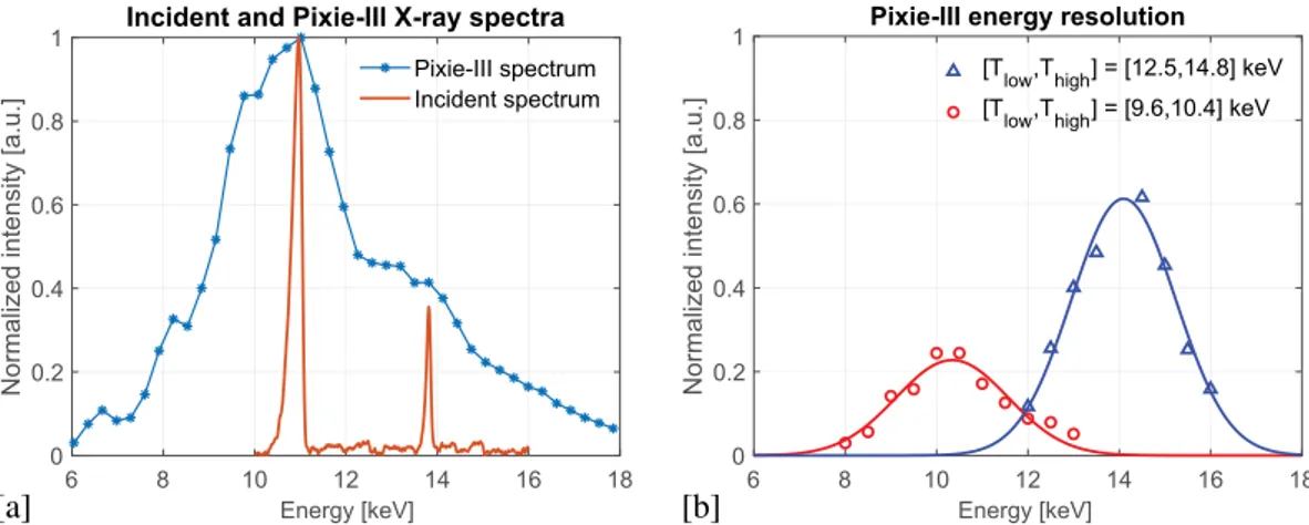
Discriminating between the two harmonics in the reported experiment requires an accurate setting of the energy thresholds. The undulator X-ray spectrum at the sample, as recorded with an imaging detector (PCO4000 camera) during an energy scan performed using the monochromator, is shown in Fig. 1a. The 4th harmonic at
11.0 keV is below the Au L-edge and the 5th harmonic at 13.8 keV is above. The bandwidth is 0.33 keV (Full Width
Half Maximum - FWHM) for both the 4th and 5th harmonics, which enforces a spatial resolution limit of 113 nm6.
Figure 1a also reports the differential X-ray spectrum detected by Pixirad-1/Pixie-III assessed via the so-called threshold scan at a few selected values. The counts above one single energy threshold were recorded for increasing values of this threshold in the range [6.0, 18.0] keV. Then, the difference between two adjacent measured values were used for the reported spectrum.
The optimal levels of Tlow and Thigh to isolate the harmonics is an optimisation between the finite energy
res-olution of the detector15,16 as reported in Fig. 1b) and the photon statistics. Narrower threshold separations
pro-duce images with a higher degree of monochromaticity but at the expense of photon statistics, thus increasing exposure times. Also, having the two thresholds too close can result in unresponsive pixels, because of single pixel miscalibration of the energy thresholds17. The following settings were found as optimal: for the “below” image,
the detector energy thresholds were set at values of Tlow = 9.6 keV and Thigh = 10.4 keV, whereas Tlow = 12.5 keV
and Thigh = 14.8 keV were used for the “above” acquisition. It is worth noting that, in order to minimise the
con-tamination between the adjacent harmonics, the energy windows were not centered onto the harmonic peaks but slightly shifted apart, to 10.8 keV and 14.3 keV, respectively.
Figure 1b shows the detector’s bandwidth at the two chosen settings. These were measured by scanning the energy of the X-ray beam with the Si(111) monochromator available at the beamline and dividing the counts between the thresholds by the total number of counts recorded by the detector. The red circles represent the band-width with the thresholds set for the harmonic “below” and the blue triangles represent the bandband-width with the thresholds set for the harmonic “above”. For both bandwidths an effective energy spread (FWHM) of 3 keV was measured, which is in agreement with previous similar independent measurements16.
pink beam LeS ptychography of a Au-ni micro-grid.
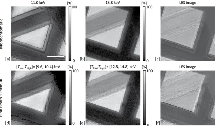
The LES imaging of a Au-Ni micro-grid test sample was performed in both monochromatic and pink beam mode. In both cases, two separate acquisi-tions were required, both above and below the Au L-edge. The illumination was defined by a 5 m diameter pinhole. The acquisitions consisted of a 40 × 40 regular snake scan, with a step size of 2 m (80% beam over-lap) and 5 s exposure. For the monochromatic images the Si(111) monochromator was placed in the beam Figure 1. (a) Filtered incident pink beam at the sample position as measured with the PCO4000 detector while performing an energy scan (orange, solid) and differential X-ray spectrum as detected by Pixirad-1/Pixie-III (blue, stars) assessed via the threshold scan at a few selected values. (b) Detector’s bandwidth when adopting the energy threshold settings to discriminate the 11.0 keV and the 13.8 keV peaks as measured at a few selected energy points.path, the 4th (11.0 keV) and the 5th harmonics (13.8 keV) were selected in the two consecutive acquisitions.
Pixirad-1/Pixie-III was configured in energy integration mode. The top row of Fig. 2 shows the recon-structed object modulus of the complex-valued ptychographs from below and above the absorption edge along with their digital subtraction. For the pink beam acquisition, the monochromator was removed from the beam path. The detector was set in colour mode and the two images were acquired consecutively with the optimised setting for Tlow and Thigh as described in the previous section. Figure 2d–f shows the
recon-structed object modulus for the below, the above, and the corresponding digital subtraction images, respec-tively. In both monochromatic and pink beam mode the Au is successfully isolated via digital subtraction of the images across the edge.
pink beam ptychography of a siemens star.
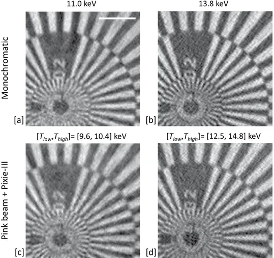
A Siemens star test sample was imaged to evaluate the achievable resolution of the experiment when considering object phase reconstructed images. The acqui-sition consisted of scanning the sample with the same parameters as for the micro-grid sample, but with 38 × 38 points. Again, for the monochromatic references images the Si(111) monochromator was placed in the beam path and the 4th (11.0 keV) and the 5th harmonic (13.8 keV) were selected in the twoconsecu-tive acquisitions by using Pixirad-1/Pixie-III in energy integration mode. The top row of Fig. 3 shows the reconstructed object phase at the two considered monochromatic energies. For the pink beam acquisition, the monochromator was removed from the beam path and compound refractive lenses (CRLs) were added into the beam path. The detector was set in colour mode and the two images were acquired consecutively with the optimised setting for Tlow and Thigh as previously described. The reconstructed object phase of the
complex-valued ptychographs is shown in Fig. 3. The spatial resolution obtained for the monochromatic and pink beam cases is similar. For each case, the Modulation Transfer Function was formed over a diagonal spoke. The best result of 141 nm was recorded for the reference monochromatic image at 13.8 keV and the worst case of 167 nm was recorded for the pink beam image with detector settings of [Tlow, Thigh] = [9.6, 10.4]
keV. The achieved resolution of approximately 150 nm is a factor of 67 greater than that resolved by the 5 m probe (beam) size alone. The Universal Quality Index (UQI) was used to estimate the similarity between the two sets of results. The UQI is in the [−1, +1] range and values towards +1 correspond to higher sim-ilarities between the images. We obtained for the monochromatic-pink beam comparison a UQI = 0.67 at 11.0 keV and a UQI = 0.70 at the 13.8 keV, which confirm the strong similarity of the results.
Figure 2. Top row: reference reconstructed images (modulus of the complex-valued ptychographs) acquired with the detector in energy integration mode and by using monochromatic beams at 11.0 keV (a) and 13.8 keV (b). The digital subtraction of (a,b) is reported in (c). Bottom row: reconstructed images acquired by using pink beam and the detector in colour mode when considering [Tlow, Thigh] = [9.6, 10.4] keV (d) and [Tlow, Thigh]
= [12.5, 14.8] keV (e). The digital subtraction of (d,e) is reported in (f). [Scale bar = 25.0 m. Color bar for the above and below images representing the X-ray transmission].
Discussion
We have presented the first results of spectral edge subtraction ptychographic imaging using multiple undulator harmonics and a multi-energy threshold XPCD. LES imaging of a test sample was performed and compared with conventional monochromatic images. The harmonic separation required depends on the specific edge of interest. The considered experiment aims at LES imaging and L-edge is generally weaker than the K-edge so a narrow sep-aration of the harmonics is required. This means that a fine tuning of the detector thresholds is needed. We have proved that, with optimized threshold settings, the quality of the pink beam reconstructed images is comparable to those acquired with monochromatic settings. The edge subtraction process is a precursor for further image seg-mentation and quantification. Although imperfections in the subtracted images are visible, simple segseg-mentation by gray-level thresholding (slightly above the background noise) accurately isolates the target material.
Pixirad-1/Pixie-III used during the experiment is coupled with a CdTe sensor, which is generally conceived for high-energy imaging18. We therefore also expect the methodology outlined here to be valid at higher energies.
The use of high-energy X-rays in ptychography is very attractive for their larger penetration power, the depth of focus increases for a given resolution, the dose delivered to the sample is potentially lowered, and shorter wavelengths give access to higher resolution information. Moreover, for the specific case of edge subtraction (ES) ptychography, higher-energy X-rays are required to reach the absorption edges of high-Z elements.
The polychromatic source used in this experiment consists of a comb-like spectrum with energy peaks of different amplitudes. The photon statistics might be significantly different for different energy windows. Filtering of the beam and the detector exposure time can be tuned to mitigate amplitude and consequent signal-to-noise ratio variations. However, in the case of pronounced asymmetry in the energy peaks, attention should be paid to avoid a non-linear response or, even worse, the paralysis of the detector with a high photon flux.
The two images required for the ES were acquired without the need of a monochromator and any mechanical scan of it. This could be beneficial since the stabilization of high-heat-load double-crystal monochromators is one of the most critical challenges for their effective utilization in diffraction-limited storage rings19. In addition,
having more than two energy thresholds20 would allow for the edge subtraction to be performed in a single
acquisition. Collecting the data in a single acquisition would be beneficial for the stability and quality of the final results. This is particularly relevant for ptychography, which typically requires longer acquisition times than direct
eixi
P +
ma
eb
kni
P-III
M
[T
low,T
high]= [12.5, 14.8] keV
[T
low,T
high]= [9.6, 10.4] keV
[a]
[b]
[c]
[d]
Figure 3. Reconstructed images (phase of the complex-valued ptychographs) of the Siemens star acquired by using monochromatic beams at 11.0 keV (a) and 13.8 keV (b). Reconstructed images by using pink beam and the detector in colour mode when considering [Tlow, Thigh] = [9.6, 10.4] keV (c) and [Tlow, Thigh] = [12.5, 14.8]
methods. The reduced scanning time of a single-acquisition approach would increase the experimental through-put. Increasing the number of energy thresholds beyond four would allow to target multiple elements at once.
The radiation damage is an important aspect for applications to biomedical imaging. In general ptychography is dose efficient, thanks to the diversity in measurements from the illumination structure and the splitting up of information across the scanning positions. The dose delivered to the sample during the 2D acquisition was estimated from the readout of an ionization chamber placed upstream of the pinhole. A dose of about 200 Gy was calculated for the ptychographic images presented in this article; a value far below the maximum tolerable dose limit described in21,22. When considering a 3D acquisition of a hundred projections the delivered dose would be
on the order of 2 × 10⁵ Gy, thus still acceptable for the spatial resolution of approximately 150 nm. The
method-ology described in this work is therefore promising also for biomedical applications of spectro-nanotomography.
Materials and methods
Source.
The experiment has been developed and conducted at the I13 beamline of Diamond Light Source (DLS). The beamline consists of two independent and complementary branchlines: the coherence branch (I13-1) and the imaging branch (I13-2). The coherence branch is a multimodal, multiscale instrument for reciprocal space imaging: including ptychography23, bragg CDI24, and bragg-ptychography. The imaging branch is focusedon real space imaging. The I13-1 ×-ray source consists of a 2.8 m long undulator with 2.5 cm period. The beam-line operates in the 7-24 keV energy range with a distance from the source of 215 m and with a sample-detector distance up to 14.5 m. The Au L-Edge imaging with the proposed technique, requires two isolated harmonics either side of the Au L2 (13.7 keV) and L3 (11.9 keV) edges. During the experiment the undulator was tuned to produce one harmonic above and one harmonic below the Au L-edge with a separation matching the detector energy resolution. The selected harmonics were the 4th and the 5th, respectively at 11.0 and 13.8 keV. This
con-figuration was achieved from a 3 GeV electron storage ring, by opening the undulator gap up to 13.64 mm. In a higher energy storage ring, for a similar insertion device, a smaller undulator gap would have been required to obtain a similar X-ray spectrum. The beam was filtered in order to reduce the contribution from lower and higher harmonics by a combination of metal filters (1.3 mm of pyrolytic graphite plus 0.7 mm of Al) and a Si mirror whose reflectivity drops quickly above 13 keV. The spectrum of the adapted incident beam at the sample is shown in Fig. 1a where two main peaks at 11.0 keV and 13.8 keV, respectively are clearly visible. For the reference images, the X-ray beam was monochromated using a Si(111) double crystal monochromator with 10−4 bandwidth.
Detector.
Pixirad-1/Pixie-III is a direct detection XPCD built with a hybrid architecture, where a semicon-ductor sensor is coupled with flip-chip bonding technique to the ASIC. The sensor is a Schottky type diode array with electron collection on the pixels composed of a thick CdTe crystal (650 m) substrate. A common problem in XPCDs is the limited energy resolution due to charge sharing effects. In Pixie-III this problem is mitigated by summing up the signals of neighbouring pixels, to correctly evaluate the total energy of any event involving up to 4 pixels. This hardware solution improves the energy resolution11, and this is beneficial for spectral imagingapplications. Pixie-III is a 0.16 mm CMOS ASIC organized as a 512 × 402 matrix of square pixels with 62 m pitch resulting in an active area of 31.7 × 24.9 mm2. The readout implements, at single pixel level, two 15-bit counters
fed by two independent discriminators with programmable thresholds. During operation the detector is cooled at −30 °C and the CdTe crystal is biased with a working voltage of 400 V. Compared with other XPCDs based on the Medipix ASIC, the Pixie-III is a large area detector (one module of Pixie-III corresponds roughly to a 2 × 2 con-figuration of Medipix). This avoids the well-known problem of dead gaps in multi-module detectors, due to the fact that up until now it has been possible to make pixel detector read-out chips that can be abutted on three sides only and the fourth side is used for on-chip peripheral logic and wire-bond pads that permit electronic read-out. Dead gaps complicate X-ray imaging because several pixels need to be excluded or inpainted during the recon-struction process25. Moreover, miscalibration of the different modules composing a large area detector sometimes
occurs and this might affect the quality of the final recorded signal, thus requiring ad hoc pre-processing26.
Samples and acquisitions.
A suitable test object composed of a regular microscale grid of Au and Ni com-ponent (see schematic in Fig. 4) was scanned to demonstrate the feasibility of Au isolation via L-Edge subtraction. Ni was chosen because the X-ray absorption edges of Ni (K = 8.3 keV and L1 = 1.0 keV) are far away from those of Au. Then a Siemens star was scanned to assess the spatial resolution achievable when considering object phase Figure 4. Sketch of the Au and Ni test grid. The values of K- and L-edge for Au and Ni are listed.chromatic image with the proposed pink beam image. For the case of the Au-Ni grid sample, after reconstruction the edge subtraction is performed on the modulus components of the complex-valued ptychographs. Since the pixel size of the reconstructed images is energy-dependent, image registration and calibration were necessary before performing the pixel-by-pixel LES. The resultant image is the difference in absorption across the selected edge.
Received: 7 January 2020; Accepted: 16 March 2020; Published: xx xx xxxx
References
1. Fredenberg, E. Spectral and dual-energy X-ray imaging for medical applications. Nucl. Instruments Methods Phys. Res. Sect. A: Accel.
Spectrometers, Detect. Assoc. Equip. 878, 74–87, https://doi.org/10.1016/j.nima.2017.07.044 (2018).
2. Suzuki, M. et al. Three-dimensional visualization of magnetic domain structure with strong uniaxial anisotropy via scanning hard X-ray microtomography. Appl. Phys. Express 11, 036601, https://doi.org/10.7567/APEX.11.036601 (2018).
3. Egan, C. K. et al. 3D chemical imaging in the laboratory by hyperspectral X-ray computed tomography. Sci. Reports 5, 15979, https:// doi.org/10.1038/srep15979 (2015).
4. Rodenburg, J. M. et al. Hard-X-Ray Lensless Imaging of Extended Objects. Phys. Rev. Lett. 98, 034801, https://doi.org/10.1103/ PhysRevLett.98.034801 (2007).
5. Holler, M. et al. Three-dimensional imaging of integrated circuits with macro- to nanoscale zoom. Nat. Electron. 2, 464–470, https:// doi.org/10.1038/s41928-019-0309-z (2019).
6. Spence, J., Weierstall, U. & Howells, M. Coherence and sampling requirements for diffractive imaging. Ultramicroscopy 101, 149–152, https://doi.org/10.1016/j.ultramic.2004.05.005 (2004).
7. Edo, T. B. et al. Sampling in x-ray ptychography. Phys. Rev. A 87, 053850, https://doi.org/10.1103/PhysRevA.87.053850 (2013). 8. Maiden, A., Morrison, G., Kaulich, B., Gianoncelli, A. & Rodenburg, J. Soft X-ray spectromicroscopy using ptychography with
randomly phased illumination. Nat. Commun. 4, 1669, https://doi.org/10.1038/ncomms2640 (2013).
9. Donnelly, C. et al. Three-dimensional magnetization structures revealed with X-ray vector nanotomography. Nature 547, 328–331,
https://doi.org/10.1038/nature23006 (2017).
10. Batey, D. J. et al. Spectroscopic imaging with single acquisition ptychography and a hyperspectral detector. Sci. Reports 9, 12278,
https://doi.org/10.1038/s41598-019-48642-y (2019).
11. Bellazzini, R. et al. PIXIE III: A very large area photon-counting CMOS pixel ASIC for sharp X-ray spectral imaging. J.
Instrumentation 10, C01032, https://doi.org/10.1088/1748-0221/10/01/C01032 (2015).
12. Veale, M., Seller, P., Wilson, M. & Liotti, E. HEXITEC: A High-Energy X-ray Spectroscopic Imaging Detector for Synchrotron Applications. Synchrotron Radiat. News 31, 28–32, https://doi.org/10.1080/08940886.2018.1528431 (2018).
13. Cipiccia, S. et al. Multi-scale multi-dimensional imaging at I13-coherence branchline in diamond light source. AIP Conf. Proc. 2054, 050005, https://doi.org/10.1063/1.5084623 (2019).
14. Brun, F. et al. Single-shot K-edge subtraction x-ray discrete computed tomography with a polychromatic source and the Pixie-III detector. Phys. Medicine & Biol. 65, 055016, https://doi.org/10.1088/1361-6560/ab7105 (2020).
15. Di Trapani, V. et al. Characterization of noise and efficiency of the Pixirad-1/Pixie-III CdTe X-ray imaging detector. J.
Instrumentation 13, C12008, https://doi.org/10.1088/1748-0221/13/12/C12008 (2018).
16. Di Trapani, V. et al. Characterization of the acquisition modes implemented in Pixirad-1/Pixie-III X-ray Detector: Effects of charge sharing correction on spectral resolution and image quality. Nucl. Instruments Methods Phys. Res. Sect. A: Accel. Spectrometers,
Detect. Assoc. Equip. 955, 163220, https://doi.org/10.1016/j.nima.2019.163220 (2020).
17. Vincenzi, A. et al. Energy characterization of Pixirad-1 photon counting detector system. J. Instrumentation 10, C04010, https://doi. org/10.1088/1748-0221/10/04/C04010 (2015).
18. Da Silva, J. et al. Overcoming the challenges of high-energy X-ray ptychography. J. Synchrotron Radiat. 26, 1751–1762, https://doi. org/10.1107/S1600577519006301 (2019).
19. Yabashi, M. et al. Optics for coherent x-ray applications. J. Synchrotron Radiat. 21, 976–985, https://doi.org/10.1107/ S1600577514016415 (2014).
20. Williams, S. et al. The ExcaliburRX-3M X-Ray Photon Counting Area Detector for Coherent Diffraction Imaging at the I13 Beamline at Diamond Light Source. 2017 IEEE Nucl. Sci. Symp. Med. Imaging Conf. NSS/MIC 2017 - Conf. Proc. 8532988, https:// doi.org/10.1109/NSSMIC.2017.8532988 (2018).
21. Howells, M. R. et al. An assessment of the resolution limitation due to radiation-damage in x-ray diffraction microscopy. J. electron
spectroscopy related phenomena 170, 4–12, https://doi.org/10.1016/j.elspec.2008.10.008 (2009).
22. Gianoncelli, A. et al. Soft x-ray microscopy radiation damage on fixed cells investigated with synchrotron radiation ftir microscopy.
Sci. Reports 5, 10250, https://doi.org/10.1038/srep10250 (2015).
23. Batey, D. et al. Reciprocal-space up-sampling from real-space oversampling in x-ray ptychography. Phys. Rev. A - At. Mol. Opt. Phys.
89, 043812, https://doi.org/10.1103/PhysRevA.89.043812 (2014).
24. Newton, M., Parsons, A., Wagner, U. & Rau, C. Coherent x-ray diffraction imaging of photo-induced structural changes in BiFeO3 nanocrystals. New J. Phys. 18, 093003, https://doi.org/10.1088/1367-2630/18/9/093003 (2016).
25. Brun, F., Delogu, P., Longo, R., Dreossi, D. & Rigon, L. Inpainting approaches to fill in detector gaps in phase contrast computed tomography. Meas. Sci. Technol. 29, 014001, https://doi.org/10.1088/1361-6501/aa91ad (2018).
26. Brombal, L. et al. Large-area single-photon-counting CdTe detector for synchrotron radiation computed tomography: A dedicated pre-processing procedure. J. Synchrotron Radiat. 25, 1068–1077, https://doi.org/10.1107/S1600577518006197 (2018).
28. Maiden, A. M. & Rodenburg, J. M. An improved ptychographical phase retrieval algorithm for diffractive imaging. Ultramicroscopy
109, 1256–62, https://doi.org/10.1016/j.ultramic.2009.05.012 (2009).
29. Thibault, P. & Menzel, A. Reconstructing state mixtures from diffraction measurements. Nature 494, 68–71, https://doi.org/10.1038/ nature11806 (2013).
30. Batey, D., Claus, D. & Rodenburg, J. Information multiplexing in ptychography. Ultramicroscopy 138, 13–21, https://doi. org/10.1016/j.ultramic.2013.12.003 (2014).
Acknowledgements
The KEST project, funded by INFN - Istituto Nazionale di Fisica Nucleare (National Scientific Committee 5 for Technological and Interdisciplinary research) is acknowledged. The research leading to this result has been supported by the project CALIPSOplus under Grant Agreement 730872 from the EU Framework Programme for Research and Innovation HORIZON 2020. Diamond Light Source is acknowledged for the funding and beamtime provided under proposal no. MG21555-1.
Author contributions
S.C. and D.B. conceived the experiment(s), F.B., V.D.T., D.B., S.C. conducted the experiment(s), F.B., D.B., S.C. wrote the article. F.B., V.D.T., D.B., S.C., C.R. reviewed the manuscript.
competing interests
The authors declare no competing interests.
Additional information
Correspondence and requests for materials should be addressed to F.B.
Reprints and permissions information is available at www.nature.com/reprints.
Publisher’s note Springer Nature remains neutral with regard to jurisdictional claims in published maps and institutional affiliations.
Open Access This article is licensed under a Creative Commons Attribution 4.0 International License, which permits use, sharing, adaptation, distribution and reproduction in any medium or format, as long as you give appropriate credit to the original author(s) and the source, provide a link to the Cre-ative Commons license, and indicate if changes were made. The images or other third party material in this article are included in the article’s Creative Commons license, unless indicated otherwise in a credit line to the material. If material is not included in the article’s Creative Commons license and your intended use is not per-mitted by statutory regulation or exceeds the perper-mitted use, you will need to obtain permission directly from the copyright holder. To view a copy of this license, visit http://creativecommons.org/licenses/by/4.0/.