i
Acknowledgments
Questa tesi mi ha dato la possibilità di vivere una delle più belle esperienze della mia vita: vivere a Ferrara. Ricorderò sempre gli anni trascorsi in questa splendida città con affetto, con un sorriso e con tantissima nostalgia. Ringrazio chi l’ha vissuta con me, ringrazio di cuore il “boss” Stefano, che mi ha dato questa opportunità di crescere e che mi ha insegnato i segreti della bioinformatica. Grazie ai miei supercompagni Marco e Marco, per aver condiviso con me il lavoro di tutti i giorni. Ringrazio di cuore tutte le altre persone fantastiche e speciali che ho incontrato qui: Ilaria, Sara, Edi.
E tutte coloro che ho incrociato anche solo per brevi istanti. Non vi dimenticherò mai!!!
Grazie al mio fantastico laboratorio dell’Ospedale Riuniti di Bergamo. Grazie alla Dott.ssa Maria Iascone perché sono la SMBI che sono grazie a lei! E ovviamente grazie a tutte le girls,e soprattutto Laura, Dani e Anna Rita, perché mi supportano (e sopportano) ogni giorno! Infine grazie alla happy family Sana, per aver creduto in me e per essere sempre i miei principali sostenitori!!!
ii
Alla mia mamma e al mio papà
A Sergio
iii
“...Credo di poter affermare che nella ricerca scientifica
né il grado di intelligenza né la capacità di eseguire e portare a
termine il compito intrapreso siano fattori essenziali
per la riuscita e per la soddisfazione personale.
Nell'uno e nell'altro contano maggiormente la totale dedizione
e il chiudere gli occhi davanti alle difficoltà:
in tal modo possiamo affrontare i problemi che altri,
più critici e più acuti, non affronterebbero...”
iv
Contents
Acknowledgments ...i
List of Tables ... vi
List of Figures ... vii
1 Introduction ... 1
1.1 Sanger capillary sequencing ... 1
1.2 Next-generation sequencing ... 2
1.2.1 Library preparation ... 5
1.2.2 DNA amplification ... 6
1.2.3 Sequencing and imaging ... 7
1.2.3.1 Roche/454 ... 8
1.2.3.2 Illumina ... 9
1.2.3.3 Life Technologies/Applied Biosystems... 11
1.2.3.4 Life Technologies/Ion Torrent ... 11
1.3 Application of next-generation sequencing ... 12
1.3.1 Analysis of cancer genome by next-generation sequencing . 13
1.4 Computational challenges ... 14
1.4.1 IT infrastructure ... 15
1.5 Bioinformatic analysis ... 15
1.5.1 Primary Analysis ... 16
1.5.2 Secondary Analysis ... 17
1.5.2.1 Alignment ... 17
1.5.2.2 Sequence Alignment/Map format... 19
1.5.2.3 Variant calling ... 20
1.5.3 Tertiary Analysis ... 21
1.5.3.1 Annotation ... 22
1.5.3.2 Prioritazion and intepretation ... 23
2 Genomic analysis in complex diseases ... 24
2.1 Ras/Raf/MAPK pathway ... 24
2.1.1 Colorectal cancer ... 28
2.1.1.1 CRC treatment ... 30
v
2.1.1.3 Kras mutation in colorectal cancer ... 32
2.1.1.4 Effect of Kras mutations on anti-EGFR therapy ... 33
2.1.2 RASopathies ... 34
2.1.2.1 Noonan syndrome ... 35
2.1.2.2 Leopard syndrome... 38
2.1.2.3 Costello syndrome ... 40
3 Aims of the present study ... 42
4 Materials and Methods ... 43
4.1 Patients... 43
4.1.1 Colorectal cancer patients ... 43
4.1.2 RASopathies patients ... 43
4.1.2.1 Patient 1 ... 44
4.1.2.2 Patient 2 ... 44
4.1.2.3 Patient 3 ... 44
4.2 Sample sequencing ... 45
4.2.1 Colorectal cancer sequencing ... 45
4.2.2. RASopathies sequencing ... 46
4.3 Bioinformatic analysis ... 46
4.4 Prioritization and intepretation ... 47
5 Results…... ... 48
5.1 Colorectal cancer ... 50
5.2 RASopathies ... 50
5.2.1 Patient 1 ... 52
5.2.2 Patient 2 ... 54
5.2.3 Patient 3 ... 57
6 Conclusion ... 57
References ... 59
Appendix ... 67
List of publication ... 67
Abbreviations... 69
vi
List of Tables
Table 1.1:Comparison of performance among different NGS platforms ...
3
Table 1.2:Applications of next-generation sequencing technologies ... 12 Table 1.3:Phred quality scores are logarithmically linked to error probabilities ... 17
vii
List of Figures
Figure 1.1:Schematic workflow of Sanger sequencing method ... 2
Figure 1.2:Reduction of cost per base of DNA sequencing ... 3
Figure 1.3:Increase of number of entries in dbSNP (2002-2012). ... 3
Figure 1.4: Cover of Nature and Time journals. ... 4
Figure 1.5: Paired-end library ... 5
Figure 1.6: Mate-paired library ... 5
Figure 1.7: DNA amplification procedures: emulsion PCR. ... 6
Figure 1.8: DNA amplification procedures:bridge amplification... 7
Figure 1.9: Roche/454 sequencing workflow ... 9
Figure 1.10: Illumina sequencing workflow ... 10
Figure 1.11: Life Technologies/Applied Biosystems workflow ... 11
Figure 1.12: Workflow of NGS data processing ... 15
Figure 1.13: FASTQ format of raw data ... 16
Figure 1.14: Graphical representation of alignment ... 19
Figure 1.15: Alignment file in SAM format. ... 20
Figure 2.1:Ras//MAPK pathway. ... 25
Figure 2.2: Multiple alignment between human RAS protein ... 27
Figure 2.3:Key events in the field of Ras research. ... 28
Figure 2.4: Colorectal cancer. A) biopsy B) histological section ... 29
Figure 2.5:Mechanism of anti-EGFR monoclonal antibodies ... 31
Figure 2.6: Ras/MAPK signalling pathway and related genetic syndromes ... 34
Figure 2.7: Clinical features in Noonan syndrome. ... 36
Figure 2.8:Phenotypic features in Leopard syndrome.. ... 40
Figure 2.9: Dysmorphic craniofacial features in Costello syndrome ... 41
Figure 4.1: Pedigree of family of Patient 3 ... 45
Figure 5.1: Structural modeling of amino acid change Gly12Val in protein ... 48
Figure 5.2: Visualization of KRAS-mutant SNV using IGV Browser. ... 50
Figure 5.3: PTPN11 mutation in Patient 1 ... 52
Figure 5.4: Patient 2 sequencing reads overlapping the missense mutation... 53
viii
Figure 5.6: Mutation in MYH7 gene in brother and sister of Patient 1.. ... 54 Figure 5.7: Identification of HRAS mutation. ... 55 Figure 5.8: Pedigrees in which the MYH7 and HRAS mutations segregated.. ... 56
1
Chapter 1
Introduction
Since the genetic information is embedded in the order of nucleotides in the DNA and RNA molecules, sequencing technology to determine the order of nucleotides in the DNA or RNA has always been one of the most powerful and fundamental tools in molecular biology. Determining the sequence of DNA can reveal the secrets contained in the genetic code of a person, his susceptibility to diseases and his response to drug treatments. The sequencing technology has been evolving and improving during the past a few decades, in terms of accuracy and throughput. Recently, several sequencing platforms have been developed, which have very high-throughput and low cost comparing to the traditional Sanger sequencing technology.
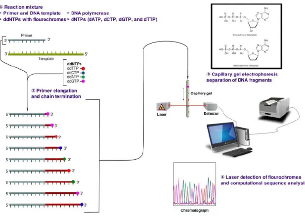
1.1 Sanger capillary sequencing
Sanger sequencing is a method of DNA sequencing, based on the selective incorporation of chain-terminating dideoxynucleotides by DNA polymerase during in vitro DNA replication [Sanger et al, 1977] (Figure 1.1). Developed by Frederick Sanger in 1977, it was the most widely-used sequencing method for approximately 25 years. The sequencing biochemistry takes place in a ‘cycle sequencing’ reaction, in which cycles of template denaturation, primer annealing and primer extension are performed. The primer is complementary to known sequence immediately flanking the region of interest. Each round of primer extension is stochastically terminated by the incorporation of fluorescently labeled dideoxynucleotides (ddNTPs). In the resulting mixture of end-labeled extension products, the label on the terminating ddNTP of any given fragment corresponds to the nucleotide identity of its terminal position. Sequence is determined by high-resolution electrophoretic separation of the single-stranded, end-labeled extension products in a capillarybased polymer gel. Laser excitation of fluorescent labels as fragments of discreet lengths exit the capillary, coupled to four-color detection of emission spectra, provides the readout that is represented in a Sanger sequencing electropherogram, that has been decoded into DNA
2
sequence [Smith et al, 1986; Ansorge et al, 1987]. Simultaneous electrophoresis in 96 or 384 independent capillaries provides a limited level of parallelization. After three decades of gradual improvement, the Sanger biochemistry can be applied to achieve read-lengths of up to ~1,000 bp, and per-base ‘raw’ accuracies as high as 99.999%.
Figure 1.1: Schematic workflow of Sanger sequencing method. Source: http://en.wikipedia.org.
1.2 Next-generation sequencing
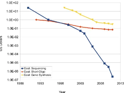
After the completion of the first human genome sequence in 2004 [Lander et al, 2001; Venter et al, 2001], the growing need to sequence a large number of individual genomes in a fast, low-cost and accurate way has directed a shift from traditional Sanger sequencing methods towards new high-throughput genomic technologies. In 2005, the first massively parallel DNA sequencing platforms emerged, ushering in a new era of next-generation sequencing (NGS) which allows sequencing at unprecedented speed in combination with low costs per base (Figure 1.2). As a consequence, the number of sequencing related data stored in public available databases has increased significantly (Figure 1.3).
3
Figure 1.2: Reduction of cost per base of DNA sequencing . Source: http://www.synthesis.cc.
Figure 1.3: Increase of number of entries in dbSNP (2002-2012). Source: http://massgenomics.org.
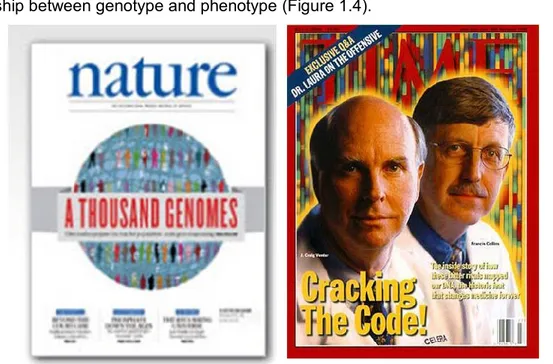
By applying genome-wide sequencing with high-throughput platforms the 1000 Genomes project (1KGP) sequenced the complete genome of 185 individuals from four populations, and analyze targeted exons of 697 individuals from seven populations within only two years [Kaiser et al, 2008; 1000 Genomes Project Consortium et al, 2010]. The aim was not only
4
provide a comprehensive resource on human genetic variation, but also investigate the relationship between genotype and phenotype (Figure 1.4).
Figure 1.4: Cover of Nature and Time journals. The impact of 1KGP on scientific community but
also in the politicl and economic world was enormous.
The variety of NGS features makes it likely that multiple platforms will coexist in the marketplace, with some having clear advantages for particular applications over others. In general, each platform embodies a complex interplay of enzymology, chemistry, high-resolution optics, hardware, and software engineering [Shendure et al, 2008; Metzker, 2010]. By different approaches, each technology seeks to amplify single strands of a fragment library and perform sequencing reactions on the amplified strands. The fragment libraries are obtained by annealing platform-specific linkers to blunt-ended fragments generated directly from a genome or DNA source of interest.
The main difference respect to traditional DNA sequencing approach is that molecules can be selectively amplified by PCR because of the presence of adapter sequences.
Because the presence of adapter sequences means that the molecules then can be selectively amplified by PCR, no bacterial cloning step is required to amplify the genomic fragment in a bacterial intermediate as is done in traditional sequencing approaches.
Specifically, the sequencing process could be grouped into library preparation, amplification reaction, sequencing and imaging, and data analysis [Metzker, 2010]. In the following sections, the experimental stages will be discussed, while paragraphs 1.3 and 1.4
5
will be dedicated to computational challenges introduces by NGS and data analysis procedures.
1.2.1 Library preparation
Library preparation is the step of preparation of DNA templates [Roe, 2004]. This process includes the random breaking of DNA sample into smaller fragments, ligating adapter sequences to allow the later use of universal primers, and amplifying the produced DNA templates.
Thre are three different protocols:
SINGLE-END library (SE) is created by randomly shearing genomic DNA (gDNA) or complementary DNA (cDNA) into fragments which are less than 1 kb in size;
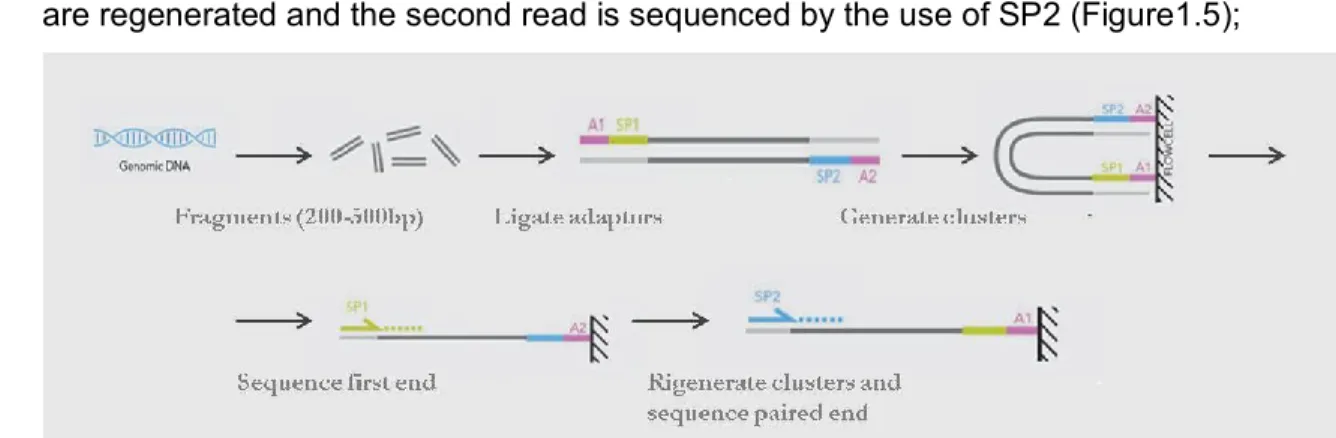
PAIRED-END library (PE) is created as the SE protocol by sequencing primer (SP) sites are ligated at each end. After analyzing the first read with SP1 the templates are regenerated and the second read is sequenced by the use of SP2 (Figure1.5);
Figure 1.5: Paired-end library. Source: www.illumina.org (adapted).
MATE-PAIRED library (MP) is created by shearing DNA with 2-5 kb in size labeled at the ends, circularized, and again linearized by cutting the cycles. Only fragments containing the label and therefore both ends of the original DNA fragment are selected and sequenced (Figure 1.6).
6
Figure 1.6: Mate-paired library. Source: www.illumina.org (adapted).
1.2.2 DNA amplification
After the library preparation, NGS technologies perform DNA amplification to increase the signal intensity for nucleotide detection. The most commonly used amplification procedures are:
Emulsion PCR in which single-stranded DNA (ssDNA) hybridizes onto oligonucleotide bound beads, ideally leaving one DNA template per bead (Figure 1.7). The beads are part of water-inoilmicro-emulsions which additionally contain all necessary components for PCR. After PCR amplification within these micro-reactors, up to thousands of complementary DNA strands are covalently bound to the beads. The original DNA template strands are washed away and the beads are purified and immobilized for later sequencing [Dressman et al, 2003; Metzker, 2010].Figure 1.7: DNA amplification procedures: emulsion PCR. [Metzker, 2010].
Bridge amplification in which the main device is a solid surface which is densely coated with forward and reverse primers. ssDNA templates anneal randomly to the surface and their complementary strands are built [Metzker, 2010]. After denaturation, a washing step removes all original template DNA. The remaining covalently bound complementary strands bind over to nearby reverse primers enabling the creation of yet another replicated strand. The DNA is denaturated
7
again to yield ssDNA and the next cycle of primer annealing and DNA replication is started. Thereby, clusters of DNA are produced all over the solid surface. As a last step prior to sequencing the template DNA is cleaved and washed away (Figure 1.8).
Figure 1.8: DNA amplification procedures: bridge amplification [Metzker, 2010].
1.2.3 Sequencing and imaging
The platforms for massively parallel DNA sequencing read production widely used up to today are distributed from Roche/454 (Eurofins MWG Operon; Huntsville, AL, USA), Illumina ((San Diego, CA, USA), Life Technologies/Applied Biosystems (Foster City, CA, USA) and Life Technologies/Ion Torrent (South San Francisco, CA, USA) (Table 1.1).
Company Platform Sequencing Throughput
Roche/454 GS FLX-T-XL Emulsion PCR/
Pyrosequencing
700Mb/23h
GS Junior 35Mb/10h
Illumina HiSeq 2000 Bridge
PCR/Sequencing-by-synthesis
105-600Gb/ 2-11d
MiSEQ 540Mb-7Gb/ 4-39h
8
Life Tech/Ion Torrent Ion PGMTM semiconductor sequencing Emulsion PCR/Ion 300Mb-1Gb/ 0.9-4.5h
Table 1.1: Comparison of performance among different NGS platforms. Abbreviations: ABI, Applied
Biosystems; d, day; h, hours.
All the instruments implement a process in which the synthesis of a complementary DNA strand is used to determine the DNA sequence and the use of a template amplification step to increase signal intensity for nucleotide identification.
Recently, three benchtop high-throughput sequencing instruments are become available, allowing the implementation of high-throughput sequencing in diagnostic settings. These are the 454 GS Junior (Roche/454), MiSeq (Illumina) and Ion Torrent PGM (Life Technologies/Ion Torrent) [Loman et al, 2012].
These platforms are smaller and cheaper that the large instruments and allow the analysis of patients in parallel in short time, since they generate a reduced throughput.
1.2.3.1 Roche/454
The Roche/454 was the first commercially available NGS platform, introduced in 2005. The platform analyzes DNA by pyrosequencing technology [Ronaghi et al, 1998; Ronaghi, 2001] in which nucleotides are detected based on the release of pyrophosphate. After the amplification of target DNA with emulsion PCR, beads, pyrosequencing enzymes, and pyrosequencing sulfates are loaded into a pico titer plate device, which places each bead into an addressable position within the plate. After the sequencing primer has been annealed the first sequencing cycle is started. Each cycle, nucleotides of one type (either dATP, dCTP, dGTP, or dTTP) are added to the plate. When incorporated, the nucleotide releases a pyrophosphate thereby triggering a series of downstream reactions. The use of luciferase in these reactions causes emission of a light signal which is proportional to the amount of integrated nucleotides. This signal is detected by a CCD camera and image information is stored for further processing. The remaining nucleotides are washed away and the next type of nucleotides is added to the plate (Figure 1.9) [Ansorge, 2009].
The latest 454 GS FLX platform with Titanium chemistry can produce approximately one million reads with lengths of up to 1000 bp per instrument run. Despite the higher costs, this platform for its long reads is best suitable for de novo assembly, metagenomic and characterization and analysis of microbiome.
9
Figure 1.9: Roche/454 sequencing workflow [Mardis, 2008].
The benchtop sequencer GS Junior System, available since 2010, is based on the chemistry of 454 Sequencing technology and was developed to allow the long read sequencing in reduced time (10 hours sequencing and 2 hours data processing), with lower set-up and running costs scaled for the needs of diagnostic laboratories.
1.2.3.2 Illumina
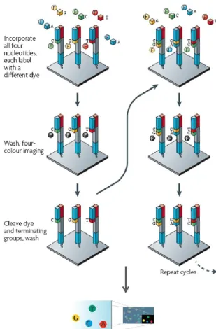
The Illumina sequencing system employs an array-based DNA sequencing-by-synthesis technology with reversible terminator chemistry. This technology analyzes different DNA samples in parallel by the use of bridge amplification and dye-terminated nucleotides (Figure 1.10).
10
Figure 1.10: Illumina sequencing workflow (adapted from Mardis, 2008).
After primer hybridization, nucleotides which are labeled by different fluorescent dyes are added to the slide. In each sequencing cycle, only one nucleotide is incorporated into the complementary strand due to its attached terminator group. A washing step removes all remaining free nucleotides before the newly added base is identified. Finally, the terminator and dye group are cleaved off, and the sequencing cycle is restarted. The first Illumina sequencing platform, the Genome Analyzer (GA), originally produced 35-bp reads and generate more than 1 Gb of high-quality sequence per run in 2–3 days. Latest platforms, GA IIX and HiSeq 2000, allow to increase fragment length up to 150bp and improve the throughput.
Recently, Illumina released a benchtop high-throughput sequencing instrument, MiSeq, developed for clinical settings and diagnostics routine. The MiSeq system is based on the existing Illumina sequencing-by-synthesis chemistry, but with a drastic reduction run times and cost, compared to other platforms.
11
1.2.3.3 Life Technologies/Applied Biosystems
The SOLiDTM system is based on ligating fluorescently labeled dinucleotide probes to the
DNA template [Tomkinson et al, 2006; McKernan et al, 2009]. After emulsion PCR, beads are covalently bound to a glass slide and universal sequencing primers, ligases, and a pool of labeled dinucleotide probes are added to the glass slide. The DNA sequence is determined by recording the color code, representing the first two bases of the dinucleotide, in several cycles of DNA ligation and cycles of primer reset. Each nucleotide (color-encoded) in the template is read twice by two fluorescent signals, reducing the error rate (Figure 1.11).
Figure 1.11: Life Technologies/Applied Biosystems sequencing workflow. A) The SOLiDTM substrate consists of an emulsion PCR bead, its covalently bound primer site (two sites for MP and PE), and the DNA template to be sequenced. b) SOLiDTM uses ‘1,2-probes’ (a version of a dinucleotide probe) where the first and second nucleotides are analyzed. The remaining six bases consist of either degenerated or universal bases [Metzker, 2010]. Each dye represents 4 of 16 possible dinucleotide sequences. (adapted from Mardis, 2008).
.
1.2.3.4 Life Technologies/Ion Torrent
Ion TorrentTM is based on semiconductor sequencing approach, which combines computer
software with integrated circuits and complementary metal-oxide semiconductors (CMOS). The technology also adopts an electrochemical detection system, the ion-sensitive field-effect transistors (ISFET), which can detect ions as they are released by DNA polymerase during sequencing by DNA synthesis. All of these electronics are focused on detecting and analyzing the release of a hydrogen ion (or proton) which occurs each time a nucleotide triphosphate is added. The proton release causes a slight pH shift which is detected by a
12
CMOS sensor. Each chip has at least 1.2 million sensors. These are composed of a well containing the dNTP and an acrylamide bead with a DNA template. Just beneath the well lies the metal oxide sensing layer, which itself lies over a sensor plate and floating metal “gate” that transmits electronic information (the pH changes) to the semiconductor. This technology differs from other sequencing technologies in that no modified nucleotides or optics are used. Ion Torrent Personal Genome Machine (PGMTM), available since 2011, is
benchtop NGS platform from Ion Torrent, which enables fast, affordable, genome-sequencing with a high throughput (80–100 Mb/h) and with up to 200bp fragment reads.
1.3 Application of next-generation
sequencing
The combination of different types of sample input and library preparations and the production of low cost reads by next-generation sequencing technologies makes them
useful in a variety of areas, such as whole genome sequencing, ChIP-Seq, metagenomics,
targeted re-sequencing (e.g. exome sequencing), RNA-Seq, Methyl-Seq, and others [Smith et al, 2008; Wold et al, 2008] (Table 1.2).
Application references
De novo sequencing of genomes Velasco et al., 2007; Bentley, 2006 Targeted resequencing (SNP, indels, CNV and structural
variations)
Hodges et al., 2007; Porreca et al., 2007
Epigenome Johnson et al., 2007; Mikkelsen et al., 2007
DNA Methylation Cokus et al., 2008
Transcriptome (RNA profiling, gene expression, alternative splicing)
Axtell et al., 2006; Berezikov et al., 2006 Metagenome (human microbiome characterization) Turnbaugh et al., 2007;
Hubert et al., 2007
Table 1.2: Applications of next-generation sequencing technologies.
Important applications include: (1) de novo genome sequencing, whole-genome resequencing or more targeted sequencing for discovery of mutations or polymorphisms; (2) transcriptome analysis for gene expression and RNA profiling; (3) large-scale analysis
13
of DNA methylation; (4) analysis of DNA-protein interactions by chromatin immunoprecipitation (ChIP-Seq); (5) characterization of human microbiome (epigenomics). A specific application of NGS is the molecular analysis of cancer genome.
1.3.1 Analysis of cancer genome by NGS
Cancers are caused by the accumulation of genomic alterations. Therefore, analyses of cancer genome sequences and structures provide insights for understanding cancer biology, diagnosis and therapy [Ross et al, 2011]. NGS technologies have allowed substantial advances in cancer genomics, since they have facilitated an increase in the efficiency and resolution of detection of each of the principal types of somatic cancer genome alterations, including nucleotide substitutions and small insertions and deletions. Furthermore, these new sequencing methods make it feasible to discover novel intrachromosomal rearrangements, including inversions, tandem duplications and deletions, reciprocal and non-reciprocal interchromosomal rearrangements and microbial infections, and to resolve copy number alterations at very high resolution.
Cancer samples have specific features requiring particular consideration in NGS processing and analysis. The main characteristic is that cancer samples differ in their quantity, quality and purity from the peripheral blood samples, commonly used for germline genome analysis. Surgical resection specimens tend to be large and have been the mainstay of cancer genome analysis [Meyerson et al, 2010]. However, diagnostic biopsies from patients with disseminated disease tend to contain few cells and the quantity of nucleic acids available may be limiting. Extracted nucleic acids from cancer are also often of lower quality than those purified from peripheral blood. This because most cancer biopsy and resection specimens are formalin-fixed and paraffin embedded (FFPE) that makes DNA impure. Moreover, cancer specimens often include substantial fractions of necrotic or apoptotic cells that reduce the average nucleic acid quality histology. Moreover, a cancer specimen contains a mixture of malignant and nonmalignant cells and, therefore, a mixture of cancer and normal genomes (and transcriptomes). For all these reasons, FFPE-derived nucleic acids can require special experimental protocols and computational approach for the processing and analysis.
Furthermore, the cancers themselves may be highly heterogeneous and composed of different clones that have different genomes. Cancer genome analytical models must take these two types of heterogeneity (cancer versus normal heterogeneity and within-cancer
14
heterogeneity) into account in their prediction of genome alterations. Specifically, cancer genomes vary considerably in their mutation frequency (degree of variation compared to the reference sequence), in global copy number or ploidy, and in genome structure.
Various computational methods have been developed to determine the presence of somatic mutations using NGS data. The detection of somatic mutations in cancer requires mutation calling in both the tumour DNA and the matched normal DNA, coupled with comparison to a reference genome and an assessment of the statistical significance of the number of counts of the mutation in the cancer sequence and its absence in the matched normal sequence. False positives and errors in variant calls can be due to inaccurate detection of mutation in tumor for insufficient coverage or for machine-sequencing biases, incorrect local alignment of individual reads and discordant alignment of pairs.
1.4 Computational challenges
Next-generation sequencing has revolutionized the study of human genetics and has immense clinical implications. The possibility to sequence different samples and different genomes in parallel has reduced the cost and increased the throughput of genomic sequencing and these technologies are still evolving [Metzker, 2010]. Using deep sequencing, for example, it is now possible to discover novel disease causing mutations [Ley et al, 2008] and detect traces of pathogenic microorganisms [Isakov et al, 2011]. For the first time, research fields such as personalized medicine for patient treatment are becoming tangible at genomic levels given advances in deep sequencing data integration. At the same time, NGS has introduced in the scientific field a computational challenge. In fact, the amount of data produced by ultra high throughput sequencing run is often tremendous and can reach hundreds of millions of reads in various lengths per experiment [Mardis, 2008]. NGS platforms in production are able to produce data of the order of giga or terabytes per machine day. The storage, processing, querying, parsing, analyzing and interpreting of such an incredible amount of data is a significant task that holds many obstacles and challenges. The emergence of NGS platforms imposes increasing demands on statistical methods and bioinformatic tools for the analysis and the management of the huge amounts of data generated by these technologies. Today a large number of softwares already exist for analyzing NGS data. These tools can be fit into many general categories including alignment of sequence reads to a reference, base-calling and/or polymorphism
15
detection, de novo assembly from paired or unpaired reads, structural variant detection and genome browsing.
1.4.1 IT infrastructure
The high amount of DNA data generated by next-generation sequencing techniques is demanding computationally intense hard- and software systems to conquer the computationally expensive tasks of NGS data analysis.
To provide an efficient system for analyzing and visualizing next-generation sequencing data, in this thesis a 64 bit computing cluster, consisting of the following components, was introduced: two Sun FireTMX4600 M2 Servers each with four Quad-Core AMD OpteronTM8356, 2.3 GHz, 80 GB RAM; Serial attached SCSI (SAS) storage of 16 TB (extendable up to 256 TB).
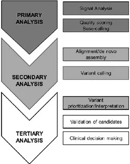
1.5 Bioinformatic analysis
There are three typical analysis stages in NGS processing (Figure 1.12): a. Primary analysis (base calling)
b. Secondary analysis (alignment and variant calling) c. Tertiary analysis.
16
1.5.1 Primary analysis
Primary analysis can be defined as the machine specific steps needed to call base pairs and compute quality scores for those calls from the image detected during the sequencing. This results in a FASTQ file, the format of raw data, which is just a combination of the sequence data, the so called read, as a string of A, C, G and T characters and an associated Phred quality score for each of those bases (Figure 1.13). This output is ready for processing in a secondary analysis pipeline.
Figure 1.13: FASTQ format of raw data.
All current commercial next-generation instruments first capture images of many parallel reactions. Analysis software is then applied to locate sequencing reactions and extract information about each reaction. Each platform analysis process is unique and proprietary and may involve several sub-steps. Data filtering may be done subsequent to or during primary analysis. Each platform applies specific rules to eliminate sets of reads and/or images that may have gross artifacts, eliminate individual reads or duplicate sequences. The user may or may not have the ability to view or change these rules.
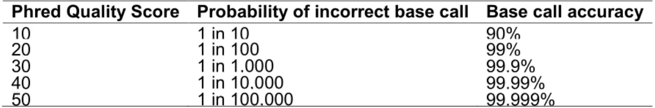
As quality parameter, the Phred score has become the standard to characterize the quality of DNA sequences and the goodness of sequencing [Ewing et al, 1998; Ewing et al, 1998]. Phred quality score Q is defined as logarithmically related to the base-calling error probabilities P:
Q=-10log
10P
(1)
This means that with a Phred of 30 assigned to a base, the chances that this base is called incorrectly are 1 in 1000 (Table 1.3).
17
Phred Quality Score Probability of incorrect base call Base call accuracy
10
1 in 10
90%
20
1 in 100
99%
30
1 in 1,000
99.9%
40
1 in 10,000
99.99%
50
1 in 100,000
99.999%
Table 1.3: Phred quality scores are logarithmically linked to error probabilities
1.5.2 Secondary analysis
1.5.2.1 Alignment
The step conducted after base calling is alignment of the reads to a reference sequence (also called “mapping”) or, if a reference sequence does not exist, for example in the case of microbiology analysis, assembly de novo of the sequence reads into contigs and scaffolds.
Alignment of next-generation reads to a reference sequence may be conducted by a variety of different algorithms and may or may not use quality values or intensity values. Mappers such as MAQ [Li et al, 2008] (developed specifically for the Illumina platform) used a hash/seed approach, wherein the reference genome was indexed by k-mers of a given size and the first k-mer of each read was searched for a near-exact match. This approach was well-suited for near-exact matches (i.e. 0-3 single base differences between read and genome) but does not treat small insertions or deletions. It also tends to be both processor and memory intensive. Another algorithms are based on the Burrows-Wheeler transform [Li et al, 2009], for example BWA, which demonstrated significant (>10-fold) improvement in mapping speed. Some mappers are platform-specific features, such as BFAST [Homer et al, 2009] for the Life Technologies/Applied Biosystems which takes advantage of the SOLiD two-base encoding scheme (color base space), and the gsMapper [Shearer et al, 2010] for the Roche/454 platform which uses full flowgram information for alignment. Due to their central role in almost all next-generation applications, mappers remain under active development.
An important feature to be considered in this step is the quality assessment of the data that can improve the mapping performance [Li et al, 2010]. The alignment output contain a Phred based quality score for each of the aligned reads, describing the probability of per-base false alignment. Combination of this quality score together with other alignment
18
parameters such as mismatches could and should be further assessed using specialized tools [Lassmann et al, 2011] in order to characterize mapped and unmapped reads for potential alignment improvement. These alignment quality scores can be re-assessed and recalibrate taking into account the given base and its quality score, the position within the read and the adjacent nucleotides to account for sequencing chemistry biases. This procedure reduces the effect of sequencing technology derived biases and improve overall variant detection accuracy [DePristo et al, 2011].
Another feature to consider is that most alignment tools allow the user to set the number of allowed mismatches between the read and a reference location and the scoring scale for gap opening and extension. Allowing more mismatches results in a higher portion of mapped reads but at the cost of increased ambiguity and reduced confidence of these alignments. Mismatch allowance should be set while considering the specific experiment at hand. For example, when undergoing microRNA expression profiling, one will want an accurate estimate of the abundance of each microRNA , and should not allow a high mismatch rate if any. On variant calling experiments however, the user should consider the possible expected size range of the variants before setting the allowed mismatch and gap penalty parameters (e.g, if one aims to find a >5nt long deletion, the mismatch limitation should allow it).
To ensure the accuracy of alignment and of the variant detection, during alignment step it is important to considered the multiple mapping. This means that a read could be mapped to multiple loci on the reference genome for sequence homology and repetitiveness. Different alignment tools flag these multiply mapped reads, and provide the user with the option to either randomly assign them to one loci. Discarding multiply mapped reads results in loss of a substantial portion of the data, with potential crucial effects on the following analysis. An important concept in NGS is depth of coverage. This is a measure of how many reads cover a given locus of the genome (Figure 1.14). After the sequencing process is complete, upper and lower depth thresholds should be applied on the sequencing data before variant calling is performed. Setting a lower coverage limit removes erroneous mismatches caused by sequencing errors and thus supported by very few reads [1000 Genomes Project Consortium et al, 2010; Li, et al, 2009]. Setting a lower limit has been shown to reduce sensitivity without increasing specificity in some tools [Goya et al, 2010] and therefore should be considered in the context of the utilized tool. Setting an upper limit removes
19
mismatches caused by copy number variations, PCR duplicates introduced by library preparation and reads mapping to paralogous sequences.
Figure 1.14: Graphical representation of alignment. Visualization by Integrative Genomics Viewer
(IGV) [Robinson et al, 2011; Thorvaldsdóttir et al, 2012].
In secondary analysis, according to the specific application, the choice of appropriate reference sequence in genome re-sequencing is also critical. The University of California Santa Cruz (UCSC) Genome Browser [Kent et al, 2002] provide in FASTA format genome assembly released from Genome Sequence Consortium (the most recent release is GRCh37/hg19 assembly). Projects such as the 1KGP, the Cancer Genome Atlas (TCGA) (http://cancergenome.nih.gov/), the 10,000 genomes project (http://www.genome10k.org/), as well as the ongoing NGS of DNA and RNA populations from human tissues may serve to enrich and expand the concept of “reference.”
1.5.2.2 Sequence Alignment/Map format
The standard output data file is in SAM (Sequence Alignment/Map) format [Li et al, 2009], which contains several information regarding the the original sequence, or the matching sequence of the reference as well as information about the match (e.g. sample name, SNP calls, or mapping quality value). Most common alignment software generate the alignment output in SAM format, with a multitude of supporting downstream analysis tools.
20
The SAM format was designed to store nucleotide alignments in a generic way and with the aims to support several sequencing platforms, allowing short and long reads up to 128 Mb, saving SE as well as PE information, and including various types of alignments (Figure 1.15).
Figure 1.15: Alignment file in SAM format.
The tab delimited text format can be divided into a header and an alignment section. Header lines can be identified by the ‘@’ character at the start of each line and contain, among others, information about the reference against the reads have been aligned, and read groups. In the alignment section, a tab delimited line describes a read alignment and contains required information about read name, read flags, reference sequence, alignment position, mapping quality, extended CIGAR, reference sequence of the paired read, position of the paired read, inferred insert size within the read pair, read sequence, and read quality. The extended CIGAR string characterizes the type of alignment operation including clipped, spliced, multi-part, and padded alignment. Allowed operations are match/mismatch (M), insertion (I), deletion (D), skipped base (N), soft clipping (S), hard clipping (H), and padding (P). Additional optional fields allow the documentation of less important or program specific data. Color space read information is also described in the optional fields. To accelerate parsing and ease data processing, the binary file format Binary Alignment/Map (BAM) equivalent to SAM was introduced [Li et al, 2009].
1.5.2.3 Variant calling
The secondary analysis includes after alignment the variant calling procedure [DePristo et al, 2011]. Variant calling is the process of accurately determining the variations between a sample and the reference genome. These may be in the form of single nucleotide variants, smaller insertions or deletions (indels), or larger structural variants of categorizations such as transversions, trans-locations, and copy number variants (CNV).
21
The process is subject to biases for low coverage, sequencing errors, misalignment caused by either low complexity and repeat regions or adjacent variants and library preparation biases. Variant calling depends on an efficient combination between an accurate alignment and sophisticated inference of variance from it.
After aligning deep sequencing reads against a reference genome, SNPs can be inferred from the results by simply denoting each base that is inconsistent between reference and read as a SNP. This straightforward inference of mismatches results in a massive amount of alleged SNPs, many of which suffer from some sort of inaccuracy such as: calling a mismatch in the wrong location, homozygousity and heterozygousity discrepancies and even calling a mismatch in the correct location but with the wrong base. Currently most SNP calling tools [Koboldt et al, 2009; Li et al, 2009; McKenna et al, 2010] apply different probabilistic based considerations and heuristics such as quality assessment and recalibration, SNP filtration, local realignment, coverage assessment, prior probability based on known SNPs, genotype based likelihood and even cancer genomics to elucidate SNPs from alignment results.
Some features, such as local realignment, base quality and transition/transversion ratio have to be considered critically to improve the goodness of variant calling step. Local alignment considers reads that support the presence of an indel in the vicinity of either detected SNPs or known SNP sites retrieved from dbSNP, results in a significant reduction in false positive SNPs [McKenna et al,2010].
The expected ratio between transitions (e.g purine-purine substitutions) and transversions (e.g purine-pyrimidine substitutions) (Ti/Tv ratio) is ~2.3 for whole-genome sequencing and around 3.3 for whole-exome sequencing (coding regions only) [1000 Genomes Project Consortium et al, 2010; DePristo et al, 2011].
After producing a list of detected SNPs, it is highly recommended to compare it against dbSNP, to rescue the number of false positives. The portion of novel SNPs detected in a deep sequencing experiment should range between 1 and 10 percent [DePristo et al., 2011].
1.5.3 Tertiary analysis
Variant calling in secondary analysis allows the detection of an extensive catalogues of human genetic variation in the samples. However, pinpointing the few phenotypically
22
causal variants among the many variants present in human genomes remains a major challenge, particularly for rare and complex traits wherein genetic information alone is often insufficient. Tertiary analysis includes all the approaches to estimate the deleteriousness of single nucleotide variants, which can be used to prioritize disease-causal variants in the context of a specific study [DePristo et al, 2011; paper VI].
Specifically, experimental or computational approaches that provide assessments of variant function can be used to better estimate the prior probability that any given variant is phenotypically important.
1.5.3.1 Annotation
Calling variants using deep sequencing data often results in a multitude of detected variations, even after strict and effective quality filtration as denoted earlier, deep sequencing data reveals thousands to millions of different variations. These ones can result in biological effects through introduction of different amino acids into protein sequences, early termination of coding sequences and alteration of regulatory elements and splice sites. The following process after variant calling is annotating the detected variants and elucidating their effect and biological significance, separating clinically, scientifically and medically relevant variations from neutral, non functional ones. In a large list spanning this many variants, manual annotation of each variant effect is neither feasible or accurate. For annotation, public databases can flexibly use, such as RefSeq, UCSC, ENSEMBL, GENCODE or many other gene definition systems. All these information can be used to determine the position of the mismatch in the gene and whether it is in a coding/non-coding, exon, intron, UTR or intron/exon junction region (intronic regions contiguous to exon starts and exon ends that are important to evaluate splice-site mutations).
To give biological context to NGS data could be useful to query public or commercial databases containing biological/functional annotation. For example OMIM (Online Mendelian Inheritance in Man, OMIM®. McKusick-Nathans Institute of Genetic Medicine, Johns Hopkins University (Baltimore, MD), {February, 2012}. URL: http://omim.org/), which catalogues genetic disorders and traits, Human Gene Mutation Database (HGMD) [Stenson et al, 2009], which reports validate gene mutations, and COSMIC, a large repository of somatic mutations in cancer [Forbes et al, 2009].
Moreover, large projects such as 1KGP, Exome Sequencing Project (ESP) (Exome Variant Server, NHLBI GO Exome Sequencing Project (ESP), Seattle, WA (URL:
23
http://evs.gs.washington.edu/EVS/) [February 2012]) allow for studies to be able to determine the relative frequencies of variants in their samples compared to common populations (MAF, minor allele frequency).
A MAF of 1% is used as cutoff to define a variants as rare [Frazer et al, 2009; paper I]. The rarity of a variant or the absence in a healthy control population is classically one of the criteria for evaluating the pathogenicity both in research and in clinical settings. Although there is need of caution in considering this parameter as prediction of deleteriousness. In fact, MAF ≥1% implies that the variant is sufficiently common in the population to be not tremendously deleterious, the opposite is not generally true.
1.5.3.2 Prioritization and interpretation
The development of methods to assess the effect of variants has been a major subject of research in the field of bioinformatics during the past decade. Currently, there are several tools available which can predict the disease-causing potential of mismatches for protein structure and function. These programs can predict the effects on the basis of different features, including evolutionary conservation, changes in the physico-chemical characteristics of the aminoacids, the sequence environment of the affected amino acid alteration in structural properties of proteins.
This “tolerance” predictors, which evaluate in silico the effect of mutations on the phenotype at protein level include Polymorphism Phenotyping 2 (PolyPhen-2) [Adzhubei et al, 2010], Align-GVGD [Tavtigian et al, 2006], SIFT [Kumar et al, 2009] and MutationTaster [Schwarz et al, 2010].
The Grantham Score, which categorizes the differences of physicochemical properties for codon replacements, , including polarity and molecular volume, can be also retrieved from the original Grantham Score Matrix [Grantham, 1974].
Moreover, the evolutionary nucleotide conservation score in vertebrates (phyloP) or in different species is a reliable method for predicting possible pathogenicity of a missense variant [Flanagan et al., 2010; Siepel et al, 2005].
24
Chapter 2
Genomic analysis in complex disease
The advent of NGS technologies has revolutionized the field of genomics, enabling fast and cost-effective generation of genome-scale sequence data with exquisite resolution and accuracy. To date, NGS technologies have been widely used for many applications, such as rare variant discovery by whole genome resequencing or targeted requencing, transcriptome profiling of cells, tissues and organisms, and identification of epigenetic markers for disease diagnosis. Studies using massive parallel sequencing have yielded surprising and important discoveries regarding the genetic bases of Mendelian and complex diseases, particularly cancer. The identification of molecular mechanism underlying tumorigenesis enables molecular diagnosis and carrier testing in the patient and his or her family. This is of great importance for patient management and family counseling, and serves as a starting point for therapeutic interventions. Furthermore, the identification of Mendelian disease genes contributes to our understanding of gene functions and biological pathways underlying health and disease in general [paper II].
In the following paragraphs, some examples of application of NGS technologies in clinical and diagnostic context will be presented.
2.1 Ras/MAPK pathway
The Ras/MAPK (mitogen-activated protein kinases) pathway is one of the most important signaling network inmplicated in growth-factor mediated cell proliferation, differentiation and death [Molina et al, 2006]. It is a chain of proteins in the cell that communicates a signal from a receptor on the surface of the cell to the DNA in the nucleus of the cell. The signal starts when a signaling molecule binds to the receptor on the cell surface and ends when the DNA in the nucleus expresses a protein and produces some change in the cell, such as cell division. The pathway includes many proteins, including MAPK, originally called ERK
25
(extracellular signal-regulated kinases), which communicate by adding phosphate groups to a neighboring protein, which acts as an "on" or "off" switch (Figure 2.1).
Figure 2.1: Ras//MAPK pathway. Source: http://journals.cambridge.org
The cascade of activation of this pathway starts when receptor-linked tyrosine kinases such as the epidermal growth factor receptor (EGFR) are activated by extracellular ligands. Binding of epidermal growth factor (EGF) to the EGFR activates the tyrosine kinase activity of the cytoplasmic domain of the receptor. The EGFR becomes phosphorylated on tyrosine residues. Docking proteins such as GRB2 contain an SH2 domain that binds to the phosphotyrosine residues of the activated receptor. GRB2 binds to the guanine nucleotide exchange factor SOS (son of sevenless) to the plasma membrane by way of the two SH3 domains of GRB2. When the GRB2-SOS complex docks to phosphorylated EGFR, SOS becomes activated. Activated SOS displaces GDP from a member of the Ras subfamily (i.e. HRas, KRas), subsequently allowing the binding between Ras and GTP [Molina et al, 2006]. Ras are membrane-associated guanine nucleotide-binding proteins which affect many cellular functions, including cell proliferation, apoptosis, migration, fate specification, and differentiation. In the GTP-bound form, Ras interacts specifically with so-called effector proteins, thereby initiating cascades of protein-protein interactions that may finally lead to
26
cell proliferation and activates a number of signaling pathways, including the Raf/MEK/ERK pathway. The activity of Ras is limited by the hydrolysis of GTP back to GDP by GTPase activating proteins (GAP) in an enzymatic process. Reactivation of Ras requires the removal of GDP by SOS. Activated Ras activates the protein kinase activity of RAF kinase. RAF kinase phosphorylates and activates MEK (MEK1 and MEK2). MEK phosphorylates and activates a mitogen-activated protein kinase (MAPK).Raf, and MAPK are both serine/threonine-selective protein kinases. MEK (also known as MAPKK) is a tyrosine/threonine kinase. In the technical sense, Raf, MEK, and MAPK are all mitogen-activated kinases.
The Ras/MAPK pathway is probably the best characterized signal transduction pathway in cell biology. The function of this pathway is to transduce signals from the extracellular milieu to the cell nucleus where specific genes are activated for cell growth, division and differentiation. MAPK regulates the activities of several transcription factors. MAPK can phosphorylate C-myc. MAPK phosphorylates and activates MNK, which, in turn, phosphorylates CREB. MAPK also regulates the transcription of the C-Fos gene. By altering the levels and activities of transcription factors, MAPK leads to altered transcription of genes that are important for the cell cycle. The Ras/MAPK pathway is also involved in cell cycle regulation, wound healing and tissue repair, integrin signaling and cell migration. Finally, the Ras/MAPK pathway is able to stimulate angiogenesis through changes in expression of genes directly involved in the formation of new blood vessels. Thus, signaling through the Ras/Raf/MAPK regulates a variety of cellular functions that are important for tumorigenesis. Dysregulation of this pathway is a common event in cancer as Ras proteins are the most frequently mutated oncogenes in human cancer [Malumbres et al, 2003]. Mutations in the Kras oncogene have been localized in codons 12, 13, 59 and 61 with those at codons 12 and 61 occurring most frequently. Kras mutations are present in 15-50% of lung cancers and in 72-90% of pancreatic cancers
Historically, the rat sarcoma (RAS) virus homologue was the first oncogene to be described in human cancer (Der et al 1982). In cancer, the most commonly mutated members of the RAS superfamily include HRAS, KRAS and NRAS, highly conserved proteins . The RAS proteins are small (21 kilodalton) G-proteins that are active with bound GTP and inactive with bound GDP. Although the GTP can self-hydrolyze, there is a class of enzymes termed GAPs (GTPase activating proteins) that facilitate this hydrolysis and terminate RAS activity.
27
The name 'Ras' is an abbreviation of 'Rat sarcoma', reflecting the way the first members of the protein family were discovered. When Ras is 'switched on' by incoming signals, it subsequently switches on other proteins, which ultimately turn on genes involved incell growth, differentiation and survival. As a result, mutations in ras genes can lead to the production of permanently activated Ras proteins. This can cause unintended and overactive signalling inside the cell, even in the absence of incoming signals. Because these signals result in cell growth and division, overactive Ras signaling can ultimately lead to cancer.Ras is the most common oncogene in human cancer - mutations that permanently activate Ras are found in 20-25% of all human tumors and up to 90% in certain types of cancer (e.g. pancreatic cancer).[2] For this reason, Ras inhibitors are being studied as a treatment for cancer, and other diseases with Ras overexpression [Fernández-Medarde et al, 2011].
All Ras protein family members belong to a class of protein called small GTPase, and are involved in transmitting signals within cells (cellular signal transduction). Ras is the prototypical member of the Ras superfamily of proteins, which are all related in 3D structure and regulate diverse cell behaviours (Figure 2.2) [Malumbres et al, 2003; Karnoub et al, 2008].
Figure 2.2: Multiple alignment between human RAS proteins. The amino acid sequences are highly
28
Figure 2.3: Key events in the field of Ras research [Malumbres et al, 2003].
.
Oncogenic lesions introduce changes in the primary sequence of RAS so that the protein is constitutively active. RAS pathway still remains one of the most investigated pathways in human cancer (Solit et al 2006), including melanoma, and our current understanding suggests that several possible mutations along this cascade lead to tumor-promoting physiology.
Although RAS proteins are frequently mutated in cancer, there is preferential targeting of specific family members in different tumor types.
2.1.1 Colorectal cancer
Colorectal cancer (CRC) is the second most common cancer in the world, accounting for approximately one million new cases each year [Markowitz et al, 2009] (Figure 2.4). The overall mortality is approximately 50%. The surgical stage at diagnosis is the most important factor for predicting patient outcome, with five year survival rates of more than 90% for stage I disease and less than 10% for stage IV disease.
29
Figura 2.4: Colorectal cancer. A) biopsy B) histological section
The surgical stage represents a classification system based on the extent and depth of tumor growth. The system most commonly used in describing CRC is the Tumor, Nodes, Metastasis (TNM) system of the American Joint Committee on Cancer (AJCC) provided in 2002.
In Stage I CRC shows invasive grown into the anatomical layers of the colon, but the tumor is not spread outside the colon wall or into regional lymph nodes. During Stage II there is tumor penetration through the bowel wall involving the serosa; however, there is no involvement of regional lymph nodes or distant metastases. In Stage III CRC have spread to nearby lymph nodes but not yet metastasized to distant sites in the body. Finally, in Stage IV the tumor has spread to distant organs such as the liver, lungs, or other sites. CRC is considered to develop through a multi-step process, originating from normal colon epithelium that develops into precursor lesions termed adenomas. Adenomas can subsequently further progress into invasive CRC with metastatic potential. Spread of CRC occurs by direct growth through the bowel wall and through invasion of lymphatic and venous channels. The most common sites for metastases are regional lymph nodes and the number of lymph node metastases influences prognosis. The liver is the most common distant site for CRC metastases. The vast majority of CRC are adenocarcinomas, with less than 10% of the cancers being distinguished by an abundant secretion of mucin. The tumors are classified according to the degree of morphological differentiation into well, moderately and poorly differentiated. About 80% are well or moderately differentiated with a growth pattern consisting of tumor cells that form irregular glandular structures present at different layers of the bowel wall. Poorly differentiated CRC show no, or only hinted, glandular formation. Overall poor differentiation with a diffuse infiltrative growth pattern is
30
associated with poor prognosis, although stringent classification systems based on morphological features are lacking
2.1.1.1 CRC treatment
Treatment decisions are thus based on the surgical stage and morphological characteristics.
At stage 0, in which the cancer has not grown beyond the inner lining of the rectum, the strategy consist in removing or destroying the cancer by polypectomy, local excision, or transanal resection. For stage I, during which cancer has grown through the first layer of the rectum into deeper layers but has not spread outside the wall of the rectum itself, surgery is usually the main treatment: a low anterior resection, colo-anal anastomosis, or an abdominoperineal resection may be done, depending on exactly where the cancer is found within the rectum
In some cases, adjuvant chemoradiation (treatment with radiation and chemo together) is advised for patients having such surgery. 5-Fluorouracil (5-FU) is the chemo drug most often used.
Stage II in which the cancer have grown through the wall of the rectum and may extend into nearby tissues is treated by low anterior resection, colo-anal anastomosis, or abdominoperineal resection (depending on where the cancer is in the rectum), along with both chemotherapy and radiation therapy. Most doctors now favor giving the radiation therapy along with the chemo drug 5-FU before surgery (neoadjuvant treatment), and then giving adjuvant chemo after surgery, usually for a total of 6 months of treatment (including the time getting chemo and radiation together). The chemo after surgery may be the FOLFOX regimen (oxaliplatin, 5-FU, and leucovorin), 5-FU and leucovorin, CapeOx (capecitabine plus oxaliplatin) or capecitabine alone, based on what's best suited to your health needs. Treatment for stage III cancers that have spread to nearby lymph nodes includes radiation therapy, given along with 5-FU chemo before surgery (called chemoradiation). This may shrink the cancer, often making surgery more effective for larger tumors. It also lowers the chance that the cancer will come back in the pelvis. Giving radiation before surgery also tends to lead to fewer problems than giving it after surgery. The rectal tumor and nearby lymph nodes are then removed, usually by low anterior resection, colo-anal anastomosis, or abdominoperineal resection, depending on where the
31
cancer is in the rectum. After surgery, chemo is given, usually for about 6 months. The most common regimens include FOLFOX, 5-FU and leucovorin, or capecitabine alone. Finally, in stage IV cancer has spread to distant organs and tissues such as the liver or lungs. and the Treatment options disease depend to the extension of metastasis. Treatment options include surgery to remove the rectal lesion and distant tumors, followed by chemo (and radiation therapy in some cases) with combination of chemo and radiation therapy before and after the surgery.
2.1.1.2 anti-EGFR monoclonal antibodies
Recently new targeted drugs have been implemented into the treatment of patients with advanced colorectal cancer. Epidermal growth factor receptor is commonly expressed in colorectal tumors and monoclonal antibodies (mAbs) inhibiting EGFR demonstrate clinical efficacy in patients with mCRC. This anti-EGFR monoclonal antibodies, cetuximab and panitumumab, were designed as effective inhibitors of the EGFR (Figure 2.5).
Figure 2.5: Mechanism of anti-EGFR monoclonal antibodies. A) EGFR binding to ligand activates
32
Cetuximab (Erbitux®, Merck KgaA, Darmstadt, Germany) is a chimeric mouse/human antibody targeted against the extracellular domain of the EGFR. Binding of cetuximab to the receptor prevents ligand binding, induces receptor internalization and causes a direct inhibition of the receptor tyrosine kinase activity.This in turn blocks downstream signal transduction via the PI3K/Akt and Ras/MAPK pathways inducing pro-apoptotic mechanisms and inhibiting cellular proliferation, angiogenesis and metastasis. Cetuximab is indicated for the treatment of patients with epidermal growth factor receptor (EGFR)-expressing, Kras wild-type metastatic colorectal cancer, in combination with chemotherapy, and as a single agent in patients who have failed oxaliplatin- and irinotecan-based therapy and who are intolerant to irinotecan. As an IgG1 antibody cetuximab may also induce antibody-dependent cell-mediated cytotoxicity (ADCC). The side effects include acne-like rash, and dermathological reactions (urticaria, pruritis), hypotension, bronchospasm, dyspnea, wheezing, angioedema, dizziness, anaphylaxis, and cardiac arrest. Photosensitivity and hypomagnesemia have been also observed in some patients. This drug is given by intravenous therapy and costs up to $30,000 for eight weeks of treatment per patient.
Panitumumab (Vectibix®, Amgen Thousand Oaks, CA, USA), by contrast, is a fully human antibody which is also directed against the EGFR but being an IgG2 MoAb lacks ADCC activity.
2.1.1.3 Kras mutation in colorectal cancer
Oncogenic mutations of the Kras genes are observed in about 40% (20–50%) of sporadic colorectal cancers [Lievre et al, 2006; Heinemann et al, 2009]. These are point mutations and are generally observed as somatic mutations. Up to 90% of activating mutations of the Ras gene are detected in codons 12 and 13, but less frequently also in codons 61 and 63. The most frequent types of KRAS mutations in colorectal cancers are G>A transitions and G>T transversions. The codons 12 and 13 code for two adjacent glycine residues located in the proximity of the catalytic site of RAS. In particular codon 12 mutations of the Kras gene were associated with a mucinous phenotype of colorectal cancer, while codon 13 mutations were rather non-mucinous, but were characterized as more aggressive tumors with a greater metastatic potential.
33
Different KRAS mutations result in an exchange of different amino acids at these catalytic sites, and therefore, may be responsible for the different levels of intrinsic GTPase activity reduction. As a consequence, variable RAS mutations may imply variable effects on the biology of disease.
For this reason, several studies support the importance of mutational activation of KRAS in the progression of CRC [Amado et al, 2008; Souliers et al, 2010].
For the detection of KRAS mutations several methods are known. The hotspots and thus the most frequent mutations g.34G>C (p.Gly12Arg), g.35G>C (p.Gly12Cys), g.34G>A (p.Gly12Ser), g.35G>A (p.Gly12Asp), g.35G>C (p.Gly12Ala), g.35G>T (p.Gly12Val), g.38G>A (p.Gly13 Asp) and rarely g.183G>T (p.Gln13His). Moreover, as the detection of KRAS mutations has become a routine diagnostic method, the protocols in use are selected on the basis of velocity, robustness and easiness. The main problems in detection of KRAS mutations involve the low quality of the DNA that often causes error in specificity and sensitivity of the analysis.
2.1.1.4 Effect of Kras mutations on response to anti-EGFR therapy
The reports on the prognostic relevance of KRAS mutations are inconsistent. While some studies have indicated a negative impact of KRAS mutations on survival (ref)others did not [Lievre et al, 2006; Souliers et al, 2010].
The KRAS mutation status has been identified as a strong predictor of response to anti-EGFR antibodies. In fact, mutations of the KRAS gene may activate downstream signal transduction and confer resistance to upstream inhibition of the EGFR by monoclonal antibodies. For this reason, EGFR directed therapy is not only not effective in KRAS mutant mCRC patients. Moreover, it may also induce unnecessary toxicity and has been associated with an inferior outcome in some clinical trials. In fact, determination of the KRAS mutation status is required in all mCRC patients who may receive anti-EGFR directed antibody therapy. Studies using cetuximab plus chemotherapy for first-line treatment of mCRC could detect an improvement of treatment efficacy only in KRAS wild-type patients. By contrast, KRAS mutant patients either had no benefit from the addition of cetuximab or even showed a worse outcome than their comparators. At present, it is not clear if additional toxicity impairs treatment intensity in KRAS mutant patients or if negative interactions between cetuximab and chemotherapy take place.
34
2.1.2 RASopathies
The Ras/MAPK pathway is critical to normal development and essential in the regulation of the cell cycle, differentiation, growth and cell senescence (Figure 2.6). A class of developmental disorders, the so called RASopathies, is caused by germline mutations in genes that encode protein components of the Ras/MAPK pathway resulting in increased signal transduction down the Ras/MAPK pathway [Tidyman et al, 2008]. The term RASopathies includes different diseases, such as capillary malformation-AV malformation syndrome, autoimmune lymphoproliferative syndrome, Costello syndrome (CS), Leopard syndrome (LS), Noonan syndrome, (NS), neurofibromatosis type 1 etc.. Each syndrome exhibits unique phenotypic features, however, since they all cause dysregulation of the Ras/MAPK pathway, there are numerous overlapping phenotypic characteristics between the syndromes, including craniofacial dysmorphisms, cardiac malformations and cutaneous, musculoskeletal and ocular abnormalities, varying degrees of neurocognitive impairment and an increased risk of developing cancer.
Figure 2.6: Ras/MAKP signalling pathway and related genetic syndromes. RASopathies are caused
by mutations in the genes which control the production of certain signal proteins.Source:
![Figure 1.7: DNA amplification procedures: emulsion PCR. [Metzker, 2010].](https://thumb-eu.123doks.com/thumbv2/123dokorg/4716919.45491/14.918.150.806.736.836/figure-dna-amplification-procedures-emulsion-pcr-metzker.webp)
![Figure 1.8: DNA amplification procedures: bridge amplification [Metzker, 2010].](https://thumb-eu.123doks.com/thumbv2/123dokorg/4716919.45491/15.918.218.768.262.566/figure-dna-amplification-procedures-bridge-amplification-metzker.webp)
![Figure 1.9: Roche/454 sequencing workflow [Mardis, 2008].](https://thumb-eu.123doks.com/thumbv2/123dokorg/4716919.45491/17.918.141.764.155.525/figure-roche-sequencing-workflow-mardis.webp)