University of Siena – Department of Medical Biotechnologies
Doctorate in Genetics, Oncology and Clinical Medicine (GenOMeC)
XXXIII cycle (2017-2020)
Coordinator: Prof. Francesca Ariani
CRISPR/Cpf1 and suicide gene as personalized approach for patients with
TP53
-mutated Chronic Lymphocytic Leukemia (CLL)
Scientific disciplinary sector: MED/03 – Medical Genetics
Tutor
PhD Candidate
Prof. Francesca Mari
Flaminia Clelia Lorenzetti
Academic Year 2019/2020
FLAMINIA CLELIA LORENZETTI 11.03.2021 14:51:39 UTC
University of Siena – Department of Medical Biotechnologies
Doctorate in Genetics, Oncology and Clinical Medicine (GenOMeC)
XXXIII cycle (2017-2020)
Coordinator: Prof. Francesca Ariani
CRISPR/Cpf1 and suicide gene as personalized approach for patients with
TP53
-mutated Chronic Lymphocytic Leukemia (CLL)
Scientific disciplinary sector: MED/03 – Medical Genetics
Tutor
PhD Candidate
Prof. Francesca Mari
Flaminia Clelia Lorenzetti
2 “To change the world It starts with one step
However small First step is hardest of all” Dave Matthews Band, You Might Die Trying
3
Acknowledgments
First of all I should thank all the Medical Genetic Unit of Siena, especially Prof. Alessandra Renieri and my tutor Prof. Francesca Mari. It has been tough but motivating working with all of them. Of
course, I need to thank Filomena Papa, she was always available to discuss, to help me with the experiments and to answer to all my questions, even the most silly ones! A big and warm thanks
goes to all my lab crew with who I spent most of my time in the past 3 years. I bring with me a lot of funny memories, thank you!
I would like to especially thank Prof. Rik Gijsbers. He is an incredible professor, researcher and most of all he is a really great person. It was inspiring discussing with him and his team about projects and experimental ideas. I am so grateful to have worked in his lab and I am very happy to
keep collaborating with him.
Lastly, I want to thank my family and friends to have always be there to listen to me and to support me in the good and especially in the bad times of this important journey.
4
Table of Contents
Table of Contents 4 Abbreviations 5 Abstract 7 Introduction 8Chronic Lymphocytic Leukemia (CLL) 8
Current treatments and clinical trials 11
Genome editing technology 14
CRISPR based therapies 17
Delivery by viral vector: lentiviral (LV) vector 19
Lentiviral vectors based therapies 20
Aim of the project 22
Results 22
Strategy of the system 22
Lentiviral vector transfer plasmid design 25
Validation of specific editing using transfection of reporter 293T target cells 26
Optimization of Ganciclovir kill curve 29
DNA analysis corroborates correct recombination of the eGFP-TK cassette 31
Lentiviral vector (LV) production and target cell transduction 32
In vivo study: establishment of human CLL xenograft mouse model 38
Discussion 40
Conclusion and Perspectives 42
Material and Methods 43
Cell culture 43
Plasmids cloning 43
Cell transfection and ganciclovir treatment 44
Viral vector production and cell transduction 44
FACS analysis and PCR analysis 45
qPCR analysis 45
Western Blot analysis 45
CLL Xenograft mice model 46
References 47
Clinical trials references 51
5
Abbreviations
CAR: Chimeric Antigen Receptor CIT: Chemoimmunotherapy
CLL: Chronic Lymphocytic Leukemia CR: Complete Response
CRISPR: Clustered Regularly Interspaced Short Palindromic Repeats DSB: Double Strand Break eGFP: Enhance Green Fluorescent Protein
FACS: Fluorescence activated cell sorting GCV: Ganciclovir
gRNA: Guide RNA
HDR: Homology-Directed Repair
HSCT: Hematopoietic Stem Cells Transplantation HSV1: Herpes Simplex Virus type 1
IDLV: Integration-deficient Lentiviral Indel: Insertion and Deletion
KO: Knock-Out
LTR: Long Terminal Repeats LV: Lentiviral
MMEJ: microhomology-mediated end joining MRD: Minimal Residual Disease
NGS: Next-Generation Sequencing NHEJ: Non-Homologous End-Joining ORR: Overall Response Rate
PAM: Protospacer Adjacent Motif PCR: Polymerase Chain Reaction PFS: Progression Free Survival
6 R/R: Relapse/Refractory
TK: Thymidine Kinase
7
Abstract
Chronic Lymphocytic Leukemia (CLL) is a very heterogenous disease caused by alterations in both chromosomes and genes, such as deletion of the 13q14, 11q22-23, 17p12, and trisomy of chromosome 12 or genetic mutations in the TP53, ATM, BRIC3, NOTCH. Within genetic abnormalities, TP53 mutations are detected in a small percentage of leukemia patients (about 10%). Currently adopted therapeutic choices (chemoimmunotherapy, targeted therapy, hematopoietic stem cell transplantation) are not effective due to the acquired abilities of TP53-mutated clones to escape the control systems. For this reason CLL patients harboring a mutated TP53 gene (point mutation, insertion or deletions) are grouped in the highest risk category according to the international prognostic index for chronic lymphocytic leukemia (CLL-IPI). Hence, one possible solution for these patients would be the development of a personalized therapy.
In my study I designed and developed a CRISPR-Cpf1 system which has proved to be an efficacious technology to get rid of only mutated cancer cells. The system called CRISPR_LV_TK+, recently patented (WO2020/079574), is based on the locus specific delivery of the Herpes Simplex Virus – Thymidine Kinase (HSV-TK) suicide gene in cells bearing the target TP53 mutation detected in a CLL patient (p.Ser183*) in follow-up in our Unit. The approach was positively tested in HEK293T cell lines engineered for the target mutation. Following administration of ganciclovir, I was able to detect a high percentage of cell death only in the samples that have properly integrated the HSV-TK. In conclusion, the results show the high efficiency and specificity of the CRISPR_LV_TK+ system, and opens avenues to be applied as personalized therapy not only for CLL but for different kinds of cancers caused by specific mutations in distinct genes which are the driver of therapy resistance.
8
Introduction
Chronic Lymphocytic Leukemia (CLL)
Chronic Lymphocytic Leukemia (CLL) is one of the most common types of leukemia in the western countries1,2. It affects mostly adults of >60 years of age who remain asymptomatic for many
years but they suddenly show lymphocytosis which is the only symptom CLL patients show at diagnosis 3. Patients post diagnosis can follow two separated ways, they can start specific therapy
such as chemotherapy or they could never require any treatment3,4. Indeed, CLL is a heterogeneous
disease characterized by several different causative events which may influence patients’ treatment choice5,6. CLL is characterized by an uncontrolled B cells proliferation due to disruption of important
cellular pathways such as the apoptotic pathway, the nuclear factor-κB (NF-κB) signaling pathway, NOTCH signaling pathway all involved in controlling cell growth and proliferation7. Chromosomal
alterations like deletion of the 13q14, 11q22-23, 17p12, and trisomy of chromosome 12 or genetic mutations in the TP53, ATM, BRIC3, NOTCH genes are the main causes of the disease7,8. The
immunoglobulin heavy variable (IGHV) gene mutational status is another crucial characteristic associated with CLL and patients can harbor two different forms of the genes, mutated and unmutated, which leads to a completely different outcome7. It has been reported that CLL patients with mutated IGHV genes sequence have higher overall survival (OS) due to a better response to chemotherapeutic
agents with respect to patients harboring the un-mutated IGHV genes4.
Patients show a different survival rate and response to treatment depending on the type of chromosomal or genetic alterations they harbor4,9. According to Dohner, CLL patients can be
classified into 5 risk groups associated with different prognosis where CLL cases harboring the 17p chromosomal deletion or a mutant p53 are grouped in the worst prognostic class and patients with 13q deletion belong to the lowest risk group10. The ranking has been recently modified by an
international consortium which was able to establish the international prognostic index for chronic lymphocytic leukemia (CLL-IPI) which helps clinicians to better predict the OS and the time-to-first treatment (TTFT) for specific patients harboring different genetic alterations6. In the CLL-IPI,
10 induces a toxic effect on cells which should activate apoptotic pathways in order to induce specific cell death5,12. However, in case of TP53 mutated cells, apoptotic pathway activation is impaired and
cancer cells will keep proliferating and occasionally acquiring new genetic alterations due to the toxicity induced by chemotherapy1.
Hence, mutations in TP53 or deletions in the 17p chromosome need to be carefully detected at diagnosis to start the correct treatment for CLL patients. In order to accurately detect abnormalities in the TP53 gene the European Research Initiative on Chronic Lymphocytic Leukemia (ERIC) suggested specific guidelines that should be followed15. First of all, the employment of the next
generation sequencing (NGS) technology in order to detect genomic variants carried by the patients at low frequency which could have been hidden using Sanger sequencing6,9,15which is less sensitive
technique than NGS16. Second, ERIC suggested to sequence TP53 exons 4 to 10 which embedded
the DBD region where most mutations occur15. Third, it is important to carefully analyze the data
acquired after DNA sequencing using specific bioinformatic tools to avoid skipping any significant variants15. Finally, they give tips about the quality of the genomic DNA that needs to be sequenced,
the databases which can be consulted (IARC or COSMIC) for the identified variants and they also give a list for clinical significance of exonic and intronic variants which are commonly detected15,17,18.
Mutations characterized by a low variant allele frequency (VAF) need to be carefully identified in order to avoid their clonal expansion which can be deleterious for the patients15,19. As
described by Landaou et al., two types of driver mutations may be present in a CLL patient, predominantly clonal and sub-clonal3,20. The former is detected at diagnosis whereas the latter and
most critical one shows later on during disease course due to its expansion20. Indeed, the starting of
chemotherapy could help the sub-clone to spread due to the toxicity of the therapy targeting the predominant clone9,20. Hence, with no competitors the sub-clone is able to proliferate and expand,
ultimately acquiring resistance for the cancer to the therapy which lead the patients to chemo-refractoriness3,21. Moreover, TP53 mutations are good markers for chemotherapy resistance in CLL
12 alkylating agents and purine analogs showed positive response in treated patients with an adequate overall response rate (ORR) and progression free survival (PFS), they are not suitable for all CLL patients24. Indeed, for CLL patients characterized by an aberrant TP53 gene the apoptotic pathway is
impaired leading to accumulation of cytotoxicity which may result in the accumulation of novel mutations1. However, chemoimmunotherapy (CIT), thus the combination of chemotherapy and
antibody-based therapy is now widely applied as a treatment of choice for almost all CLL patients. Rituximab is the first developed type of anti-CD20 monoclonal antibody (mAb) which show promising results in CLL patients23. Upon binding to CD20 antigen which is expressed on both
neoplastic and healthy B cells, Rituximab will activate different cellular pathways in order to eliminate cancer cells, such as antibody-dependent cellular cytotoxicity (ADCC), complement-dependent cytotoxicity (CDC), programmed cell death (PCD) or adaptive cellular immunity25. During
ADCC, immune effector cell macrophages and natural killer (NK) cells recognize and kill CD20 target cells by releasing cytotoxic enzymes26,27. When the CDC pathways is activated, cancer cells
are lysed by cytotoxic enzymes released by the complement system25. The biding of Rituximab to
CD20 can also trigger the adaptive immunity, thus dendritic cells (DC) will present tumor antigens to T cells which will differentiate into cytotoxic T cells able to eliminate target cells25,26. Lastly,
Rituximab can also lead cancer cells to apoptosis by activation of the caspase 327.
CIT treatment with fludarabine, cyclophosphamide and rituximab (FCR) have demonstrated increase in ORR, complete response (CR) and a long PFS28. Rituximab combined with bendamustine
(BR) is another possible CIT regime23. However, when compared with FCR, BR showed less toxicity
but a decrease in PFS, whereas the ORR was identical in both the CITs23. Unfortunately, CIT did not
show significant increase in the positive response rate in CLL patients harboring a TP53 mutation or a del17p6,9,24.
Targeted agents acting on the apoptotic pathway regardless of the TP53 status have shown great positive outcomes in several clinal trials. The first FDA approved targeted therapy in 2016 was ibrutinib which acts as inhibitor of the Bruton’s tyrosine kinase (BTK) involved in the B cell receptor
13 (BCR) signaling pathway1. Another powerful kinase inhibitor is idelalisib targeting the
phosphoinositide 3-kinase δ (PI3Kδ)23,24. The FDA allowed its use together with ibrutinib but due to
the high toxicity detected in a group of treated patients, idelalisib should not be considered as first therapeutic choice23. Venetoclax is a third type of targeted agent able to induce cellular apoptosis by
inhibiting the anti-apoptotic protein BCL-21. It was approved by the FDA and it is considered the
first line therapeutic agent for CLL due to the outstanding positive results detected in the clinical trials23. When combined with ibrutinib, venetoclax showed an increase in ORR, PFS, CR and the
total absence of minimal residual disease (MRD) or toxicity compared to all the other type of available therapies1. Moreover, in the MURANO Phase III study venetoclax combined with rituximab
revealed a deep decrease in MRD and prolonged PFS when compared with bendamustine and rituximab therapy29. Interestingly, ibrutinib, idelalisib and venetoclax all demonstrated a strong
performance also in relapse/refractory (R/R) patients harboring an aberrant TP53 gene1,23,29.
The newly developed chimeric antigen receptor (CAR) therapy was recently tested in CLL patients. CAR therapy is an ex vivo gene- and cell-therapy in which patients T cells are isolated, genetically modified and re-infused into the patients30. The resulting modified T cells express on their
surface receptors that specifically recognize cancer-specific antigens present on neoplastic cells31.
Patients receiving CAR treatment may develop cytokine release syndrome or neurotoxicity as adverse events1,30. However, in the next few years results from the ongoing clinical trials will give a more
general safety assessment for CLL patients treatments (NCT03960840, NCT02935257, NCT01853631).
Allogenic hematopoietic stem cells (HSC) transplantation (HSCT) is considered the ultimate treatment for CLL especially for R/R patients bearing TP53 abnormalities but it is not recommended for elderly patients1. HSC transplantation should be considered only when the other therapeutic
options failed or when patient biomarkers are particularly favorable for the transplant32. About 50%
of patients receiving allogenic HSCT showed stable negative MRD during follow up32. However,
14 disease (GvHD)32,33. The group of M. Hahn stated that pre- or post-therapeutic strategies should be
also explored in order to overcome the GvHD in relapse CLL patients who undergo HSCT34.
However, TP53 mutated CLL patients still gain little benefits from the aforementioned therapies showing relapse, thus a personalized, cancer cell specific therapy could be an important option to consider in order to help these patients.
Genome editing technology
It was a woman, named Barbara McClintock, the pioneer in genome editing. In the 1950 she studied corn (Zea mays) and discovered particular genomic sequences called transposons or mobile genetic elements able to move from one region of the genome to another leading to the insertion of random mutations35. Later on, in the 1980s, homologous recombination (HR) was described by
Capecchi, Smithies and Evans. When foreign double stranded DNA (dsDNA) having homologies with a target sequence was added into target cells, the exogenous molecule was inserted at that particular place36. The era of the first generation of genome editing technologies ends in the late 90ies
when RNA interference (RNAi) was first reported as a method to knock-down specific RNA sequences, thus controlling gene expression37. At the beginning of the 21st century the second
generation of genome editing technologies started to be explored. Zinc Finger Nuclease (ZFN) and TAL effector nuclease (TALEN) were the first tools to be employed in the targeting of specific genomic sequences36. They consist of a DNA-binding moiety that defines the specificity and is
coupled to one half of the bacterial FokI endonuclease38,39. Thus, two ZFNs or two TALENs (one left
and one right of the target site) dimerize at the target sequence, and produce a double strand break (DSB) at the desired locus40–42. The protein domains that bind the target sites on the genomic DNA
(ZnF or TALE) can be modulated in order to recognize different nucleotides with the possibility of editing several different genomic sequences43,44. Unfortunately, it is very laborious and expensive to
modify TALE or ZnF protein structures. In 2005 the discovery of the clustered regularly interspaced short palindromic repeats (CRISPR) was still to completely revolutionize the research in life science.
17 a third type of repairing mechanism called the microhomology-mediated end joining (MMEJ) is described (Figure 04), which occurs in the M, G1 and early S phase of the cells cycle when HR is not active. For MMEJ to occur, 5 to 20 bp of homology arms need to be present at the edited site which is fixed by the annealing of the microhomologies61–63.
The PAM sequence in the end limits the applicability of CRISPR. However, Cas orthologs have been identified in various bacterial strains able to recognize different PAM sites, thus allowing the targeting of several unique loci for particular experimental aims64. CRISPR-Cas system is largely
employed to study gene functions by knocking-out specific genes and it is also used for knock-in experiments aimed at correcting detrimental mutations in order to restore a wild type and healthy cellular state. Hence, CRISPR applications described so far have been applied in different areas of research from plant biotechnologies to biofuels production and also in the biomedical fields with the development of powerful gene and cellular therapies tested for a wide range of diseases.
CRISPR based therapies
Several studies are testing CRISPR editing technology in different genetic disorders such as Duchenne Muscular Dystrophy (DMD), ß-thalassemia and sickle cell disease65. DMD is caused by a
nonsense mutation on the dystrophin gene responsible for maintaining the correct integrity of muscle fibers. Mutations occurs on the exon 23 leading to the complete loss of protein expression. Hence, the excision of exon 23 by CRISPR-Cas9 tool could theoretically restore the expression of a functional dystrophin protein. Systemic delivery of the CRISPR system showed great efficiency in
vivo studies performed in both mice and dogs66,67. Moreover, positive results were achieved in vitro
and in vivo on animal models for the hemoglobinopathy ß-thalassemia for which a clinical trial is ongoing (NCT03655678). ß-thalassemia is caused by mutations in the ß-globin gene which inhibit the translation of the ß-globin protein. The therapy is based on the in vitro editing of autologous hematopoietic stem cells (HSC) by CRISPR in order to knock out the BCL11A a repressor of the γ-globin allowing its expression which can restore a healthy condition in affected patients who will
18 receive the edited cells back by transfusion68. This strategy could also be applied for sickle cell disease
characterized by the synthesis of an aberrant ß-globin due to mutation in the ß-globin gene65,68. The
first human clinical trial using CRISPR technology as an in vivo approach started in 2019 and aims at restoring a healthy condition for people with Leber Congenital Amaurosis 10 (LCA10). The trial sponsored by Editas Medicine and Allergan is based on the research held by the group of H. Jiang et al.69 who developed EDIT-101, a CRISPR Adeno Associated Virus (AAV)-based therapy that uses
two gRNAs to target and excise an intronic mutation (c.2991+1655A>G) within the CEP290 (NCT03872479). Indeed, the endonuclease Cas9 and two sgRNAs are delivered into patients by subretinal injection targeting two sites adjacent to the mutation. Once Cas9 cleaves both target sites the mutated intron gets removed and correct splicing will occur leading to the translation of a functional CEP290 protein69. Finally, CRISPR has found its way also in the fight against cancer with
the development of T cell therapies: CAR and T cell receptor (TCR). Indeed, CRISPR is employed to edit T cells isolated from patients in order to specifically drive the engineered T cells toward cancer cells. Clinical trials testing the safety and feasibility of T therapies are ongoing for both hematologic and solid tumors. The company CRISPR Therapeutics AG tried for the first time to overcome a huge obstacle related to CAR therapies by engineering allogenic CAR-T cells (http://www.crisprtx.com). One of the main issues related with CAR generation is the availability of enough autologous T cells to be edited70. CRISPR Therapeutics AG is testing the CTX110 which applies CRISPR-Cas9 edited
allogenic CAR-T expressing an anti-CD19 receptor (http://www.crisprtx.com; NCT04035434). The phase I clinical trial started July 2019 and is still recruiting patients. Currently, it is running in the US and in Germany enrolling up to 95 participants with relapsed or refractory B-Cell malignancies (NCT04035434). Moreover, a group from the University of Pennsylvania developed an innovative type of TCR therapy which entered the first clinical trial on January 2018 for patients with multiple myeloma, melanoma, synovial sarcoma and myxoid/round cell liposarcoma (NCT03399448). The group of C.H. June71 was able to perform the knock-out of 3 genes in autologous T cells by employing
19 for regulating immune system cell response, and toward TCRα and TCRβ genes essential for TCR synthesis71. Hence, deletion of these genes will increase persistence of engineered T cells expressing
the mutated TCR71. However, T cell therapies still present some issues related to time and cost needed
for production, long persistence of the engineered T cells once infused back into the patients and the development of possible side effects such as cytokine release syndrome into treated patients30.
Delivery by viral vector: lentiviral (LV) vector
Physical, chemical and biological delivery methods can be selected to introduce the CRISPR-Cas system into target cells. Electroporation, lipofectamine or polyethylenimine (PEI) are commonly used physical and chemical delivery techniques for transient in vitro experiments72,73. However, viral
vectors are employed to efficiently deliver CRISPR tool in vivo. Adeno-associated virus (AAV) and lentivirus (LV) are the most widely adopted biological delivery tools. They show high transduction efficiency in various dividing and non-dividing cell types and tissues74. The disadvantages of viral
vectors are the low packaging capacity and the possible immune reaction72.
Lentiviral vectors (LV) are derived from lentiviruses, which belong to the Retroviridae viral family and they are characterized by a single stranded RNA (ssRNA) genome which is reverse transcribed once lentivirus infects a target cell75. The newly transcribed viral DNA flanked by specific
viral sequences called long terminal repeats (LTR) is then integrated into the host genome leading to viral gene expression76. LV are able to efficiently transduce both dividing and non-dividing cells as
described for the first time in 1996 by Naldini et al., who employed a modified version of the HIV-1 vector for gene therapy purposes77 . Indeed, HIV-1 based vectors are the most widely used LV vectors
in research. However, some concerns arise due to the ability of LV to integrate in the genome of target cells. Indeed, following lentiviral integration insertion of random mutations which may results in the activation or repression of genes important in regulating a healthy cellular state may lead to cancer development75,78. Hence, for safety reasons first generation LV vectors were re-designed in
20 target genome75,76. Second generation LV is characterized by the lack of vif, vpr, vpu and nef
accessory genes and tat gene important for viral replication and pathogenicity75,76. Further safety
improvements were applied for the production of the third generation of LV also referred to as self-inactivated (SIN) LV vectors79. The transcriptional role of the LTR was edited by introducing a
mutation in the U3 region of 3’LTR sequence, thus decreasing the chance of generating replication competent lentivirus (RCL)75. The production of LV relies on the expression of essential genes
located in the transfer plasmid and in the helper plasmids. Indeed, interaction between LTR, RNA packaging signal (ψ), Rev-response element (RRE), woodchuck-hepatitis virus post-transcriptional regulatory element (WPRE) and gag, pol and env is important for regulating RNA reverse transcription, RNA stabilization and export from the nucleus and for viral envelope construction78,80.
Lentiviral vectors based therapies
LV can be pseudotyped by changing the env gene naturally present in the HIV-1 viral genome with one originating from another family of viruses in order to allow the proper transduction of different target cells. The most employed env gene is the VSV-G from the vesicular stomatitis virus glycoprotein. VSVG binds the low density lipoprotein receptor (LDLR) and shows a great tropism for the majority of mammalian cells75. However, the hemagglutinin (H) and the fusion (F)
glycoproteins from the measles virus (MV) were used by two different groups which both showed the high efficiency of MV glycoproteins compared to VSV-G for the transduction of primary B lymphocytes81,82. The group of Funke added on the H domain either epidermal grow factor (EGF)
receptor or an anti-CD20 antigen in order to make the B cells transduction even more specific81.
However, they stimulate B cells with a cocktail of cytokines prior transduction. The group of Verhoeyen showed the ability of H/F-LV to transduce even unstimulated B lymphocytes with a greater efficiency than VSV-G82. Moreover, the envelope from the baboon endogenous retrovirus
(BaEV-LV) deriving from the ß-retroviruses was also employed to efficiently pseudotype LV vectors83. A relevant quality characterizing BaEV-LV is its ability to escape the human complement
21 system decreasing the risk of starting an immune reaction in the host organism. Girard-Gagnepain et al. tested BaEV-LV into hematopoietic stem cells (HSC) which are important therapeutic targets for several disorders. They show the great specificity of BaEV-LV to transduced HSC without the need of cellular stimulation which may alter HSC phenotype84.
Due to their broad tropism and the stable integration in the host cell genome ensuring persisting expression in daughter cells, lentiviral vectors are largely employed for the treatment of a variety of disorders such as cancer, immunodeficiency or hematological disorders. CAR-T are developed by ex vivo transduction of patient-derived autologous T cells using lentiviral vectors (or MLV-based retroviral vectors) carrying cDNA encoding for the CAR directed against the desired cancer-specific antigens30. Lentiviral vectors have been used to carry out an ex vivo gene therapy
aimed at modifying HSC isolated from patients with Wiskott-Aldrich Syndrome (WAS) caused by mutation in WAS gene85. Indeed, autologous HSCs are in vitro transduced with LV harboring the
wild-type ORF sequence of WAS and later on re-injected into the patient restoring a functional WAS protein86. A similar strategy was adopted in the phase I/II clinical trial to modify autologous HSC for
the treatment of Fanconi anemia caused by mutation in the FANCA gene (NCT03157804). Indeed, LV were selected as vector to drive a wild-type FANCA gene into the HSC isolated from patients87.
All the aforementioned trials are based on the ex vivo delivery of LV which will integrate into the host genome leading to the expression of the desired cDNA. Even though several studies are testing the injection of LV into animal models, in vivo LV delivery in humans has not been tested yet. Hence, next step in gene therapy would be the development of safe LV based vaccines to be tested in vivo in order to treat patients with life threatening disorders which cannot benefit enough from ex-vivo strategies.
22
Aim of the project
The presented study aimed at specifically targeting cancer cells holding mutation in one of the genes responsible for cancer development such as TP53 or KRAS. We focused on a TP53 stop mutation (NM_000546.5(TP53):c.548C>G(p.Ser183*)) reported as pathogenic in ClinVar database (https://www.ncbi.nlm.nih.gov/clinvar/variation/634701/) and detected in a patient with Chronic Lymphocytic Leukemia (CLL) who is in follow up at our unit. As stated in the introduction CLL patients bearing TP53 alterations, either genomic mutations or chromosomal deletions of chromosome 17p, belong to the very-high-risk category according to CLL-IPI ranking6 showing short
overall survival and high chance of relapse post treatments. Hence, we aimed at developing a precise gene therapy system for these patients by designing an all-in-one lentiviral vector (LV) carrying the CRISPR-AsCpf1 components and the sequence of the suicide gene HSV-TK.
Results
Strategy of the system
A lentiviral vector harboring the CRISPR_LV_TK+ system, composed of both the CRISPR-AsCpf1 and the suicide gene sequence TK deriving from the herpes simplex virus type 1 (HSV1), is employed to edit and kill only mutated cells once the antiviral prodrug Ganciclovir (GCV) is provided to the cells. Indeed, GCV is phosphorylated and converted into a toxic compound by HSV-TK. The resulting GCV triphosphate will act as inhibitor of DNA elongation disrupting DNA synthesis and leading to cell death.
Two sgRNAs (Table SI) are designed which are able to recognize different target sites: the target mutation on the cell genome and a sequence on the viral vector itself (Figure 05).
25
Lentiviral vector transfer plasmid design
Three different LV transfer plasmids are employed in our experiments. The pCRISPR_eGFP-TK+ is composed by the Acidaminococcus sp (As) Cpf1 endonuclease, two sgRNAs targeting both the target genomic site and the vector genome itself, two PAM sites mandatory for the proper recognition by the AsCpf1 and the HSV-TK gene which is linked by a 2A peptide sequence to the eGFP gene sequence. The two PAM sites are located at the edge of the eGFP-TK cDNA sequence which is cut out from the vector after AsCpf1 activity (figure 07 A). However, the AsCpf1 employed in the study is the variant produced by F. Zhang group88. Indeed, it is able to bind an alternative PAM
site (TATV) which was the only PAM sequence available for our target mutation. Moreover, since the eGFP-TK construct lacks a promoter, both the proteins are encoded only when correctly integrated at the target mutated site on the cell genome by taking advantage of the TP53 promoter. eGFP is used as reporter protein, thus target cells will turn green when the eGFP-TK construct is properly integrated. The second designed LV is the pCRISPR_eGFP-TK- (figure 07 B). It carries the same features as the pCRISPR_eGFP-TK+ but it lacks the eGFP-TK cassette. The last LV employed in all the performed analysis is the pCMV_eGFP-TK+ (figure 07 C). It misses the CRISPR system but it carries the eGFP-TK sequence which is constitutively expressed by the CMV promoter. The
Figure 06. Strategy of CRISPR_LV_TK+ system employed for the editing of TP53-mutated target cells. A-B) LV vector harboring the CRISPR_LV_TK+ system within its genome, binds and releases viral RNA into target mutated cells. C) Reverse transcription is initiated to convert viral RNA into viral DNA. D) The viral DNA is imported into the nucleus. E) sgRNAs and AsCpf1 complex together and move toward the target sites (viral DNA and target genomic DNA) to perform site specific cleavage. DSBs are generated at the target DNA and eGFP-TK sequence is cut out from the viral genome. F) Cleaved DNA is repaired by MMEJ resulting in the integration of the eGFP-TK sequence at the target site. G) Ganciclovir is added to the edited cells. H-I) GCV-TK interaction leads target mutated cells to apoptosis.
28 HEK#20 receiving the control pCMV_eGFP-TK+ due to the transient eGFP expression which would have been lost a few days after cell expansion. Once the sorted eGFP positive clones were expanded to confluency, a second FACS analysis was performed to check the stable insertion of our construct. We detected 67% of cells to be positive for eGFP expression after cell expansion confirming the proper stable integration of the eGFP-TK cassette which was transmitted to daughter cells and validating the high specificity of our system (figure 09 B).
We hypothesized that even if the percentage of eGFP+ cells and thus the integration of the cassette was low, indels may have been generated at the target site. Hence, a new transfection with pCRISPR_eGFP-TK+ was carried out on HEK#20 following the same procedure described above. Total DNA was extracted 3 days post transfection and amplified using specific primers designed on the target mutation. The amplicons were sequenced by Sanger sequencing and the ICE analysis tool (by Synthego) was ran following the protocol. Indeed, the ICE analysis tool is a straightforward system employed to know the amount of indels generated at the target site. It is essential just to upload the Sanger sequencing data of the edited and the control/wild-type samples together with the sgRNA sequence used in the experiment. ICE algorithms will then match the sgRNA to the wildtype sequence and to the edited samples calculating the amount of indels generated at the target locus.
As expected, 36% of indels were generated at the target site underscoring that the AsCpf1(TATV) is able to specifically cleave the target region but the integration of the cassette remains low (figure 10). This may be due to either the length of the eGFP-TK sequence (2kb) or due to the fact that cells prefer repairing DSB by NHEJ mechanism instead of HDR or MMEJ.
40 However, on week 4 a drop in B-CLL cells and an increase in T cells was observed, still in line with what previously observed93. Moreover, on week 4 post injection mice were sacrificed and organs
collected. As expected, B neoplastic lymphocytes were detected in mice lymphoid organs such as spleen and bone marrow (figure 20 B). This is also in line with the disease course in which neoplastic B cells accumulate in lymphoid organs.
Finally, the results showed that we were able to generate a CLL xenograft mouse model which will be employed to study LV vector transduction followed by GCV treatment.
Discussion
Cancer remains always a very hard player to defeat. This is mostly due to the high heterogeneity among cancer types that can originate from different kind of deleterious mutations. Specifically, CLL can be caused by either chromosomal or genetic alterations. The worst scenario is represented by patients carrying a mutation in the TP53 gene or/and a deletion in the corresponding chromosome 17p. By now two are the most effective treatments for CLL patients holding a mutant p53 or a del17p, a combination of chemoimmunotherapy and target therapies such as ibrutinib and venetoclax or the development of CAR-T. Unfortunately, these therapies cannot be considered as universal treatments for CLL patients who still show relapse and a short overall survival when treated with CIT and target therapies or important side effects could arise in patients treated with CAR-T (e.g. cytokine release syndrome).
Figure 20. CLL xenograft mice analysis. A) Flow cytometry analysis on blood samples collected every week for 4 weeks from xenograft mice. B-CLL and T cells were detected on week 3. An increase in T cells and a drop in B-CLL cells count was observed on week 4. B) Flow cytometry analysis on spleen and bone marrow (BM) organs collected on week 4 from sacrificed xenograft mice. B-CLL and T cells were detected in both organs showing the successful engraftment.
41 The aim of my research was the development of a personalized therapy which can be beneficial for CLL patients and hopefully it will be applied to other critical cancer types. The developed tested CRISPR_LV_TK+ system is an all-in-one lentiviral vector carrying the CRISPR-AsCpf1 tool and the suicide gene HSV thymidine kinase (HSV-TK). The HSV-TK is harmless by itself but it is able to lead cells to apoptosis by phosphorylating the antiviral prodrug Ganciclovir (GCV). Indeed, cells are treated with GCV which once activated by the TK will inhibit the DNA replication ultimately leading cells to death. TK was selected as suicide gene for the developed system due to the affordability and availability of GCV which is usually employed in order to treat patients with cytomegalovirus (CMV) infection. Indeed, other suicide genes are employed for gene therapy such as the inducible Caspase9 (iCasp9) described by Straathof et al. (2005)94. However, iCasp9 gets
activated after the interaction with the synthetic drug AP1903 which is more expensive compared to Ganciclovir (AP1903 (Bio-techne): 325 euro for 5mg vs GCV (Roche): 57.67 euro for 500mg).
While I am finalizing the lentiviral vectors production, an in vivo model is ready to test the feasibility of the system. Indeed, we were able to properly generate a CLL xenograft mice model starting from NOD-scid IL2Rγnull mice. Experiments are on-going to investigate both viral infection and the optimal GCV dosage. To study lentiviral transduction we will first check for GFP expression in T and B cells. Indeed, xenograft mice will be i.v. injected with a control LV vector [10^8 IU/ml] encoding only GFP driven by CMV internal promoter. The transduction will take place the day after PBMC injection and mice will be monitored every week for 4 weeks when mice are finally sacrificed. Whereas, to determine the best GCV dosage, mice will receive different concentrations of the drug at different time points. However, this last experiment still needs to be fully discussed, for now the utmost importance is to investigate the infection efficiency.
42
Conclusion and Perspectives
With the presented study, I wanted to show the potential, strength and efficiency of our developed CRISPR_LV_TK+ system which was recently patented with the international publication number: WO2020/079574. The system based on the combination of CRISPR-AsCpf1 technology and HSV-TK gene delivery followed by ganciclovir administration revealed its potential when tested on target mutated cells. Indeed, a 80% decrease in cell viability was detected only in mutated cells carrying the HSV-TK and treated with GCV. The obtained results made me confident about the great specificity of the CRISPR_LV_TK+ system which could be applied to several cancer types characterized by different mutations in different cancer driver genes. Thanks to the advance in the NGS analysis we are now able to detect mutations in genes that can be drivers of cancer progression which can become the perfect target for the described and tested gene editing tool. The developed system is versatile and can be easily employed for multiplexing, theoretically targeting multiple mutations. It would be enough to just change a single sgRNA within the LV vector to adapt the system to target different mutations. We are now developing the system to target 3 additional TP53 variants
detected in 2 CLL patients (NM_000546.5(TP53):c.C637T:p.R213*,
NM_000546.5(TP53):c.675delT:p.V225fs) and one patient with thyroid cancer
(NM_000546.5(TP53):c.742C>G:p.Arg248Gly). I truly believe the CRISPR_LV_TK+ tool is a possible solution to get rid of mutated cancer cells thus bringing hopes for a possible personalized cancer therapy.
43
Material and Methods
Cell cultureHEK293T and HEK#20 are grown in high glucose Dulbecco’s Modified Eagle Medium (DMEM), 10% Fetal Bovine Serum (FBS) and 1% L- glutamine. Engineered HEK#20 are produced by stable transfection of wild-type HEK293T with a plasmid (pBML5) harboring the target mutation, the mCherry ORF sequence and blasticidin S-resistance (bsr) gene sequences to select positive clones, as reported in a previous work89. PBMC are isolated from CLL patients’ blood samples by Pancoll
solution. Freshly isolated PBMC are either incubated at 37°C and 5% CO2 or stored in liquid nitrogen. Primary and stable cell lines were incubated at 37°C and 5% CO2. Experiments were conducted in a laminar flow hood.
Plasmids cloning
The plasmid employed in the study is cloned using a third generation (SIN) lentiviral vector backbone. Two sgRNAs are selected from MIT website (http://crispr.mit.edu) and ordered from Addgene. The plasmid pCRISPR_eGFP-TK+, derived from two plasmids: pBML4 and pAsCpf1(TATV)(BB) (pY221) purchased from Addgene. First the sgRNAs and the U6 promoter were cloned into pBML4. The AsCpf1 from the pAsCpf1(TATV)(BB) (pY221) was added into the pBML4 as well. All the sequence U6, sgRNAs, Cph and AsCpf1 were later cloned into a lentiviral plasmid from Conticello’s lab (ISPRO, Florence, Italy). Finally, the construct eGFP-TK together with the PAM were cloned into the lentiviral plasmid. The main features are: U6 promoter for sgRNAs expression and Cbh promoter for AsCpf1(TATV) protein expression cloned within the 3’LTR and 5’LTR. The eGFP-TK construct does not have a promoter.
44 pCRISPR_eGFP-TK+ is digested with ClaI and the homology arms are ligated back together in order to delete Cbh and AsCpf1(TATV) from the plasmid. The resulting plasmid is called: pCRISPR_eGFP-TK+_AsCpf1-.
Cell transfection and ganciclovir treatment
Both Lipofectamine2000 and Lipofectamine3000 are used to transfect HEK293T and HEK#20 with pCRISPR_eGFP-TK+, pCRISPR_eGFP-TK- and pCMV_eGFP-TK+. The transfection with Lipofectamin2000 is done by following the protocol by ThermoFisher. 8x10^4 cells are counted the day of transfection. 1ug of DNA and 1ul of Lipofectamine2000 is selected for transfection in a final Optimem volume of 50ul. The mix is incubated for 20 minutes and aliquot into each sample. Cells are plated into p24 well plate. Lipofectamine 3000 reagent protocol (by Invitrogen ThermoFisher) was followed in order to select the proper DNA concentration, Lipofectamine and P3000 amount. 4x10^5 and 1x10^4 cells were plated a day before transfection into a p6 or p96 well plate, respectively.
Ganciclovir (GCV) (Citovirax 500 mg, Roche) is added to positively transfected HEK293T, HEK#20 and control samples with a final concentration of 0.1 ug/ml. Cells receive one dosage of GCV each day for 3 days. Medium is removed each day prior GCV treatment.
Viral vector production and cell transduction
All the LV vectors used in the study are produced following the protocol from the group of Prof. R. Gijsbers (Leuven Viral Vector Core and Molecular Virology and Gene Therapy Laboratory at the KU Leuven, BE). Briefly, HEK293T are transfected with LV transfer plasmid, PAX plasmid and VSV-G plasmid using PEI as transfection method. Medium is changed 24hr post transfection. Virus is collected on day 2 and 3 post transfection using a 45uM filter. On day 3 post transfection collected virus is concentrated by Vivaspin concentration method. Viral vector production was assessed by p24 ELISA. Produced viral vector was stored at -80°C.
45 HEK293T and HEK#20 cells are plated in a p96 well plate a day before transduction. A 3-fold serial dilution of the LV vectors is performed. 3 days post transduction cells are FACS analyzed and the correct TU is calculated for each produced LV vector.
FACS analysis and PCR analysis
Transfected and transduced HEK293T and HEK#20 are FACS analyzed to test the expression of eGFP. Ganciclovir treated cells are FACS analyzed 3 days post treatment to evaluate the percentage of cell death. The BD FACSAria™ is used to sort eGFP positive cells. PCR is perform to evaluate the proper integration of the construct eGFP-TK into target HEK#20 after transfection with pCRISPR_eGFP-TK+. Primers are designed on the mCherry sequence and on the eGFP sequence: (FW) 5’- CTCCCACAACGAGGACTACACC-3’, (RV) 5’- GACTGGGTGCTCAGGTAGTG-3’.
qPCR analysis
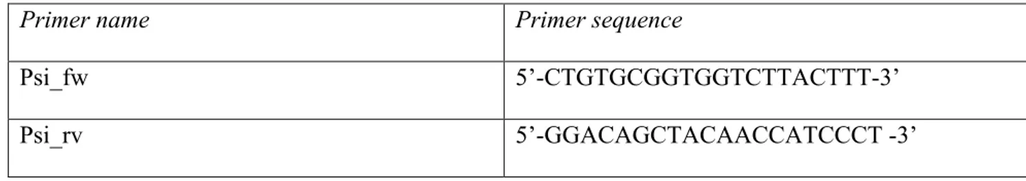
qPCR is performed on gDNA extracted from transduced HEK293T in order to test the correct integration of the produced viral vectors. Specific primers were designed (Table I). β-actin gene was the selected housekeeping gene. All data was later analyzed by calculating the Ct mean, the Delta Ct = Ct gene test – Ct housekeeping gene and finally the ∆∆Ct = ∆Ct sample – ∆Ct control.
Primer name Primer sequence
Psi_fw 5’-CTGTGCGGTGGTCTTACTTT-3’
Psi_rv 5’-GGACAGCTACAACCATCCCT -3’
Western Blot analysis
Proteins are extracted from expanded HEK#20 and HEKAcrVa1 respectively transduced and
transfected with p/LV_CRISPR_eGFP-TK+, p/LV_CRISPR_eGFP-TK-, both
46 p/LV_CRISPR_eGFP-TK+/TK- and p/LV_CMV_eGFP-TK+. Cells pellet is resuspended in 80ul of 1% SDS, left for 10 min at 95°C and later sonicated. BCA assay (ThermoFisher) is performed in order to quantify the proteins concentration and to load an equal amount of protein for immunoblotting. Antibody used: anti-HA and anti-Vinculin.
CLL Xenograft mice model
Mice (male, 8-10 weeks) NOD-scid IL2Rγnull (NSG) are purchased from CHARLES RIVER LABORATORIES ITALIA s.r.l. and kept in the animal facilities of Toscana Life Science (TLS) in Siena, Italy. Animals are kept in isolation for 2 weeks before the starting of the study. 100 ul of blood samples are collected in EDTA from the animals a day before the starting of the experiments. Mice are i.v. injected with Busulfan [25mg/Kg] (Myleran, Busulfex, 50mg) in a final volume of 500ul. 1x10ˆ8 PBMCs from CLL patient are thaw and counted the day of injection. The final pellet is resuspended in PBS in a final volume 100ul ready to be injected in each animal. Every week for 4 weeks blood sample (100ul EDTA) is collected from the mice and quickly analyzed by flow cytometry using human antibodies: CD45+,CD5+,CD19- (T cells), CD45+,CD5+,CD19+(B-CLL cells), CD45+,CD5-,CD19+ (B cells). On week 4, mice are sacrificed and organs collected. Spleen, BM and lymph nodes are homogenized by mechanically cutting the tissues by scalpel and needles (21G and 23G) in order to isolate cells for flow cytometry analysis. Tissues are stored in optimal cutting temperature (OCT) compound at -80°C for further analysis.
47
References
1. Aitken, M. J. L., Lee, H. J. & Post, S. M. Emerging treatment options for patients with p53-pathway-deficient CLL. Ther. Adv. Hematol. 10, 204062071989135 (2019).
2. Muñoz-Novas, C. et al. The International Prognostic Index for Patients with Chronic Lymphocytic Leukemia Has the Higher Value in Predicting Overall Outcome Compared with the Barcelona-Brno Biomarkers Only Prognostic Model and the MD Anderson Cancer Center
Prognostic Index. BioMed Res. Int. 2018, 1–8 (2018).
3. Sutton, L.-A. & Rosenquist, R. Deciphering the molecular landscape in chronic lymphocytic leukemia: time frame of disease evolution. Haematologica 100, 7–16 (2015).
4. Rozovski, U., Keating, M. J. & Estrov, Z. Why Is the Immunoglobulin Heavy Chain Gene Mutation Status a Prognostic Indicator in Chronic Lymphocytic Leukemia? Acta Haematol. 140, 51–54 (2018).
5. Malcikova, J. et al. Detailed analysis of therapy-driven clonal evolution of TP53 mutations in chronic lymphocytic leukemia. Leukemia 29, 877–885 (2015).
6. Gaidano, G. & Rossi, D. The mutational landscape of chronic lymphocytic leukemia and its impact on prognosis and treatment. Hematology 2017, 329–337 (2017).
7. Moia, R. et al. Precision Medicine Management of Chronic Lymphocytic Leukemia.
Cancers 12, 642 (2020).
8. Cohen, J. A. et al. An Updated Perspective on Current Prognostic and Predictive
Biomarkers in Chronic Lymphocytic Leukemia in the Context of Chemoimmunotherapy and Novel Targeted Therapy. Cancers 12, 894 (2020).
9. Campo, E. et al. TP53 aberrations in chronic lymphocytic leukemia: an overview of the clinical implications of improved diagnostics. Haematologica 103, 1956–1968 (2018).
10. Döhner, H. et al. Genomic Aberrations and Survival in Chronic Lymphocytic Leukemia. N.
Engl. J. Med. 343, 1910–1916 (2000).
11. Gentile, M. et al. Validation of the CLL-IPI and comparison with the MDACC prognostic index in newly diagnosed patients. Blood 128, 2093–2095 (2016).
12. Rossi, D. et al. The Prognostic Value of TP53 Mutations in Chronic Lymphocytic Leukemia Is Independent of Del17p13: Implications for Overall Survival and Chemorefractoriness. Clin.
Cancer Res. 15, 995–1004 (2009).
13. Aubrey, B. J., Strasser, A. & Kelly, G. L. Tumor-Suppressor Functions of the TP53 Pathway. Cold Spring Harb. Perspect. Med. 6, a026062 (2016).
14. Harris, S. L. & Levine, A. J. The p53 pathway: positive and negative feedback loops.
Oncogene 24, 2899–2908 (2005).
15. on behalf of the European Research Initiative on Chronic Lymphocytic Leukemia (ERIC) — TP53 network et al. ERIC recommendations for TP53 mutation analysis in chronic lymphocytic leukemia—update on methodological approaches and results interpretation. Leukemia
32, 1070–1080 (2018).
16. Kosicki, M. et al. Dynamics of Indel Profiles Induced by Various CRISPR/Cas9 Delivery Methods. in Progress in Molecular Biology and Translational Science vol. 152 49–67 (Elsevier, 2017).
17. Petitjean, A. et al. Impact of mutant p53 functional properties on TP53 mutation patterns and tumor phenotype: lessons from recent developments in the IARC TP53 database. Hum. Mutat.
28, 622–629 (2007).
18. Tate, J. G. et al. COSMIC: the Catalogue Of Somatic Mutations In Cancer. Nucleic Acids
Res. 47, D941–D947 (2019).
19. Pinto, A. M. et al. Low‐level TP 53 mutational load antecedes clonal expansion in chronic lymphocytic leukaemia. Br. J. Haematol. 184, 657–659 (2019).
20. Landau, D. A. et al. Evolution and Impact of Subclonal Mutations in Chronic Lymphocytic Leukemia. Cell 152, 714–726 (2013).
48 21. Rossi, D. et al. Clinical impact of small TP53 mutated subclones in chronic lymphocytic leukemia. 123, 10 (2014).
22. Stilgenbauer, S., Schnaiter, A., Paschka, P., Zenz, T. & Rossi, M. Gene mutations and treatment outcome in chronic lymphocytic leukemia: results from the CLL8 trial. 123, 8 (2014). 23. Boddy, C. S. & Ma, S. Frontline Therapy of CLL: Evolving Treatment Paradigm. Curr.
Hematol. Malig. Rep. 13, 69–77 (2018).
24. A New Era is Coming up in the Treatment of Chronic Lymphocytic Leukemia. Lymphoma
Chronic Lymphocytic Leuk. 4, 9–19 (2014).
25. Alduaij, W. & Illidge, T. M. The future of anti-CD20 monoclonal antibodies: are we making progress? Blood 117, 2993–3001 (2011).
26. Boross, P. & Leusen, J. H. W. Mechanisms of action of CD20 antibodies. 15.
27. Smith, M. R. Rituximab (monoclonal anti-CD20 antibody): mechanisms of action and resistance. Oncogene 22, 7359–7368 (2003).
28. Thompson, P. A. et al. Fludarabine, cyclophosphamide, and rituximab treatment achieves long-term disease-free survival in IGHV-mutated chronic lymphocytic leukemia. Blood 127, 303– 309 (2016).
29. Kater, A. P. et al. Fixed Duration of Venetoclax-Rituximab in Relapsed/Refractory Chronic Lymphocytic Leukemia Eradicates Minimal Residual Disease and Prolongs Survival:
Post-Treatment Follow-Up of the MURANO Phase III Study. J. Clin. Oncol. 37, 269–277 (2019). 30. June, C. H., O’Connor, R. S., Kawalekar, O. U., Ghassemi, S. & Milone, M. C. CAR T cell immunotherapy for human cancer. Science 359, 1361–1365 (2018).
31. Mancikova, V. et al. Performance of anti-CD19 chimeric antigen receptor T cells in genetically defined classes of chronic lymphocytic leukemia. J. Immunother. Cancer 8, e000471 (2020).
32. Dreger, P. et al. Managing high-risk CLL during transition to a new treatment era: stem cell transplantation or novel agents? Blood 124, 3841–3849 (2014).
33. Sorror, M. L. et al. Five-Year Follow-Up of Patients With Advanced Chronic Lymphocytic Leukemia Treated With Allogeneic Hematopoietic Cell Transplantation After Nonmyeloablative Conditioning. J. Clin. Oncol. 26, 4912–4920 (2008).
34. Hahn, M. et al. Allogeneic hematopoietic stem cell transplantation for poor-risk CLL: dissecting immune-modulating strategies for disease eradication and treatment of relapse. Bone
Marrow Transplant. 50, 1279–1285 (2015).
35. Fedoroff, N. V. McClintock’s challenge in the 21st century. Proc. Natl. Acad. Sci. U. S. A.
109, 20200–20203 (2012).
36. Fernández, A., Josa, S. & Montoliu, L. A history of genome editing in mammals. Mamm.
Genome 28, 237–246 (2017).
37. Unniyampurath, U., Pilankatta, R. & Krishnan, M. RNA Interference in the Age of CRISPR: Will CRISPR Interfere with RNAi? Int. J. Mol. Sci. 17, 291 (2016).
38. Urnov, F. D. et al. Highly efficient endogenous human gene correction using designed zinc-finger nucleases. Nature 435, 646–651 (2005).
39. Miller, J. C. et al. A TALE nuclease architecture for efficient genome editing. Nat.
Biotechnol. 29, 143–148 (2011).
40. Smith, J. Requirements for double-strand cleavage by chimeric restriction enzymes with zinc finger DNA-recognition domains. Nucleic Acids Res. 28, 3361–3369 (2000).
41. Li, T. et al. TAL nucleases (TALNs): hybrid proteins composed of TAL effectors and FokI DNA-cleavage domain. Nucleic Acids Res. 39, 359–372 (2011).
42. Mahfouz, M. M. et al. De novo-engineered transcription activator-like effector (TALE) hybrid nuclease with novel DNA binding specificity creates double-strand breaks. Proc. Natl. Acad.
Sci. 108, 2623–2628 (2011).
43. Carroll, D. Genome Engineering With Zinc-Finger Nucleases. Genetics 188, 773–782 (2011).
49 44. Boch, J. et al. Breaking the Code of DNA Binding Specificity of TAL-Type III Effectors.
Science 326, 1509–1512 (2009).
45. Doudna, J. A. & Charpentier, E. The new frontier of genome engineering with CRISPR-Cas9. Science 346, 1258096 (2014).
46. Mojica, F. J. M., D
Sequences of Regularly Spaced Prokaryotic Repeats Derive from Foreign Genetic Elements. J. Mol.
Evol. 60, 174–182 (2005).
47. Barrangou, R. et al. CRISPR Provides Acquired Resistance Against Viruses in Prokaryotes.
Science 315, 1709–1712 (2007).
48. Wright, A. V., Nuñez, J. K. & Doudna, J. A. Biology and Applications of CRISPR Systems: Harnessing Nature’s Toolbox for Genome Engineering. Cell 164, 29–44 (2016).
49. Nishimasu, H. et al. Crystal Structure of Cas9 in Complex with Guide RNA and Target DNA. Cell 156, 935–949 (2014).
50. Wu, X., Kriz, A. J. & Sharp, P. A. Target specificity of the CRISPR-Cas9 system. Quant.
Biol. 2, 59–70 (2014).
51. Ran, F. A. et al. Genome engineering using the CRISPR-Cas9 system. Nat. Protoc. 8, 2281– 2308 (2013).
52. Fonfara, I., Richter, H., Bratovič, M., Le Rhun, A. & Charpentier, E. The
CRISPR-associated DNA-cleaving enzyme Cpf1 also processes precursor CRISPR RNA. Nature 532, 517– 521 (2016).
53. Świat, M. A. et al. FnCpf1: a novel and efficient genome editing tool for Saccharomyces cerevisiae. Nucleic Acids Res. 45, 12585–12598 (2017).
54. Zetsche, B. et al. Cpf1 Is a Single RNA-Guided Endonuclease of a Class 2 CRISPR-Cas System. Cell 163, 759–771 (2015).
55. Fagerlund, R. D., Staals, R. H. J. & Fineran, P. C. The Cpf1 CRISPR-Cas protein expands genome-editing tools. Genome Biol. 16, 251 (2015).
56. Wyman, C. & Kanaar, R. DNA Double-Strand Break Repair: All’s Well that Ends Well.
Annu. Rev. Genet. 40, 363–383 (2006).
57. Ma, Y., Zhang, L. & Huang, X. Genome modification by CRISPR/Cas9. FEBS J. 281, 5186–5193 (2014).
58. Gaj, T., Gersbach, C. A. & Barbas, C. F. ZFN, TALEN, and CRISPR/Cas-based methods for genome engineering. Trends Biotechnol. 31, 397–405 (2013).
59. Wyman, C., Ristic, D. & Kanaar, R. Homologous recombination-mediated double-strand break repair. DNA Repair 3, 827–833 (2004).
60. Lee, J., Chung, J.-H., Kim, H. M., Kim, D.-W. & Kim, H. Designed nucleases for targeted genome editing. Plant Biotechnol. J. 14, 448–462 (2016).
61. Taleei, R. & Nikjoo, H. Biochemical DSB-repair model for mammalian cells in G1 and early S phases of the cell cycle. Mutat. Res. Toxicol. Environ. Mutagen. 756, 206–212 (2013). 62. Lin, D.-W. et al. Microhomology-based CRISPR tagging tools for protein tracking, purification, and depletion. J. Biol. Chem. 294, 10877–10885 (2019).
63. Sakuma, T., Nakade, S., Sakane, Y., Suzuki, K.-I. T. & Yamamoto, T. MMEJ-assisted gene knock-in using TALENs and CRISPR-Cas9 with the PITCh systems. Nat. Protoc. 11, 118–133 (2016).
64. Hsu, P. D., Lander, E. S. & Zhang, F. Development and Applications of CRISPR-Cas9 for Genome Engineering. Cell 157, 1262–1278 (2014).
65. Wu, S.-S., Li, Q.-C., Yin, C.-Q., Xue, W. & Song, C.-Q. Advances in CRISPR/Cas-based Gene Therapy in Human Genetic Diseases. Theranostics 10, 4374–4382 (2020).
66. Nelson, C. E. et al. In vivo genome editing improves muscle function in a mouse model of Duchenne muscular dystrophy. Science 351, 403–407 (2016).
67. Tabebordbar, M. et al. In vivo gene editing in dystrophic mouse muscle and muscle stem cells. Science 351, 407–411 (2016).
50 68. Wu, Y. et al. Highly efficient therapeutic gene editing of human hematopoietic stem cells.
Nat. Med. 25, 776–783 (2019).
69. Maeder, M. L. et al. Development of a gene-editing approach to restore vision loss in Leber congenital amaurosis type 10. Nat. Med. 25, 229–233 (2019).
70. Halim, L. & Maher, J. CAR T-cell immunotherapy of B-cell malignancy: the story so far.
Ther. Adv. Vaccines Immunother. 8, 251513552092716 (2020).
71. Stadtmauer, E. A. et al. CRISPR-engineered T cells in patients with refractory cancer.
Science 367, eaba7365 (2020).
72. Glover, D. J., Lipps, H. J. & Jans, D. A. Towards safe, non-viral therapeutic gene expression in humans. Nat. Rev. Genet. 6, 299–310 (2005).
73. Gori, J. L. et al. Delivery and Specificity of CRISPR/Cas9 Genome Editing Technologies for Human Gene Therapy. Hum. Gene Ther. 26, 443–451 (2015).
74. Giacca, M. & Zacchigna, S. Virus-mediated gene delivery for human gene therapy. J.
Controlled Release 161, 377–388 (2012).
75. Pauwels, K. et al. State-of-the-Art Lentiviral Vectors for Research Use: Risk Assessment and Biosafety Recommendations. Curr. Gene Ther. 9, 459–474 (2009).
76. Merten, O.-W., Hebben, M. & Bovolenta, C. Production of lentiviral vectors. Mol. Ther. -
Methods Clin. Dev. 3, 16017 (2016).
77. Naldini, L. et al. In Vivo Gene Delivery and Stable Transduction of Nondividing Cells by a Lentiviral Vector. Science 272, 263–267 (1996).
78. Vink, C. A. et al. Eliminating HIV-1 Packaging Sequences from Lentiviral Vector
Proviruses Enhances Safety and Expedites Gene Transfer for Gene Therapy. Mol. Ther. 25, 1790– 1804 (2017).
79. Schmidt, F. & Grimm, D. CRISPR genome engineering and viral gene delivery: A case of mutual attraction. Biotechnol. J. 10, 258–272 (2015).
80. Cockrell, A. S., van Praag, H., Santistevan, N., Ma, H. & Kafri, T. The HIV-1 Rev/RRE system is required for HIV-1 5’ UTR cis elements to augment encapsidation of heterologous RNA into HIV-1 viral particles. Retrovirology 8, 51 (2011).
81. Funke, S. et al. Targeted Cell Entry of Lentiviral Vectors. Mol. Ther. 16, 1427–1436 (2008). 82. Frecha, C. et al. Efficient and stable transduction of resting B lymphocytes and primary chronic lymphocyte leukemia cells using measles virus gp displaying lentiviral vectors. Blood 114, 3173–3180 (2009).
83. Levy, C. et al. Baboon envelope pseudotyped lentiviral vectors efficiently transduce human B cells and allow active factor IX B cell secretion in vivo in NOD/SCIDγc -/- mice. J. Thromb. Haemost. 14, 2478–2492 (2016).
84. Girard-Gagnepain, A. et al. Baboon envelope pseudotyped LVs outperform VSV-G-LVs for gene transfer into early-cytokine-stimulated and resting HSCs. Blood 124, 1221–1231 (2014). 85. Sereni, L. et al. Lentiviral gene therapy corrects platelet phenotype and function in patients with Wiskott-Aldrich syndrome. J. Allergy Clin. Immunol. 144, 825–838 (2019).
86. Aiuti, A. et al. Lentiviral Hematopoietic Stem Cell Gene Therapy in Patients with Wiskott-Aldrich Syndrome. Science 341, 1233151–1233151 (2013).
87. Diez, B. et al. Therapeutic gene editing in CD 34 + hematopoietic progenitors from Fanconi
anemia patients. EMBO Mol. Med. 9, 1574–1588 (2017).
88. Gao, L. et al. Engineered Cpf1 variants with altered PAM specificities. Nat. Biotechnol. 35, 789–792 (2017).
89. Niccheri, F., Pecori, R. & Conticello, S. G. An efficient method to enrich for knock-out and knock-in cellular clones using the CRISPR/Cas9 system. Cell. Mol. Life Sci. 74, 3413–3423 (2017). 90. Chen, F. et al. Targeted activation of diverse CRISPR-Cas systems for mammalian genome editing via proximal CRISPR targeting. Nat. Commun. 8, 14958 (2017).
91. Watters, K. E., Fellmann, C., Bai, H. B., Ren, S. M. & Doudna, J. A. Systematic discovery of natural CRISPR-Cas12a inhibitors. Science 362, 236–239 (2018).
51 92. Bagnara, D. et al. A novel adoptive transfer model of chronic lymphocytic leukemia
suggests a key role for T lymphocytes in the disease. Blood 117, 5463–5472 (2011).
93. Herman, S. E. M. et al. Modeling tumor–host interactions of chronic lymphocytic leukemia in xenografted mice to study tumor biology and evaluate targeted therapy. Leukemia 27, 2311–2321 (2013).
94. Straathof, K. C. et al. An inducible caspase 9 safety switch for T-cell therapy. Blood 105, 4247–4254 (2005).
Clinical trials references
ClinicalTrials.gov Identifier:NCT03960840.
CD19-specific CAR-T Cells in CLL/SLL and DLBCL (2019)
https://clinicaltrials.gov/ct2/show/NCT03960840
ClinicalTrials.gov Identifier:NCT02935257.
Immunotherapy for High Risk/Relapsed CD19+ Acute Lymphoblastic Leukaemia, B-cell Non-Hodgkin's Lymphoma (B-NHL) and Chronic Lymphocytic Leukaemia (CLL)/ Small Lymphocytic Lymphoma (SLL) Using CAR T-cells to Target CD19 (ALLCAR19).
https://clinicaltrials.gov/ct2/show/NCT02935257
ClinicalTrials.gov Identifier: NCT01853631.
Activated T-Cells Expressing 2nd or 3rd Generation CD19-Specific CAR, Advanced B-Cell NHL, ALL, and CLL (SAGAN) (SAGAN). https://clinicaltrials.gov/ct2/show/NCT01853631
ClinicalTrials.gov Identifier: NCT03655678.
A Safety and Efficacy Study Evaluating CTX001 in Subjects With Transfusion-Dependent β-Thalassemia. https://clinicaltrials.gov/ct2/show/NCT03655678
ClinicalTrials.gov Identifier: NCT03872479.
Single Ascending Dose Study in Participants With LCA10.
https://clinicaltrials.gov/ct2/show/NCT03872479
52 A Safety and Efficacy Study Evaluating CTX110 in Subjects With Relapsed or Refractory B-Cell Malignancies. https://clinicaltrials.gov/ct2/show/NCT04035434
ClinicalTrials.gov Identifier:NCT03399448
NY-ESO-1-redirected CRISPR (TCRendo and PD1) Edited T Cells (NYCE T Cells). https://clinicaltrials.gov/ct2/show/NCT03399448
ClinicalTrials.gov Identifier:NCT03157804
Lentiviral-mediated Gene Therapy of Fanconi Anemia Patients Subtype A (FANCOLEN-1). https://clinicaltrials.gov/ct2/show/NCT03157804