- 1 -
SCUOLA NORMALE SUPERIORE PISA
and
THE INTERNATIONAL CENTRE for GENETIC ENGINEERING and BIOTECHNOLOGY (ICGEB)
TRIESTE
The Cytoplasmic Tail
of the Notch Ligand Jagged-1: Intrinsic
Disorder, Induced Order and Molecular
Interactions
Thesis submitted for the Degree of Doctor of Philosophy
Perfezionamento in Genetica Molecolare e Biotecnologie
Candidate: Matija Popović
Supervisor: Sándor Pongor, Ph.D., D.Sc.
- 2 -
Abstract
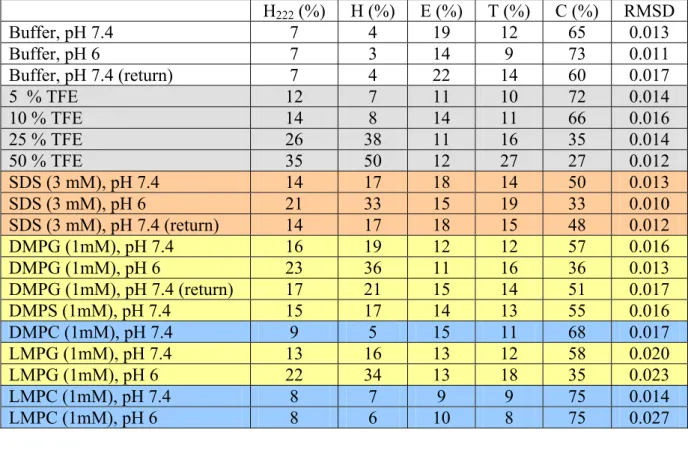
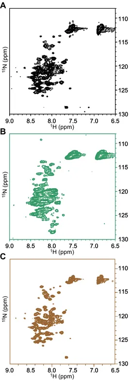
Notch signaling plays a key role in cell differentiation in all metazoans. As both receptors and ligands are cell-surface proteins, Notch signaling is restricted to nearby interacting cells. Notch ligands are membrane-spanning proteins made of a large extracellular region, a transmembrane segment, and a 100–200 residue cytoplasmic tail. The sequence of the intracellular region of Jagged-1, one of the five ligands to Notch receptors in man, is very well conserved throughout evolution but does not encode any globular domain. The cytoplasmic tail of Jagged-1 mediates protein–protein interactions through the C-terminal PDZ binding motif, is involved in ligand endocytosis triggered by mono-ubiquitination, and, as a consequence of regulated intramembrane proteolysis, can be released into the cytosol as a signaling fragment. The intracellular region of Jagged-1 may then exist in at least two forms: as a membrane-tethered protein located at the interface between the membrane and the cytoplasm, and as a soluble nucleocytoplasmic protein. To investigate its structural properties, a recombinant protein corresponding to the human Jagged-1 intracellular region (J1_tmic) was expressed, purified, and characterized in different environments using various biophysical methods such as circular dichroism, tryptophan fluorescence, size-exclusion chromatography, and NMR.
In solution, J1_tmic behaves as an intrinsically disordered protein, but displays a significant helical propensity. In the presence of SDS micelles or negatively charged phospholipid micelles and vesicles, used to mimick the interface between the plasma membrane and the cytosol, J1_tmic gains partial helical structure. The partial folding and association of the intracellular region of Jagged-1 with the membrane is expected to reduce its “capture radius” towards target proteins and to make selected residues unavailable for post-translational modifications or binding.
Binding of Jagged-1 intracellular region to the PDZ domain of afadin, a protein located at cell-cell adherens junctions, couples Notch signaling with the adhesion system and the cytoskeleton. The interaction between the PDZ domain of afadin (AF6_PDZ) and a series of polypeptides comprising the Jagged-1 PDZ-binding motif (EYIV) was investigated using NMR chemical shift perturbation and surface plasmon resonance. It was shown that binding of Jagged-1 intracellular region to
- 3 -
AF6_PDZ is strictly local, involving only the last six residues of the binding motif and the PDZ binding groove, and that it does not trigger global folding of J1_tmic.
In the C-terminal region of Jagged-1 cytoplasmic tail, four potential phosphorylation sites can be identified, one of them (Y1216) located in the PDZ-binding motif. It was found that, while phosphorylation at any of these sites disrupts binding of the C-terminal peptides to lipid micelles, phosphorylation at (Y1216) also affects the interaction with AF6_PDZ, with a reduction in the binding affinity. Phosphorylation thus provides a potential way to modulate the interaction of Jagged-1 C-terminal region not only with the membrane but also with the partner PDZ. It was also shown that the R1213Q mutation in the PDZ binding motif associated with a congenital obstruction of the bile ducts, increases the affinity for AF6_PDZ.
In summary, this work presents the first biochemical and structural characterization of Jagged-1 cytoplasmic tail in solution and in environments that mimic the membrane/cytoplasm interface, and the first biophysical study on its interaction with the afadin PDZ domain.
- 4 -
List of publications
Publications related to this thesis:
Popovic M, Bella J, Zlatev V, Hodnik V, Anderluh G, Barlow PN, Pintar A, Pongor S. The interaction of Jagged-1 cytoplasmic tail with afadin PDZ domain is local, folding independent, and tuned by phosphorylation. J. Mol. Recognit. (2010), in press.
De Biasio A, Guarnaccia C, Popovic M, Uversky VN, Pintar A, Pongor S. Prevalence of intrinsic disorder in the intracellular region of single-pass transmembrane proteins: the case of the Notch Ligand Delta-4. J. Proteome Res. (2008) 7:2496–2506
Pintar A, De Biasio A, Popovic M, Ivanova N, Pongor S. The intracellular region of Notch ligands: does the tail make the difference? Biol. Direct. (2007) 2:19.
Popovic M, De Biasio A, Pintar A, Pongor S. The intracellular region of the Notch ligand Jagged-1 gains partial structure upon binding to synthetic membranes. FEBS J. (2007) 274(20):5325-36.
Popovic M, Coglievina M, Guarnaccia C, Verdone G, Esposito G, Pintar A, Pongor S. Gene synthesis, expression, purification, and characterization of human Jagged-1 intracellular region. Protein Expr. Purif. (2006) 47:398–404.
- 5 -
Acknowledgments
This thesis was prepared in the Protein Structure and Bioinformatics Group at the International Centre for Genetic Engineering and Biotechnology (ICGEB) in Trieste, Italy.
First, I am very grateful to my supervisor, Sándor Pongor, for giving me the opportunity to carry out my work in his lab. I want to thank him for his support and helpful tips, which were necessary for the completion of this thesis.
Special gratitude goes to Alessandro Pintar who supervises the studies on Notch signaling within the lab, for his continuous help and sincere patience in taking me throughout the entire project. Thanks to him, now I can say that I am much closer to becoming an independent researcher in the field of structural biology and protein NMR spectroscopy.
I am very grateful to all the people of the Protein Structure and Bioinformatics Group that contributed to this thesis: Maristella Coglievina, who trained me in the molecular biology techniques, Corrado Guarnaccia for continuous help with HPLC, mass spectrometry, peptide synthesis, and peptide and protein purification, Ventsislav Zlatev for the synthesis and purification of many peptides, and Sotir Zahariev for helpful advice in the peptide chemistry field.
I am also grateful to all external collaborators that contributed, directly or indirectly, to this thesis: Doriano Lamba (CNR-ELETTRA, Basovizza, Italy) for extensive use of the CD spectropolarimeter, Fabio Calogiuri (CERM, Sesto Fiorentino, Italy) and Nicola D’Amelio (CBM, Basovizza, Italy) for technical assistance with the acquisition of NMR spectra, Juraj Bella, Paul Barlow and Dusan Uhrin (Biomolecular NMR Unit, University of Edinburgh, UK) for technical assistance and support in the acquisition and processing of the NMR spectra acquired in Edinburgh, Vesna Hodnik and Gregor Anderluh (Biotechnical Faculty, University of Ljubljana, Slovenia) for technical assistance and support in the SPR experiments.
- 6 -
I also acknowledge the EU (European Network of Research Infrastructures for Providing Access and Technological Advancements in Bio-NMR) for providing access to the CERM NMR facility, EMBO (European Molecular Biology Organization) for a Short Term Fellowship, and ICGEB for support with a pre-doctoral fellowship.
I want to thank my friend and colleague Vjeko, with whom my work at the institute became more interesting and amusing.
My greetings go to the young Buddhist group of Trieste, with whom I felt at home and started to love this town.
I am taking this opportunity to express my deepest gratitude to the most important people in my life:
my parents, my sister,
and Vanessa.
- 7 -
Table of Contents
Abstract
2List of publications
4Acknowledgments
51. Introduction
11 1.1 Notch Signaling 111.1.1 Molecular mechanisms of Notch signaling 11 1.1.2 Notch signaling and cell-fate decisions 14 Notch signaling in development 14
Notch signaling in cancer 15
Notch signaling in genetic disorders 16
1.1.3 Notch signaling regulation 17
Glycosylation 17
Endosomal trafficking 19
Transcriptional modulation 20
1.2 Jagged-1 20
1.2.1 Jagged-1 and other Notch ligands 20
1.2.2 The intracellular region of Notch ligands 21
1.3 Jagged-1 and AF6 24
1.3.1 PDZ domains 25
1.3.2 Afadin/AF6 28
1.3.3 AF6_PDZ domain 30
1.4 Notch structural biology 31
1.5 Aim of the thesis 37
2. Results
382.1 Structural characterization of J1_tmic 38
2.1.1. Expression and purification of the recombinant Jagged-1
intracellular region 38
Gene synthesis 38
Protein expression and purification 39 2.1.2 J1_ic is mainly disordered in solution 40
- 8 -
2.1.3 The additional intramembrane residues do not promote
folding of J1_ic 45
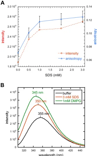
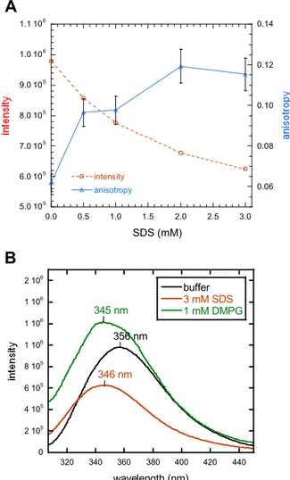
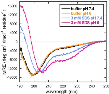
2.1.4 J1_tmic exhibits intrinsic helical propensity 49 2.1.5 J1_tmic binds to SDS micelles and phospholipid vesicles 51 2.1.6 J1_tmic gains helical structure upon binding to SDS micelles 55 2.1.7 J1_tmic gains helical structure upon binding to negatively
charged phospholipid vesicles 59
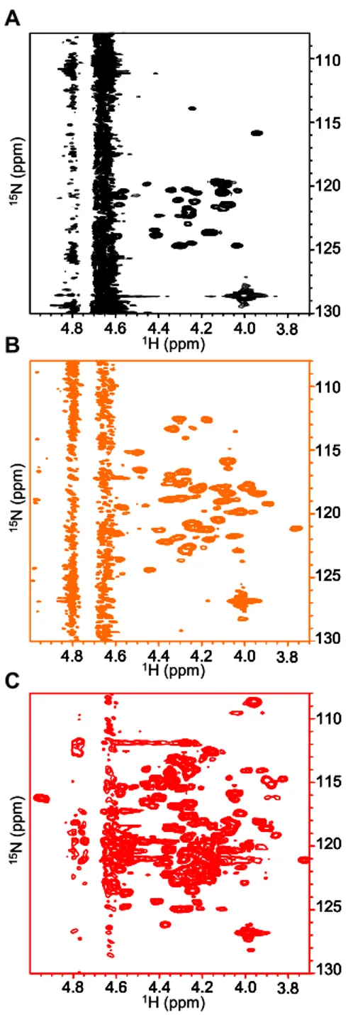
2.1.8 J1_tmic in presence of lipid micelles – NMR studies 61 2.1.9 J1_tmic as a potential zinc-binding protein 68
2.2 The interaction between J1_tmic and AF6_PDZ 69
2.2.1 Expression and purification of AF6_PDZ 69 2.2.2 NMR experiments – AF6_PDZ resonance assignments 70 2.2.3 Jagged-1 C-terminus binds into the βB/αB groove of AF6_PDZ 75 2.2.4 Jagged-1’s binding is strictly local 82
2.2.5 J1_tmic does not fold upon binding 85
2.2.6 Tyrosine phosphorylation at P-2 reduces affinity 85 2.2.7 The R1213Q mutation at P-5 increases affinity 91 2.2.8 Phosphorylation affects binding to lipid micelles 92
3. Discussion
973.1 Life on the membrane: Jagged-1 intracellular region as an
interfacial protein 97
3.2 Leaving the membrane: Jagged-1 intracellular region as a
nucleocytoplasmic protein 103
3.3 Social life: the interaction with afadin 110
3.3.1 A qualitative analysis of the binding 111 3.3.2 A quantitative analysis of the binding 113 3.3.3 J1_tmic does not fold upon binding 115 3.3.4 Tyrosine phosphorylation at P-2 reduces affinity 115 3.3.5 The R1213Q mutation at P-5 increases affinity 119
3.4 All together now: phosphorylation may affects both binding to
the membrane and to the PDZ 119
- 9 -
4. Materials and Methods
1234.1 Production of the recombinant Jagged-1 intracellular
region 123
4.1.1 Gene synthesis of J1_ic 123
4.1.2 Preparation of the J1_tmic gene construct 124 4.1.3 J1_ic expression and purification 125 4.1.4 J1_ic purification in native conditions 126 4.1.5 J1_tmic expression and purification 127
4.2 Peptide synthesis 127
4.3 Production of the recombinant AF6 PDZ domain 128
4.3.1 Expression and purification 128
4.4 Determination of protein and peptide concentrations 129
4.5 Size exclusion chromatography 130
4.6 Limited proteolysis 130
4.7 Preparation of phospholipid vesicles and micelles 130
4.8 Circular dichroism 131
4.9 Fluorescence spectroscopy 131
4.10 NMR spectroscopy 132
4.10.1 NMR of the Jagged-1 intracellular region 132
NMR of unlabeled J1_ic 132
NMR of 15N-labeled J1_tmic 132 NMR of 15N-labeled J1_tmic bound to lysophospolipid micelles 133
4.10.2 NMR of AF6_PDZ 134
Resonance assignments of AF6_PDZ 134
Chemical shift mapping 134
NMR of J1_tmic 135
4.11 Surface Plasmon Resonance 135
5. Biophysical methods
1375.1 Circular Dichroism 137
5.2 Fluorescence spectroscopy 139
- 10 -
5.4 NMR spectroscopy 143
5.4.1 Physical background 143
5.4.2 Protein NMR 145
5.4.3 Mapping protein-ligand interactions by NMR 149
5.4.4 In-cell NMR 152
5.5 Membrane mimicking models 154
5.5.1 Micelles 155 5.5.2 Liposomes 156 5.5.3 Bicelles 156 5.5.4 Nanodiscs 157
Abbreviations
158References
161Supplementary material
181- 11 -
1. INTRODUCTION
1.1 Notch signaling
1.1.1 Molecular mechanisms of Notch signaling
Notch signaling mediates cross-talk between two contacting cell. The main components in this signaling pathway are Notch receptors and their corresponding ligands, which are both membrane-bound proteins [1], [2] (Figure 1.1). Notch receptors, four members of which have been identified in man (Notch1, 2, 3 and -4), are membrane-spanning glycoproteins assembled in a non-covalent heterodimeric complex. After their expression, Notch receptors are proteolytically processed (at the S1 cleavage site) by a furin-like convertase in the trans-Golgi to give an extracellular (N_ec) and a transmembrane subunit (N_tm) [3]. The N_ec contains an array of up to 36 EGF tandem repeats followed by three LIN-12 repeats that maintain Notch in a resting conformation before ligand-induced activation [4]. The EGF 11-12 couple in Notch receptor constitutes the binding site for the ligand DSL domain [5]. The N_tm has a short extracellular region, involved in the dimerization with the N_ec, and a ~900 residue long intracellular region (N_ic). The second one consists of a one RAM domain followed by seven ankyrin repeats with two lateral nuclear localization signals, a transactivation domain (TAD), and a C-terminal PEST region. Upon ligand binding, an extracellular ADAM type metalloproteinase cleaves the N_tm at the S2 site; 12 residues before the membrane-spanning segment. This removes the N_ec from the membrane, rendering the N_tm subunit susceptible to the cleavage at the S3 site, close to the inner leaflet of the plasma membrane. This proteolytic processing is mediated by the presenilin/γ-secretase complex and releases the intracellular domain (N_ic) from the membrane into the cytoplasm. This mechanism is known as "regulated intramembrane proteolysis" (RIP) [6] and is also present in other signal transduction pathways, such as those mediated by CD44, LRP, ErbB-4 and others [7]. Once released into the cytoplasm, N_ic translocates to the nucleus and associates with the CSL family of DNA-binding proteins (CBF-1/suppressor of Hairless/Lag-1) [8].
This association, along with the recruitment of Mastermind and other co-activators, converts the CSL-containing complex from a transcriptional repressor to a transcriptional activator of target genes. The best known Notch target genes are the
- 12 -
basic helix-loop helix (bHLH) transcriptional repressors of the HES and HEY families [8], [9] CSL CoR CSL MamN-ic N-ic N-ec N-ic N-ec
S1 Furin N-ic N-ec S3 N-ic S2 ADAM
γ-secretase complex Jagged Delta Notch N-ec CSL CoR CSL MamN-ic CSL CoR CSL CSL CoR CSL CSL MamN-iN-icc N-ic N-ec N-ic N-ec
S1 Furin N-ic N-ec S3 N-ic S2 ADAM ADAM
γ-secretase complex Jagged Delta Notch N-ec N-ec
Figure 1.1. The Notch signaling pathway. Notch receptors are cleaved at the S1 site by furin, to be presented as non-covalent heterodimers at the cell surface (ec and N-ic, extra-and intracellular region of the receptor, respectively). Upon ligand binding, the receptor is cleaved by ADAM and γ-secretase at the S2 and S3 sites, respectively. In the nucleus, N-ic displaces the co-repressor (CoR) to form a transcriptional activator shown as a complex of N-ic, mastermind (MAM) and CSL bound to DNA.
These are basic helix-loop helix (bHLH) DNA binding proteins that, acting within a complex and strongly context dependent transcriptional network, interact with other bHLH proteins, bind to specific gene promoter regions, and, through the recruitment of transcriptional corepressors, inhibits transcription of the target genes. [10],[11]. Several DNA target sequences have been identified so far, including HES1 own promoter, the promoter regions of the transcription factor Achaete Scute Homolog 1 (ASH1) [12], and the promoters of enzymes involved in cell cycle, like the cyclin-dependent kinase inhibitors p21WAF-1/CIP1 and p27kip1 [13].
- 13 -
In man, there are two families of Notch ligands, homologues of Drosophila Serrate (Jagged1 and 2) and homologues of Drosophila Delta (Deltalike1, 3 and -4) [14] (Figure 1.2). All five ligands share the same architecture and belong to the so called DSL family (Delta/Serrate/Lag-2, the latter being the C. elegans ligand). They are type I membrane spanning proteins consisting of a long extracellular region, a transmembrane segment and a relatively short cytoplasmic tail (100-150 amino acids). The ligand extracellular region has an N-terminal DSL domain followed by a variable number of EGF-like repeats. Ligands of the Jagged group have an additional cysteine rich region close to the transmembrane segment, which is not present in the Delta group ligands. EGF-like repeats (29-36) LNR modules HD RAM ANK repeats PEST EGF-like repeats (6-8) DSL MNNL Cys-rich N1 N2 N3 N4 D4 D3 D1 J2 J1
cytoplasmic tails of Notch ligands (105-155 residues) plasma membrane TAD EGF-like repeats (29-36) LNR modules HD RAM ANK repeats PEST EGF-like repeats (6-8) DSL MNNL Cys-rich N1 N2 N3 N4 D4 D3 D1 J2 J1
cytoplasmic tails of Notch ligands (105-155 residues)
plasma membrane
TAD
Figure 1.2. Domain organization of Notch receptors and ligands in man. The four Notch receptors (Notch-1 (N1), Notch-2 (N2), Notch-3 (N3) and Notch-4 (N4)) and five Notch ligands (Delta-like-4 (D4), Delta-like-3 (D3), Delta-like-1 (D1), Jagged-2 (J2) and Jagged-1 (J1)) are schematically shown. EGF, Epidermal Growth Factor repeat; LNR, Lin-Notch repeat; HD, hetero-dimerization region; RAM, RBP-Jkappa-associated module; ANK, Ankirin repeat; TAD, transcription activation domain; PEST, region containing P, E, S, T residues; MNNL, domain Mainly found in the N-terminal region of Notch Ligands; DSL, Delta-Serrate-Lag2 domain; Cys-rich, cysteine rich region.
- 14 - 1.1.2 Notch and cell-fate decisions
Development of a multicellular organism requires cell-fate decisions that are intimately associated with the determination of a cell-specific gene expression pattern. Notch signaling, having a direct effect on gene expression, acts in various development decisions either through lateral-inhibition or induction [15] (Figure 1.3). Lateral inhibition occurs between two equipotent neighbouring cells, which can signal one to another, as ligands and receptors are expressed in both of them. Due to fluctuations in the steady-state expression levels, the concentrations of these proteins between neighbouring cells start to differ. In cells with the higher receptor activation, the Notch feedback loop slightly inhibits the ligand expression. In this way, the initial small differences are amplified, which drives neighbouring cells into different developmental fates. Inductive signaling occurs when two non-equivalent groups of cells confront one another. Keeping the two populations distinct, Notch signaling is involved in establishing boundaries at the interface between them. In addition to this inductive role, Notch signaling also plays an important role in lineage decision between two daughter cells. A typical example is the development of sensory bristles in Drosophila, where sensory organ precursors (SOP) are initially selected from equivalent pro-neural cells by the Notch lateral inhibition [16]. SOP cells undergo multiple asymmetric cell divisions in which the Notch antagonist, Numb, is asymmetrically distributed. In each division step, only one daughter cell holds the Notch activity, eventually giving rise to a glial cell on one hand and cells that form the bristle on the other.
Notch signaling in development. Notch receptors and ligands are widely expressed during organogenesis in mammalian embryos, where they play a key role in establishing cell-lineage decisions in tissues derived from all the three primary germ layers: the endoderm (for ex. the pancreas), the mesoderm (skeleton, mammary gland, the vascular system and hematopoietic cells), and the ectoderm (neuronal cell lines) [17]. In the pancreas, where different cell types appear with different timing, yet stemming from the same early cells, Notch-1 appears to delay both endocrine and exocrine development, trapping progenitor cells in an undifferentiated state. In the presomitic mesoderm that will differentiate into the axial skeleton, muscles, tendons and dermis, Notch signaling plays a role as a molecular clock that controls regular segmentation of the mesoderm. Notch is also required in the later steps of vascular development, which includes proliferation and branching of the newly formed vessels
- 15 -
[18]. In the hematopoietic system, enforced activation of Notch-1 suppresses the differentiation of stem cells into myeloid, erythroid, or lymphoid lineages, and plays a role at a number of stages of lymphocyte development in the bone marrow and thymus. One of the essential functions of Notch-1 is the suppression of B cell development in the thymus. In the nervous system, Notch activation is required for the self-renewal of neural stem-cells, although it is not necessary for their generation [19], [20]. Furthermore, Notch signaling controls the differentiation of glial cells and the length and organization of dendritic extensions from neurons (neurite arborization).
signal sending cell Numb A B
Figure 1.3. Notch signaling in cell-fate decisions. Receptors are colored in orange, ligands in green, and orange arrows represent the Notch signaling direction in (A) lateral inhibition and (B) line lineage decision between two daughter cells in the development of sensory bristles in Drosophila. The intensity of the orange color is proportional to the receptor activation.
Notch signaling in cancer. At least two direct links between alterations in Notch signaling and human cancer have been established to date. A rare form of T cell acute lymphoblastic leukemia (T-ALL) is associated with a translocation that fuses the intracellular portion of Notch-1 with the promoter/enhancer region of the T-cell receptor beta locus, leading to constitutive activation of Notch-1 signaling [21]. The majority of T-ALL cases have been recently associated with activating mutations
- 16 -
in Notch-1 [22]. Another chromosomal translocation, which is altering the function of Mastermind, a nuclear regulatory protein in the Notch signaling pathway, has been linked to mucoepidermoid carcinoma, a common type of malignant salivary gland tumor [23]. High levels of Delta-1 have been observed in neuroblastoma cell lines. High expression levels of Notch have also been reported in some breast cancers and in human colon adenocarcinomas. Intriguingly, Notch can behave both as an oncogene or a tumor suppressor, depending on the cellular context and on the interactions with other signaling pathways [14].
Notch signaling in genetic disorders. The importance of the Notch pathway in cell fate control and development is further confirmed by the association of several diseases with mutations in genes involved in this complex signaling network [24]. Alagille syndrome [25] (AGS, MIM #118450) is a rare autosomal dominant disorder characterized by a variety of clinical abnormalities, including a reduction in the number of bile ducts eventually leading to the obstruction of biliary flow, and cardiac, musculoskeletal, ocular, facial defects. Although no clear genotype-phenotype correlation has been defined, AGS is caused by mutations in JAG1 [26], [27]. While the majority of the mutations causing AGS are related to the generation of stop codons leading to unstable mRNA or truncated proteins, many missense point mutations either introduce or delete cysteine residues that are critical for proper folding of the mature protein. Most of these mutations are located in the DSL domain and in the EGF tandem repeats. Similarly to AGS, but with a different pathogenesis, extrahepatic biliary atresia (EHBA) is related to mutations in JAG1 and recognized as abnormalities in the development of the bile duct [28]. In the EHBA patients, besides mutations in the extracellular region, one missense mutation was also detected in the cytoplasmic tail of Jagged-1 (R1213Q). Familial tetralogy of Fallot (TOF, MIM #187500) is the most common form of complex congenital heart disease (~1/3000 births). It is characterized by ventricular septal defects, obstruction to right ventricular outflow, aortic dextroposition and right ventricular hypertrophy. A familial form of TOF was found to be associated with a missense G274D mutation [29], [26] occurring in the second EGF repeat of Jagged-1.
Spondylocostal dysostosis (SD, MIM #277300) is a vertebral malsegmentation syndrome characterized by multiple hemivertebrae, rib fusions and deletions. Mutations correlated with autosomal recessive SD have been identified in DLL3 [30]. Two of these mutations are expected to lead to truncated forms of the protein, while
- 17 -
the third is a missense mutation in one of the EGF tandem repeats, G385D. Interestingly, this is the same kind of mutation observed in JAG1 and for which the genotype has been correlated to the TOF phenotype.
Cerebral autosomal dominant arteriopathy, with subcortical infarcts and leukoencephalopathy [31] (CADASIL, MIM #125310) is associated with strokes and dementia. It is caused by mutations in the Notch-3 member of the Notch receptor family [23]. Most of the mutations involve the removal or insertion of cysteine residues in the EGF repeats and are likely to affect receptor folding, trafficking, maturation, or signaling.
1.1.3 Notch signaling regulation
Notch signaling controls cell differentiation, proliferation and survival and is used in a diversity of developmental processes. Despite the relatively simple core machinery of its signaling pathway, Notch has a very complex regulation system. The signal transduction between two neighbouring cells implies the necessity of regulation at the extra- and intra-cellular level. Notch is regulated at three major levels, glycosylation and cleavage control at the extracellular level, endosomal trafficking at the cytoplasmic level and transcriptional modulation in the nucleus (Figure 1.4). Glycosylation. Glycosylation of the Notch extracellular domain is essential for its activity and can also regulate its binding affinity for different Notch ligands. Two different glycosylation modes were found in Notch receptors, O-linked fucosylation and O-linked glucosylation [32]. The O-linked fucosylation has a role in the modulation of Notch signaling. The O-linked glucosylation seems to be involved in the folding and the trafficking of Notch receptors, but its real function still has to be established. There are two enzymes involved in O-linked fucosylation of Notch receptors, O-FucT-1 and Fringe. The O-FucT-1 adds the first O-fucose on specific Serine/Threonine sites of EGF repeat, while Fringe uses fucose-O-EGF as a substrate for further glycosylation. O-fuct-1 is essential for Notch signaling as proved by mutation and knock-out experiments in Drosophila and mice (they all result in loss of Notch signaling) [33]. In Drosophila, Fringe regulates the Notch interaction with two different classes of ligands. It facilitates Delta and inhibits Serrate activation of Notch receptor. The O-fucosylation of Notch takes place in the Golgi and influences the receptor-ligand binding either by conformational changes of the N_ec or by creating recognition sites for ligands.
- 18 -
Notch ligands are also modified by O-fucose glycans, which is not surprising since they possess conserved O-fucosylation motifs in their EGF modules. However, there is no direct evidence for any functional relevance of these modifications. In vertebrates, the biological function of Fringe is more difficult to assess, as there are three different enzymes (Lunatic-, Manic- and Radical-Fringe) and several Notch proteins (four receptors and five ligands). The present experimental results suggest that the influence of Fringe on Notch signaling depends not only on the ligand, but also on the receptor. The expression of different Fringes is cell-type dependent and they glycosylate Notch receptors in a different way, indicating that the distribution of different Notch forms is determined by transcriptional regulation of fringe genes.
Hes N_ic degra dation Sel10 Delta Ub Fringe Fuc Jagged Delta Ub Itch degradation Numb endocytosis endocytosis Deltex Ub recycling Neur Mib Ub endocytosis recycling Hes N_ic degra dation Sel10 Delta Ub Hes N_ic degra dation Sel10 Delta Ub Hes N_ic degra dation Sel10 Sel10 Delta Delta Ub Ub Fringe Fringe Fringe Fuc Jagged Delta Ub Ub Itch Itch degradation Numb Numb endocytosis endocytosis Deltex Deltex UbUb recycling Neur Neur Mib Mib Ub Ub endocytosis recycling
Figure 1.4. Regulation of Notch signaling. For simplicity, the signal sending and the signal receiving cells are shown as presenting only ligands (green) or only receptors (orange) at the cell surface, respectively. Ubiquitine ligases that up-regulate Notch signaling through recycling of receptors and ligands (Neur, Neuralized; Mib, Mind Bomb; Deltex) are in green. Ubiquitine ligases that down-regulate Notch signaling through degradation of receptors (Itch and Sel10) are in dark-orange. Ub, Ubiquitine (grey); Fuc, fucose (light blue). In the cell nucleus (green), CSL and Mastermind co-activators are colored in blue.
- 19 -
Endosomal trafficking. The endocytic regulation is essential for Notch signaling and both ligands and receptors are involved. It was shown that ligand endocytosis promotes activation of Notch signaling. The cytoplasmic tail of Notch ligands is ubiquitinated by E3 ligases (Mind Bomb, Neuralized 1 and 2 in mammals) and consequently internalized by endocytosis. The role of ligand endocytosis can be explained by three general models based on experimental data. Two of these models consider ligand recycling as the main role of endocytosis. In the first one, the ligand becomes activated by post-translational modifications during the endosomal trafficking and than recycled back to the cell surface. In the other model, recycling promotes ligand clustering, which was shown to be required to activate signaling [34]. In the third model, the internalization of the ligand bound to the receptor creates a pulling force on the Notch extracellular domain (N_ec) (Figure 1.1). Trans-endocytosis of N_ec induced by the ligand in Drosophila [35] and man [36] are in support of this theory. It was proposed that after the cleavage at the S2 site, the Notch heterodimer is mechanically dissociated allowing its S3 site to be unmasked and thus more accessible for the final cut. Later, it was found that the N_ec trans-endocytosis occurs before the proteolysis at S2, indicating that the separation of N_ec is a prerequisite for both proteolytic processes and thus necessary in Notch activation.
Endocytosis of Notch receptors can promote either negative or positive regulation of Notch signaling, depending on the context [37]. Unlike the ligands, endocytosis of the receptors generally down-regulates Notch signaling. Nedd4, Itch/AIP4 and Cbl are E3 ligases which target Notch to lysosomal degradation and thus regulate its basal activity on the cell surface. Deltex is another E3 ligase involved in Notch endosomal trafficking, and can behave as positive or negative Notch regulator, depending on the cell environment. In Drosophila, Deltex protects Notch receptors from entering the degradation pathway, possibly by promoting sorting of the receptor in endosomal compartments [38]. Endocytosis can regulate Notch activity also in an indirect way. One example is the interaction between two Notch E3 ligases, in which Nedd4 marks Deltex for lysosomal degradation, showing once more its antagonism towards Notch activity. The other case involves Numb, a membrane associated protein and a negative regulator of Notch signaling. Numb actually removes Sanpodo from the membrane, a protein which plays a yet unknown, but essential role in Notch signaling.
- 20 -
Transcriptional modulation. Once located in the nucleus, the Notch intracellular domain (N_ic) can switch on its target genes. N_ic binds to CSL, a DNA binding protein which actually confers specificity to the expression of Notch target genes. In the absence of N_ic, CSL recruits various co-repressors like SMRT, SHARP, CtBP, SKIP and CIR [39]. Moreover, to keep the chromatin in a silent mode, these co-repressors further associate with histone deacetylases. The binding of N_ic to CSL removes the co-repressors and recruits the co-activator Mastermind. This ternary complex recruits other co-activators such as p300, SKIP, the histone acetyl-transferase GCN5, the chromatin-remodelling enzyme Brahma and others. This activator complex has a short life because Mastermind and SKIP recruit the CDK8 kinase which specifically phosphorylates N_ic rendering it a substrate for the nuclear ubiquitin ligase SEL10. Once ubiquitinated, N_ic is targeted for degradation, which is a very common mechanism for the turnover of nuclear effectors. It was shown that N_ic is necessary in small concentrations and has a short half-life in the nucleus. This is to be expected considering the dynamics and constant activity of Notch signaling during the cell lifetime. The most commonly induced Notch target genes are proteins of the Hes and Hey families, which belong to the basic helix-loop-helix (bHLH) class of transcriptional repressors. These proteins regulate the maintenance of stem cells and binary cell-fate decision in the development of many organs. In Drosophila, one of HES repressor’s targets is the Delta locus, which explains the mechanism of the negative feed-back loop in the above mentioned Notch lateral inhibition. Beside transcriptional repressors, N_ic/CLS can induce genes of cyclin D1 [40] and p21 [41], indicating the direct impact of Notch on the cell-cycle progression. Many of the above mentioned transcriptional modulators are tissue-specific and can have cross-talk with other signaling systems.
1.2 Jagged-1
1.2.1 Jagged-1 and other Notch ligands
Five different Notch ligands, divided into two families, and four different Notch receptors have been identified in man. In vivo experiments showed that Notch receptors and ligands can perform distinct, but also overlapping functions. This may occur in a different, but also in the same developmental context. For example, Notch-1 and Notch-4 can play partially redundant functions during embryonic vascular
- 21 -
development, while Notch-3 is unable to compensate for the loss of Notch-1 during T cell development [14]. Delta-1 is involved in T cell development, whereas Jagged-1 has influence on peripheral T cell differentiation [42]. On the other hand, the above mentioned ligands seem to have identical signaling pathways, and thus redundant role, during the osteoprogenitor cells differentiation in bone regeneration [17]. Many ligand knock-out experiments showed developmental defects and embryonic lethality in mice, indicating a unique role of each Notch ligand [43], [44], [45].
The molecular basis for ligand specificity is still awaiting to be discovered and described. The two ligand families display some differences in the domain architecture of the extracellular region. Yet, it is not known how these differences may affect the binding specificity for the receptors, nor is the binding mechanism well determined. In Drosophila, the DSL domain is required for the binding to Notch, but it was shown that the whole ligand sequence is required for the transmission of a specific signal by a given ligand [46].
Interestingly, the amino acid sequence of ligand intracellular regions is evolutionary well conserved within the same ligand type and, at the same time, different ligand types display different sequences [47], [48]. This fact provides new opportunities to study specificities of ligand-receptor interactions, but also opens up a new view on the Notch signaling in general.
1.2.2 The intracellular region of Notch ligands
While the role of the intracellular region has been carefully investigated in Notch receptors, we are still at the beginning in the understanding of the function of the ligands cytoplasmic tail. The cytoplasmic tail of Notch ligands is involved in ligand endocytosis, acts like a signaling fragment in the ligand bearing cell and is a mediator in the cross-talk between Notch and other signaling pathways (Figure 1.5). The cytoplasmic tail of all Notch ligands, with the exception of Delta-3, contains multiple lysine residues that can serve as potential sites for the ubiquitination by E3 ligases. Indeed, these ligands are ubiquitinated by Neuralized (Neur) and Mind bomb (Mib) E3 ligases, and then internalized by endocytosis. The different ubiquitination states of ligands mediated by these structurally distinct E3 ligases can explain their different roles in Notch signaling. It was demonstrated that monoubiquitination of Jagged-1 by Neur1 leads to the ligand degradation and thus attenuation of Notch signaling induced by Jagged-1 [49]. On the other hand, the ubiquitination of Jagged-2
- 22 -
by Mib2 seems to be associated with activation of Notch signaling [50]. It can be speculated that the differences in the cytoplasmic tails of Notch ligands may have an impact on the specificity of the regulation mechanism.
PDZ Ub γ-secretase complex ADAM Mib Neur PDZ Ub Ub γ-secretase complex ADAM Mib Neur
Figure 1.5. Role of the ligand intracellular region. From top: interaction with PDZ domain proteins (PDZ binding motif in red); endocytosis upon ubiquitination by Neuralized and Mind bomb; regulated intramembrane proteolysis and release of the ligand intracellular region in the cytoplasm.
Notch ligands undergo a proteolytic processing with the consequent release of their intracellular regions from the cell-membrane. The cleavage of rat Jagged-1 by metalloprotease ADAM 17 was demonstrated by in vitro experiments in cultured cells, and further proven by in vivo experiments [51]. Rat Jagged-1 is also subjected to intramembrane proteolysis carried out by the presenilin/γ-secretase complex. The released intracellular region (rat_J1_ic) was found in the cytoplasm and in the nucleus. The intramembrane cleavage site, even if not precisely determined, was proposed to be placed at the first valine residue close to the cytoplasmic region, in analogy with the position of the cleavage site found in Notch receptors [51]. Cytoplasmic tails of Notch ligands contain positively charged residues that could function as nuclear localization signals (NLS) [52]. In each of the two ligand families, this NLS sequence is conserved from flies to man. In the Jagged family, the KRRK amino acid sequence is located in the N-terminal region of the cytoplasmic tail. It was
- 23 -
shown that rat J1_ic cannot enter into the nucleus without this localization motif. In co-transfection studies, it was demonstrated that rat J1_ic specifically binds the transcription factor AP1 (Activator Protein 1, p39 jun) and thereby activates transcription of the reporter gene in COS, CHO, and HEK cells [51]. Evidences of the above described ligand processing were found also for human Jagged-2 [53] and mouse Delta-1 [54]. When released from the cell membrane, the intracellular region of Delta-1 (D1_ic) translocates to the nucleus, where it specifically binds to the Smad transcription factors. This binding enhances the transcription of genes involved in neuronal differentiation of mouse neuronal stem cells (NSC) [55]. Moreover, the development of neurons from NSC was enhanced by co-culture with Notch1-expressing cells, and depends on presenilin activity. These results indicate that the Delta-1 processing is enhanced through the interaction of the neighbouring cell with Notch-1, implying the existence of a bi-directional signaling pathway. Notch-related signal transduction pathways are thus active not only in the receptor bearing cell, but also in the ligand bearing one. The molecular mechanism of the latter, however, remains largely uncharacterized, and its role in Notch signaling feed-back and cell differentiation is still unknown.
Sequence alignments of the Notch ligand cytoplasmic tails show evident lack of homology between different ligands, but high evolutionary conservation within the same ligand type [48]. The Jagged-1 cytoplasmic tail has a remarkably well conserved terminal region (last 25 residues) while the same is not true for Jagged-2. The C-terminal region of 1 and 4 is evolutionary more conserved than in Delta-3. The cytoplasmic tails of Jagged-1, Delta-1 and Delta-4 contain a PDZ binding motif at their C-terminus. Jagged-1, with its C-terminal EYIV sequence, was shown to interact with the PDZ domain of the protein AF6 in a PDZ-dependent manner [56], [57]. Delta ligands share the same PDZ recognition motif (ATEV) at the C-terminus, and were shown to bind PDZ domains of Dlg1 [58] and MAGI proteins [59]. Jagged-1, on the contrary, does not bind to the PDZ-domain of MAGI-1 and Dlg1 [58], [60]. AF6, together with E-cadherin/catenin belongs to an adhesion system that plays a role in the organization of cell-cell junctions [61]. Dlg1 is a membrane-associated guanylate kinase involved in the maintenance of cell adhesion, cell polarity, growth control and cell invasion, and is essential for the assembly of multiprotein complexes at cell-cell junctions. The presence of PDZ binding motifs, together with the experimentally confirmed interactions with PDZ containing proteins, suggest that
- 24 -
Notch ligands are involved in a cell-autonomous, Notch-independent signal transduction pathway or that Notch signaling is coupled to other signaling networks [57]. Delta-1 reduces the motility of 3T3 cells and was shown to co-localize with Dlg1 at cell-cell contacts [58]. The Delta-1 mutant lacking its PDZ-recognition motif does not show the above characteristics, but still maintains its ability to activate the Notch signaling pathway. These results point to a PDZ-dependent activity of Delta-1 in the regulation of cell-adhesion.
1.3 Jagged-1 and AF6
The first evidence of the interaction between the C-terminal hexapeptide (RMEYIV) of Jagged-1 intracellular region (J1_ic) and the PDZ domain of AF6 was found using a yeast-two-hybrid assay [56]. The interaction was then confirmed in pull down experiments [57]. Expression of human Jagged-1 in RKE cells (rat kidney epithelial cells) mediates their neoplastic transformation in a dose-dependent manner. Interestingly, deletion of the Jagged-1 C-terminal PDZ-recognition motif results in a loss of its transformation activity, yet does not impair its ability to induce Notch receptors in the neighbouring cells (a coculture luciferase assay). Additionally, human Jagged-1 up-regulates gene transcriptions of Jagged-1 and Delta-1, as it was seen by the increase of the endogenous rat mRNA of these two ligands in RKE transformed cells. The induction of the Jagged-1 mRNA is due to the transcriptional activation of its gene, as it was demonstrated by luciferase assays using JAG1 promoter reporter constructs. Jagged-1 with a deleted PDZ-binding motif does not increase the endogenous mRNA level of Jagged-1 and Delta-1 neither activates JAG1 promoter. Moreover, the blockage of the PDZ-binding motif by the myc-tag on its C-terminus abrogates the promoter activation as well, indicating that signaling downstream Jagged-1 involves PDZ-domain proteins. Additionally, the Jagged-1 promoter could not be activated by the overexpression of the Notch receptor intracellular region. These results indicate that the above cellular transformation is a consequence of, at least in part, a PDZ-dependent signaling pathway intrinsic to Jagged-1 and probably independent of Notch activation. The main question is how this interaction between Notch ligands and specific PDZ proteins affects gene expression. Even if interactions with PDZ proteins mainly occur at the interface between the membrane and the cytosol, there are some PDZ-containing proteins acting like transcriptional activators
- 25 -
(CASK, Bridge-1 or GRIPtau) [62], [63], [64]. However, Notch ligands could affect the expression of the target genes indirectly, via signal transducers, without being released from the cell surface. One example is the binding of Delta-1 to Acvrinp1. Acvrinp1 is a PDZ containing protein that binds to Smad3, inhibiting in that way the Smad3-dependent transcription [65]. Moreover, AF6 interacts with RAS [66], [57] which further activates signaling to the nucleus promoting changes in gene expression. This suggests that Jagged-1 can indirectly transduce the signal to the nucleus. How the RIP mechanism of proteolytic cleavage occurring in Jagged and Delta proteins can affect their interaction with the partner PDZ proteins remains unknown, as well as the role of Notch receptors in these interactions.
1.3.1 PDZ domains
PDZ domains, one of the most abundant protein interaction modules in metazoans, play an essential role in the control of cell signaling. The PDZ acronym comes from the initials of the proteins where this domain type was originally identified; PSD-95 (proteins postsynaptic density 95), Dlg (disc large) and ZO-1 (zonula occludens). These ~90 residue long globular domains are composed of six β−strands (βA–βF) forming two opposing antiparallel sheets, and two flanking α−helices (αA and αB), with the N- and C-termini close in space (Figure 1.6).
Figure 1.6. Structure of a PDZ domain bound to a peptide. Ribbon representation of the solution structure of AF6 PDZ domain complexed with the LFSTEV peptide (PDB code 2AIN) (Zhou 2005). The peptide (red) forms an additional antiparallel β-strand with the PDZ βB β-strand (orange) and interacts with the PDZ αB helix (blue) and the carboxylate-binding loop (green).
- 26 -
PDZ domains bind a short C-terminal peptide sequence of their partner proteins, usually transmembrane receptors and channel proteins. Binding of the peptide ligand takes place in an extended groove between the βB-strand and the αB-helix and does not significantly affect the overall PDZ structure. By extensive hydrogen bonding with the βB-strand, the peptide forms an additional β-strand, a mechanism known as β-strand addition. The peptide carboxylate group binds to the carboxylate-binding loop which precedes the βB and contains the conserved (R/K)xxxGψGψ motif (where ψ is a hydrophobic residue). The interactions between the peptide and the βB-strand are sequence-independent, involving only backbone bonds. On the other side of the binding groove, the side chains of the βA-helix residues have specific interaction with the side chains of the peptide residues. Generally, the last four C-terminal peptide residues are necessary and sufficient for the binding. Mutational experiments showed that residues in positions 0 and -2 are crucial for the binding specificity (where 0 is referred to the C-terminal residue, while preceding residues towards the N terminus are numbered as -1, -2 etc., hereafter referred as P-0, P-1, P-2, etc.). Structural studies confirmed the specific binding of these two residues with the PDZ and revealed that also residues at P–1 and P–3 and those at further upstream positions can contribute to the binding [67], [68], [69], [70].
PDZ domains are classified on the basis of their preferred peptide ligand sequence, or more precisely, on the peptide residues at P-0 and P-2. Class-I and class-II PDZ domains recognize S/T-x-Φ-COOH and Φ-x-Φ-COOH motifs, respectively (where φ is a
hydrophobic amino acid and x is any amino acid). Ligand peptides were classified in the same way as PDZ domains with which they bind. In Class I, the side chain hydroxyl group of S/T at P-2 forms a hydrogen bond with a highly conserved histidine at position 1 of the αB helix of the PDZ (αB1). Class II PDZ domains have a hydrophobic residue (methionine or leucine) at the αB1 position, which interacts with the peptide hydrophobic P-2 residue [71]. In both PDZ classes, the hydrophobic C-terminal residue (usually valine, but also isoleucine or leucine) enters in a hydrophobic pocket which is separated from the pocket hosting residue P-2. Additionally, class III PDZ domains bear a tyrosine residue in the αB1 position, whose hydroxyl group forms an hydrogen bond with the side chain carboxylate of the peptide P-2 residue (aspartic or glutamic acid) [72], [73]. There are also PDZ domains that recognize peptides with a x-x-C-COOH binding motif, which are some time
- 27 -
considered as class III PDZ [74]. However, the above classification for the specificity should be taken with caution, because there is an increasing number of binding promiscuity cases whereby the same PDZ domain can recognize the first as well as the second class of ligands [75], [76], [77], [78], [79], [80].
The class I peptides have a Serine or Threonine residue at P-2, which can be phosphorylated by kinases. It was shown that the interaction between the PDZ domain of PSD-95 and its binding partners, the inward rectifier K+ channel Kir2.3 and the NMDA receptor, is abolished upon serine phosphorylation of the ligand at P-2 [81],
[82]. Phosphorylation can also occur at other positions of the ligand peptide. For example, the serine at P-3 of the AMPA receptor subunit GluR2 is phosphorylated by protein kinase C, disrupting in that way its binding to the PDZ domain protein GRIP [83]. Besides the negative regulation of the PDZ interaction, phosphorylation of the peptide ligand residues can also have the opposite effect. An example is given by serine phosphorylation at P-3 in the MRP2 C-terminus, which increases the binding affinity to its binding partner, the PDZ domain of EBP50 protein [84]. Even more
intriguingly, phosphorylation at the same position in a certain ligand does not necessarily have the same effect on all its PDZ binding partners, but can rather serve as a partner switching regulator [85].
The repertoire of the PDZ binding motifs is not limited only to the C-terminal regions, but encompasses also specific internal sequences which can structurally mimic the free C-terminus. This binding mode was observed in the case of the nNOS PDZ which can heterodimerize either with the second PDZ domain of PSD-95 or with the syntrophin PDZ domain. Both PDZ domains can recognize the β-finger structure formed in the C-terminal extension of the nNOS PDZ. The β-finger enters in the peptide binding groove (αB/βB groove) and interacts in a similar way as a C-terminal peptide would do [86]. PDZ domain can dimerize using also other interfaces. GRIP1, a protein with 7 consecutive PDZ domains, can dimerize by its sixth PDZ domain, using the βA-strand and αA-βD loop from each domain. In this way, the peptide binding grooves are oriented in the opposite direction, which gives the possibility of simultaneous interactions with several ligand [87]. PDZ domains can also interact with lipids, in particular with phosphatidylinositol. It was shown that the PDZ domains of syntenin and CASK can bind to phosphatidylinositol phosphate (PIP) with a binding constant (10-50 µM) comparable to that of other known lipid-binding
- 28 -
domains (PH and C2 domains), suggesting a possible biological significance of this interaction [88]. Structural analysis showed that the PDZ-PIP interaction is mainly hydrophobic, but involves also some positively charged PDZ residues that are in contact with the negative PIP phosphate group. The PDZ residues involved in this interaction were found in the βB strand, αB helix and the βD-βE loop, which indicates a partial overlap between peptide and lipid binding regions [89].
PDZ-containing proteins can be divided into three groups. The first one comprises proteins containing only PDZ domains, from a single PDZ in PICK1 up to 13 PDZ domains in MUPP1. The second group includes MAGUK (membrane-associated guanylate kinase) proteins which contain between one and three PDZ domains, one SH3 domain and a guanylate kinase homology (GuK) domain [90]. There are 24 MAGUK proteins in the human genome and several of them works in the organization of signaling complexes (PSD-95, Dlg, ZO-1 and others). The third group is formed by proteins (AF6, Scribble and others) having single/multiple PDZ domain combined with other protein-interacting domains. Proteins containing PDZ domains are generally considered scaffold proteins; proteins that bind with multiple members of a signaling pathway, tethering them into complexes. PDZ scaffold proteins also play an important role in the establishment of cell polarity. One of the best studied examples of PDZ scaffolds is the MAGUK protein PSD-95, which organizes glutamate receptors and their associated signaling proteins to determine the size and strength of synapses [91].
1.3.2 Afadin/AF6
Afadin/AF6 is a multidomain protein that plays a scaffolding role in the organization of cell-cell junctions [61], [92]. In epithelial cells there are three types of cell-cell junctions: tight junctions, adherens junctions and desmosomes (Figure 1.7). In these junctions, neighbouring cells are linked by transmembrane adhesion molecules which are further connected to the actin cytoskeleton by cytoplasmic scaffolding proteins. Adherens junctions (AJ) regulate tissue formation during development and maintain the solid tissue in the adult organism [93]. There are two major adhesive protein complexes at adherent junctions, cadherin-catenin and nectin-afadin. Cadherins are transmembrane glycoproteins that mediate Ca2+-dependent intercellular adhesion through homophilic interactions. Cadherins are linked to the cytoskeleton through catenins, cytoplasmic proteins associated with their cytoplasmic
- 29 -
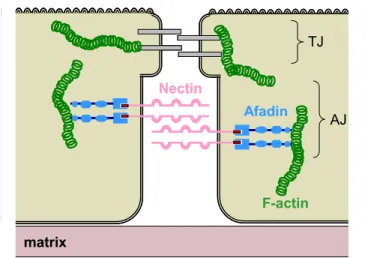
tail and required for the adhesive function. Nectins are adhesion proteins that can form homo- and heterodimers in a Ca2+-independent manner. There are four members in the nectin family, most of which are transmembrane Ig-like proteins and have a C-terminal PDZ binding motif at their cytoplasmic tail. Nectins couple adherent junctions to the cytoskeleton through the interaction with the PDZ domain of afadin, a protein that binds actin filaments.
Nectin Afadin F-actin matrix TJ AJ Nectin Afadin F-actin matrix TJ AJ
Figure 1.7. The Afadin-Nectin interaction in adherent cell junctions. In adherent cell junctions (AJ), the PDZ binding motif of Nectin (red) interacts with the PDZ domain of afadin (blue), coupling Nectin to F-actin (green).
There are two main splicing variants of this protein, a longer one, l-afadin, and a shorter one, s-afadin (Figure 1.8). L-afadin, simply called afadin, consists of two N-terminal Ras-association domains, one forkhead association domain (FHA), one dilute domain (DIL), one PDZ domain, three proline rich segments and one F-actin-binding domain. The smaller variant, which lacks the third proline-rich region and the F-actin–binding domain, is named AF6, like the afadin’s gene AF-6 (ALL-1 fusion partner on chromosome 6), which was found to be fused to the ALL-1 gene in acute myeloid leukemias caused by chromosomal translocation (t 6;11) [94]. Afadin is ubiquitously expressed in epithelial and endothelial cells, whereas AF6 is mainly expressed in neural tissue [61], [95]. Afadin has also other binding partners involved in cell-cell junctions. For example, it was shown that afadin can interact directly and indirectly with catenins, clustering cadherin and nectin complexes and thus making the adhesion in adherent junctions stronger [96]. AF6, although unable to bind the
- 30 -
actin cytoskeleton, is however involved in cell-cell junctions. AF6, through its Ras-binding domain, can bind Z0-1, a scaffold protein implicated in clustering of several adhesion proteins in tight junctions. Using the same binding domain, AF6 interacts with several members of the Ras family, small GTPases involved in the regulation of cell proliferation and differentiation. Activated Ras inhibits the interaction between AF6 and Z0-1, which further results in the perturbation of cell–cell contacts. As a multidomain scaffold protein, AF6/afadin recruits different protein complexes near the plasma membrane cell-cell junctions. Through its single PDZ domain, AF6/afadin was shown to bind, beside nectins, some other transmembrane proteins like Bcr kinase (negative regulator of Ras signaling pathway [97], Neurexin (neuronal cell surface protein, [56]), JAM (junctional adhesion molecule in TJs, [98]), SPA-1 (GTPase-activating protein, [99]), Eph-related receptor tyrosine kinase ([56], [100]) and Jagged-1([56]). PDZ PDZ RA RA FHA DIL PR PR F-Actin PR
l-Afadin
s-Afadin (AF-6)
PDZ PDZ PDZ PDZ RA RA FHA DIL PR PR F-Actin PRl-Afadin
s-Afadin (AF-6)
Figure 1.8. Domain architecture and splicing variants of afadin. The long (l-Afadin) and short (s-(l-Afadin) variants are shown. RA, Ras-association domain; FHA, Forkhead associated domain; DIL, Dilute domain; PDZ, domain present in PSD-95, Dlg, and ZO-1/2; PR, proline-rich region; F-actin, F-actin binding domain
1.3.3 AF6_PDZ domain
The NMR 3D structure of AF6_PDZ is similar to X-ray structures of other PDZ domains [75]. Its structure is very close to the third PDZ domain of PSD-95 protein (RMSD 2.1 Å; sequence identity = 30.4%; sequence similarity = 50%). Even if sorted in the class-II by oriented peptide library technique, AF6-PDZ can also interact with class-I ligands. The amino acid sequence shows that the crucial residue for the peptide binding specificity, the first residue of the αB helix (αB1), is
- 31 -
glutamine. Generally, the αB1 position is occupied by a hydrophobic amino acid or by histidine, in Class II and Class I PDZ domains, respectively [68], [101]. Glutamine in this position is unique and is not found even in Class III PDZ domains, where αB1 residue is tyrosine. The side chain of the glutamine (αB1) points towards the peptide binding groove in a similar way the histidine (αB1) in the PSD-95 PDZ3 does, which is one typical Class I PDZ domain. Even if AF6-PDZ and PSD-95 PDZ3 share similar βB/αB grooves, there are length differences in the βB-strand flanking loops, βA/βB and βB/βC. Another feature that distinguishes the AF6 PDZ domain is its carboxyl-binding loop with a GMGL sequence, which is slightly different from the standard GLGF motif. However, the structure analysis shows that the hydrophobic residues, methione and leucine, have their side chains pointed toward the interior of the protein making a hydrophobic pocket for the ligand carboxyl group [75].
NMR chemical shift mapping of the AF6_PDZ titrated with C-terminal peptides from Bcr and Neurexin, ligands of Class I and II, respectively, showed in both cases binding via the βB/αB groove, with slightly differences in the binding mode. Regardless of its structural characteristics and binding promiscuity, AF6 PDZ has a greater tendency to bind Class II ligands [68]. Additionally, mutational analysis by two-hybrid assays showed that a minimum of six sequence specific C-terminal residues from the EphB2 and EphB3 ligands are required for binding to AF6-PDZ [56]. All together, AF6_PDZ is a very flexible interaction domain, which makes interesting to study how binding to the ligands is regulated.
1.4 Notch structural biology
Current structural analyses in Notch signaling are mainly focused on the receptor-ligand recognition, the activation of the receptor proteolysis and the nuclear complex assembly. Analysis of the receptor-ligand binding interface, carried out in
Drosophila by cell-aggregation experiments, determined the pair of EGF-like repeats
11-12 in Notch and the DSL domain in Delta as a minimal region for the their association [5].
The first structural insights were the NMR and X-ray structures of the human Notch-1 tandem EGF-like repeats 11th -13th (N1_EGF11-13) and the X-ray structure of
the human Jagged-1 region corresponding to the DSL domain and the first three EGF-like repeats (J1_DSL_EGF1-3) [102], [103] (Figure 1.9 A, B). It was shown that each
- 32 -
EGF-like repeat has its characteristic fold consisting of a two-stranded antiparallel β-sheet and three disulfide bonds. The X-ray structure of N1_EGF11-13 showed that a
coordinated Ca2+ ion fixes the orientation between adjacent repeats, creating an extended rod-like structure. In the NMR analyses of N1_EGF11-13, the dynamic
behavior of the EGF13 is distinctly different from that of the other two repeats,
suggesting that the position of the EGF13 is not rigidly fixed with respect to the
EGF11-12 repeats.
A
B
S1C
S2A
B
S1C
S2Figure 1.9. Structures of functionally relevant regions in Notch receptors and ligands. Cartoon representation of (A) the tandem EGF-like repeats 11th -13th of human Notch-1 (PDB code 2VJ3), (B) the region comprising the DSL domain and the first three EGF-like repeats from human Jagged-1 (PDB code 2VJ2), and (C) the negative regulatory region (NRR) of human Notch-2 (PDB code 3I08). The NRR region consists of three Lin/Notch repeats (LNR, blue) and of a heterodimerization domain (HD, N-terminal region in orange, C-terminal region in raspberry red) that is cleaved at S1. Cleavage sites S1 and S2 are in light green.
From the structure of these three tandem repeats, two distinct models for the structure of the Notch-1 extracellular region comprising all 36 EGF-like repeats were proposed. In the first model, the region has an extended conformation, mainly rigid along its length, but with flexible parts between some EGF-like repeats that do not bind Ca2+ ions. In the second model, the extracellular region folds onto itself to form a
- 33 -
flexibility in the linkers that connect non-Ca2+- binding EGF-like repeats. Using the second model, we can speculate how the extracellular region of Notch receptors, having a different number of EGF-like repeats, can adopt different structures and thus can have different affinity for different Notch ligands. The X-ray structure of the Jagged-1 polypeptide corresponding to the DSL domain and the first three EGF-like repeats (J1_DSL_EGF1-3) revealed an extended conformation that resembles the
structure of the N1_EGF11-13 [103]. The DSL domain shows similarities with an EGF
module, but has a recognizably different disulfide bond pattern. EGF1 and EGF2
exhibit a truncated version of the EGF-fold, with no canonical secondary structure and a more distant structural homology to other EGF-like domains while EGF3 has a
classical EGF-like fold. Recently, it was shown that exons 5 and 6 of the human JAG1 gene, which together encode EGF1 and EGF2 repeats, are out of phase with respect to
the domain boundaries [104]. Exon 5 encodes a truncated EGF with only four half-cysteines and exon 6 encodes the C-terminal half of EGF1 and the entire EGF2. It has
been proposed that the DSL domain may have evolved from the truncation of tandemly connected, short EGF repeats [103]. Interestingly, the region encoded by exon 6, J1ex6, can be viewed in itself as two truncated tandem EGF repeats and has an amino acid sequence and a disulfide pattern reminiscent to those of a DSL domain. Two models for the Notch-1/Jagged-1 binding interface were proposed on the ground of in-silico docking of these two structures, confirming the DSL domain as the minimal and indispensable unit for the binding. Moreover, the first two EGF-like repeats of Jagged-1 also participate in the formation of the interface. This is in agreement with findings obtained from deletion mutants, where it was demonstrated that the EGF-like repeats of mouse Jagged-1 can modulate the affinity of the binding to mNotch-2 [105].
Besides the pair of EGF-like repeats 11-12, the most important part of the Notch extracellular region is the so-called negative regulatory region (NRR). It is composed of three LIN-12 repeats and the heterodimerization domain (HD), which holds together the extracellular and transmembrane subunits of the receptor (Figure 1.9 C). The HD domain contains the S2 cleavage site which becomes exposed to the ADAM-metalloprotease upon ligand-receptor binding, but the mechanism is still not clearly described. The crystal structure of the NRR showed for the first time the autoinhibited conformation of the human Notch-2 [106]. Extensive interdomain interactions between the LIN-12 and HD domains bury the S2 site, suggesting that a
- 34 -
drastic conformational change in this region is required to expose this site upon activation triggered by the ligand. It has been proposed that ligand binding relieves this autoinhibition either by inducing an allosteric conformational change in the receptor or by the pulling force caused by the ligand endocytosis, but further experiments are necessary for the exact description of this activation switch.
The crystal structure of the Notch transcriptional complex highlighted how the Notch intracellular region (N_ic) binds CSL and then both recruit Mastermind to provide stability for the complex [107], [108] (Figure 1.10). It was shown that the Ankyrin repeats (ANK) of N_ic are indispensable for the interaction with Mastermind [109] while for the binding with CSL N_ic mainly uses its RAM region. ANK domains of Notch consists of seven ANK repeats, each of which is about 33 residues long and typically folds into a pair of antiparallel helices followed by a β-hairpin that connects to the next repeat [110]. RAM domains of Notch are approximately 100 residues long and defined as a region that starts from the γ−secretase cleavage site and ends at the first ankyrin repeat. RAM has very little or no secondary structure but becomes ordered upon complex formation [109], [111]. CSL consists of an N-terminal Rel-homology region (RHR-N), a central β-trefoil domain (BTD) and a C-terminal Rel-homology region (RHR-C). The DNA is bound by the BTD and the RHR-N and makes no contacts with the distal RHR-C domain. Mastermind proteins are ~1000 residues long proteins predicted to be of low structural complexity, suggesting that they function as scaffolds for further recruitment of additional co-activators and/or the transcription machinery [109]. Only a short N-terminal fragment (55 residues) of Mastermind is required for the interaction with CSL and N_ic [112]. The C-terminal portion of Mastermind is important for the interaction with CBP/p300 and to activate transcription [113]. Mastermind adopts a bent helical structure when complexed with CSL and N_ic. The N-terminal helical region interacts with RHR-C of CSL and ANK of N_ic, while its C-terminal helix binds RHR-N of CSL [107], [108]. In the CSL-DNA complex, when not bound to N_ic and Mastermind, the β-hairpin loop of RHR-N is closed. In order to accommodate binding of the C-terminal Mastermind helix, the β-hairpin loop needs to adopt an open conformation. It was demonstrated [114] that RAM binding to the BTD of CSL produces an allosteric change in the RHR-N, opening the β-hairpin loop and in that way creating one-half of the docking site required for Mastermind to bind to the complex. The CSL-ANK
- 35 -
interface, created upon the interaction between RAM to BTD, creates the complete docking site for Mastermind. Despite the structural and biochemical information about the RAM and Ankyrin repeats, less is known about the overall structure of the entire N_ic, especially the less conserved C-terminal region of N_ic. The future challenge is obtaining structural insights into the cooperativity between the Notch transcription complex and other transcription factors.
Figure 1.10. Crystal structure of the Notch transcriptional complex. Ankyrin repeats of N_ic in orange, the N-terminal region of Mastermind is in blue, CSL is in cyan and dsDNA is in red.
The cytoplasmic tails of the Notch ligands still need to be characterized biophysically. The cytoplasmic tail of Jagged-1 is 125 residues long region and, based on its amino acid composition, is predicted to have a high degree of intrinsic disorder. J1_ic has a positive mean net charge and is rich of lysines, arginines, glutamines and prolines, residues that predominates in disordered proteins. Moreover, J1_ic is significantly depleted of residues that usually form the hydrophobic core of globular proteins (Ile, Leu, Val, Trp, Tyr and Phe) [115]. Many intrinsically disordered proteins and protein domains have been identified, and regions predicted to be intrinsically disordered are quite common, especially in eukaryota [116]. Although these regions were considered in the past of little importance by structural biologists, they often play an important role in protein-protein interactions, especially in signaling networks [117]. The role of intrinsic disorder in these contexts is well
- 36 -
explained in terms of the plasticity that disorder confers to proteins, which allows proteins to bind different partners through disorder to order transitions [118], [119]. Intrinsically disordered regions can work as docking stations hosting several different protein interactions motifs on the same polypeptide chain. In protein/protein interactions, they potentially offer a very large interaction surface/volume ratio compared to globular proteins, which makes them capable of binding their target molecules with high specificity but low affinity. Detailed biophysical studies are crucial to clarify the relationship between biological function and structural characteristics of intrinsically disordered proteins. The application of increasingly sophisticated methods has revealed that disordered proteins are far from being homogeneous statistical random coil polymers. Intrinsically disordered proteins exhibit a rich diversity of local and even long-range structural preferences as well as dynamics, that are likely to be of functional significance.