DEPARTMENT OF BIOMOLECULAR SCIENCES
PH.D. COURSE IN:
LIFE SCIENCES, HEALTH AND BIOTECHNOLOGIES
CURRICULUM:
BIOCHEMICAL AND PHARMACOLOGICAL SCIENCES AND BIOTECHNOLOGY
XXXIII° CYCLE
UBIQUITIN EXPRESSION IN PRIMARY 23132/87 AND METASTATIC MKN45
GASTRIC CANCER CELL LINES AND DIFFERENTIAL RESPONSE TO POLYUBIQUITIN
UBB AND UBC GENE SILENCING
SSD: BIO/11Supervisor Ph.D. Student
Prof. Marzia Bianchi Dr. Filippo Tasini
Abstract
Gastric cancer is one of the most lethal tumors worldwide; it ranks as the 5th most common malignancy and the 3rd most lethal, with over 1 million new diagnoses and over 780’000 deaths in 2018 alone. The outcome of the disease is greatly influenced by tumor stage at the time of diagnosis; unfortunaterly, most patients are diagnosed only after the tumor has already metastasized, which dramatically reduces their chances of survival. Therefore, additional knowledge regarding gastric cancer is needed to define possible biomarkers and therapeutic targets that may respectively improve early detection protocols and treatment.
To this end, we compared the asset and regulation of ubiquitin pools in two gastric cancer cell lines: the primary line 23132/87 and the metastatic MKN45. The two cell lines were analyzed in various aspects, such as the relative ubiquitin content and the expression patterns of the four ubiquitin genes UBC, UBB, UBA52 and RPS27A; the mRNA and protein levels of three transcription factors, HSF1, YY1 and SP1, were also measured in light of their involvement in the regulation of some of the ubiquitin genes.
Then we performed a series of siRNA-mediated knockdown experiments targeting the two polyubiquitin genes UBB and UBC to investigate if and how different alterations of the ubiquitin content would affect the two cell lines. To briefly summarize our results, the primary gastric cancer cell line 23132/87 exhibits a higher reliance on its endogenous ubiquitin production for survival than the metastatic line MKN45, identifying UBB and UBC as pro-survival genes in 23132/87 primary cells.
Chapters 1 and 2 respectively contain an introduction to gastric cancer and the ubiquitin system, the major topics addressed in this thesis.
Chapter 3 lists the research objectives.
Chapter 4 includes the rationale, methods and results concerning the characterization and comparison of the two gastric cancer cell lines 23132/87 and MKN45, regarding the expression of ubiquitin genes and their reliance on the intracellular content of ubiquitin. The article reported therein summarizes the greater part of the research work I have done during my Ph.D. Chapter 5 comprehends the knowledge base that constituted the foundations of our investigation of gastric cancer. My contribution to the displayed manuscript consisted in performing a series of transfection experiments in several cell lines (both normal and tumor-derived), in order to find out if the downregulation of UBC caused by the ectopic expression of ubiquitin could be found in cell lines other than HeLa. Mycoplasma detection was also performed by me in all cell lines tested.
TABLE OF CONTENTS
CHAPTER 1: AN INTRODUCTION TO GASTRIC CANCER 1
1.1 Epidemiology 1
1.2 Classification 1
1.3 Environmental Risk Factors 2
1.4 Inheritable genetic components 3
1.5 Acquired genetic factors 5
1.6 Importance of prevention 7
1.7 Importance of early detection 7
1.8 Treatment 8
CHAPTER 2: AN INTRODUCTION TO THE UBIQUITIN SYSTEM 9
2.1 Ubiquitin, a complicated molecule 9
2.2 Ubiquitination and its mechanism 10
2.3 Ubiquitination is highly specific 11
2.4 Deubiquitinating enzymes 12
2.5 Ubiquitin and substrate degradation 12
2.6 Ubiquitin and transcription regulation 14
2.7 Ubiquitin and DNA damage repair 15
2.8 Ubiquitin and apoptosis 15
2.9 Ubiquitin and immunity 16
2.10 Ubiquitin and cell cycle regulation 16
2.11 Ubiquitin genes 17
2.12 UBC gene structure 18
2.13 UBC transcriptional regulation in basal conditions 18
2.14 UBC transcriptional regulation in stress conditions 19
2.15 UBC downregulation upon overexpression of exogenous ubiquitin 20
2.16 Ubiquitin and cancer 20
CHAPTER 3: AIMS OF THE THESIS 22
CHAPTER 4: THE UBIQUITIN GENE EXPRESSION PATTERN AND 23
SENSITIVITY TO UBB AND UBC KNOCKDOWN DIFFERENTIATE PRIMARY 23132/87 AND METASTATIC MKN45 GASTRIC CANCER CELLS
4.1 Abstract 24
4.2 Introduction 24
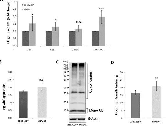
4.3 Results 26
MKN45 GC Cells
4.3.2 Cytosolic and nuclear distribution of YY1, HSF1 and SP1 in 23132/87 and 28 MKN45 GC cell lines
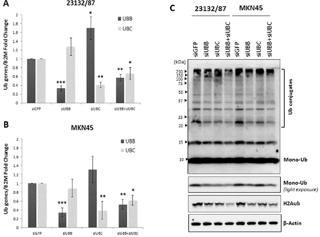
4.3.3 Effect of YY1, HSF1 and SP1 transcription factor silencing on Ub gene 29 expression
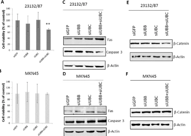
4.3.4 Role of UBB and UBC in gastric adenocarcinoma cell proliferation and 30 survival
4.4 Discussion 34
4.5 Materials and Methods 38
4.5.1 Cell cultures and chemicals 38
4.5.2 Small interfering RNA transfection in 23132/87 and MKN45 cells 38
4.5.3 Real-Time quantitative Polymerase Chain Reaction (RT-qPCR) 39
4.5.4 Cell extracts 40
4.5.5 Western blot analysis 40
4.5.6 Cell viability assay 40
4.5.7 Proteasome activity assay 41
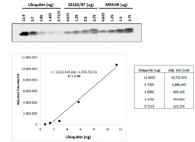
4.5.8 Ubiquitin carboxyl-terminal hydrolase 2 (Usp2) digestion and Mono- 41 Ubiquitin quantification
4.5.9 Statistical analysis 41
4.6 Supplementary Data 44
4.7 References 53
CHAPTER 5: A NEGATIVE FEEDBACK MECHANISM LINKS UBC GENE 57
EXPRESSION TO UBIQUITIN LEVELS BY AFFECTING RNA SPLICING RATHER THAN TRANSCRIPTION
5.1 Abstract 58
5.2 Introduction 58
5.3 Results 60
5.3.1 Overexpression of ubiquitin downregulates the endogenous UBC gene 60 expression
5.3.2 A conjugation competent ubiquitin is required for UBC downregulation 60
5.3.3 Promoter analysis by transfection of reporter constructs reveals the 63 importance of the UBC intron for the downmodulation effect
5.3.4 Role of HSF1 and HSF2 in UBC downregulation 65
5.3.5 Role of Sp1 in UBC downregulation 68
5.3.6 Role of YY1 in UBC downregulation 68
5.3.7 H2A and H2B histone ubiquitination signatures of UBC promoter do not 69 change upon ubiquitin overexpression
5.3.8 Ubiquitin overexpression does not impact the transcriptional activity of 69 the UBC promoter, but rather the splicing of nascent UBC transcripts
5.5 Materials and Methods 78
5.5.1 Cell lines and treatments 78
5.5.2 Plasmid constructs and transfections 78
5.5.3 Luciferase reporter assay 79
5.5.4 RNA preparation and quantitative real-time RT-PCR (RTqPCR) 80
5.5.5 Cell extracts 80
5.5.6 Western blotting 80
5.5.7 USP2 digestion and solid phase immunoassay 81
5.5.8 Electrophoretic mobility shift assay (EMSA) 81
5.5.9 Chromatin immunoprecipitation (ChIP) 81
5.5.10 Metabolic labeling of nascent transcripts 82
5.5.11 Nuclei purification and detection of unspliced transcripts 82
5.5.12 Statistics 83
5.6 Supplementary Data 84
5.7 References 95
CHAPTER 6: Conclusions 99
ORIGINAL PAPERS
This Thesis is based on the following original research articles, which will be referred to by their Roman numerals.
I. Scarpa ES, Tasini F, Crinelli R, Ceccarini C, Magnani M, Bianchi M. The Ubiquitin Gene Expression Pattern and Sensitivity to UBB and UBC Knockdown Differentiate Primary 23132/87 and Metastatic MKN45 Gastric Cancer Cells. Int. J. Mol.
Sci. 2020;21(15):5435.
II. Bianchi M, Crinelli R, Giacomini E, Carloni E, Radici L, Scarpa ES, Tasini F, Magnani M. A negative feedback mechanism links UBC gene expression to ubiquitin levels by affecting RNA splicing rather than transcription. Sci. Rep. 2019;9(1):18556.
CHAPTER 1: AN INTRODUCTION TO GASTRIC
CANCER
1.1 Epidemiology
Gastric cancer (GC) is one of the most lethal tumors worldwide. According to the most recent estimates it ranks as the 5th most common malignancy and the 3rd most lethal, with over 1 million new diagnoses and over 780’000 deaths in 2018 alone.
Gastric cancer is the leading cause of cancer death in several Western Asian countries, such as Iran, Turkmenistan and Kyrgyzstan, while its incidence rates are especially elevated in Central and Eastern Asia (e.g. in Mongolia, China, Japan and Republic of Korea, which exhibits the highest incidence in the world). Other high risk zones are Russia and Central and South America (Bray F et al. 2018; Rawla P, Barsouk A. 2019).
1.2 Classification
While more than 90-95% of gastric cancer cases are adenocarcinomas (Balakrishnan M et al. 2017; WCRF/AICR; 2018), which arise from the glands of the most superficial layer of the stomach (the mucosa), they are individually very heterogeneous and their classification into subtypes is not straightforward. Gastric adenocarcinomas are primarily classified as cardia and non-cardia based on their anatomic site. Cancers of the gastric cardia arise in the region adjoining the esophageal-gastric junction and share epidemiological characteristics with esophageal adenocarcinoma. Non-cardia cancer is more common and arises in the lower (or distal) portion of the stomach (Rawla P, Bardouk A. 2019). Over the years several systems have been devised for the classification of non-cardia gastric cancer, but the most widely utilized is Lauren’s classification, which divides gastric cancer in two major subtypes, namely intestinal and diffuse.
Intestinal type gastric cancer grows in more shallow fashion, is significantly larger in size before infiltrating the serous membrane and has a higher incidence of blood vessel invasion and liver metastases. It shows a predominance of glandular epithelium with well-differentiated cells, similar to intestinal columnar cells, good cellular cohesion and a pushing margin at the invasive edge (Grabsch HI, Tan P. 2013). Patients with intestinal-type gastric cancer are on average 7 years older at the time of diagnosis (Grabsch HI, Tan P. 2013) and the disease generally arises from premalignant lesions, such as atrophic gastritis followed by intestinal metaplasia and dysplasia. The progression into cancer may take several years or even decades and is aided by the chronic inflammatory state induced by Helicobacter pylori infection, often found in patients affected by intestinal-type GC (McLean MH, El-Omar EM. 2014).
Diffuse type GC tends to spread more commonly via the lymphatics to the pleura and peritoneum. The tumor mass is composed of scattered poorly cohesive cells or small clusters of cells with little or no gland formation and a diffuse infiltrative margin. Tumor cells are
undifferentiated, may contain mucus and can have a signet ring cell appearance (Grabsch HI, Tan P. 2013). Diffuse type GC does not seem to follow any neoplastic progression but arises from normal gastric mucosa with no definite pre-malignant stage. H. pylori infection is often negative (McLean MH, El-Omar EM. 2014).
There are also GC cases that exhibit mixed or unidentifiable features and cannot therefore be classified as diffuse or intestinal.The relative frequencies are approximately 54% for intestinal type, 32% for diffuse, and 15% for indeterminate type, although this distribution varies in different geographical regions, age groups and socioeconomic conditions.
Intestinal types are more common in males and older adults, whereas diffuse types may occur in all age groups with equal sex distribution and show more rapid progression and poorer prognosis.
It is important to underline that all information and experimental data contained in this thesis regard only the non-cardia subtype of gastric cancer.
1.3 Environmental Risk factors
Gastric cancer pathogenesis shows a strong environmental component. Helicobacter pylori infection has been proposed as one of the causes of GC since the 1990s (Forman D et al. 1991), when it was declared a class I carcinogen (IARC. 1994), and is now considered the main risk factor, as the risk of developing gastric cancer was found to be 20-fold, or even higher, in the presence of H. pylori infection (Lazăr DC et al. 2016; Sitarz R et al. 2018). In particular, the infection has a very strong association with intestinal-type gastric cancer, while it is less likely to be found in diffuse-type cases. The chronic inflammatory background induced by H. pylori infection fosters premalignant changes in the gastric epithelium, starting with atrophic gastritis, followed by intestinal metaplasia and dysplasia, which in turn develops into gastric tumor. This progression is typical of intestinal-type gastric cancer (McLean MH, El-Omar EM; 2014). According to recent estimates, 84% of all GC patients test positive for H. pylori infection (WCRF/AICR; 2018) and as many as 90% of all cases can be attributed to H. pylori (Balakrishnan M et al. 2017).
H. pylori is a gram-negative bacterium able to colonize the human stomach; the infection is predominantly acquired in early infancy, remains present indefinitely if not treated with antibiotics (Correa P. 2013), and elicits life-long inflammatory responses, including the release of various bacterial and host-produced cytotoxic substances (Feldman M et al. 2020).
The infection is usually asymptomatic, but it is also widely spread: H. pylori prevalence has demonstrated great variability determined by factors such as geographic location, age, ethnicity and socioeconomic conditions. For these reasons, it is usually high in developing regions, where H. pylori infection represents a public health issue, and lower in developed countries. Its prevalence can show variability within regions of different countries and between more crowded urban and rural areas, mostly due to socioeconomic differences between the inhabitants (Lazăr DC et al. 2016). Globally, H. pylori infects 50% of the population and prevalence
increases with age (Ando T et al. 2006; Singh K, Ghoshal UC. 2006). Average H. pylori prevalence is 35% in high-income countries and 85% in low-income countries (Ang TL, Fock KM. 2014). The highest prevalence is in Asia; in South Korea, the infection reaches 90% at age 20 years (Youn HS et al. 1998).Regions with high stomach cancer incidence rates tend to have high seroprevalence rates for H. pylori infection. However, in some regions of Africaand South Asia, particularly India, H. pylori infection rates are high, but stomachcancer incidence rates remain low (Singh K, Ghoshal UC. 2006). This could be explained by studies on genomic sequencing and post-genomic analyses of H. pylori, which hyphothesize that the bacterium might once have been a symbiotic component of the human biome.
While H. pylori infection increases the risk of non-cardia gastric cancer, it also reduces acid secretion in the proximal portion of the stomach, thus having protective effects on gastro-esophageal reflux, gastro-esophageal adenocarcinoma and cardia GC, as it reduces risk of gastro-esophageal inflammation (Rawla P, Barsouk A. 2019).
Although international variation in H.pylori prevalence correlates reasonably with that of stomach cancer incidence, other risk factors have been proposed (Bray F et al. 2018); among these are consumption of salt-preserved foods, Epstein-Barr virus (EBV) infection, active tobacco smoking, alcohol consumption, obesity and certain occupations (WCRF/AICR, 2018; IARC, 2012). Evidence from preclinical studies suggests that H. pylori interacts with dietary factors such as salt to affect cancer risk (Gaddy JA et al. 2013). Research mainly relates to high-salt foods and high-salt-preserved foods, including pickled vegetables, processed meat and high-salted or dried fish (WCRF/AICR, 2018).
Epstein-Barr virus, member of the Herpesviridae family, is mainly known as the cause of infectious mononucleosis and for its association to various lymphoproliferative diseases such as Burkitt’s and Hodgkin’s lymphomas, but may also have a causal role in gastric carcinogenesis (Sitarz R et al. 2018). According to recent estimates, about 10% of all GC cases present EBV infection. The association between EBV and gastric cancer is highest in Germany and USA (16-18%) and lowest in China (4,3%) (Iizasa H et al. 2012).
Regarding tobacco smoking, both current and former smokers have an increased risk of gastric cancer compared with people who have never smoked, with estimates of increased risk ranging from 1.5–2.5 times that of never-smokers. The risk increase is larger in men than in women, but a dose-response relationship is apparent in both (Freedman ND et al. 2007; Sjodahl et al. 2007; Tredaniel J et al. 1999).
Occupational exposure to dusty and high-temperature environments, such as in wood processing and food machine-operating occupations, has been associated with increased risk of stomach cancer, particularly diffuse type cancer (Santibañez M et al. 2012). Exposure to other dusty environments, such as in rubber manufacturing, coal mining and metal processing, has also been implicated (Raj A et al. 2003). Finally, male sex is also a risk factor: males exhibit a 2-fold higher risk of developing gastric cancer than females (Karimi P et al. 2014).
1.4 Inheritable Genetic components
Gastric cancer carcinogenesis and progression result from a combination of both environmental factors and accumulation of specific genetic alterations, including activation of oncogenes,
overexpression of growth factors and receptors, inactivation of tumor suppressor genes, DNA repair genes and cell adhesion molecules. Most of these alterations are acquired randomly in the population, while only few have been documented to be hereditary. It follows that the majority of GC cases arise sporadically and show no apparent inherited component.
Familial clustering is observed in 10 to 15% of gastric cancer cases and most of these are not associated with a definite germline mutation.
Only less than 3% of gastric carcinomas arise from inherited gastric cancer predisposition syndromes, such as Hereditary Diffuse Gastric Carcinoma (HDGC), familial adenomatous polyposis, Hereditary Nonpolyposis Colorectal Carcinoma (HNPCC or Lynch syndrome), juvenile polyposis syndrome, Peutz-Jeghers syndrome, Li-Fraumeni syndrome and gastric hyperplastic polyposis (Hu B et al. 2012).
The most frequent is Hereditary Diffuse Gastric Cancer (Carneiro F. 2012; Oliveira C et al. 2013), an automosomal dominant pathology characterized by high penetrance and a heterozygous germline mutation. Approximately 30% of HDGC patients and 45% of the identified HDGC families harbor germline mutations of CDH1, the tumor suppressor gene that encodes cadherin 1 (or E-cadherin), a protein with an essential role in cell-cell adhesion (Hu B et al. 2012; Paredes J et al. 2012).
The mutation in CDH1 can take many forms (such as a deletion, frameshift, splice-site or missense mutations) that may involve a variety of sites in the gene and is not only restricted to coding regions but could include untranslated regions as well. A loss-of-function mutation in the remaining allele can be caused by a number of mechanisms, such as loss of heterozygosity or promoter hypermethylation, and can lead to gastric cancer (McLean M, El-Omar EM. 2014). The estimated lifetime risk for HDGC in proven mutation carriers is more than 80% in both men and women by age 80 (Oliveira C et al. 2013). Given the high penetrance of CDH1 mutation, prophylactic total gastrectomy after confirmation through CDH1 molecular testing is the only recommended way to save patients’ lives (Hu B et al. 2012).
CDH1 mutations have only been found in hereditary and sporadic diffuse type, but not in intestinal type GC (Grabsch HI, Tan P. 2013).
Patients with hereditary nonpolyposis colon cancer suffer an increased risk of intestinal-type gastric cancer, which arises from mutations in DNA mismatch repair genes such as MSH2 or MLH1. These events in turn increase the mutation rate in oncogene and tumour suppressor genes, which leads to cancer initiation and progression. Microsatellite instability is also a key characteristic of this pathology. Gastric cancer seems to be a common extracolonic manifestation of HNPCC, with a 2 to 19-fold increased risk of GC in patients with this syndrome compared to the general population; geographical location correlates with the level of increased risk (McLean MH, El-Omar EM. 2014). According to a study conducted in Northern Europe and the USA, the lifetime risk of gastric cancer in families with HNPCC is around 7%, occurring primarily in subjects over 50 years of age (Watson P et al. 2008).
In contrast, GC patients with germline mutations in one of the DNA mismatch repair genes (hereditary nonpolyposis colon cancer patients) show intestinal type morphology in 79% of cases (Grabsch HI, Tan P. 2013; Gylling A et al. 2007).
1.5 Acquired genetic factors
As most cases of GC are sporadic, acquired mutations play a great role in GC carcinogenesis. These genetic abnormalities can result from several mutagenic events, such as chromosomal and microsatellite instability (CSI and MSI; respectively), alterations in the epigenetic landscape and somatic gene mutations (McLean M, El-Omar EM. 2014). Given the highly variable nature of genetic mutations and the fact that many can occur and be accumulated by a single individual or even by a single tumor, gastric cancer, as all other malignant pathologies, exhibits intrapatient and interpatient heterogeneity (Renovanz M, Kim EL. 2014).
Chromosomal instability is defined by a change in the DNA content, with loss or gain of whole or large portions of chromosomes leading to altered DNA copy number and consequentially to loss or gain of function of oncogenes and tumor suppressors (McLean M, El-Omar EM. 2014). Chromosomal Instability has been detected as the most common feature of sporadic gastric cancers and has been reported in up to 84% of gastrointestinal tumours (Hudler P. 2012). A large variety of chromosomal aberrations have been identified in GC patients and different anomalies have been associated to patient age, geographical background and prognosis; particularly relevant in this context is the association of certain aberrations to GC histology: for example, intestinal type GC is associated with copy number gains at 8q, 17q and 20q, while diffuse type GC has been linked with copy number gains at 12q and 13q (Hudler P. 2012; Tomioka N et al. 2010; Tsukamoto Y et al. 2008).
Microsatellite instability is a condition arising from deficiency or inactivation of one or more DNA mismatch repair genes (Li K et al. 2020) of the DNA mismatch repair genes, such as MLH1, MSH2, MSH6 and PMS2, which in turn renders the naturally occurring DNA replication errors impossible to repair. In this scenario, hundreds of thousands of somatic mutations (e.g. insertions or deletions) accumulate in microsatellite DNA, which consists of short, repeated sequences randomly interspersed in the human genome. GC tumors can be categorized into those with high or low levels of MSI, depending on the frequency of mutations within a specific set of microsatellite markers. Tumors that show no instability in any of the markers are defined as stable.
MSI can lead to genetic anomalies in hundreds to thousands of genes and cause the appearance of new alleles not normally present: this condition takes the name of “MSI phenotype” or “DNA replication phenotype” (Oliveira C et al. 1998; Grabsch HI, Tan P. 2013). The reported frequency of MSI for GC varies between 11,68 and 33,82%, strongly depending on the number of loci examinated (Zhu L et al. 2015).
GC tumors can be categorized into those with high or low levels of MSI, depending on the frequency of mutations within a specific set of microsatellite markers. Surprisingly, according to a meta-analysis study conducted in 2015, gastric cancer patients with a high rate of MSI show improved prognosis and reduced risk of lymphnode metastasis, tumor invasion and mortality (Zhu L et al. 2015).
The most common cause of MSI in GC cases is the hypermethylation of the MLH1 gene promoter, with consequent impairment of its transcription (Fleisher AS et al. 1999). As a matter
fact, aberrant methylation of CpG regions and silencing of tumor suppressor genes can be found in up to 50% of all GC cases (Grabsch HI, Tan P. 2013), and takes the name of CpG Island Methylator Phenotype (CIMP). In addition to MLH1, many other genes are silenced due to aberrant methylation, such as CDK2A and APC (Liu JB et al. 2012; Toyota M et al. 1999). Tumors that test positive for H. pylori infection also show a generally higher CpG methylation rate compared to H. pylori negative tumors, as do EBV-positive tumors (Cancer genome atlas research network, 2014; Liu JB et al. 2012).
There any many genes known to be subject of somatic mutations in gastric cancer. A comprehensive list would be beyond the scope of this thesis, thus the ones reported below will be limited to the most relevant examples.
Human Epidermal Growth Factor Receptor 2 (HER2) is a tyrosine kinase without any known direct activating ligand and is classified as an oncogene. HER2 overexpression has been reported in up to 27% of intestinal type GC cases, but only rarely in diffuse type GC cases (Gravalos C, Jimeno A. 2008). While HER2 amplification has been proposed as a marker of poor prognosis in GC patients, the relation is still regarded as controversial, with several studies reporting contradicting findings (Grabsch H et al. 2010; Okines AF et al. 2013).
K-Ras is GTPase mainly known for its role in the RAS/MAPK pathway and its high mutation rates in various cancers, including colorectal, pancreatic and lung cancer; the most common mutation sites in K-ras are codons 12 and 13. However, KRAS mutations are not as common in gastric cancer, in fact several studies report low percentages of cases with KRAS mutations ranging from 1,6 to 17,5% and a single study (conducted in Northeast Iran, a high risk area for GC) with a peak of 40% (Ayatollahi H et al. 2018; van Grieken NC et al. 2013). KRAS mutations are preferentially present in well-differentiated intestinal type GCs and are more frequently associated with microsatellite instability.
KRAS mutations on codons 12 and 13 have been found on average in 5% of GC and were preferentially present in well-differentiated intestinal-type GC (Corso G et al. 2011; Lee KH et al. 1995). In contrast to colorectal cancer, KRAS mutations in GC are more frequently seen in GC with MSI (Corso G et al. 2011; Wu M et al. 2004).
P53 is a nuclear protein involved in cell cycle control, DNA repair and programmed cell death, which is frequently inactivated in tumors by loss of heterozygosity (LOH) or point mutations of its encoding gene (TP53); more than one mutation may be present in a single tumor, resulting in heterogeneity of the p53 mutational status. TP53 mutation is one of the most frequent genetic alterations in GC: there are conflicting results regarding the prevalence of TP53 mutations and their relationship to histological type or tumor stage of GC. Some studies show that mutations tend to affect mainly intestinal-type tumors, while others found that the incidence of mutation is similar in both intestinal and diffuse-type tumors, ranging between 25% and 40% of the cases presented. Regarding tumor stage, p53 abnormalities appear to occur early in intestinal-type cancers, but some studies have shown that the frequency of TP53 mutation in both early and advanced intestinal-type is consistent around 40% each, similar to that observed in the advanced diffuse-type, while TP53 mutations are rare in early diffuse-type (Fenoglio-Preiser CM et al. 2003; Iwamatsu H et al. 2001; Liu XP et al. 2001).
APC (Adenomatous Polyposis Coli) is a multidomain protein with binding sites for numerous proteins including the Wnt pathway components β-catenin and Axin. APC plays a major role in cell adhesion, cell migration, spindle formation, and chromosome segregation. APC mutations are the second most frequent mutations in GC and have been observed in 30–40% of well- and moderately differentiated intestinal type GC and in up to 13% of diffuse type GC (Fang DC et al. 2002; Horii A et al. 1992). APC mutations have also been described in adenomas of the stomach and intestinal metaplasia, indicating that they occur during early stages of GC development (Nishimura K et al. 1995).
1.6 Importance of prevention
Globally, the incidence of gastric cancer has been steadily declining in the past 50 years, a trend that has been attributed to changes in lifestyle and environment, such as increasing smoking cessation, adoption of healthier dietary habits and, above all, H. pylori eradication (McLean M, El-Omar EM. 2014). In turn, the decline in prevalence of H. pylori infection is linked to improved food preservation practices such as refrigeration, which also directly favor consumption of fresh produce and reduce consumption of salt-preserved foods (WCRF/AICR 2018).
1.7 Importance of early detection
The 5-year survival rate is defined as the percentage of cancer patients that survive for at least 5 years after diagnosis; it is a very useful index to determine the trend over time of cancerous diseases. There have been great improvements in the 5-year survival rates for GC since the 1970s, but only minor progress has been made in recent years: for example, in the United States it went up from 15% in 1975 to 29% in 2009, to 32% in 2020 (Karimi P et al. 2014; ACS. Cancer facts & figures 2020). This statistic is heavily influenced by the fact that most GC patients are diagnosed after the cancer has already spread to other parts of the body. If the diagnosis happens before tumor metastatization, the 5-year survival rate is higher, but it depends on cancer stage. According to the most recent American estimates, the 5-year survival rate is 69% if the cancer is found and treated when it is still localized, 31% if it has spread to the surrounding tissues and organs, and 5% if it has already undergone distant metastatization (ACS Cancer facts & figures 2020). In most areas of the world, the average 5-year survival rate is currently around 20%. Survival rates are generally higher in high-income countries and other parts of the world where there are established services for screening and early detection of cancer as well as well-established treatment facilities (WCF/AICR 2018). A bright example is set by Japan, where rates above 70% have been reported; these results are attributed to the effectiveness of the mass screening programs employed by Japan (Katai H et al. 2018).
It is thus easy to see how early diagnosis of GC greatly improves patient survival, but diagnosis is usually delayed by a lack of early specific symptoms. Most patients are still diagnosed in advanced stages, resulting in poor 5-year survival rates, but also in short median survival times, less than 1 year for metastatic disease (Lazăr DA et al. 2016).
1.8 Treatment
Patient treatment regimens vary according to the stage of the tumor and the presence of metastases. When possible, surgical resection of the tumor mass (partial or total gastrectomy) and of the nearby lymphnodes is the main approach to gastric cancer treatment, supported by adjuvant chemotherapy or chemoradiation (Sitarz R et al. 2018).
Stage 0 and IA cancers can be treated by surgery alone and chemo- or radiotherapy is usually not required. Cancers that range from stage IB to III are similarly treated with surgical resection and chemo- or radiotherapy can be administered before and/or after the surgical procedure, depending on the severity of the disease, to reduce the size of the tumor and lower the chances of recurrence. Stage IV cancers, by definition, have spread to distant locations, such as the liver, lungs, brain or the peritoneum, and are usually incurable. In these cases, surgery and cycles of chemo- and/or chemoradiation therapy can still be administered, but treatment is palliative and aims at keeping the disease under control, relieving its symptoms, improving quality of life and extending life expectancy of patients (Smyth EC et al. 2016; ACS. Cancer Facts & Figures 2020). However, even after complete resection, gastric cancer exhibits high rates of recurrence, ranging from 20 to even 50%; in most cases (60-70%) recurrence manifests within 2 years after surgery (Barchi LC et al. 2016; D’Angelica M et al. 2004; Marrelli D et al. 2005; Shin CH et al. 2016). Targeted therapy is also being explored in the treatment of gastric cancer and a number of preclinical studies and clinical trials investigated several possible therapeutic targets, such as Vascular Endothelial growth Factor Receptor (VEGFR), Epidermal Growth Factor Receptor (EGFR), Poly-ADP-Ribose-Polymerase (PARP) among others (Lazăr DA et al. 2016). Trastuzumab (also known as Herceptin) is a monoclonal antibody specific for the Human Epidermal Growth Factor Receptor Type2 (HER2); originally employed for breast cancer therapy, its use is now permitted for gastric cancer, but only in cases that exhibit HER2 overexpression. Trastuzumab is usually administered in combination with capecitabine or 5-fluorouracil and cisplatin (Lazăr DA
et al. 2016; Sitarz R et al. 2018). Another application of moleclular biology to the treatment of gastric cancer is the testing of the CDH1 gene status to screen high risk patients for Hereditary Diffuse Gastric carcinoma (Hu B et al. 2012).
CHAPTER 2: AN INTRODUCTION TO THE
UBIQUITIN SYSTEM
2.1 Ubiquitin, a complicated molecule
Ubiquitin (Ub) is a small 8,6 kDa regulatory protein composed of 76 aminoacids. Discovered in 1975 by Goldstein et al. (Goldstein et al. 1975), it takes its name from its “ubiquitous” and abundant presence in all eukaryotic cells, where it constitutes 0,1-5% of the total protein content (Ryu KY et al. 2006).
The attachment of one or more ubiquitin molecules to a protein substrate is called ubiquitination (also known as ubiquitylation or ubiquitinylation) and it consists in a series of sequential reactions catalyzed by enzymes belonging to 3 (or even 4) different classes.
Ubiquitin is also highly conserved among different species: for example, the yeast and human analogues of ubiquitin only differ in 3 aminoacids. This remarkable grade of conservation is attributed to a strong selective pressure on the entire molecule as a result of the many functions it serves in the cell (Pickart CM, Eddins MJ. 2004) such as cell-cycle progression, DNA transcription and repair, apoptosis, modulation of cell surface receptors, cellular differentiation, response to cellular stresses and proteolysis (Pickart CM, Eddins MJ. 2004).
The attachment of a single ubiquitin molecule to a target substrate is called monoubiquitination, while the addition of two or more ubiquitin monomers to just as many residues of a substrate is defined as multi-monoubiquitination. However, additional ubiquitin moieties can be linked to any of the seven lysine residues or to the aminoterminal methionine of the ubiquitin molecule to form chains through subsequent rounds of conjugation; this process is known as polyubiquitination. Said chains may also be branched or linear and, in the latter case, the nature of the linkages can be homogeneous or heterogeneous. Taking into account the presence of multiple linkage sites on each ubiquitin molecule that is part of a chain and the possibility to attach more than one ubiquitin molecule to a given target, the versatility of this modification becomes apparent and is the reason of ubiquitin involvement in so many cellular functions (Guo HJ, Tadi P. 2020; Kim HC, Huibregtse JM. 2009; Lòpez-Mosqueda J, Dikic I. 2014).
Different kinds of ubiquitin modifications result in different outcomes for the target protein, such as proteosomal degradation, changes in cellular location, enzymatic activity or interactions with other proteins. However, the ultimate fate of a ubiquitylated protein depends not only on chain topology, but also on other factors like activity and availability of ubiquitylating and deubiquitylating enzymes, as well as of ubiquitin-binding proteins (Komander D, Rape M. 2012): whereas other post-translational modifications like phosphorylation produce an on-off binary signal, ubiquitylation could be defined as “analogical”, given how tunable and diverse it can be (Mevissen TET, Komander D. 2017).
In the case of polyubiquitin chains, the specific linkages connecting the single ubiquitin monomers determine the spatial conformations of the chain. K29- K33-, K63- and M1-linked chains exhibit “open” linear and highly flexible conformations, where the ubiquitin molecules do not interact with each other except for the isopeptide bonds linking them. K6-, K11- and K48-linked chains have “closed” conformations which allow interaction between ubiquitin moieties. These conformations hide and expose different portions of the ubiquitin molecules, and constitute a vast array of geometries that can be specifically recognized by ubiquitin-conjugating enzymes, deubiquitinases (DUBs) and proteins that contain Ubiquitin-Binding Domains (UBDs) (Komander D, Rape M. 2012; Ye Y et al. 2012).
In the cell, the total ubiquitin pool is divided into free and conjugated ubiquitin pools which coexist in a dynamic equilibrium that adjusts in accordance to cellular needs (Park CW, Ryu KY. 2014). Indeed, due to the variety of its functions and large number of ubiquitylated substrates, ubiquitin is not constantly produced in excess, but its de novo synthesis increases only when cellular demand does, such as during proteotoxic stress. Ubiquitin recycling, mediated by deubiquitinating enzymes, also plays an important part in ubiquitin homeostasis (Kimura Y, Tanaka K. 2010; Bianchi M et al. 2018).
2.2 Ubiquitination and its mechanism
On a chemical standpoint, ubiquitination comprises 3 stages, each catalyzed by a specific class of enzymes, E1 (ubiquitin-activating enzymes), E2 (ubiquitin-conjugating enzymes) and E3 (ubiquitin ligases):
1) Activation – ubiquitin is activated in a two step ATP-dependent reaction. E1 binds to both a ubiquitin and an ATP molecule. The enzyme then catalyses the acyl-adenylation of the C-terminus of the ubiquitin molecule. Then, in the second step, the ubiquitin molecule is transferred to an active site on the E1 enzyme, constituted by a cysteine residue, with release of AMP and the formation of a thioester link between the C-terminal carboxyl group of ubiquitin and the E1 cysteine sulfhydryl group.
2) Conjugation – thanks to the action of an E2 enzyme, the activated ubiquitin is transferred from the E1 enzyme to the cysteine in the active site of E2 via a trans(thio)esterification reaction in which the E2 binds to both the E1 enzyme and the activated ubiquitin.
3) Ligation – at this point an E3 enzyme catalyzes the formation of an isopeptide bond between the C-terminal glycine of the ubiquitin molecule and a lysine residue on the target protein. The exact mechanism of the reaction differs according to the type of E3 involved and the binding domain it possesses: these mainly include the HECT (Homologous to the E6AP Carboxyl Terminus) domain and the RING (Really Interesting
New Gene) domain. HECT domain E3s transiently bind ubiquitin during the reaction, while RING domain E3s do not form a catalytic intermediate with ubiquitin and instead function as a scaffold that allows interaction between E2 and substrate (Kim HC, Huibregtse JM. 2009; Metzger MB et al. 2012). Other types of E3s contain U-Box domains, which function similarly to RING E3s, or RBR (RING in-between RING), which is a RING-type domain capable of forming a catalytic intermediate with ubiquitin in a similar manner to HECT domains (Nakagawa T, Nakayama K. 2015). Ligation is also the limiting step of the entire ubiquitination process.
2.3 Ubiquitination is highly specific
Ubiquitination is an extremely complex, but also highly specific modification; these characteristics depend on several factors:
First, there is great disproportion in the numerosity of E1, E2 and E3 enzymes. In humans, there are only 2 known ubiquitin-specific E1s (UBA1 and UBA6), whereas there are 35 different E2s and hundreds of E3s. However, these enzyme families also differ in their binding specificities: both E1s can bind many E2s, which in turn can bind hundreds of E3s; finally, every E3 can interact with a wide array of protein substrates (Pickart CM, Eddins MJ. 2004). The hierarchic flow of the process and the enormous variety of protein substrates determine the process specificity and also explain the wide array of cell functions that are affected by ubiquitination. The existence of E2 enzymes as a bridge between E1s and E3s also offers an additional layer of regulation, dependent on E2s concentration, activity and specificity for different E3s. Other Ubiquitin-like proteins (UBLs), such as SUMO, ISG15, Atg8 and Nedd8, are also modified through similar E1-E2-E3 cascades (Kerscher O et al. 2006).
E4s are a less known category of ubiquitin-related enzymes. E4s function as ubiquitin-chain elongation factors as, in the absence of E4s, the E1-E2-E3 cascade is capable of assembling only short ubiquitin chains on target substrates; the presence of an E4 is necessary to elongate those chains into longer ones, yielding substantially longer chains than those obtained with E1, E2 and E3 enzymes alone (Koegl M et al. 1999). E4 enzymes share a conserved “U-Box” domain, which is similar to the RING domain, but lacking its characteristic metal-chelating residues (Aravind L, Koonin EV. 2000).
E4-mediated ubiquitin chain elongation is also associated with efficient targeting of protein substrates to proteosomal degradation (Koegl M et al. 1999): for example, Shi D et al. demonstrated that the E3 activity of MDM2 also requires the E4 activity of the p300/CBP protein complex to achieve polyubiquitination and subsequent degradation of p53. The distinction between E3s and E4s however can be blurry, since E4s can posses E3 activity, as in the case of p300 (Shi D et al. 2009).
2.4 Deubiquitinating enzymes
Enzymes capable of removing or editing ubiquitin molecules attached to proteins are known as deubiquitinases or deubiquitinating enzymes (DUBs) and constitute a large group of proteases. The human genome encodes nearly a 100 DUBs, 79 of which are functional. DUBs are divided in 6 families: Ubiquitin C-terminal Hydrolases (UCH), Ubiquitin-Specific Proteases (USP), Ovarian Tumor Proteases (OTU), Machado-Josephin Domain proteases (MJD) and the recently discovered Motif Interacting with Ubiquitin-containing Novel DUB (MINDY) (Rehman ASA et al. 2016) are classified as cysteine proteases, while Jab1/Mov34/Mpr1 Pad1 N-terminal+ proteases (JAMM) are metalloproteases (Reyes-Turcu FE et al. 2009). Cysteine proteases are characterized by the presence of a cysteine residue in the active site which forms a covalent intermediate with ubiquitin during its removal from a protein substrate; JAMM metalloproteases employ a Zn2+ ion, stabilized by an aspartate and 2 histidine residues, to form a non-covalent intermediate with ubiquitin (Ambroggio XI et al. 2004; Nijman SM et al. 2005). The USP family, with its 54 members, is the most represented in humans (Mevissen TET, Komander D. 2017).
To regulate ubiquitin modifications, DUBs must be able to recognize ubiquitin; every DUB has at least one ubiquitin-binding site, named S1, that guides the ubiquitin C-terminus and the scissile bond into the active site. Some DUBs also exhibit additional ubiquitin-binding sites that contribute to linkage specificity. Indeed, some DUBs recognize and cleave specific types of ubiquitin chain, but they are often unable to remove the proximal ubiquitin, that is to say, the one that forms an isopeptide bond with the protein itself (Mevissen TET, Komander D. 2017); an example of a linkage-specificic DUB is OTULIN, which only targets linear Met1-linked chains (Keusekotten K et al. 2013). Conversely, other DUBs, like most USPs, lack said additional ubiquitin-binding sites and remove ubiquitin modifications in their entirety aspecifically, regardless of linkage and chain type (Faesen AC et al. 2011).
Another distinction between DUBs is the capability of endo- or exo-proteolysis: endoproteolytic activity results in the production of unanchored chains from substrates, which can be further processed into monoubiquitin, while exoproteolytic activity directly releases single ubiquitin monomers from substrates.
DUBs represent yet another layer of ubiquitin regulation, as they counteract the activity of ubiquitinating enzymes and are able to trim existing ubiquitin modifications without removing them entirely and thus modify their function. In a manner befitting their role in regulating all processes dependent on ubiquitin, the availability, localization and catalytic activity are kept under strict control (Sahtoe DD, Sixma TK. 2015).
2.5 Ubiquitin and substrate degradation
In eukaryotic cells, the 26S proteasome is the major contributor to protein degradation, both in the cytosol and in the nucleus. It allows a strict control of regulatory proteins such as cyclins,
CDK inhibitors, IκB and p53, while also disposing of misfolded and aberrant proteins (Finley D. 2009).
A complete proteasome is formed by three parts: a barrel shaped 20S core particle, in which the proteasome peptidase activity resides, and two 19S regulatory particles placed at both ends of the barrel-like structure of the 20S core (Bard JAM et al. 2018).
Proteosomal degradation is the most frequent result of ubiquitin tagging; this function is prevalently mediated by K48-linked Ub chains, which are the most abundant type of chain since many E3s, like the SCF complex or E6AP, regulate protein turnover through their synthesis (Petroski MD, Deshaies RJ. 2005; Kim HC, Huibregtse JM. 2009). Other chain types can also induce proteasomal degradation: K11-linked chains are required for the degradation of cell cycle regulators during mitosis (Matsumoto ML et al. 2010), while K29- and K63-linked chains have also been reported as capable of inducing substrate degradation (Johnson ES et al. 1995; Saeki Y et al. 2009).
The proteasome generally recognizes proteins targeted to degradation through the ubiquitin chains they are attached to; it is also important to note that the proteasome has a strong preference for polyubiquitin chains rather than monoubiquitin; longer the chain, better the interaction with the proteasome will be (Lee MJ et al. 2011; Thrower JS et al. 2000). After recognition, the substrate is processed by three deubiquitinases associated with the regulatory particle: Rpn11 cuts entire ubiquitin chains at the base, where the linkage with the substrate is (Yao T, Cohen RE. 2002), USP14 works in similar manner, but only removes supernumerary chains (Lee BH et al. 2016), while UCH37 can edit chains by cleaving single K48-, K11- and K6-linked ubiquitin moieties form the distal end (Lam YA et al. 1997; Lee MJ et al. 2011). Importantly, the editing or removal of substrate-attached ubiquitin chains by USP14 and UCH37 seem to have a regulatory role, since it may result in substrate detachment from the proteasome; thus USP14 and UCH37 might antagonize Rpn11 activity, which results only in substrate degradation (Bard JAM et al. 2019). The ubiquitin molecules that have been released from a substrate are for the most part available for utilization in the cell.
After the substrate has been committed to degradation, its tertiary structure is unraveled by six ATP-dependent proteases, organized in a ring structure named the AAA+ ATPase motor. The structure itself is constituted by three heterogenous protein dimers: Rpt1/Rpt2, Rpt6/Rpt3 and Rpt4/Rpt5. A particular feature of the heterohexamer is the N-ring, a pore that acts as a bottleneck through which the AAA+ motor pulls protein substrates to induce their unfolding through a series of conformational changes dependent on ATP hydrolysis (Bard JAM et al. 2019; Navon A, Goldberg AL. 2001). This channel is sufficiently narrow to allow entry to the core particle only to unfolded polypeptides and thus to prevent spurious degradation of cytoplasmic proteins and incorrectly processed substrates (Finley D. 2009; Lee C et al. 2002). Thanks to the AAA+ motor, each unfolded polypeptide is pulled into the 20S core cavity, in an act termed as translocation.
The core particle is formed by 28 subunits, derived from 14 gene products, and arranged into four heteroheptameric rings. Each ring contains three subunits, named β1, β2 and β5, with
endoproteolytic activity and each capable of processing a wide variety of peptide sequences. Based on their cleaving site, the catalytic activities of the subunits have been classified as caspase-like for β1 as it cleaves on the C-terminal site of acidic residues, trypsin-like for β2 as it cleaves after basic residues and chymotrypsin-like for β5 as it cleaves after hydrophobic residues. The cleavage specificity however does not depend solely on amino acid preference (Borissenko L, Groll M. 2007; Goldberg AL et al. 2002; Groll M et al. 1997).
Once inside the cavity, every polypeptide is degraded by the proteolytic β subunits into a mixture of heterogeneous peptides, generally 7-9 residues long, although they can range from 4 to 25 residues depending on substrate and organism (Finley D. 2009; Voges D et al. 1999).
Moreover, mono ubiquitylation and attachment of K63-linked chains to membrane proteins induce their endocytosis, which may result in lysosomal degradation, another mode of protein disposal. However, lysosomes contain many low-specificity proteases, merge with autophagic vesicles and degrade any protein contained therein, a process which stands in stark contrast to proteasome-mediated degradation, where each protein is recognized and processed individually (Duncan LM et al. 2006; Finley D. 2009; Mukhopadhyay D, Riezman H. 2007).
2.6 Ubiquitin and transcription regulation
Ubiquitin influences gene transcription in many ways. To cite a few:
1) Histones can be monoubiquitylated: H2A monoubiquitylation is associated with transcriptional repression, whereas monoubiquitylation of H2B and H1 usually leads to transcriptional activation.
2) Transcription factors can also be regulated through ubiquitylation. For example the activity of NF-κB, an anti-apoptotic and proinflammatory factor, is regulated by ubiquitin on 4 levels: I𝜅Bα, one of the inhibitory partners of NF-κB, can be monoubiquitylated to prevent its phosphorilation by the IκB Kinase complex (IKK), thus indirectly suppressing NF-κB activity (Da Silva-Ferrada et al. 2011); one of IKK subunits, NEMO (NF-κB Essential Modulator), requires monoubiquitylation by cIAP1 to be exported from the nucleus and assembled into the functional IKK complex (Jin HS et al. 2009), while another PKCε (Protein Kinase Cε) is monoubiquitylated by the E3 ligase RINCK1 (Yang W et al. 2012). Finally, the NF-κB precursor p105 must be monoubiquitylated at multiple Lys residues by CRL1β-TrCP in order to be matured into p50 (Kravtsova-Ivantsiv Y et al. 2009). Through its regulation of NF-κB activation, ubiquitin plays an important part in inflammation.
3) Mouse Double Minute 2 (MDM2) is a RING E3 that regulates the levels of the tumor suppressor p53, the famous “guardian of the genome”. First of all, MDM2 binds the N-terminal trans-activation domain of p53, inhibiting p53 interaction with DNA and
blocking p53-mediated transcriptional activation (Moll UM, Petrenko U. 2003); importantly, MDM2 transcription is activated by p53, thus MDM2 and p53 are regulated through a negative feedback loop.
Secondly, MDM2 ubiquitylates both itself and p53; the latter is monoubiquitylated on several residues on the C-terminus. However, different levels of MDM2 have different effects on p53.
Low levels of MDM2 result in p53 monoubiquitylation, which induces in p53 a conformational change that exposes its nuclear export signal. Conversely, high levels of MDM2 trigger p53 polyubiquitination (Nakagawa T, Nakayama K. 2015); in the nucleus, MDM2 is mostly bound to the p300/CBP (CREB-Binding Protein) complex, which catalyzes polyubiquitination of p53 and ends in its degradation. To achieve this outcome, both the activities of MDM2 and p300 are required (Grossman SR et al. 2003; Moll UM, Petrenko U. 2003).
2.7 Ubiquitin and DNA damage repair
PCNA (Proliferating Cell Nuclear Antigen) is a processivity factor for DNA polymerases and essential for DNA replication. In the event of DNA damage, PCNA is monoubiquitylated on lysine 164; this modification allows recruitment of Y family DNA polymerases, such as Pol η (eta), Pol ι (iota), and Pol κ (kappa), that, unlike other polymerases, can synthesize over and past damaged nucleotidic bases at the cost of fidelity (Freudenthal BD et al. 2010; Jackson SP, Durocher D. 2013). After the DNA lesion has been repaired, Y polymerases are deubiquitylated by USP1, allowing for normal replication to resume (Huang TT et al. 2006).
2.8 Ubiquitin and apoptosis
Apoptosis is a form of programmed cell death normally employed by multicellular organisms for normal development, tissue homeostasis, defense from autoimmunity, persistent viral infections and insurgence of tumors (Green D et al. 2011). The levels of many pro- and anti-apoptotic proteins are regulated through ubiquitylation. For instance, under normal conditions the largest isoform of the pro-apoptotic factor Bim, namely BimEL, is constitutively phosphorylated, subsequently ubiquitylated and thus degraded to keep its levels low (Broemer M, Meier P. 2009; Ley R et al. 2003).
XIAP and cIAP1 are two RING E3s who are members of the apoptic inhibitor family IAP (Inhibitors of Apoptosis Proteins). XIAP is capable of inhibiting caspases 3, 7 and 9 (Deveraux QL et al. 1997), while cIAP1 ubiquitinates RIP1, stopping its association with FADD and caspase-8, and resulting in apoptosis avoidance (Bertrand MJ et al. 2008; Graber TE, Holcik M. 2011). Caspase-3 in particular has also been reported to be both poly- and mono-ubiquitinated by XIAP and cIAP1, respectively (Bromer M, Meier P. 2009). However, mutation of XIAP RING
domain does not significantly impact apoptosis (Duckett CS et al. 1998). In these as in many cases, it is unclear to what extent the ubiquitination of caspases is necessary for their inhibition and degradation (Bader M, Steller H. 2009).
2.9 Ubiquitin and immunity
The immunoproteasome is a variant of the normal proteasome that is expressed by immune cells, such as Antigen Presenting Cells (APC), but can also be induced even in non-immune cells (Arellano-Garcia ME et al. 2014; Keller I et al. 2015; Kimura HJ et al. 2009); its main function is processing “foreign” proteins, like those of viral origin, and allowing their presentation on Major Histocompatibility Complex I (MHC I) molecules. During inflammatory events, such as viral infections or autoimmune diseases, the inflammation-related cytokine Interferon-γ (IFN-γ) triggers the expression of 5 specialized proteasome subunits that assemble on the core particle and form the immunoproteasome: β1, β2 and β5 are replaced by iβ1, iβ2 and iβ5 respectively, while the 19S regulatory particle is substituted by the 11S regulator (also known as Protein Activator αβ, PA28αβ), formed by 2 subunits PA28α and PA28β, which lack ubiquitin-binding domains and ATPase activity (Kloetzel P. 2001; Finley D. 2009). Importantly, the binding of PA28αβ on the proteasome dramatically increases its proteolytic activity, thus boosting the efficiency of substrate degradation (Wang J, Maldonado MA. 2006); iβ1, iβ2 and iβ5 have different cleavage preferences than the canonical β subunits and produce small peptides that are further trimmed at their N-termini by cytosolic aminopeptidases into fragments 8-10 residues long, which are suitable for loading into MHC I molecules (Goldberg AL et al. 2002). These Peptides are transported into the endoplasmic reticulum by the specialized Transporter associated with Antigen Processing (TAP) where they bind with MHC I molecules. The MHC I-peptide complexes are then exposed onto the cellular surface where they can be recognized by specific receptors on CD8+ T lymphocytes (Finley D. 2009).
2.10 Ubiquitin and cell cycle regulation
The cell cycle can be defined as a highly controlled series of events that ultimately results in a cell dividing into 2 daughter cells; the process is unidirectional and irreversible. The cell cycle comprises 4 phases:
- The G1 phase, where the cell grows and the biosynthetic activity is high - The S phase, where chromosome duplication occurs
- The G2 phase, where the cell continues to grow to prepare for mitosis
- The M phase, where the cell undergoes chromosome and cytosol repartition and division into two daughter cells.
Progression through the phases is regulated primarily by the phosphorylation of numerous proteins, catalyzed by a group of kinases known as Cyclin-Dependent Kinases (CDKs), that are
constantly expressed throughout the entire cell cycle. CDK are activated through binding of cyclins, which act as positive regulatory subunits. Different CDK-cyclin complexes form at different stages of the cell cycle to specifically phosphorylate their target substrates, which participate in chromosome replication and segregation, as well as mitotic spindle assembly. The activity of CDK-cyclin complexes is in turn suppressed by the CDK inhibitors (CKIs). Unlike CDKs, cyclins and CKIs are expressed only at well defined points in the cell cycle (Vermeulen K et al. 2003). The temporally controlled degradation of these proteins, orchestrated by the ubiquitin-proteasome system, is crucial to cell cycle control; in particular, the proteasome is tasked with the elimination of CKIs to favor the transition from G1 to S phase and the degradation of cyclin B, to allow chromosomal separation, and of the anaphase inhibitor Securin (Teixeira LK, Reed SI. 2013).
The Anaphase-Promoting Complex/Cyclosome (APC/C) and the Skp/Cullin/F-box-containing (SCF) complexes are both members of the Cullin-RING Ligases (CRLs) subfamily of E3s and are responsible for the ubiquitylation of key cell cycle regulatory proteins. In humans, the core APC/C complex is composed of at least 14 different proteins; Apc2 serves as the complex scaffold, Apc11 is the E3 enzyme that is responsible for the recruitment of the E2s necessary for ubiquitylation, while Cdc20 and Cdh1 are the adaptors that bind to the complex in a mutually exclusive manner, activate it and contribute to substrate specificity. Once activated, the APC/C complex ubiquitylates mitotic cyclins, anaphase regulators, spindle assembly factors and DNA replication-related proteins; in particular APC/C determines mitotic exit by mediating the degradation of cyclin B and securin, an inhibitor of the protease responsible for sister chromatid separation (Gilberto S, Peter M. 2017; Hirano T. 2015; Teixeira LK, Reed SI. 2013). SCF complexes always contain three components: Cul1, Skp1 (S-phase Kinase-associated Protein 1) and Rbx1. Cul1 is the scaffold protein that binds both the RING E3 Rbx1, responsible in turn for E2 recruitment, and the adaptor protein Skp1 which determines substrate specificity through recruitment of additional proteins. The SCF complex controls S phase and mitosis entry by ubiquitylating CKIs like p27 and WEE1 respectively, G1 and S Phase cyclins, and mitotic inhibitors (Gilberto S, Peter M. 2017; Teixeira LK, Reed SI. 2013).
2.11 Ubiquitin genes
In humans, Ubiquitin is encoded by 4 different genes, namely UBC, UBB, UBA52 and RPS27A, whose transcripts are translated into different ubiquitin precursors: UBB and UBC are translated as linear polyproteins, respectively formed by 3 and 9 head-to-tail ubiquitin repeats; each repeat has the same aminoacidic sequence except for one additional residue at the C-terminal end: a cysteine for UBB and a valine for UBC. No spacer sequences are present in-between repeats. UBA52 and RPS27A code for a fusion protein constituted by a single ubiquitin molecule attached to a ribosomal subunit, L40 and S27a respectively (Kimura Y, Tanaka K. 2010; Radici L et al. 2013; Wiborg O et al. 1985). These ubiquitin precursors are processed into ubiquitin monomers by DUBs (Kimura Y, Tanaka K. 2010). Under basal conditions, UBB and
UBC contribute the most to the ubiquitin cellular pool thanks to the higher number of ubiquitin monomers encoded per transcript (Bianchi M al. 2015).
Even though all ubiquitin produced in this way is functionally identical, the 4 ubiquitin genes do not seem to be redundant in function as impairment of one of the genes is not compensated by overexpression of the others (Ryu KY et al. 2007; Ryu KY et al. 2008). Ryu KY et al. demonstrated that while disruption of a single UBC allele does not result in any apparent phenotype, double knockout of UBC is lethal in mice embryos, probably due to severe impairment of liver cell proliferation. Moreover, mouse embryonic fibroblasts obtained from said embryos show reduced growth rate, premature senescence, increased apoptosis, delayed cell-cycle progression and a 40% decrease in ubiquitin content. UBC -/- fibroblasts are also unable to adequately increase their endogenous Ub levels when challenged with cellular stresses such as heat shock and proteasome inhibitors. Moreover, midgestation embryonic lethality, liver development impairment and delayed cell-cycle progression were partially rescued by providing extra genomic copies of ubiquitin, suggesting that observed defects are likely due to ubiquitin deficiency (Ryu KY et al. 2007). Further work from the same authors also established that impairment of one or both copies of UBB results in viable mice, but UBB -/- mice of both sexes are infertile due to failure of the germinal cells to progress through meiosis I and subsequent hypogonadism (Ryu KY, Garza JC et al. 2008). Moreover, UBB -/- mice exhibit smaller size and adult-onset obesity, attributed to a 30% reduction of the ubiquitin levels in hypothalamic neurons, which control energy balance and feeding behavior (Ryu KY, Sinnar SA et al. 2008), and interestingly, degeneration of the retina, a tissue which originates during embryonic development from the diencephalon, same as the hypothalamus (Lim D et al. 2019).
2.12 UBC gene structure
The polyubiquitin gene UBC is located on 12q24.3. Its DNA sequence is 3005 base pairs (bp) long and it contains a 64 bp untranslated exon, an 812 bp intron and a second exon encoding 9 tandem repeats of the ubiquitin molecule (RefSeq NG_027722.2; Radici L et al. 2013).
Even though there is extensive evidence of the functional importance of UBC (Ryu KY et al. 2007; Ryu KY, Garza JC et al. 2008; Ryu KY, Sinnar SA et al. 2008), the mechanisms responsible for its regulation have not been fully discovered.
2.13 UBC transcriptional regulation in basal conditions
Bianchi et al. showed how the 812 bp intron located next to the first untranslated exon is essential for UBC expression in basal conditions (Bianchi M et al. 2009). In plants, the polyubiquitin genes are characterized by the presence of introns in the 5’-UTR (untranslated region), which enhance gene expression through a mechanism named Intron-Mediated Enhancement (IME) that partially depends on splicing (Akua T et al. 2010; Sivamani E, Qu R. 2006).
To determine if the same mechanism could affect the human UBC gene, luciferase-expressing reporter constructs were prepared with several different portions of the UBC promoter and transfected in HeLa cells; a drastic reduction in luciferase expression was detected in constructs which lacked the intron compared to those that had it. Similarly, moving the intron upstream of the proximal promoter region, inverting its orientation or replacing it with a chimeric intron yielded the same results, thus demonstrating that UBC expression relies on the presence of its specific intron sequence, in the correct location and orientation. To investigate the relevance of splicing capability, site-directed mutagenesis was again employed to obtain splicing-impaired constructs; HeLa cells expressing the splicing-defective constructs showed an almost null luciferase expression, proving that intron splicing is a significant contributor of UBC expression (Bianchi M et al. 2009; Bianchi M et al. 2013).
Electrophoretic Mobility Shift Assay (EMSA) analysis also revealed the presence, within the intron, of multiple binding sites for three ubiquitously expressed transcription factors: SP1, SP3 and YY1. Knockdown of YY1 levels through use of small interfering RNA (siRNAs) and especially the mutagenesis of one or both YY1 binding sites resulted in a substantial decrease in UBC promoter-driven luciferase expression; thus, YY1 is one of the determinants of UBC transcription. Conversely, mutagenesis of SP1 and SP3 binding sites did not significantly alter luciferase transcription, suggesting that, despite in vitro evidence, they are not necessary for UBC transcription in vivo (Bianchi M et al. 2013). However, SP1 is also capable of binding to two additional sites placed in the upstream UBC promoter, as proven by Marinovic et al., and may thus have a role in basal UBC transcriptional regulation (Marinovic C et al. 2002).
2.14 UBC transcriptional upregulation in stress conditions
UBC is a stress responsive gene, as its transcription can be induced by stressors such as heat shock (Finley D et al. 1987; Fornace AJ et al. 1989; Vihervaara A et al. 2013), UV irradiation (Nenoi M. 1992), oxidative stress (Fernandes R et al. 2006) and translation blockade through use of cycloheximide and canavanine (Hanna J et al. 2003). UBB also responds to various stressors, although to a lesser degree than UBC, while UBA52 and RPS27A do not (Bianchi M et al. 2015). The polyubiquitin genes upregulation as a consequence of stress conditions can be explained as a method of supplying the cell with the extra ubiquitin required to face the challenge; in the case of UBC, its promoter houses three Heat Shock Elements (HSEs), two distal and one proximal to the Transcription Start Site (TSS). All three are bound by Heat Shock Factors (HSFs), a family of transcription factors responsible for the induction of stress-responsive genes, and in particular by HSF1; indeed, siRNA-mediated knockdown of HSF1 severely compromises stress-induced UBC upregulation (Bianchi M et al. 2018). Crinelli et al. discovered that while the distal HSEs upregulate UBC in response to proteasome inhibition, the proximal HSE suppresses stress-induced transcriptional activity, which constitutes the first report of HSE with a transcription-repressive function. Moreover, the UBC intron does not seem to participate in UBC upregulation under stressful conditions (Crinelli R et al. 2015).