Interleukin-13 and -4 expression in the central airways of smokers with
chronic bronchitis
D. Miotto
*
, M.P. Ruggieri
#, P. Boschetto
*
, G. Cavallesco
}, A. Papi
*
, I. Bononi
*
, C. Piola
#, B. Murer
z,
L.M. Fabbri
#, C.E. Mapp
*
Interleukin-13 and -4 expression in the central airways of smokers with chronic bronchitis. D. Miotto, M.P. Ruggieri, P. Boschetto, G. Cavallesco, A. Papi, I. Bononi, C. Piola, B. Murer, L.M. Fabbri, C.E. Mapp. #ERS Journals Ltd 2003.
ABSTRACT: The aim of this study was to determine whether the T-helper 2-type cytokines interleukin (IL)-13 and -4 are involved in mucus hypersecretion, the hallmark of chronic bronchitis (CB).
Surgical specimens were examined from 33 subjects undergoing lung resection for localised peripheral malignant pulmonary lesions: 21 smokers with symptoms of CB, 10 asymptomatic smokers (AS) and two nonsmokers with normal lung function. The number of IL-4 and -13 positive (z) cells in the central airways was quantified. To better assess the cytokine profile, a count was also made of IL-5zand interferon (IFN)-cz cells.
Compared to AS, the CB group had an increased number of IL-13z and -4z cells in the bronchial submucosa, while the number of IL-5z and IFN-cz cells were similar in all the groups. No significant associations were found between the number of cells expressing IL-13 or -4 and the number of inflammatory cells. Double labelling showed that 13.2 and 12.9% of IL-13zcells were also CD8zand CD4z, whereas 7.5 and 5% of IL-4z cells were CD8z and CD4z, respectively.
In conclusion, T-helper-2 and -1 protein expression is present in the central airways of smokers and interleukin-4 and -13 could contribute to mucus hypersecretion in chronic bronchitis.
Eur Respir J 2003; 22: 602–608.
*Dept of Clinical and Experimental Medicine, University of Ferrara, Ferrara, #Dept of
Medicine, Oncology and Radiology, Univer-sity of Modena and Reggio Emilia, Modena,
}Dept of Surgery, Anesthesiology and
Radi-ology, University of Ferrara, Ferrara, and
z
Dept of Pathology, General Hospital of Mestre, Mestre, Italy.
Correspondence: C.E. Mapp, Dipartimento di Medicina Clinica e Sperimentale, Sezione di Igiene e Medicina del Lavoro, Via Fossato da Mortara 64/b, 44100 Ferrara, Italy.
Fax: 39 0532205066 E-mail: [email protected]
Keywords: Chronic bronchitis, cytokines, mucus hypersecretion
Received: May 30 2002
Accepted after revision: June 6 2003 This work was supported by the MURST (Minister of University and Scientific Research, Italy; 60%, 40%), ARCA (Associazione per la Ricerca e la Cura dell9Asma, Padova, Italy) and Ferrararicerche (Ferrara, Italy).
Chronic productive cough is the clinical hallmark of chronic bronchitis (CB) [1]. The mechanisms of the develop-ment of mucus hypersecretion are still unclear, even though mediators such as neutrophil elastase, arachidonic acid metabolites, platelet-activating factor, macrophage-derived mucin secretagogue-68 and neuropeptides have been shown to have a potential role in the increased production of sputum [2–6]. Recently, a role of the T-helper (Th)-2 cytokines interleukin (IL)-13 and -4 in the development of mucus hyper-secretion and allergen-induced goblet cell metaplasia has also been suggested [7–9]. As key cytokines in the develop-ment of allergic inflammation, IL-4 and -13 have been studied mainly with regard to the pathogenesis of asthma [10–13], so their role in CB and chronic obstructive pulmonary disease (COPD) is still unclear, even if their expression seems different in CB compared with asthma [14]. The difference is evident mainly in the different cellular infiltrates observed in the two diseases [15], although similarities do exist [16].
MUELLERet al. [17], by using a semi-quantitative score to assess the expression of Th-1 and -2 cytokines in bronchial biopsies, have previously found an increased expression of IL-4z cells in the airway submucosa of patients with CB, as compared with a group of normal healthy controls [17]. More recently, ZHU et al. [18], have reported strong IL-4 gene
expression in the submucosal bronchial glands and in the subepithelium of smokers with CB [18].
To the best of the current authors9 knowledge, however, a precise quantification of IL-4-expressing cells has not been carried out in the airways of subjects with CB and no data are available on IL-13 expression in CB and in COPD.
The aim of the present study was to quantify the number of IL-13z and -4z cells in the epithelium, submucosa and glandular compartments of the central airways of smokers with CB symptoms. In order to better assess the cytokine profile in CB, the expression of the Th-2 cytokine IL-5 and of the Th-1 cytokine interferon (IFN)-c were also quantified. For this purpose, surgical specimens were examined from 33 subjects undergoing lung resection for localised pulmonary lesions: 21 smokers with symptoms of CB, 10 AS and two nonsmokers with normal lung function.
Methods Subjects
A total of 33 patients undergoing lung resection for localised peripheral malignant lung lesions were studied (table 1). The study population was composed of 21 smokers with symptoms of CB, 10 AS and two nonsmokers with normal lung function. CB was defined as cough and sputum production occurring on most days of the month for at least 3 months a year, during the 2 yrs preceding the study [1]. Fixed
DOI: 10.1183/09031936.03.00046402 Printed in UK – all rights reserved
airflow limitation was defined as a forced expiratory volume in one second (FEV1)/forced vital capacity (FVC) v70% [19], with a reversibility v12% after inhalation of 200 mg salbuta-mol. None of the patients had had any exacerbations, as previously defined [16], during the month preceding the study. All the subjects had been free of acute upper respiratory tract infections and none had received glucocorticoids or anti-biotics within the month preceding surgery, or bronchodila-tors within the previous 48 h. They were nonatopic, i.e. they had negative skin-prick tests for common allergen extracts including tree pollens, animal danders, moulds, house dust mites and storage mites (Lopharma, Milan, Italy), and had no past history of asthma or allergic rhinitis.
The study was carried out pursuant to the Declaration of Helsinki and an informed written consent was obtained for each subject undergoing surgery. Each patient under-went interview, chest radiography, electrocardiogram, routine blood tests, skin-prick tests with common allergen extracts and pulmonary function tests in the week before surgery.
Pulmonary function tests
Pulmonary function tests included measurements of base-line FEV1 and FVC in all subjects examined and were performed within the week before surgery, as previously described [16].
Sample processing: histology and immunohistochemistry Lung tissue was obtained from patients who underwent surgery for limited lung lesions. After excision, rings of bronchi (lobar or segmental bronchus) were taken at least 2 cm from the tumour and were then fixed in 4% formalde-hyde in phosphate buffered saline at pH 7.2. After dehydra-tion, they were embedded in paraffin. Tissue specimens were oriented and 5-mm-thick serial sections were cut for immuno-histochemical analysis.
Sections were immunostained with polyclonal antibodies specific for human IL-13, -4 and -5, and IFN-c (Peprotech EC Ltd, London, UK; code 500P13, 500P24, 500P25 and 500P32, respectively). Negative controls were obtained by omission of the primary antibody. Immunoreactivity was assessed through the streptavidin-biotin-complex/alkaline phosphatase method using fast red as substrate. Briefly, sections were hydrated, immersed in citrate buffer 5mM, pH 6, and high-power incubated in a microwave oven for 10 min. Slides were incubated in normal swine serum 1:30 in tris-buffered saline (TBS) (X0901; Dako Ltd, Glostrup, Denmark). Primary antibodies (1:100 in TBS) were applied overnight at 4uC.
Sections were incubated for 30 min with biotinylated swine antirabbit immunoglobulin G antibody (E431; Dako Ltd) and subsequently with streptavidin-biotin complex conju-gated to alkaline phosphatase (Strept AB complex/AP, K0391; Dako Ltd) for 30 min. Immunoreactivity was visua-lised with fast red (K699; Dako Ltd). Sections were counter-stained with haematoxylin and mounted in Glycergel (C563; Dako Ltd).
Serial sections were immunostained with anti-CD68z (M814; Dako Ltd), -CD4z (M834; Dako Ltd) and -CD8z T-cell (M7103; Dako Ltd) mouse monoclonal antibodies, respectively, using the alkaline phosphatase-antialkaline phos-phatase method, as previously described [16].
To determine the phenotype of the inflammatory cells expressing IL-13 and -4 immunoreactivity, a double immuno-histochemistry technique was used. Briefly, the endogenous peroxidase activity was blocked by incubating the sections with 0.3% H2O2in methanol for 30 min. Slides were treated in a microwave and incubated with Dako protein block (X0909; Dako Ltd) for 30 min. Sections were incubated overnight with a mixture of primary antibodies (polyclonal antibody anti-IL-13 or anti-IL-4 and the appropriate monoclonal for CD4z or CD8z cells). Immunoreactivity for cytokines was determined as previously described, while immunoreactivity for CD4z and CD8z cells was determined by incubating slides with Envision, conjugated to horseradish peroxidase (Envision HRP K4000; Dako Ltd), and by developing with 0.5 mg?mL-1 diaminobenzidine. As a negative control pro-cedure, the analyses were repeated omitting one or both of the primary antibodies.
Quantification
Light-microscopic analysis was performed on the coded slides with a Zeiss microscope (Zeiss, Oberkochen, Germany) at a magnification of 5006. The number of IL-13z, -5z or -4z, or IFN-cz cells and the number of CD68z, CD4z or Table 1. – Characteristics of the subjects examined
Subject no. Sex M:F Age yrs Smoking history pack-yrs Baseline FEV1 % pred Baseline FEV1/ FVC % CB 1 M 69 25 89 66 2 M 70 49 56 61 3 M 63 45 79 69 4 M 68 50 73 64 5 M 72 100 57 54 6 M 66 78 68 67 7 M 84 61 62 69 8 M 57 88 78 69 9 M 74 58 56 64 10 M 75 100 77 62 11 M 63 16 78 75 12 M 80 18 130 90 13 M 57 94 91 70 14 M 65 52 80 84 15 M 75 55 97 71 16 M 58 46 83 72 17 M 67 49 99 74 18 M 58 32 105 82 19 M 72 42 115 101 20 M 68 43 68 72 21 M 58 31 75 70 Mean¡SD 68¡7 52¡24 81¡19* 71¡10* AS 22 M 72 65 108 72 23 M 66 45 108 76 24 M 65 48 86 76 25 M 58 22 94 87 26 M 81 60 116 91 27 M 65 47 97 80 28 F 44 21 104 81 29 M 70 54 86 82 30 M 56 29 101 75 31 M 64 47 93 74 Mean¡SD 64¡10 44¡15 99¡10 79¡6 NS 32 F 82 0 102 80 33 F 50 0 120 79
M: male; F: female; FEV1: forced expiratory volume in one second; FVC: forced vital capacity; CB: smokers with symptoms of chronic bronchitis; AS: asymptomatic smokers; NS: nonsmokers. Subjects 1–10: fixed airflow limitation. Subjects 11–21: no fixed airflow limitation. *: pv0.05 versus AS.
CD8zT-cells were determined in the area 100 mm beneath the epithelial basement membrane (as defined by a squared eyepiece graticule) in nonoverlapping fields excluding glands, smooth muscle and disrupted areas until all the available area was covered. Quantification was performed blind. Results were expressed as number of positive cells per mm2bronchial submucosa.
Light microscopic analysis of the bronchial glands and of epithelium was performed with an Olympus BX41 microscope (Olympus Optical Co., Hamburg, Germany) connected to a video recorder linked to a computerised image analysis system (Image-Pro plus; Media Cybernetics, Inc., Silver Spring, MD, USA) at a magnification of 4006. The bronchial glands included the entire gland area, comprising acini plus inter-stitium between acini. All the available area of gland was measured and results were expressed as number of IL-13z or -4z cells per mm2 gland, as previously described [20]. The length of intact nonmetaplastic bronchial epithelium was measured under the basement membrane and results were expressed as number of IL-13z or -4z cells infiltrating the epithelium per mm basement membrane.
The data for double staining for IL-13/CD8z, IL-13/CD4z, IL-4/CD8z and IL-4/CD4z were expressed as percentage of double-stained cells of the total positive cells stained for CD8z or CD4z, or each of the cytokines in the area lying 100 mm beneath the epithelial basement membrane at a magnification of 6006 [21].
Statistical analysis
Group data were expressed as mean¡SD or as median
(interquartile range) for normally and non-normally distrib-uted data, respectively. Differences between groups were analysed using the unpaired t-test for clinical data and the Mann-Whitney U-test for morphological data. The two nonsmokers were not included in the statistical analysis. Spearman9s rank correlation test was used to examine the association between cytokines and inflammatory cells. Prob-ability values of pv0.05 were accepted as significant. At least three replicate measurements were performed by the same observer on 10 randomly selected slides and the intra-observer reproducibility was assessed with the coefficient of variation. The intra-observer coefficient of variation ranged 9–14% for the cytokines and 6–14% for the inflammatory cells examined.
Results Clinical findings
Table 1 shows the characteristics of the subjects examined. The two groups of smokers were similar with regard to age
and smoking history (pack-yrs). The two nonsmokers were also in the same age group. CB subjects had a longer smoking history (48¡10 yrs) than AS (37¡11 yrs) (pv0.05). Nineteen current smokers were included, four in the group of AS and 15 in the group of CB, whereas the 12 exsmokers included six subjects among AS and six among CB. CB subjects had significantly lower baseline FEV1% predicted and FEV1/FVC % (pv0.05) values, as shown in table 1. In smokers with fixed airflow limitation, the average response to bronchodilator was 4.5%.
Immunohistochemical findings
IL-13z and -4z cells were located in the bronchial submucosa, around the glandular acini and in the epithelium, and immunoreactivity was intracellular. Furthermore, IL-13z and -4zimmunostaining was also located in some acini of the bronchial glands, in the endothelium of some bronchial vessels and, occasionally, in the bronchial smooth muscle. IL-4 immunoreactivity was also detected in some basal or ciliated bronchial epithelial cells while IL-13 immunoreactivity was rarely noted in the structural cells of the epithelium.
Quantification of IL-13z and -4z cells in the bronchial submucosa was carried out satisfactorily in all the subjects. Quantification of IL-4z cells in the bronchial epithelium could not be performed in subject 2 and quantification of IL-13z cells in the epithelium and in the glands could not be performed in subject 28 because of the presence of disrupted epithelium and the scarcity of bronchial glands (less than five microscopic fields).
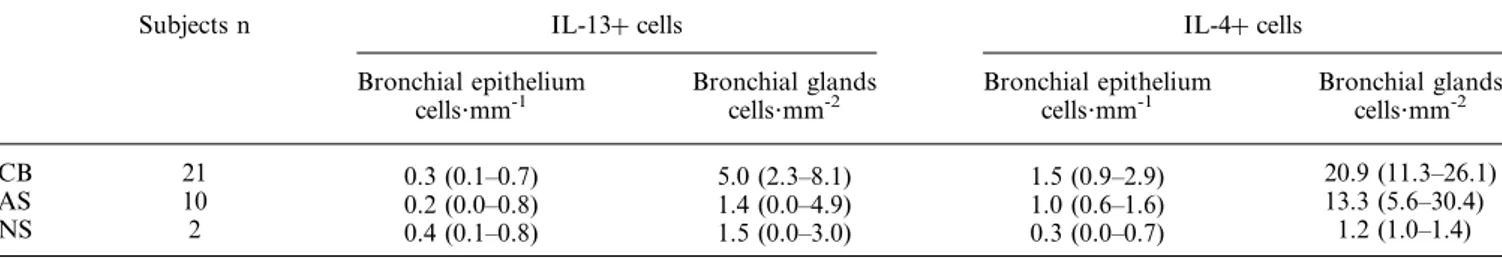
Low numbers of IL-13z and -4z cells were found in the bronchial submucosa (14.0 (7.4–20.7) and 12.8 (9.5–16.1), respectively), glands and epithelium of nonsmokers (table 2). IL-13z and -4z cells were increased in CB as compared to AS (56.8 (42.8–111.2) versus 36.9 (24.5–48.6) and 83.0 (55.3– 123.2) versus 46.5 (25–73), respectively; pv0.05) (fig. 1 and fig. 2). No significant differences were observed in the IL-4 and -13 expression in the epithelium between smokers with CB and AS (table 2). In the bronchial glands there was a trend towards increased IL-13 and -4 expression in smokers with CB, as compared to AS (table 2, fig. 3).
Double labelling was used to identify and count the IL-4/ CD8z, IL-13/CD8z, IL-4/CD4zand IL-13/CD4zdouble-labelled cells present. Approximately 13% of IL-13z cells were also CD8z and CD4z, while 7% of IL-4z cells were CD8z and 5% were also CD4z cells. Furthermore, no associations by correlation were found between IL-13z and -4z cells and CD68z, CD4z or CD8z in the two groups. There were also no associations found between IL-13z and -4z cells and lung volumes.
In the submucosa of the central airways, quantification of IL-5z cells was satisfactory in all the subjects with the exception of subject 2, whereas in the majority of the subjects Table 2. – Immunohistochemical assessment of interleukin (IL)-13z and -4z cells in the bronchial epithelium and glands of smokers and nonsmokers
Subjects n IL-13z cells IL-4z cells
Bronchial epithelium
cells?mm-1 Bronchial glandscells?mm-2 Bronchial epitheliumcells?mm-1 Bronchial glandscells?mm-2
CB 21 0.3 (0.1–0.7) 5.0 (2.3–8.1) 1.5 (0.9–2.9) 20.9 (11.3–26.1)
AS 10 0.2 (0.0–0.8) 1.4 (0.0–4.9) 1.0 (0.6–1.6) 13.3 (5.6–30.4)
NS 2 0.4 (0.1–0.8) 1.5 (0.0–3.0) 0.3 (0.0–0.7) 1.2 (1.0–1.4)
Data are expressed as median (25th–75th percentile). CB: smokers with symptoms of chronic bronchitis; AS: asymptomatic smokers; NS: nonsmokers. Epithelium: IL-4, CB, number of observations=20; IL-13, AS, number of observations=9. Glands: IL-13, AS, number of observations=9. No significant differences were observed between the groups.
very few IFN-cz cells were observed. Low numbers of IL-5z and an absence of IFN-cz cells were found in the bronchial submucosa of nonsmokers (5.9 (0.0–11.8) and 0.0 (0.0–0.0), respectively). No significant differences were observed in the IL-5 and IFN-c expression in the bronchial submucosa between CB and AS (fig. 1). Similarly, there were no sig-nificant differences between the numbers of CD68z, CD4z and CD8z cells in the two groups (table 3).
Discussion
The main finding of the present study is the evidence for IL-13 and -4 immunoreactivity in the central airways of smokers. The experiments presented here indicate that the number of IL-13z and -4z cells is increased in the bronchial submucosa of smokers with CB as compared to AS. IFN-c and IL-5 immunoreactivity was also present, but the protein expression for these cytokines was similar in smokers with CB and in AS. To the best of the current authors9 knowledge, this is the first study assessing, with quantitative methods, the number of cells expressing both Th-2 and -1 cytokines in the central airways of smokers with symptoms of CB. One previous study, which demonstrated an increase of IL-4zcells in the airway submucosa of subjects with CB compared to healthy nonsmokers by using a semiquantitative score, supports the present findings [17]. The current results also confirm and extend the results of ZHU et al. [18], who demonstrated an increased IL-4 messenger ribonucleic acid expression in the bronchial submucosa of smokers with CB.
This study suggests a potential role for Th-2 cytokines in CB. The fact that a parallel increase in the expression of both IL-13 and -4 was observed is consistent with the hypothesis that these two cytokines have many overlapping biological activities due to the shared receptor subunit, the IL-4Ra chain, required for signal transduction [22]. Apart from their role in allergic inflammation [10–13], IL-13 and -4 also appear to be relevant in CB. SHIM et al. [7] found that, in rats,
intratracheal administration of IL-13 induces mucin gene expression and goblet cell metaplasia. Moreover, IL-13 converts human bronchial epithelial cells from an absorptive to a secretory phenotype and increases the portion of secretory cells in primary cultures of human nasal epithelial cells [23, 24]. Recently, it has been shown that CD4z T-cells can only stimulate mucus production through a common, IL-13-mediated, pathway, and that IL-13 acts not through intermediate inflammatory cells but on lung structural cells, probably those of the airway epithelium itself [25]. An effect on the secretion of airway mucus for IL-4 has been shown in an animal model [8]. The instillation of this cytokine into mouse airways resulted in mucin gene expression and goblet cell metaplasia. Goblet cell metaplasia also occurs in the IL-4 transgenic mice characterised by the specific expression of IL-4 in the airways [9].
The current authors speculated that the increased protein expression of both IL-13 and -4 could play a role in the hypersecretion of mucus in smokers with symptoms of CB. Once inflammatory cells have released these cytokines, the latter can contribute to mucus hypersecretion in several ways. IL-13 may act indirectly, through the activation of an
l AS CB l l l l l l l l l l l l ll ll l l l ll l l l l l l l l l l l l 0 100 200 a) l ll l lll l l l l l l l l l l ll l l l l l l l ll l 0 150 750 b) * n+cells·mm -2 * l l ll ll l l l l l l l l l l l l l l l l l lll ll l 0 150 300 c) AS CB n+cells·mm -2 l l l l l l l ll l l l lll l ll l l l l l l l l l l l 300 150 0 d) l l
Fig. 1. – Individual values of cytokineszcells?mm-2in the bronchial submucosa. a) Interleukin (IL)-13, b) IL-4, c) IL-5, and d) interferon-c. AS:
asymptomatic smokers; CB: smokers with symptoms of chronic bronchitis. Horizontal bars represent median values. *: pv0.05. IL-5, CB, number of observations=20.
epidermal growth factor receptor cascade [7]. According to this hypothesis, SINGER et al. [26] recently showed that, in mice, granulocyte depletion inhibited mucus accumulation induced by IL-13, suggesting a supportive role of these cells in mediating the effects of IL-13 on mucus. Mucus hypersecre-tion is believed to result, at least in part, from inflammahypersecre-tion. COHNet al. [27] have showed that IL-4 is crucial for Th-2 cell recruitment to the lung and for induction of inflammation, but that it has no direct role in mucus production. Mucus production could be induced by Th-2 cells in the absence of 4 and -5, eosinophils, and mast cells, but not without IL-4Ra signalling [27, 28]. The data of WHITTAKER et al. [25]
showed that several cytokines, including IL-4, -5, -9 and -10, and inflammatory cells, including mast cells and eosinophils,
activate mucus and the mucin gene muc5ac expression through the increased secretion of IL-13. It has been proposed that these effects are dependent on IL-4Ra expression in structural lung cells, probably those of the airway epithelium, that express both IL-4Ra and IL-13Ra1 [29]. Other studies suggest that IL-4 directly modulates ion transport in the human bronchial epithelium [30].
The current authors predicted that IL-4 and -13 could play a role in mucus hypersecretion in smokers with CB, but no differences could be found in IL-4 and -13 immunoreactivity in glands and epithelium in the two groups. However, this
a)
b)
Fig. 2. – Light microscopic images of immunostained bronchial sub-mucosa from a) an asymptomatic smoker and b) a smoker with symptoms of chronic bronchitis. Interleukin (IL)-13-positive (z) cells were immunostained with a polyclonal antibody specific for human IL-13. Arrows: IL-13z cells. Scale bar=20 mm.
a)
b)
Fig. 3. – Light microscopic images of a) immunostained bronchial epithelium and b) bronchial glands from a smoker with symptoms of chronic bronchitis. Interleukin (IL)-13 positive (z) cells were immu-nostained with a polyclonal antibody specific for human IL-13. Arrows: IL-13z cells. Scale bar=20 mm.
Table 3. – Immunohistochemical assessment of inflammatory cells in the bronchial submucosa of smokers and nonsmokers
Subjects n CD8z cells?mm-2 CD4z cells?mm-2 CD68z cells?mm-2 CB 21 131.0 (59.5–316.5) 123.0 (73.8–233.0) 101.0 (63.2–166.3) AS 10 119.5 (42.0–192.0) 106.0 (58.0–164.0) 45.5 (26.0–103.0) NS 2 68.8 (49–88.7) 57.2 (19.7–94.8) 65.5 (51.3–79.6)
Data are expressed as median (25th–75th percentile). CB: smokers with symptoms of chronic bronchitis; AS: asymptomatic smokers; NS: nonsmokers. No significant differences were observed between the groups.
could take place without an increase in IL-4z and -13z cells in the epithelium and glands of bronchitics being observed. Immunohistochemistry gives a picture of the mediators produced in the airways, but it does not demonstrate the mechanisms of action of mediators. Moreover, IL-4 and -13 may act directly and indirectly on structural cells within the lung, probably on the airway epithelium itself, and the absence of an increase of these cytokines close to the structures involved in mucus secretion is not surprising. The fact that mediators released in the bronchial submucosa may diffuse and act directly on the structures involved in mucus secretion cannot be excluded. It is also possible that cells expressing IL-4 and -13 at the level of the epithelium and glands could play a regulatory role under physiological conditions, whereas, under pathological conditions, the pre-sence of inflammatory cells that can express these cytokines is more pronounced in the bronchial submucosa, and this could change the environment in the airways and indirectly promote mucus hypersecretion. Finally, a variety of inflam-matory mediators have been shown to stimulate mucus secretion and IL-4 and -13 may be only two of these.
The most abundant sources of IL-13 and -4 are CD4zTh-2 and CD8z T-cytotoxic (c) 2 lymphocytes, and IL-13 is also expressed by alveolar macrophages [31, 32]. In the present study it has been demonstrated, with a double-labelling technique, that IL-13 and -4 were co-localised with the CD8z and CD4z T-cell phenotype, but the low percentages of double-labelled cells indicate that cells other than the CD8z and the CD4z phenotype produce IL-13 and -4 in CB. Other candidate cells for the origin of IL-13 include macrophages [32], mast cells, which can also secrete IL-4, dendritic cells and B-cells [31, 33, 34]. In this study, the authors failed to find significant correlations between IL-13 and -4 protein expres-sion and the numbers of CD8zin the bronchial submucosa of bronchitics. In smokers with CB, ZHUet al. [18] found similar
results and they also demonstrated that the CD8z cells present in the bronchial submucosa did not express the IL-4 gene. As with CD8zcells, no correlations were found between IL-13 and -4 expression and the number of CD4zand CD68z cells. A possible explanation for this lack of correlation is that a mixed population of inflammatory cells contributes to their release in the airways. Alternatively, the limited number of subjects examined could account for the lack of associations. In conclusion, T-helper-2 (interleukins-13, -4 and -5) and T-helper-1 (interferon-c) protein expression is present in the central airways of smokers. Smokers with symptoms of chronic bronchitis have an increased number of interleukin-13 and -4-expressing cells in the bronchial submucosa, suggesting a potential role for these T-helper-2 cytokines in mucus hypersecretion, the clinical hallmark of chronic bronchitis. Among the many mechanisms by which mucus is produced, the interleukin-13-mediated pathway seems to be important. Future studies will help to establish whether the inhibition of interleukin-13 may block or modulate mucus production in chronic bronchitis.
Acknowledgements. The authors would like to thank M. Padovan for expert collaboration, G. Fulgeri for typing the manuscript and F. Parise for editing the manuscript.
References
1. American Thoracic Society. Standards for the diagnosis and care of patients with chronic obstructive pulmonary disease. Am J Respir Crit Care Med 1995; 152: S77–S120.
2. Marom Z, Shelhamer JH, Sun F, Kaliner M. Human airway
monohydroxyeicosatetraenoic acid generation and mucus release. J Clin Invest 1983; 72: 122–127.
3. Rieves RD, Goff J, Wu T, Larivee P, Logun C, Shelhamer JH. Airway epithelial cell mucin release: immunologic quanti-tation and response to platelet-activating factor. Am J Respir Cell Mol Biol 1992; 6: 158–167.
4. Sperber KE, Gollub S, Goswami S, Kalb TH, Mayer L, Marom Z. In vivo detection of a novel macrophage-derived protein involved in the regulation of mucus-like glycocon-jugate secretion. Am Rev Respir Dis 1992; 146: 1589– 1597.
5. Lucchini RE, Facchini F, Turato G, et al. Increased VIP-positive nerve fibers in the mucous glands of subjects with chronic bronchitis. Am J Respir Crit Care Med 1997; 156: 1963–1968.
6. Voynow JA, Rosenthal Young L, et al. Neutrophil elastase increases MUC5AC mRNA and protein expression in respiratory epithelial cells. Am J Physiol Lung Cell Mol Physiol 1999; 276: L835–L843.
7. Shim JJ, Dabbagh K, Ueki IF, et al. IL-13 induces mucin production by stimulating epidermal growth factor receptors and by activating neutrophils. Am J Physiol Lung Cell Mol Physiol 2001; 280: L134–L140.
8. Dabbagh K, Takeyama K, Lee HM, Ueki IF, Lausier JA, Nadel JA. IL-4 induces mucin gene expression and goblet cell metaplasia in vitro and in vivo. J Immunol 1999; 162: 6233– 6237.
9. Rankin JA, Picarella DE, Geba GP, et al. Phenotypic and physiologic characterization of transgenic mice expressing interleukin 4 in the lung: lymphocytic and eosinophilic inflammation without airway hyperreactivity. Proc Natl Acad Sci USA 1996; 93: 7821–7825.
10. Wenzel SE, Trudeau JB, Barnes S, et al. TGF-beta and IL-13 synergistically increase eotaxin-1 production in human airway fibroblasts. J Immunol 2002; 169: 4613–4619. 11. Taube C, Duez C, Cui ZH, et al. The role of IL-13 in
established allergic airway disease. J Immunol 2002; 169: 6482–6489.
12. Venkayya R, Lam M, Willkom M, Gru¨nig G, Corry DB, Erle DJ. The Th2 lymphocyte products IL-4 and IL-13 rapidly induce airway hyperresponsiveness through direct effects on resident airway cells. Am J Respir Cell Mol Biol 2002; 26: 202–208.
13. Willis-Karp M, Luyimbazi J, Xu X, Schofield B, Neben TY, Karp CL, Donaldson DD. Interleukin-13: central mediator of allergic asthma. Science 1998; 282: 2258–2261.
14. Chung KF. Cytokines in chronic obstructive pulmonary disease. Eur Respir J 2001; 18: Suppl. 34, 50s–59s.
15. Saetta M, Turato G, Maestrelli P, Mapp CE, Fabbri LM. Cellular and structural bases of chronic obstructive pulmo-nary disease. Am J Respir Crit Care Med 2001; 163: 1304– 1309.
16. Saetta M, Di Stefano A, Maestrelli P, et al. Airway eosinophilia in chronic bronchitis during exacerbations. Am J Respir Crit Care Med 1994; 150: 1646–1652.
17. Mueller R, Chanez P, Campbell AM, Bousquet J, Heusser C, Bullock GR. Different cytokine patterns in bronchial biopsies in asthma and in chronic bronchitis. Respir Med 1996; 90: 79–85.
18. Zhu J, Majumdar S, Qiu Y, et al. Interleukin-4 and interleukin-5 gene expression and inflammation in the mucus-secreting glands and subepithelial tissue of smokers with chronic bronchitis. Lack of relationship with CD8z cells. Am J Respir Crit Care Med 2001; 164: 2220–2228. 19. Global initiative for chronic obstructive lung disease. Global
strategy for the diagnosis, management and prevention of chronic obstructive pulmonary disease. NHLBI/WHO work-shop report. National Institutes of Health. National Heart, Lung, and Blood Institute. 2001; p. 7.
20. Saetta M, Turato G, Facchini FM, et al. Inflammatory cells in the bronchial glands of smokers with chronic bronchitis. Am J Respir Crit Care Med 1997; 156: 1633–1639.
21. Sousa A, Parikh A, Scadding G, Corrigan CJ, Lee TH. Leukotriene-receptor expression on nasal mucosal inflam-matory cells in aspirin-sensitive rhinosinusitis. New Engl J Med 2002; 347: 1493–1499.
22. Zurawski SM, Vega F Jr, Huyghe B, Zurawski G. Receptors for interleukin-13 and interleukin-4 are complex and share a novel component that functions in signal transduction. EMBO J 1993; 12: 2663–2670.
23. Danahay H, Atherton H, Jones G, Bridges RJ, Poll CT. Interleukin-13 induces a hypersecretory ion transport phe-notype in human bronchial epithelial cells. Am J Physiol Lung Cell Mol Physiol 2002; 282: L226–L236.
24. Laoukili J, Perret E, Willems T, et al. IL-13 alters mucociliary differentiation and ciliary beating of human respiratory epithelial cells. J Clin Invest 2001; 108: 1817– 1824.
25. Whittaker L, Niu N, Temann UA, et al. Interelukin-13 mediates a fundamental pathway for airway epithelial mucus induced by CD4 T cells and interleukin-9. Am J Respir Cell Mol Biol 2002; 27: 593–602.
26. Singer M, Lefort J, Vargaftig BB. Granulocyte depletion and dexamethasone differentially modulate airways hyperreac-tivity, inflammation, mucus accumulation, and secretion induced by rmIL-13 or antigen. Am J Respir Cell Mol Biol 2002; 26: 74–84.
27. Cohn L, Homer RJ, Marinov A, Rankin J, Bottomly K. Induction of airway mucus production by T helper 2 (Th2)
cells: a critical role for interleukin 4 in cells recruitment but not mucus production. J Exp Med 1997; 186: 1737–1747. 28. Cohn L, Homer RJ, MacLeod H, Mohrs M, Brombacher F,
Bottomly K. Th2-induced airway mucus production is dependent on IL-4Ra, but not eosinophils. J Jmmunol 1999; 162: 6178–6183.
29. Akaiwa M, Yu B, Umeshita-Suyama R, et al. Localization of human interleukin 13 receptor in non-haematopoietic cells. Cytokine 2001; 13: 75–84.
30. Galietta LJ, Pagesy P, Folli C, et al. IL-4 is a potent modulator of ion transport in the human bronchial epithelium in vitro. J Immunol 2002; 168: 839–845.
31. de Waal Malefyt R, Abrams JS, Zurawski SM, et al. Differential regulation of IL-13 and IL-4 production by human CD8zand CD4zTh0, Th1 and Th2 T cell clones and EBV-transformed B cells. Int J Immunol 1995; 7: 1405–1416. 32. Hancock A, Armstrong L, Gama R, Millar A. Production of interleukin 13 by alveolar macrophages from normal and fibrotic lung. Am J Respir Cell Mol Biol 1998; 18: 60–65.
33. Paul WE, Seder RA, Plaut M. Lymphokine and cytokine production by Fc epsilon RIz cells. Adv Immunol 1993; 53: 1–29.
34. de Saint-Vis B, Fugier-Vivier I, Massacrier C, et al. The cytokine profile expressed by human dendritic cells is dependent on cell subtype and mode of activation. J Immunol 1998; 160: 1666–1676.