Presenilin-1 regulates the neuronal threshold to
excitotoxicity both physiologically and
pathologically
M. Grilli*†, E. Diodato*, G. Lozza*, R. Brusa*, M. Casarini*, D. Uberti‡, R. Rozmahel§, D. Westaway§, P. St George-Hyslop§, M. Memo‡, and E. Ongini*
*Schering-Plough Research Institute, Department of Central Nervous System兾Cardiovascular Research, San Raffaele Science Park, Milan, Italy;‡Department
of Biomedical Sciences, University of Brescia, Brescia, Italy; and§Center for Research on Neurodegenerative Diseases, Department of Neurology,
University of Toronto, Toronto, ON, Canada
Edited by Floyd E. Bloom, The Scripps Research Institute, La Jolla, CA, and approved August 28, 2000 (received for review May 12, 2000) A direct pathophysiological role of Familial Alzheimer’s Disease
(FAD)-associated Presenilin 1 (PS1) mutations in neuronal vul-nerability remains a controversial matter. We evaluated the relationship between PS1 and excitotoxicity in four different experimental models of neurotoxicity by using primary neurons from (i) transgenic (tg) mice overexpressing a human FAD-linked PS1 variant (L286V mutation), (ii) tg mice overexpressing human wild-type (wt) PS1, (iii) PS1 knockout mice, and (iv) wt mice in which PS1 gene expression was knocked down by antisense treatment. We found that primary neurons overexpressing mu-tated PS1 showed an increased vulnerability to both excitotoxic and hypoxic-hypoglycemic damage when compared with neu-rons obtained from either mice overexpressing human wt PS1 or in wt mice. In addition, reduced excitotoxic damage was ob-tained in neurons in which PS1 expression was absent or diminished. Data obtained in in vivo experimental models of excitotoxicity partially supported the in vitro observations. Accelerated neuronal death was demonstrated in the hippocam-pus of mice overexpressing mutated PS1 after peripheral ad-ministration of kainic acid in comparison with wt animals. However, measurement of the infarct volume after middle cerebral artery occlusion did not show significant difference between the two animal groups. The results altogether suggest that expression of FAD-linked PS1 variants increases the vulner-ability of neurons to a specific type of damage in which exci-totoxicity plays a relevant role. In addition, they support the view that reduction of endogenous PS1 expression results in neuroprotection.
A
subset of early-onset cases of autosomal dominant familial Alzheimer’s disease (FAD) is caused by mutations in three different loci: the amyloid precursor protein gene (1, 2) and the Presenilin 1 (PS1) and Presenilin 2 (PS2) genes (3, 4). Of the three loci, mutations in the PS1 gene are most frequent and associated with early onset of the disease. PS1, widely expressed throughout the central nervous system and in peripheral tissues, is a multipass membrane protein localized predominantly in the endoplasmic reticulum but also in the nuclear membrane and coated transport vesicles (5–7). More recently, it has been suggested that PS1 is constitutively expressed at the cell surface of mammalian cells (8–10). On the basis of the predicted structure, it has been proposed that PS proteins may function as signal receptors, form channels, and兾or participate in protein trafficking (3). Yet, the physiological functions of PS1 in adult life remain largely unknown.Because Alzheimer’s disease (AD) is a neurodegenerative disease characterized by the death of vulnerable neurons in selective brain areas, much effort has been dedicated to find a correlation between PS1 mutant isoforms and neuronal death. Recently, Guo et al. (11) found that hippocampal neurons from PS1 mutant knock-in mice show increased sensitivity to gluta-mate-induced cell death. In addition, PS1 mutations have been
shown to increase the vulnerability of neural cells to apoptosis induced by trophic factor withdrawal,-amyloid, hyperosmotic stress and aging, so as to suggest that PS1 mutations may lower the threshold to neurotoxicity (11–15). The proposed mecha-nism for such a role is that PS1 mutations contribute to disrupt calcium homeostasis and increase free radical production (12) and兾or to influence intracellular signaling pathways (16, 17). These results are apparently in contrast with the data of Bursz-tajn et al. (18), who showed that overexpression in mouse cortical neurons of human PS1 both in its normal and FAD-linked isoform does not enhance apoptosis. Furthermore, down-regulation of PS1 was associated with apoptosis of a series of nonneuronal cell lines (19). Thus, a direct physiopathological role of FAD-associated PS1 mutations in cellular vulnerability to toxic stimuli is still a controversial matter.
Experimental Procedures
Animals.Generation of the transgenic (tg) lines overexpressing human wild-type (wt) PS1 (PS1 wt tg) and FAD PS1 L286V (PS1 L286V tg) is described elsewhere, therein referred to as lines 195 and 198, respectively (20). In all studies, both lines were used as homozygotes in a pure FVB兾N background. Non-tg littermates of corresponding age兾weight were used as control animals and referred to as wild-type (wt) mice. The PS1 knock-out mice used in these experiments are described elsewhere (R.R., J. Huang, F. Chen, C. Bergeron, Y. Liang, V. Nguyen, G. Wong, C. McKerlie, M. Ikeda, G. Levesque, G. Yu, M. Nishimura, D.W., and P. Segh, unpublished data). In short, these animals have a modification of PS1 exon 6 such that neither intact transcript nor functional protein is produced and is propagated on a mixed CD1 ⫻ 129兾SvJ strain background. All of the experiments were carried out in accordance with the National Institutes of Health guide for the care and use of laboratory animals.
PS1ⴙ/ⴙ, PS1ⴙ/ⴚ, and PS1ⴚ/ⴚGenotyping.Mouse tail genomic DNA was purified by using standard methods followed by PCR amplification. Primer sequences were as follows: 5⬘-AGT TGG TAG GAC CAC TTT GT-3⬘ and 5⬘-AGA CAC TTC CTC TTT GCT AG-3⬘ (endogenous mouse PS1) and 5⬘-AGT TGG TAG
This paper was submitted directly (Track II) to the PNAS office.
Abbreviations: AD, Alzheimer’s disease; CCA, common carotid artery; FAD, familial Alzhei-mer’s disease; KA, kainic acid; LDH, lactate dehydrogenase; MCA, middle cerebral artery; MK-801, dizolcipine maleate; NMDA, N-methyl-D-aspartate; ODNs, oligodeoxynucleotides; OGD, oxygen-glucose deprivation; PS1, Presenilin 1; PS2, Presenilin 2; tg, transgenic; wt, wild-type; AS, antisense; SCR, scrambled; MIS, mismatched.
†To whom reprint requests should be addressed at: Schering-Plough Research Institute, Via Olgettina, 58, 20132 Milan, Italy. E-mail: [email protected].
The publication costs of this article were defrayed in part by page charge payment. This article must therefore be hereby marked “advertisement” in accordance with 18 U.S.C. §1734 solely to indicate this fact.
GAC CAC TTT GT-3⬘ and 5⬘-ATG CCT GCT CTT TAC TGA AG-3⬘ (targeted allele). The wt allele product was 127 bp, whereas the targeted allele product was 480 bp. The PCR mixture contained 0.2 mM dNTP mixture, 2 mM MgCl2, 0.2M
of each of the primer sets, and 1.5 units Taq DNA polymerase. Running conditions were as follows: 94°C for 30 s, followed by 35 cycles at 94°C for 30 s, 58°C for 30 s, and 72°C for 30 s. Primary Neuronal Cultures. Mixed cortical cell and cerebellar granule cell cultures were prepared from homozygotes PS1 L286V tg, PS1 wt tg, or from wt mice at 14–16 days of gestation and at postnatal day 8, respectively, as previously described (21). Pure cortical neurons were prepared from individual E16 em-bryos derived from PS1⫺/⫹⫻ PS1⫺/⫹breeding pairs. Genotyping
was subsequently performed on tail DNA to identify PS1⫹/⫹,
PS1⫺/⫹, and PS1⫺/⫺ embryos. All primary neuronal cultures
were used after 12–14 days in vitro.
In Vitro Neurotoxicity Assay.N-Methyl-D-aspartate (NMDA) (Sig-ma) was added to culture medium in the absence or presence of 1 M dizolcipine maleate (MK-801) (Sigma). For oxygen-glucose deprivation (OGD), culture medium was exchanged with deoxygenated glucose-free minimum essential medium (MEM). Cultures were kept in an anaerobic chamber saturated with N2
for 60 min at 37°C. OGD was terminated by removal of the cultures from the chamber and replacement of the medium with oxygenated MEM supplemented with 1% horse serum. In both experimental paradigms, neuronal viability was assessed 24 h after NMDA addition or after removal from the anaerobic chamber, as previously described (21). Briefly, in all mixed neuron-glia experiments, neuronal cell death was expressed as the percentage of total lactate dehydrogenase (LDH) release elicited by 100 M NMDA, a concentration that results in maximal neuronal cell death. All values were corrected for basal LDH release in each group. In both cerebellar and pure cortical neuronal cultures, cell viability was determined by intravital staining with fluorescein diacetate (15g兾ml) and propidium iodide (80 g兾ml) for 3 min at 25°C, and expressed as the percentage of the total cell number.
Antisense Oligonucleotide Synthesis and Treatment. Phosphorothio-ated PS1 oligodeoxynucleotides (ODNs) (PRIMM, Milan, Italy) were 18 nt long and comprised the ATG codon corresponding to the initiator methionine in the mouse gene. For each ODN, acronimus, position relative to the ATG start codon, and sequence were as follows: (i) PS1兾AS-1 (antisense), ⫺9 to ⫹9, 5⬘-CTC TGT CAT TGG AGC AGC-3⬘; (ii) PS1兾SENSE-1 (sense),⫹9 to ⫺9, 5⬘-GCT GCT CCA ATG ACA GAG-3⬘; (iii) PS1兾SCR-1 (scramble), ⫹9 to ⫺9, 5⬘-TCT CTG ACT GTG AAG CCG-3⬘; (iv) PS1兾MIS-1 (mismatched), ⫺9 to ⫹ 9, 5⬘-CTC
GTG CAT GTT AGC AGC-3⬘; (v) PS1兾AS-2 (antisense), ⫺15 to⫹3, 5⬘-CAT TGG AGC AGC TGG ACA-3⬘; and (vi) PS1兾 MIS-2 (mismatched)⫺15 to ⫹3 5⬘-CAT TGG AGC AGC TGT CAT-3⬘. ODN sequences exhibited no similarity to any other known mammalian genes (BLASTsearch). As indicated, ODNs (0.1, 0.3, 1, and 3M) were added to culture medium 2 h before and during NMDA exposure.
Northern Blot Analysis.Total RNA was prepared from 5⫻ 106 cortical neurons treated as described, and 8- to 10-g aliquots were fractionated on 1.2% agarose-formaldehyde gels and trans-ferred to nylon filters. A 2.0-kb XhoI fragment of a mouse cDNA clone was used to measure PS1 mRNA transcripts. The 18S rRNA DECAprobe template (Ambion, Austin, TX) was used to generate an internal control so as to normalize for total RNA. Hybridization was quantified by densitometry and expressed as PS1兾18S ratio.
Western Blot Analysis.The brain lysate (25g) was mixed with an equal volume of 2% SDS, electrophoresed on 12% Tris-glycine gels, and transferred to nitrocellulose. The C-terminal-specific anti-PS1 polyclonal antibody (kind gift of N. Brindle, St. James University Hospital, Leeds, U.K.) raised against residues 290– 304 of PS1 was used at a dilution of 1:5000, and proteins were visualized by enhanced chemiluminescence (ECL, Amersham Pharmacia) by using horse-radish peroxidase-conjugated goat anti-rabbit secondary antibody.
Induction of Focal Cerebral Ischemia.Three-month-old (25–30 g) PS1 L286V tg mice and non-tg littermates were subjected to permanent occlusion of the left middle cerebral artery (MCA) (20 tg and 20 wt) or of the left MCA plus the ipsilateral common carotid artery (CCA) (17 tg and 18 wt) as previously described (21). The body temperature was monitored and maintained at 37°C⫾ 1°C during surgery and during the immediate postop-erative period until the animals recovered fully from anesthesia. Twenty-four hours after combined MCA and CCA occlusion, and 96 h after MCA occlusion only, brains were removed, embedded in paraffin, and stained with cresyl violet. Eight sections for each brain were digitized with a color video camera, and total brain and infarct areas were measured on a blind basis, by using an image analyzer system (Image Pro-Plus, Media Cybernetics, Silver Spring, MD), as previously described (21). Kainic Acid (KA)-Induced Neurodegeneration.Twenty-five wt and 24 PS1 L286V tg mice (27–30 g) were used. Seventeen wt and 17 tg mice were given KA, 30 mg兾kg, i.p. Within this group, 14 mice (7 wt and 7 tg) died during the first 24 h after the treatment. Among survivors, 12 mice (6 wt and 6 tg) were killed at 3 days, and 8 mice (4 wt and 4 tg) at 6 days. A separate group of 6 animals (3 wt and 3 tg) were given KA at 15 mg兾kg, i.p. They all survived until day 3, when they were killed. Nine animals (5 wt and 4 tg) were treated i.p. with saline and served as controls. Mice behavior was monitored continuously for 6 h and rated according to Racine’s scale (22). Serial coronal sections from paraffin-embedded brain were cut at 5m, stained for hema-toxylin-eosin, and examined for pathological changes. Profile counting was confined to the anterior portion of the hippocam-pus (bregma level⫺1.70 to ⫺2.18 mm, figures 45–49 in Franklin and Paxinos’ Atlas). One in every five serial sections was analyzed by using a camera adapted to a Nikon microscope with a⫻40 objective. The image processing and quantitative analysis of histological data were performed by using IMAGE PRO-PLUS software. Images were used for constructing an algorithm to define ‘‘viable cells.’’ Viable cells were selected by size (diameter greater than 7m) and color, automatically defined, encircled, numbered, and counted. The mean profile number兾mm2 was
obtained from at least 15 sections per each brain.
Data Analysis. Data in text and figures referred to studies in cultures from wt and PS1 L286V tg mice, are expressed as mean⫾ SEM, and were analyzed by factorial ANOVA design. Data relative to the study performed in cultures from PS1⫹/⫹,
PS1⫹/⫺, and PS1⫺/⫺embryos and in cultures from wt versus PS1
wt tg mice are expressed as mean and confidence limits, and statistically analyzed by logistic regression for factorial design.
In in vivo studies, the infarct size was calculated with Cava-lieri’s estimator of morphometric volume (23). A two-tailed Student’s t test was used to determine statistical significance between the wt and PS1 L286V tg mice. P⬍ 0.05 was considered statistically significant. ‘‘Non-responder’’ rate, defined as in ref. 21 was compared by using Fisher’s exact test. KA data were expressed as the percentage of control and analyzed by ANOVA with Student’s t test for simple comparison.
Results
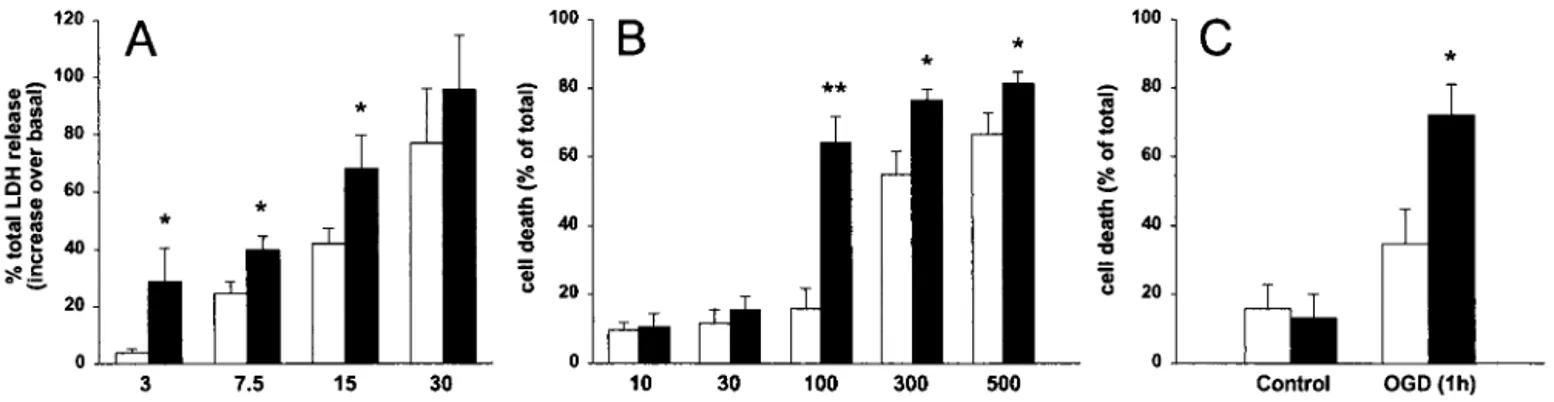
Relationship Between PS1 and Excitotoxicity in Primary Neurons from tg Mice.Primary cortical cultures were established from human PS1 L286V tg and wt embryos at E16 and maintained in vitro for 12–14 days before experimental use. During this period, no overt difference in rate of maturation and differentiation was noted in the two neuronal cultures (data not shown). As shown in Fig. 1A, neurons overexpressing L286V PS1 showed a significant increase in their sensitivity to NMDA-induced cell death, as demon-strated by a leftward shift in the NMDA dose-response curve. At the concentration of 3 M, NMDA induced less than 10% neuronal death (percentage of maximal cell death induced by 100 M NMDA) in wt but almost 30% in the PS1 L286V tg mice-derived cultures. A similar pattern of results was found by using NMDA concentrations of 7.5M and 15 M.
Exposure of cerebellar granule cells from wt mice to concen-trations of NMDA ranging from 10M to 500 M resulted in a concentration-dependent cell death (Fig. 1B). Neurons from PS1 L286V tg mice were significantly more sensitive to NMDA treatment than their wt counterpart. In particular, concentra-tions of NMDA that were below those required to cause neuronal death in cerebellar cultures from wt mice (i.e., 100M) were almost maximally neurotoxic in cultures overexpressing mutated PS1.
The increased sensitivity of neurons from PS1 L286V tg mice to excitatory amino acid-associated neuronal death was con-firmed in the OGD paradigm (24). As shown in Fig. 1C, cerebellar granule cells from PS1 L286V tg mice were signifi-cantly more vulnerable (3-fold difference) to OGD than cells from wt mice (20% and 60% over the corresponding basal value, respectively). To dissect the contribution of human PS1 overex-pression per se in our experimental setting, a parallel study was then performed in cerebellar granule cells from PS1 wt tg mice and FVB兾N non-tg littermates. No significantly different re-sponse to NMDA-induced toxicity could be observed at con-centrations ranging from 10 to 500M between the two exper-imental groups. As shown in Fig. 2 Inset, PS1 wt and PS1 L286V tg mice overexpress the human protein at comparable levels, with a human vs. murine protein ratio of about 1.
Neurodegeneration in PS1 tg Mice Afterin Vivo Experimental Models of Excitotoxicity. We then evaluated whether expression of a FAD-associated PS1 gene mutation was able to affect neuronal
sensitivity to excitotoxicity in vivo. The wt and PS1 L286V tg mice were treated with either two different doses of KA, 15 and 30 mg兾kg i.p. or saline. In animals treated with KA 15 mg兾kg, only sporadic seizures were observed and the behavioral changes consisting mainly of wet dog shakes, whereas administration of 30 mg兾kg induced convulsions in most animals. Treatment with 30 mg兾kg KA resulted in a comparable behavioral pattern in both wt and PS1 L286V tg mice (data not shown). Both strains
Fig. 1. Effect of PS1 FAD mutation on in vitro excitotoxicity. (A) Primary cortical neurons from wt (open bars) or PS1 L286V tg (filled bars) mice were exposed
to increasing concentrations of NMDA, as indicated. Cell death was quantified by measuring the release of LDH enzyme in the culture media and was expressed as the percentage of total LDH release elicited by 100M NMDA. (B) Primary cerebellar neurons from wt (open bars) or PS1 L286V tg (filled bars) mice were exposed to increasing concentrations of NMDA, as indicated. Cell death was quantified by determining intravital staining with fluorescein diacetate and propidium iodide and was expressed as the percentage of dead cells over total cell number. (C) Primary cerebellar neurons from wt (open bars) or PS1 L286V tg (filled bars) mice were subjected to OGD for 1 h. Neuronal viability was assessed 24 h later by intravital staining with fluorescein diacetate and propidium iodide. Data are expressed as the percentage of dead cells over total cell number. Numbers represent mean⫾ SEM of at least six different experiments, run in triplicate, each from different cell preparations.**, P⬍ 0.01, and*, P⬍ 0.05, vs. the corresponding values of wt group.
Fig. 2. Effect of human PS1 wt overexpression on in vitro NMDA-induced neurotoxicity. Primary cerebellar neurons from wt mice (open bars) or human PS1 wt overexpressing mice (filled bars) were exposed to increasing concen-trations of NMDA, as indicated. Cell death was measured by intravital staining with fluorescein diacetate and propidium iodide and was expressed as the percentage of dead cells over total cell number. Where indicated, MK-801 was added to the culture media 10 min before NMDA. Values are expressed as mean and confidence limits and are from nine separate cell preparations with each point run at least in quintuplicate. (Inset) Representative Western blot analysis of PS1 protein levels in cerebellar extracts from wt mice and mice overexpressing human wt (PS1 wt) or mutated (L286V) PS1. The C-terminal fragment of both human (h) and mouse (m) PS1 protein was detected with an anti-PS1 antibody. The calculated apparent molecular mass was 21 kDa and 20 kDa for human and mouse PS1 fragments, respectively.
displayed a mortality rate of 41%. At 3 days after injection, the lower KA dose produced minor histological damage whereas the higher dose led to selective hippocampal cell loss in the CA3 subregion of both wt and PS1 L286V mice. We found tg mice to be more sensitive to KA-induced neurodegeneration. Fig. 3
shows representative stainings of coronal brain sections from wt (Fig. 3 A and C) and tg mice (Fig. 3 B and D) treated with saline (Fig. 3 A and B) or 30 mg兾kg KA (Fig. 3 C and D), killed 3 days after the injection. The degree of KA-induced lesion in the different experimental groups was estimated by computer-assisted measurement of profile counts as an unbiased index of cell number, and shown in Fig. 3E. At 3 days, the percentage of surviving neurons in the CA3 hippocampal region was about 70% and 45% of the corresponding saline-treated groups, in wt and tg mice, respectively. At longer time points, i.e., 6 days, the number of CA3-surviving neurons was similar in both experi-mental groups (50% and 42% for wt and tg mice, respectively). When focal cerebral ischemia was used as in vivo model of neurodegeneration associated with excitotoxicity, we did not observe significant differences in the responsiveness to the injury in PS1 L286V tg mice compared with wt animals. The wt and tg mice were subjected to two different paradigms of focal cerebral ischemia in the territory of the MCA. We used both the ‘‘tandem’’ occlusion model (MCA plus CCA occlusion, 20 animals for each group), which produces larger infarct volumes, and the single MCA occlusion model (18 wt and 17 PS1 L286V tg mice), which results in smaller brain damage. After MCA⫹ CCA occlusion, the infarct volumes were 26.0 ⫾ 1.9 mm3and
29.9 ⫾ 2.3 mm3 (18.9⫾ 1.3% and 21.2 ⫾ 1.6% of ipsilateral
hemisphere) for wt and PS1 L286V tg mice, respectively. After single MCA occlusion, lesion volumes were 5.6⫾ 0.6 mm3and
6.8 ⫾ 0.8 mm3 (9.5 ⫾ 1.1% and 11.1 ⫾ 1.2% of ipsilateral
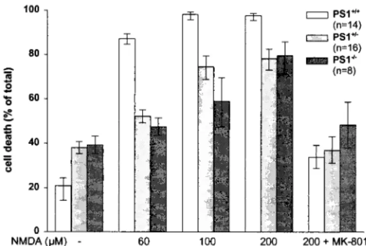
hemisphere) for wt and PS1 L286V tg mice, respectively. No difference was observed in ‘‘non-responders’’ rate (about 25%). Relationship Between PS1 and Excitotoxicity in Primary Neurons from PS1 Knockout Mice.Pure cortical neurons were established from PS1⫹/⫹, PS1⫹/⫺, and PS1⫺/⫺single embryos at E16 and
main-tained in vitro for 12–13 days. During this period, there was no obvious morphological difference in differentiation rate be-tween neurons from the different genotypes (data not shown). PS1⫹/⫺and PS1⫺/⫺neurons showed a significant reduction in
their sensitivity to NMDA-induced cell death compared with PS1⫹/⫹(Fig. 4). At a concentration of 60M, NMDA induced
nearly 90% neuronal death in the wt neurons whereas only 42% and 48% in the PS1⫺/⫺and PS1⫹/⫺genotypes, respectively. The Fig. 3. Effect of KA injection on neuronal viability in hippocampi from wt and
PS1 tg mice. (Upper) Representative micrographs of CA3 hippocampal region from wt FVB兾N (A and C) and PS1 L286V tg (B and D) mice treated with saline (A and B) or 30 mg兾kg KA (C and D) and killed 3 days after the lesion. (E) Quantitative analysis of KA-induced cell loss in the CA3 and dentate gyrus (DG) hippocampal region of wt FVB兾N and PS1 L286V tg mice treated with either 15 mg兾kg or 30 mg兾kg KA and killed after 3 days (3d) or 6 days (6d), as indicated. Viable cells were evaluated by estimating the mean profile number per mm2. Data are expressed
as the percent of the corresponding control values (black bar) and represent the mean⫾ SEM of at least 15 sections per each brain. Absolute values were 7.5 ⫾ 0.9⫻ 105and 6.9⫾ 1.3 ⫻ 105profile number per mm2in dentate gyrus of wt and
PS1 tg mice, respectively, and 2.1⫾ 0.4 ⫻ 105and 2.5⫾ 0.5 ⫻ 105profile number
per mm2in CA3 region of wt and PS1 tg mice, respectively.
*, P⬍ 0.05 vs. the corresponding values of wt group.
Fig. 4. NMDA-induced neurotoxicity in primary cortical neurons from
PS1⫹/⫹, PS1⫹/⫺, and PS1⫺/⫺mice. Primary neurons from single embryos were exposed to increasing concentrations of NMDA, as indicated. MK-801 (1M) was added to the culture media 10 min before NMDA. Cell death was quan-tified by intravital staining with fluorescein diacetate and propidium iodide and expressed as the percentage of dead cells over total cell number. Values are expressed as mean and confidence limits. P⬍ 0.01 PS1⫹/⫹vs. PS1⫹/⫺, P⬍
0.01 PS1⫹/⫹vs. PS1⫺/⫺. n, Number of cell preparations.
reduced sensitivity to NMDA was less apparent but still statis-tically significant at maximally effective concentrations of the excitotoxin. MK-801 (1 M) completely reversed NMDA-induced toxicity in all different genotypes.
Relationship Between PS1 and Excitotoxicity in Primary Neurons with Reduced PS1 Gene Expression.Two 18-mer antisense ODNs com-plementary to sequences flanking the initiation codon of the mouse PS1 gene, PS1兾AS-1 (⫺9 to ⫹9) and PS1兾AS-2 (⫺15 to ⫹3), were individually added to the culture media of 12-day in
vitro cortical neurons, 2 h before and during NMDA treatment.
Incubation of neurons with 15M NMDA resulted in about 60% of total LDH release in antisense-untreated cells. When cells were pretreated with different concentrations of PS1兾AS-1, a dose-dependent reduction in NMDA-induced cell death was observed (Fig. 5A). Maximal inhibition (⫺50%) was found by using 3M PS1兾AS-1. At the same concentration, correspond-ing sense (PS1兾SENSE-1), scrambled (PS1兾SCR-1), and mis-matched (PS1兾MIS-1) ODNs were devoid of effect. Under similar experimental conditions, 3 M PS1兾AS-2, but not its
mismatched version (PS-1兾MIS-2), prevented NMDA-induced neuronal loss (⫺50%). Both ODNs did not affect cell viability
per se (data not shown). A correlation was established between
ODN treatment and PS1 mRNA levels. Exposure of cortical neurons to NMDA resulted in a concentration-dependent in-crease of PS1 mRNA levels. Fig. 5B shows the results obtained by measuring the effects of 30M NMDA on PS1 mRNA levels in cell cultures either untreated or pretreated with 3 M PS1兾AS-1 and PS1兾SENSE-1. In untreated cells, 30M NMDA induced a 2-fold increase in PS1 mRNA levels, detectable within 1 h, which was prevented by 1M MK-801, PS1兾AS-1 ODN, but not PS1兾SENSE-1. Comparable inhibitory effects were obtained with PS1兾AS-2, but not with PS1兾MIS-2, PS1兾MIS-1, and PS1兾SCR-1 (data not shown).
Discussion
The first indication that PSs may regulate apoptotic cell death emerged from a functional screening for cDNAs that could rescue T cell receptor-induced apoptosis in a mouse T cell hybridoma (25). Subsequent studies further indicated that both PS1 and PS2 might participate in neuronal apoptosis (12, 13, 26–28). Recent work in hippocampal neurons derived from mice generated by knock-in of a FAD-linked PS1 mutation also supported this hypothesis (29). Finally, direct in vivo evidence for a role of PS1 in neurodegeneration came from quantitative stereological analysis, revealing a significant decrease in the frontal cortex and hippocampal CA1 fields of aged mice pressing human FAD-linked variants compared with mice ex-pressing wt PS1 (14).
In the present study, we found that neurons from PS1 L286V tg mice have a significant increase in their sensitivity to excito-toxicity. This phenomenon was observed in three different in
vitro experimental models, i.e., (i) cortical neurons exposed to
NMDA, (ii) cerebellar granule cells exposed to NMDA, and (iii) cerebellar granule cells exposed to OGD. Because primary cultures of cerebellar granule cells are 95–98% neuronal in nature, it is reasonable to infer that the putative factor(s) responsible for the PS1 mutation-related hypersensitivity to excitotoxicity is constitutively expressed in neuronal cells. Based on an extensive analysis of the sensitivity to excitotoxic death in primary neurons from a transgenic line overexpressing wt human PS1 at levels comparable to those of the PS1 L286V line, we could rule out the possibility that human PS1 overexpression per
se could contribute to such effects.
Our in vivo data support the concept that expression of mutant PS1 lowers the threshold to excitotoxicity. In line with recent findings (11), we found PS1 L286V tg mice were more sensitive to peripheral administration of KA than wt mice. This effect was particularly evident at 3 days after the treatment but disappeared at 6 days, suggesting that the FAD-linked PS1 mutation accel-erates the neurodegenerative process. However, when animals were subjected to cerebral ischemia, no difference was found between wt and PS1 L286V tg mice. The possibility that different results might be seen at different time points after the lesion cannot be completely ruled out. However, it should be noted that the molecular mechanisms promoting neuronal cell death (i.e., apoptosis) in the permanent focal ischemia and KA experimen-tal models are not superimposable (30, 31). It is also well known that different mechanisms contribute to the neurodegeneration associated with animal models of permanent and transient cerebral ischemia. In line with this concept, it was recently reported that the extent of brain injury after ischemia兾 reperfusion injury is indeed worsened in PS1 mutant knock-in mice (32). Overall, our in vivo data suggest that predisposition to excitotoxic damage induced by L286V PS1 mutation may not be a generalized phenomenon, being affected by the type of neu-rons that are involved, the extension of the lesion, and the kinetics of the injury. The metabolic state and the intracellular
Fig. 5. Effects of PS1 ODN treatment on NMDA-induced neurotoxicity (A)
and PS1 mRNA expression (B) in primary cortical neurons from wt mice. (A) Cortical neurons were exposed to increasing concentrations of different ODNs, ranging from 0.1M to 3 M, for 2 h before and during the NMDA treatment. Cell death was quantified by measuring the release of LDH enzyme and was expressed as the percentage of total LDH release elicited by 100M NMDA. (B) Primary cortical neurons were exposed to different ODNs for 2 h before the NMDA treatment. PS1 mRNA levels were measured by Northern blot analysis 1 h after NMDA. Data represent mean⫾ SEM of at least three different experiments and are from three separate cell preparations. MK-801 was added to the culture media 10 min before NMDA. ODN sequence and acronimus are reported in Experimental Procedures.*, P⬍ 0.01 vs. NMDA alone treated values.
pathways triggered to induce neuronal death in a specific cell phenotype may also contribute to different thresholds of vul-nerability in the presence of mutated PS1. In this regard, Bursztajn et al. (18) found that etoposide- and staurosporine-induced apoptosis of cortical neurons is not affected by overex-pression of either wt or mutated human PS1.
FAD-associated PS1 mutations are commonly believed to be associated with a gain of function although the nature of the putative function(s) is still under investigation. We addressed the question of whether the PS1 mutation amplifies a physiological function or, more specifically, whether wt PS1 is normally involved in determining the neuronal threshold for excitotoxic-ity. We therefore used primary neuronal cultures from PS1⫺/⫺,
PS1⫹/⫺, and PS1⫹/⫹and found that the absence or reduction of
PS1 results in a greater resistance of neurons to NMDA-induced cell death. Because it has been shown that PS1, possibly via Notch-1, is required for neuronal differentiation and neurite outgrowth (33, 34), one may argue that the effects of lack of PS1 expression on threshold of vulnerability to NMDA are merely due to delayed neuronal differentiation. In addition, this lower vulnerability could be explained with the biased selection of a subpopulation of cortical neurons that survive the severe devel-opmental changes described in PS1⫺/⫺mice in vivo (35). At least
two observations argue against these possibilities. First, reduced susceptibility to NMDA-induced cell death occurs not only in PS1⫺/⫺- but also in PS1⫹/⫺-derived primary neurons, even
though the heterozygous mice do not show developmental alterations. Second, greater resistance to excitotoxic-damage is demonstrated also in neurons in which PS1 gene expression is knocked-down after differentiation (antisense-treatment).
Our results support the view that endogenous PS1 is physio-logically involved in determining the threshold for excitotoxicity as well as playing an active role in the neurodegenerative process. The proposed mechanisms for such a role include the possibility that PS1 mutations may perturb calcium homeostasis and in-crease free radical production in affected cells (12), and兾or may influence signaling pathways involving Akt兾protein kinase B (PKB) (16), Jun kinase (17), or catenin (28, 36). The molecular mechanisms by which PS1, either physiologically or pathologi-cally, when mutated, may interfere with neuronal survival pro-grams therefore deserve further investigation.
We thank Dr. Catherine Strader for support throughout the study and critically reviewing the manuscript, and Harsha Basudev and Ilaria Barbieri for experimental assistance.
1. Chartier-Harlin, M. C., Crawford, F., Houlden, H., Warren, A., Hughes, D., Fidani, L., Goate, A., Rossor, M., Roques, P. & Hardy, J. (1991) Nature
(London) 353, 844–846.
2. Goate, A., Chartier-Harlin, M. C., Mullan, M., Brown, J., Crawford, F., Fidani, L., Giuffra, L., Haynes, A., Irving, N. & James, L. (1991) Nature (London) 349, 704–706.
3. Sherrington, R., Rogaev, E. I., Liang, Y., Rogaeva, E. A., Levesque, G., Ikeda, M., Chi, H., Lin, C., Li, G. & Holman, K. (1995) Nature (London) 375, 754–760. 4. Levy-Lahad, E., Wasco, W., Poorkaj, P., Romano, D. M., Oshima, J., Pettingell, W. H., Yu, C. E., Jondro, P. D., Schmidt, S. D. & Wang, K. (1995) Science 269, 973–977.
5. Cook, D. G., Sung, J. C., Golde, T. E., Felsenstein, K. M., Wojczyk, B. S., Tanzi, R. E., Trojanowski, J. Q., Lee, V. M. & Doms, R. W. (1996) Proc. Natl. Acad.
Sci. USA 93, 9223–9228.
6. Kovacs, D. M., Fausett, H. J., Page, K. J., Kim, T. W., Moir, R. D., Merriam, D. E., Hollister, R. D., Hallmark, O. G., Mancini, R., Felsenstein, K. M., et al. (1996) Nat. Med. 2, 224–229.
7. Lah, J. J., Heilman, C. J., Nash, N. R., Rees, H. D., Yi, H., Counts, S. E. & Levey, A. I. (1997) J. Neurosci. 17, 1971–1980.
8. Dewji, N. N., Do, C. & Singer, S. J. (1997) Proc. Natl. Acad. Sci. USA 94, 14031–14036.
9. Schwarzman, A. L., Singh, N., Tsiper, M., Gregori, L., Dranovsky, A., Vitek, M. P., Glabe, C. G., St George-Hyslop, P. H. & Goldgaber, D. (1999) Proc. Natl.
Acad. Sci. USA 96, 7932–7937.
10. Ray, W. J., Yao, M., Mumm, J., Schroeter, E. H., Saftig, P., Wolfe, M., Selkoe, D. J., Kopan, R. & Goate, A. M. (1999) J. Biol. Chem. 274, 36801–36807. 11. Guo, Q., Fu, W., Sopher, B. L., Miller, M. W., Ware, C. B., Martin, G. M. &
Mattson, M. P. (1999) Nat. Med. 5, 101–106.
12. Guo, Q., Sopher, B. L., Furukawa, K., Pham, D. G., Robinson, N., Martin, G. M. & Mattson, M. P. (1997) J. Neurosci. 17, 4212–4222.
13. Czech, C., Lesort, M., Tremp, G., Terro, F., Blanchard, V., Schombert, B., Carpentier, N., Dreisler, S., Bonici, B., Takashima, A., et al. (1998)
Neuro-science 87, 325–336.
14. Chui, D. H., Tanahashi, H., Ozawa, K., Ikeda, S., Checler, F., Ueda, O., Suzuki, H., Araki, W., Inoue, H., Shirotani, K., et al. (1999) Nat. Med. 5, 560–564. 15. Tanii, H., Ankarcrona, M., Flood, F. M., Nilsberth, C., Mehta, N. D.,
Perez-Tur, J., Winblad, B., Benedikz, E. & Cowburn, R. F. (1999) Neuroscience 95,593–601.
16. Weihl, C. C., Ghadge, G. D., Kennedy, S. G., Hay, N., Miller, R. J. & Roos, R. P. (1999) J. Neurosci. 19, 5360–5369.
17. Imafuku, I., Masaki, T., Waragai, M., Takeuchi, S., Kawabata, M., Hirai, S., Ohno, S., Nee, L. E., Lippa, C. F., Kanazawa, I., et al. (1999) J. Cell Biol. 147, 121–134.
18. Bursztajn, S., DeSouza, R., McPhie, D. L., Berman, S. A., Shioi, J., Robakis, N. K. & Neve, R. L. (1998) J. Neurosci. 18, 9790–9799.
19. Roperch, J. P., Alvaro, V., Prieur, S., Tuynder, M., Nemani, M., Lethrosne, F., Piouffre, L., Gendron, M. C., Israeli, D., Dausset, J., et al. (1998) Nat. Med. 4, 835–838.
20. Citron, M., Westaway, D., Xia, W., Carlson, G., Diehl, T., Levesque, G., Johnson-Wood, K., Lee, M., Seubert, P., Davis, A., et al. (1997) Nat. Med. 3, 67–72.
21. Grilli, M., Barbieri, I., Basudev, H., Brusa, R., Casati, C., Lozza, G. & Ongini, E. (2000) Eur. J. Neurosci. 12, 2565–2572.
22. Racine, R. J. (1972) Electroencephalogr. Clin. Neurophysiol. 32, 281–294. 23. Rosen, G. D. & Harry, J. D. (1990) J. Neurosci. Methods 35, 115–124. 24. Goldberg, M. P., Weiss, J. H., Pham, P. C. & Choi, D. W. (1987) J. Pharmacol.
Exp. Ther. 243, 784–791.
25. Vito, P., Wolozin, B., Ganjei, J. K., Iwasaki, K., Lacana, E. & D’Adamio, L. (1996) J. Biol. Chem. 271, 31025–31028.
26. Keller, J. N., Guo, Q., Holtsberg, F. W., Bruce-Keller, A. J. & Mattson, M. P. (1998) J. Neurosci. 18, 4439–4450.
27. Guo, Q., Robinson, N. & Mattson, M. P. (1998) J. Biol. Chem. 273, 12341– 12351.
28. Zhang, Z., Hartmann, H., Do, V. M., Abramowski, D., Sturchler-Pierrat, C., Staufenbiel, M., Sommer, B., van de Wetering, M., Clevers, H., Saftig, P., et al. (1998) Nature (London) 395, 698–702.
29. Guo, Y., Livne-Bar, I., Zhou, L. & Boulianne, G. L. (1999) J. Neurosci. 19, 8435–8442.
30. Geddes, J. W., Schwab, C., Craddock, S., Wilson, J. L. & Pettigrew, L. C. (1994)
J. Cereb. Blood Flow Metab. 14, 554–564.
31. Liu, H. M., Lei, D. L. & Yang, D. L. (1996) J. Neuropathol. Exp. Neurol. 55, 787–794.
32. Mattson, M. P., Zhu, H., Yu, J. & Kindy, M. S. (2000) J. Neurosci. 20, 1358–1364.
33. Berezovska, O., Frosch, M., McLean, P., Knowles, R., Koo, E., Kang, D., Shen, J., Lu, F. M., Lux, S. E., Tonegawa, S. & Hyman, B. T. (1999) Brain Res. Mol.
Brain Res. 69, 273–280.
34. Dowjat, W. K., Wisniewski, T., Efthimiopoulos, S. & Wisniewski, H. M. (1999)
Neurosci. Lett. 267, 141–144.
35. Davis, J. A., Naruse, S., Chen, H., Eckman, C., Younkin, S., Price, D. L., Borchelt, D. R., Sisodia, S. S. & Wong, P. C. (1998) Neuron 20, 603–609. 36. Nishimura, M., Yu, G., Levesque, G., Zhang, D. M., Ruel, L., Chen, F., Milman,
P., Holmes, E., Liang, Y., Kawarai, T., et al. (1999) Nat. Med. 5, 164–169.