BIOS
Research Doctorate School in
BIOmolecular Sciences
Ph.D. in BIOMATERIALS – XXIII Cycle (2009-2011)
“POLYMERIC AND CERAMIC BIOMATERIALS IN BONE REGENERATION”
Dr. Francesca Veronesi
Supervisor: Prof. Roberto Giardino
Tutor: Dr. Milena Fini
Laboratory of Preclinical and Surgical Studies Rizzoli Orthopaedic Institute
-CONTENTS-
1. SKELETAL SYSTEM………...1
1.1 BONE STRUCTURE ... 11.2 BONE CELLS………...7
1.2.1 Osteoblasts………....7 1.2.2 Osteocytes ... …………8 1.2.3 Osteoclasts... 9 1.2.4 Osteoprogenitor cells………...101.3 BONE MODELING AND REMODELING………...12
1.4 BONE HEALING ... 14
1.4.1 Bone healing complications ... 16
2.BONE GRAFTS ... 19
3.BIOMATERIALS ... 25
3.1 BIOCOMPATIBILITY ... 26
3.2 OSTEOINDUCTION, OSTEOCONDUCTION AND OSSEOINTEGRATION ... 31
4. TISSUE ENGINEERING ... 33
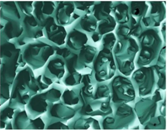
4.1 SCAFFOLDS ... 36
4.2 CHEMICAL AND PHYSICAL SCAFFOLD PROPERTIES ... 44
4.2.1. Porosity ... 49
5. AIM OF THE PROJECT ... 52
6. TESTED SCAFFOLDS ... 55
6.1 PCL MANUFACTURE AND CHARACTERIZATION ... 55
6.2 PCL/HA MANUFACTURE AND CHARACTERIZATION ... 60
7. MICROTOMOGRAPHY (MICRO-CT) ... 66
7.1 MICRO-CT EVALUATION OF SCAFFOLD POROSITY: MATERIALS AND METHODS ... 72
7.2 MICRO-CT SURFACE VISUALIZATION AND QUANTIFICATION OF CELLS ON THE PCL SCAFFOLD: MATERIALS AND METHODS ... 74
7.3 MICRO-CT EVALUATION OF SCAFFOLD POROSITY: RESULTS ... 76
7.4 MICRO-CT SURFACE VISUALIZATION AND QUANTIFICATION OF CELLS ON THE PCL SCAFFOLD: RESULTS ... 81
8. IN VITRO STUDY
... 848.1 MATERIALS AND METHODS ... 84
8.1.1 Cell adhesion and morphology ... 85
8.1.2 Cell proliferation and viability ... 86
8.1.3 Lactate dehydrogenase release (LDH) ... 87
8.1.4 Bone-specific alkaline phosphatase activity (BAP) ... 87
8.1.5 Osteocalcin measurements (OC) ... 87
8.1.6 Type I pro-collagen production (CICP) ... 88
8.1.7 Transforming growth factor β1 release (TGF-β1) ... 88
8.1.8 Tumor necrosis factor α release (TNF-α) ... 89
8.1.9 Interleukin 6 release (IL-6) ... 90
8.1.10 Total protein quantification ... 91
8.2 STATISTICAL ANALYSIS ... 91
8.3 RESULTS ... 92
8.3.1 Cell adhesion and morphology ... 92
8.3.2 Cell proliferation and viability ... 93
8.3.3 Lactate dehydrogenase release (LDH) ... 94
8.3.4 Bone-specific alkaline phosphatase activity (BAP) ... 94
8.3.5 Osteocalcin measurements (OC) ... 95
8.3.6 Type I pro-collagen production (CICP)... 95
8.3.7 Transforming growth factor β1 release (TGF-β1) ... 96
8.3.8 Tumor necrosis factor α release (TNF-α) ... 96
8.3.9 Interleukin 6 release (IL-6) ... 97
9. IN VIVO STUDY
... 989.1 MATERIALS AND METHODS ... 99
9.1.1 Histology and histomorphometry ... 100
9.2 STATISTICAL ANALYSIS ... 102
9.3 RESULTS ... 103
9.3.1 Histology and histomorphometry ... 103
10. DISCUSSION AND CONCLUSIONS ... 110
12. REFERENCES
... 1241
1. SKELETAL SYSTEM
1.1 BONE STRUCTURE
Bone is a dynamic, vascular, living tissue that changes throughout life, is one of the so-called
“connective tissues” of the body and thus comprises cells that become embedded in their own
extracellular matrix. Bone is highly specialized with a complex, hierarchical structure over multiple
levels (Standring S, 2008). Given bone tissue’s ability to adapt its mass and morphology to
functional demands, its ability to repair itself without leaving a scar, and its capacity to rapidly
mobilize mineral stores on metabolic demand, bone is considered the ultimate “smart” material
and a dynamic example of “form follows function” in biological systems. The principal role of the
bone is to provide structural support for the body and while the bone also serves as the body’s
mineral reservoir and producer of blood cells, the mineralized structure is the very basis of
posture, opposes muscular contraction resulting in motion, withstands functional load bearing,
and protects internal organs (Kneser U, et al., 2006).
It is composed of an extracellular matrix (ECM) which has been represented as a two-phases
composite, characterized by an organic phase (35%) composed by 90% of collagenous proteins
(97% collagen I and 3% collagen III, IV and V) and 10% of non collagenous proteins (mainly
osteocalcin, osteonectin, sialoproteins, proteoglycans, osteopontin, fibronectin, Growth Factors
and bone morphogenetic proteins), reinforced with inorganic (65%) component, characterized by
calcium phosphate hydroxyapatite [(Ca10(PO4)6(OH)2)] crystals (HA, TCP) with a Ca:P ratio of 5:3,
sodium (Na), magnesium (Mg) and potassium (K) (Figure 1). Apatite crystals are nanoscale,
2
Fig. 1. Bone extracellular matrix (ECM) composition.
Bone growth factors (GFs) and cytokines influence the synthesis and resorption of bone by acting
on the local cell population present in bone marrow and on bone. They are local regulators of cell
growth, function and angiogenesis. Bone matrix contains a great number of GFs and cytokines
such as, Interleukin I , VI and IX (IL-I, VI, IX), Tumor necrosis factor α (TNF-α), Transforming growth
factor beta supergene family (TGF-β1-5 and Bone Morphogenetic Proteins-BMPs), Platelet-derived
growth factors (PDGFs), insulin-like growth factor I and II (IGF-I and-II), fibroblast growth factors
3
Tab. 1. Cytokine and GF functions and sources in bone.
CYTOKINES AND GFs
BONE FORMATION
BONE
RESORPTION POTENTIAL SOURCES IN BONE
IL-I + +++ Osteoblasts IL-VI ? +/- Osteoblasts IL-IX ++ ++ Osteoblasts TNF-α + +++ Osteoblasts TGF-β and BMPs
++ ++ Osteoblasts, bone matrix
PDGFs ++ ++ Platelets, osteoblasts, bone matrix
IGF-I +++ Osteoblasts, bone matrix
IGF-II +++ Osteoblasts, bone matrix
FGFs +++ Osteoblasts, bone matrix
Histologically (Figure 2), bone is identified as lamellar bone, consisting of lamellae (3-7 μm in
thickness) composed of bone matrix and osteocytes each occupating a cavity (lacuna). Canaliculi
penetrate the lamellae of adjacent lacunae. The osteon or Haversian system is formed by
arranged lamellae around a longitudinal vascular channel. The osteons are parallel to one another
in skeletrical districts where tension forces prevail. Vascular anastomosis are present between
vascular channels of adjacent osteons, as branches that run perpendicular to Haversian channels
(Volkmann channels) (Laz PJ, et al., 2007).
4
Fig.2. Bone structure. (Image from http://training.seer.cancer.gov/anatomy/skeletal/tissue.html).
Bone tissue is arranged in two macroarchitectural forms-cortical (or compact) and cancellous (or
trabecular, or spongy)- which are employed in various proportions and geometries to form the
individual bones of the body (Figure 3). The latter can be broadly subdivided into four groups: long
bones (femur, tibia, fibula, humerus, radius, and ulna); short bones such as those of the carpus
(wrist) and tarsus (ankle); flat bones such as those of the calvaria (skull vault); and irregular bones
(the remaining bones of the skull, the scapula, and pelvic bones).
In cortical bone, densely packed collagen fibrils form concentric lamellae, and the fibrils in
adjacent lamellae run in perpendicular planes as in plywood, whereas, cancellous bone has a
loosely organized, porous matrix. Cortical bone is almost solid with less than 10% of porosity, in
contrast, cancellous bone is organized in a porous sponge-like pattern. Cortical bone has loading
fibers orientated along the loading axis with highly organized structures such as the Haversian
system. Cancellous bone has a cortex of cortical tissue consisting of longitudinally oriented
5
has greater apparent modulus of elasticity, tensile strength, and fracture toughness than
cancellous bone. However, owing to the highly porous structure (cancellous bone 75-90%; cortical
bone 5-10%), relative properties of the ECM are similar (Davies JE, 2003). In general, strength of
bone depends on the hardness of the compact cortical bone and on the underlying scaffolding
effect of the trabeculae of cancellous bone.
The cortical bone has evolved to withstand torsional loading, while the function of cancellous
bone is to withstand predominantly compressive loading.
About 3-5 % of one’s skeleton is being eaten away by osteoclasts and replaced by osteoblasts, throughout life from six weeks in utero, when bone first forms, until we die. This constant remodeling of bone tissue provides a mechanism for scar-free healing and regeneration of damaged bone tissue, and results in the exquisite lamellar microarchitecture of both cortical and trabecular mature bone.
From this perspective, therefore, trabecular bone represents a biologically superior tissue, ideally evolved for rapid bone healing, when compared to the slowly remodeling healing pattern typical of cortical bone. On the other hand, cancellous bone is finer and more delicate in appearance. Its
physical arrangement of broad plates, connected by thin struts, provides for maximum support
but with a minimum of raw material. The metabolic activity of cancellous bone is thought to be
considerably higher than that of cortical bone, with the result that diseases, which are caused by
aberrations of metabolism, are invariably seen in the cancellous compartment first.
Cancellous bone has a very high surface area, which is contiguous with the marrow compartment. Since marrow contains not only mesenchymal progenitor cells, that can give rise to osteoblasts, but also a rich vasculature that can supply both the circulating mononuclear precursors to osteoclasts (needed for remodeling) and the endothelial population needed for angiogenesis, it is
6
not surprising that trabecular bone can remodel more quickly than cortical bone (Henriksen K, et al., 2009).
Fig.3. Bone structure. a) Cancellous and b) cortical bone. Toluidine blue, fast green staining. Magnification 10x. (Image
from Laboratory of Preclinical and Surgical Studies-IOR).
The entire surface of bone, except where articular cartilage is present, is covered by specialized
dense connective tissue known as periosteum. This layer is attached to the cortical bone below by
a series of collagenous bundles known as Sharpey fibers and the strength of these attachments
varies between different bones. The internal surface of bone, which includes the medullary cavity,
cavities of the haversian system of compact bones and the trabeculae of cancellous bone, is lined
7
1.2 BONE CELLS
Bone is composed of four different cell types: osteoblasts, osteocytes, osteoclasts and
osteoprogenitor cells.
1.2.1 Osteoblasts
Osteoblasts (Figure 4) are epithelial-like cells that are fully differentiated and polarized cells
(20-30µm of diameter) within bone and derived by the differentiation of osteoprogenitor cells.
Fig.4. Osteoblasts. Scanning electron Microscopy (SEM). Magnification 1950x. (Image from laboratory of Preclinical
and Surgical Studies-IOR).
They have a spherical or ovoid nucleus, lay down the extracellular matrix and regulate
mineralization, depositing osteoid, nonmineralized organic matrix and initiating the subsequent
mineralization of the osteoid. They secrete the type I Collagen and the non collagenous proteins of
the bone matrix such as osteocalcin, osteopontin, bone sialoproteins and GFs such as BMPs.
Morphologically, they are cuboidal or columnar in shape and located at the bone surface together
with their precursors, where they form a tight layer of cells. The lifespan of an osteoblast ranges
8
µm osteoid per day. Eventually, some osteoblasts may become trapped in their own calcified
matrix, changing their phenotype and developing into osteocytes. In fact, when bone formation is
completed, osteoblasts flatten out and transform into osteocytes (Henriksen K, et al., 2009).
1.2.2 Osteocytes
Osteocytes (Figure 5) are the most abundant resident cells (>90%) in bone tissue. They are
regularly spaced in lacunae throughout the calcified bone matrix and communicate with each
other as well as with the cells on bone surface, osteoblasts and osteoprogenitor cells, through a
large number of cytoplasmic processes running in bone canaliculi. Osteocytes are no longer
involved in bone formation. Due to their inaccessibility and difficulty in cell culture, osteocytes
have not received enough attention and little is known about their function for a long time.
However, in recent years, these mysterious cells shed new light on their pivotal function in
regulation of new bone formation and remodeling (Huang CP, et al., 2008; Takai E, et al., 2004).
The large population, ideal location and unique morphologies make osteocytes ideal candidates
for detection of external stimulations and generation of signals that affect osteoblasts, osteoclasts
and their progenitors in the bone marrow. An analogy might be made between the bone cells
network and neuronal system.
Osteocytes may play the role as the ‘‘brain center’’, and their principal function is thought to be in
intercellular signaling by the extensive cellular network (Huang CP, et al., 2008).
Osteocytes form extensive interconnected 3D cellular networks that position them to be suitable
sensors of changes in the local mechanical and hormonal environment in bone tissue.
Mechanical stimuli such as hydrostatic pressure, fluid shear, and load-induced strain have been
shown to elicit a response from osteocytes both in vitro and in vivo. Unlike other in vitro
9
flow-induced effects (e.g., shear stress, streaming potentials, nutrient convection), and fluid shear
stress, which is driven by a pressure gradient, hydrostatic pressure can be applied uniformly to all
cells without other modes of mechanical stimulation (Warden SJ, 2006).
Osteocytes are not capable of division and are lost when the bone in which they reside is
degraded, so the osteocyte’s lifespan is dictated by the lifespan of the bone and they can remain
alive for years provided vascularization is continuous.
Fig.5. Osteocytes in lacunae. Scanning electron Microscopy (SEM). Magnification 1950x. (Image from laboratory of Preclinical and Surgical Studies-IOR).
1.2.3 Osteoclasts
Osteoclasts (Figure 6) are large (from 20 to 100µm in diameter), highly migratory and
multinucleated cells that resorb bone and when active, they rest directly on the bone surface and
have two plasma membrane specializations. They are highly polarized cells that occupy the
Howship’s lacuna.
The main feature of osteoclasts is their ability to resorb fully mineralized bone at Howship’s
lacunae. Both macrophages and osteoclasts are derived from hematopoietic stem cells
10
lysosomal enzymes. A resorption bay is formed underneath the cell into which the lytic enzymes
are secreted. In addition, proton pumps lower the pH in this subosteoclastic space to values
between 2 and 4, activating the secreted enzymes such as tartrate-resistant acid phosphatase.
Osteoclasts are transiently active in response to a metabolic demand for the mobilization of
calcium from bone into blood. An activated osteoclast, with an average lifespan of 15-20 days, is
able to resorb 200,00 µm3/day, an amount of bone formed by seven to ten generations of osteoblasts (Standring S, 2008).
Fig.6. Osteoclast. Scanning electron Microscopy (SEM). Magnification 1950x. (Image from laboratory of Preclinical and
Surgical Studies-IOR).
1.2.4 Osteoprogenitor cells
The regenerative potential of our body depends on specialized stem cells, characterized by their
functions to differentiate into different cell lineages and to self-renew for the maintenance of the
stem cell pool. Since the late 19th century, bone marrow (BM) has been known to host mesenchymal stem cells (MSCs), also called osteoprogenitor cells or bone-lining cells, which are
able to differentiate into multiple mesodermal tissues such as bone, cartilage, adipose, muscle,
11
These cells have a direct role in the maintenance of bone balance. They act as a source of
progenitors for osteoblasts and moreover, constitutively secrete a distinct set of cytokines,
sug-gesting that they serve specific supportive functions in the microenvironment of BM. MSCs are the
cells competent for bone tissue maintenance and regeneration: their self-renewal and
differen-tiating properties in an osteogenic lineage make them very useful tools in tissue engineering and
regenerative medicine. Osteoprogenitor cells differentiation toward a specific lineage is
dependent on hormonal and local factors, commonly called the microenvironment (e.g., leptin,
cell-cell-signaling, extracellular matrix, and cytokines), which can activate specific transcription
factors. Although it has been reported their use as a part of bone grafting procedures, that have a
clinical potential, many clinical variables (e.g., the anatomic site of BM harvest, recent trauma,
local or systemic bone diseases, menopausal status, use of tobacco or pharmaceutical agents, age
and gender) might contribute to differences in MSC samples.
Fig. 7. Osteoprogenitor cells. Scanning electron Microscopy (SEM). Magnification 1950x. (Image from laboratory of Preclinical
12
Osteoprogenitor cells persist throughout postnatal life as bone-lining cells and they are
reactivated in the adult, during the repair of bone fractures and other injuries (Veronesi F, et al.,
2011).
1.3 BONE MODELING AND REMODELING
Already in 1892, Wolff found that the orientation of trabeculae coincides with the direction of the
stress trajectories. He proposed that bone loading is somehow sensed and that the bone adapts its
structure accordingly. This principle of functional adaptation is generally known as “Wolff’s Law”
(Wolff J, 1892). The ability of bone to adapt to mechanical loads is brought about by continuous
bone resorption and bone formation. If these processes occur at different locations, the bone
morphology is altered. Frost defined this as modeling (Frost HM, 2001).
In a homeostatic equilibrium, resorption and formation are balanced and, in that case, old bone is
continuously replaced by new tissue. This ensures that the mechanical integrity of the bone is
maintained but it causes no global changes in morphology. Frost defined this as remodeling (Frost
HM, 2001). The modeling and remodeling processes are not very different at the cellular level.
They are based on the separate actions of bone resorbing cells (osteoclasts) and bone forming
cells (osteoblasts). Approximately 10% of the skeleton is renewed each year by remodeling. This
process is necessary to repair skeletal microfractures, to prevent accumulation of older and
weaker bone, and to maintain mineral homeostasis (Fini M, et al., 2010).
This process is complex and characterized by the coordinated actions of osteoclasts and
osteoblasts, organized in basic multicellular units (BMU) that follow a cycle of resorption, reversal,
formation, and resting phases (Figure 8). Remodeling begins with the migration of partially
13
osteoclasts. The signal that initiates the resorption phase is thought to be the mechanical stress
that alters local bone architecture and is sensed and transduced by osteocytes. After osteoclastic
resorption, which takes about 3 weeks, there is a reversal phase when mononuclear cells on the
bone surface provide signals for osteoblast differentiation and migration and this phase lasts up to
4-5 weeks. In the formation phase, osteoblasts lay down bone until the resorbed bone is
completely replaced by new bone; this phase may last for about 3 months. Eventually, bone
surface is covered with flattened lining cells and a resting period follows. This dynamic process
occurs in BMUs at multiple sites simultaneously throughout the skeleton (Gallagher CJ, 2008).
14
1.4 BONE HEALING
A fracture or a bone defect means that the continuity of a bone is disrupted. During the course of
healing, different types of tissues can be seen in a lesion area, whereby one substitutes the other
(Figure 9).
The striking feature of bone healing, compared to healing in other tissues, is that repair doesn’t
create a scar tissue. This is linked to the capacity for remodeling which intact bone possesses.
The healing of bone fracture and defect has traditionally been described in four phases.
1) Haematoma formation (inflammation or granulation) phase
Initially, a hematoma is found between the fragment ends. The hematoma might well act as a
guiding structure which, as a spacer, determines the size and shape of the callus.
Activated platelets release a variety of products, including fibronectin, PDGF and TGF-β, which
trigger the influx of inflammatory cells. The subsequent cytokine cascade brings the cells of repair
(fibroblasts, endothelial cells and osteoblasts) into the fracture gap.
2) Soft callus formation (proliferative) phase.
This is characterised by the formation of connective tissues, including cartilage, and formation of
new capillaries from pre-existing vessels (angiogenesis). In detail, during the first few days the
hematoma changes to become granulation tissue. Capillary sprouts, mononuclear cells,
fibroblasts, and fibrocytes are present. During the process of healing, a maturation of this
granulation tissue is observed and it is transformed into connective tissue with its collagen fibers.
This maturation results in an increase in stiffness.
3) Hard callus formation (maturing or modeling) phase.
This phase leads to woven bone, either directly from mesenchymal tissue (intramembranous) or
15
woven bone rapidly, but it is randomly arranged and mechanically weak. During this step, in both
fibrous tissue and fibrocartilage, mineralization occurs.
4) Remodeling phase.
Woven bone is remodeled into stronger lamellar bone by the orchestrated action of osteoclast
bone resorption and osteoblast bone formation. In this phase the mineralization is followed by
local resorption of the mineralized cartilage, whereby new vessels enter the fracture area. The
walls of the resorption spaces are then covered with lamellar bone, thus forming new bone
trabeculae. A further bone deposition, combined with local resorption, leads to a reshaping of
those trabeculae (Marsh DR and Li G, 1999).
Thus, a gradual increase in strength and stiffness occurs, along with a reduction of the ability to
elongate (Dimitriou R, et al., 2005; Gerstenfeld LC, et al., 2003).
In terms of extracellular matrix formation, type III collagen predominates at the inflammatory
stage, followed by type II collagen in the cartilaginous phase and type I collagen production at the
ossification and remodeling stages.
16
Bone regeneration is a highly efficient and tightly regulated process that involves all the
above-mentioned components of bone tissue. Bone regeneration is the result of a continuous interplay
between GFs and cytokines for both initiation and regulation of the remodeling process. The
majority of fractures heals well under standard conservative or surgical therapy. However,
extended bone defects, following trauma or cancer resection or non-unions of fractures, may
require more sophisticated treatments. In these cases bone grafting procedures, segmental bone
transport, distraction osteogenesis or biomaterials are applied for reconstruction (Kneser U, et al.,
2006).
1.4.1 Bone healing complications
Bone tissue, usually, has the ability to repair itself, but when complicated fractures and large bone
defects have to be bridged, the healing process fails in many cases. Despite the advent of modern
techniques, individual fractures occasionally refuse to heal and a non-union or pseudoarthrosis
can occur. Insufficient blood supply and infections of the callus or the surrounding tissue or even
systemic diseases can have further negative effects on bone regeneration, resulting in the
formation of a non-union (Janicki P and Schmidmaier G, 2011).
The healing process may be classified into 4 separate entities: union, malunion, non-union, and
pseudoarthrosis. Union is used to described a fracture that has united; in malunion the fracture
has united but the union is incorrectly aligned; non-union occurs when the apposed ends of the
fracture have failed to unite and to ossify. The amount of healing, that has occurred, may vary
from a cartilaginous bridge, which has failed to ossify, to fibrous-connective tissue, or to an
absolute lack of bridging. Non-unions can also occur after the bone resection caused by cysts,
(Sumner-17
Smith G, 2002). Pseudoarthrosis indicates a true false-joint formation when bone needs are joined
by a fibrous joint-capsule-like structure, which contains synovial-like fluid originating from local
serum production.
Nearly 5-10% of all fractures is associated with impaired healing, resulting in delayed union or
non-union.
Bone defects are very challenging in orthopaedic practice; they can result from a high-energy
traumatic event, from large bone resection for different pathologies such as tumor or infection, or
from the treatment of complex non-unions (en-bloc resection). They can be considered critical in
relation to the skeletal segment involved and the length of bone loss: 3 cm for the forearm, 5 cm
in the femur and tibia, 6 cm in the humerus (Janicki P and Schmidmaier G, 2011).
The main clinical pathologies that could require a graft or a biomaterial implantation are, as well
as the above mentioned non-unions and pseudoarthrosis, the bone fractures with loss of
substance. For these defects, there is a constant demand for substitutes which can effectively
bridge long segmental defect or fill loss of substances with minimal morbidity and accelerating
healing times. Tissue regeneration has become a great challenge for many illness, traumatic
lesions or reconstructive needs (Fini M, et al., 2005).
The amount of repair, however, depends on the size of the bone defect, the lesion site and the
patient’s health status, the age and lifestyle. For critical size defects (would not fully heal
spontaneously) and for the above mentioned defects, or even to accelerate or geode the repair
process, the use of bioactive scaffolding materials could be of great advantage. The use of
bone-filling materials with the properties of adsorption, osteopromotion, safety and ease of sterilizing,
manufacture and surgical use may improve the healing rate, making the difference between
18
Nevertheless, it is still a great challenge in reconstructive surgery to treat large bone defects or
non-unions. The therapy concept, based on bone substitutes, includes the possibility to rebuild
bone lacking structure and to allow migration, proliferation and differentiation of bone cells and to
promote vascularisation (Nandi SK, et al., 2010).
Approximately 2.2 million bone graft procedures are performed each year worldwide to repair
bone defects in orthopaedics, neurosurgery and oral and maxillofacial surgery with a yearly
19
2. BONE GRAFTS
A tissue graft is a medical procedure in which tissue from a donor is used to replace missing or
damaged tissue on a patient. Numerous types of human tissue can be used including veins, skin,
tendons, bone, and ocular materials. In general, tissue grafting can be divided into 3 major
categories according to the genetic relationship between the donor and the recipient. They are a)
autogenous tissue graft (autograft): a tissue graft from one site to another within the same
individual; b) allogenous tissue graft (allograft): a tissue graft between individuals of the same
species; d) xenogenous tissue graft (xenograft): donor and recipient are individuals from different
species (Delloye C, et al., 2003).
In orthopaedic field, regarding bone healing, bone grafts can be osteogenic, osteoinductive and
osteoconductive.
A graft that supplies and supports bone forming cells is termed osteogenic.
Osteoinductive agents (proteins) induce proliferation or differentiation of undifferentiated stem
cells to osteogenic cells. A graft that has the capacity to induce bone formation when placed into a
site where no bone formation will occur is termed osteoinductive. Osteoconduction is the process
whereby scaffolding is provided for inward migration of cellular elements involved in bone
formation (mesenchymal cells, osteoblasts, osteoclasts and vasculature) (Kneser U, et al., 2006).
Autogenous bone graft is the transplantation of bone taken from one anatomic site to another
site in the same individual. It is defined as the 'gold standard' for regeneration, is a safe solution for compatibility and the absence of immune response, and it is considered to be the most suitable material, because the graft has osteogenic (marrow-derived osteoblastic cells as well as
preosteoblastic precursor cells), osteoinductive (noncollagenous bone matrix proteins, including
20
Moreover, the differences in histocompatibility and the risk of transferring a disease are not
existent. On the other hand, the autograft drawbacks include: an insufficient amount of graft
material necessary to replace the missing portion, particularly in children or in bone loss due to tumors; a significant postsurgical morbidity at the donor site; increased surgical time and blood
loss and an additional cost due to a possible prolongation of the hospitalization. The different
types of autogenous bone grafts are represented by autologous cancellous bone, autologous
cortical bone and vascularized autografts. Autologous cancellous bone plays a dominant role in
bone-transplant surgery because osteogenic stem cells can survive in an autogenic spongiosa and
it has a clinical success in the treatment of a wide range of diseases, such as delayed fracture
healing, pseudoarthrosis, bone defects and infections. It has a robust biological activity in inducing
and producing new bone (Sen MK and Miclau T, 2007). It has been considered more osteogenic as
compared to cortical bone graft because the presence of spaces within its structure, allowing the
diffusion of nutrients and limited revascularization, but it does not provide substantial structural
support. The principle advantage of autologous cancellous grafts is the potential to transfer
osteoprogenitor cells combined with matrix and signals, provided by the cancellous particles,
yielding a mixture that contains other factors of bone regenerative strategy.
Another example of an osteogenic material is bone marrow. Bone marrow aspirates contain MSC,
cells already committed to osteogenic or chondrogenic lineage, and some biologically active
proteins that stimulate bone regeneration in similar manner to naturally occurring fracture clot,
many from platelet degranulation. However, use of bone marrow does not yield the magnitude of
osteogenesis observed with cancellous bone for several reasons. Aspiration results in varying
21
low and most importantly, bone marrow lacks the necessary scaffold or osteoconductive material
to be efficacious on its own (Kolk A, et al., 2012).
Autologous cortical bone graft has little or no osteoinductive properties, is mostly
osteoconductive, and the surviving osteoblasts provide some osteogenic properties as well. It is
much less important, for bone regeneration, than cancellous bone due to its poor vascularity and
its low content of osteogenic stem cells. Thus, the only role for free cortical autografts in bone
regeneration is the temporary increase in mechanical stability (Beaman FD, et al., 2006).
Autologous cortical bone grafts are good choices for segmental defects of bone of 5 to 6 cm,
which require immediate structural support.
Vascularized cortical autograft heals rapidly at the host-graft-interface, and its remodeling is
similar to that of normal bone. It improves osteocyte survival and enhances bony incorporation. It
appears to demonstrate less bony necrosis with no subsequent trabecular collapse and no
architectural disorganization, when compared with the non-vascularized grafts (Kawamura K, et
al., 2008).
The limitation associated with the procurement of autograft for bone grafting and the
development of modern tissue banks have resulted in a supply of high-quality allogenic tissue for
reconstructive orthopaedic surgery. Allograft overcomes the limitations associated with the
procurement of autografts for bone grafting. It may be derived from cadaveric bone sources or
from living donors harvested during hip arthroplasty, and has both osteoinductive (they release
bone morphogenic proteins that act on bone cells) and osteoconductive properties, but it lacks
osteogenic properties because of the absence of viable cells. Beside limitations in the essential
bone graft characteristics, there is an ongoing controversial discussion about the association of
22
immunodeficiency virus (HIV), hepatitis B and C viral infection (HBV/HCV), malignancies, systemic
disorders (autoimmune disease), or toxins. Removal of osteoarthritic femoral heads throughout
hip arthroplasty shows an 8% evidence of diseases not previously known. Elimination of this major
concern of allogenic material requires tissue-processing, sterilization and a deactivation process of
proteins in the extracellular matrix which contains bone growth factors, proteins, and other
bioactive substances necessary for osteoinduction and, ultimately, successful bone healing.
Although the risk of transmission of disease is much lower than with blood products, it is still
possible. The more aggressive the allograft processing, the less intense immunologic responses
will occur, but this results in a decrease of the osteoinductive properties. For this reason fresh
allografts are clinically no longer used. Frozen allografts induce stronger immune responses than
freeze-dried allografts (Habibovic P and de Groot K, 2007).
Compared with autologous cancellous bone, allograft cancellous bone is a poor promoter of bone
healing. Allogenic cortical bone incorporation occurs by sporadic formation of new appositional
bone and the lack of vascularization leads to weakness of the graft. It is used in replacing segments
of bone after tumor excision, in lengthening bones and in treating selected non-unions.
Massive osteochondral allografts (diaphyseal cortical bone, metaphyseal cancellous bone and
articular cartilage) are used in joint reconstruction after limb salvage procedures for tumor
resection and in traumatic bone loss. Some complications are represented by allograft fracture,
articular cartilage degeneration, osteoarthritis, deep infections and non-unions.
Xenograft is a tissue graft between two different species and despite its wide availability, the
untreated xenogenic bone is an unsuitable transplant material because it provokes a strong
immunodefensive reactions. Porous natural hydroxyapatite (HA) can be obtained from animal
23
concerning absorption (i.e. dependent on the porosity value, crystallinity, crystal structure, etc.)
(Kolk A, et al., 2012).
In the last few years, Demineralized Bone Matrix (DBM) has attracted attention as bone graft.
DBM consists of sponge-like collagen from human, bovine or equine origin that has undergone
decalcification and sterilization, so it can be classified as allogenic or xenogenic material
dependent on the origin. The trabecular structure of the original tissue remains, therefore
maintaining its biological structure. DBM has been shown to have osteoconductive and
osteoinductive properties, but it provides no structural stability and therefore should only be
applied in a structurally stable environment (Naujoks C, et al., 2011).
It is mainly used as a ‘‘bone graft extender’’, revascularizes quickly and acts as suitable carrier for
autologous bone marrow, it does not evoke any appreciable local foreign body reaction as the
antigenic surface structure of the bone is destroyed during demineralization. More growth factors
are available after the removal of bone minerals, so the bone inductance of DBM is higher than
that associated with mineralized allogenic transplants. DBM is a derivative of allograft bone and it
could be prepared by pulverization of allogenic bone to a consistent size, followed by mild acid
extraction of the mineralized phase of bone. This process, principally developed by Urist et al in
1965, results in a composite of non-collagenous proteins, growth factors, and collagen (Urist MR
and Dawson E, 1981).
Nowadays, bone graft materials with completely different origins are commercially available for
many applications throughout the human body. They are variable in their composition, their
mechanism of action and, therefore, their indications. In conclusion, problems related to the
availability of graft material, donor-site morbidity, immunogenicity and biomechanical integrity
24
Bone substitute materials are generally considered to be a highly important alternative to bone
grafting.
Due to the numerous disadvantages present in all kinds of grafts, there is a rationale for the
designing and developing of artificial supports (scaffolds) for tissue engineering applications and
their demand is growing steadily. The alternative to tissue grafting is the use of artificial designed
scaffolds or implants, fabricated using various materials (biomaterials). Nowadays, materials,
including metals, polymers, ceramics and composites, are used clinically for implants and medical
25
3. BIOMATERIALS
The term “Biomaterial” can be defined as “a material intended to interface with biological systems
to evaluate, treat, augment or replace any tissue, organ or function of the body”. This is still a
good conceptual definition, but should be modified to take into account the extended applications
of biomaterials. Moreover, the variety and complexity of these applications is such that a
classification of biomaterials might be considered. A slightly modified definition of biomaterial is
proposed as follows: “ a material intended to interface with biological systems as an integral part
of a process designed to evaluate, monitor or treat tissues of the body, to replace or augment
tissues or to facilitate the regeneration of tissues” (ESB satellite Consensus Conference, Sorrento,
September 2005).
Current requirements for an ideal biomaterial as bone substitute are rigorous as listed below:
- Resorbability/degradability and sterility
- Biocompatibility
- Osteoinduction, osteoconduction, osseointegration
Resorption of the material and replacement by normal bone are either biologically based on the
influence of cells or by chemical-physical dissolving processes, and should occur simultaneously in
the ideal case. The biomaterial should be easy to use, should withstand sterilization and should
come in sufficient quantities. In a time of global economic downturns, costs are an important issue
26
3.1 BIOCOMPATIBILITY
Biocompatibility ensures the absence of toxicity, teratogenicity or carcinogenicity. The lack of
antigenicity guarantees the avoidance of pro-inflammatory and immunogenic reactions. All such
requirements serve as a basis for effective long-term tolerance and such criteria are mainly
fulfilled by available synthetic materials (Williams DF, 2008).
Biocompatibility is a fundamental requirement of biomaterials and is defined as “the ability of a
material to perform with an appropriate host response in a specific application” (Williams DF,
1987). It is the essential requisite for the employment of a material in the biomedical field, is
strictly related to the specific application and location of the biomedical device and can be defined
as its substantial property of not alter the physiologic environment and not cause pathological
outcomes. Since at the present time no absolutely inert materials exist, so it is probably more
correct to describe biocompatibility as biotolerability. In 1995 Wintermantel and Mayer modified
the definition of biocompatibility into surface and structural compatibility of implants, involving
appropriate chemical, biological and physical characteristics, including surface morphology (for cell
anchorage and guidance) (Wintermantel E and Mayer J, 1995).
The assessment of biocompatibility is a mandatory prerequisite for all materials and devices
before their clinical application. The evaluation of the biocompatibility of a material, assigned to
clinical use, is a long trial that requires a well planned sequence of steps. The assessment of
biocompatibility of a particular biomaterial entails the consecutive execution of well accepted
scientific evaluations. Considered together, the results of these evaluations help to provide an
objective picture of the associated composite biocompatibility and in general, the sequence of
27
cytotoxicity evaluation to in vivo large animal anatomically relevant evaluations of biocompatibility
and biofunctionality.
These procedures are now standardized at an international level, in particular in the European
Community, the rules of EN ISO 10993 provide the guide-lines for each investigation in this field.
The International Organization for Standardization (ISO) is a worldwide federation of national
standards bodies for the biological evaluation of medical devices. ISO 10993 refers to the
fundamental principles governing the biological evaluation of medical devices, the definition of
categories of devices based on the nature and duration of the contact with the body, and the
selection of appropriate tests to be performed. Biocompatibility tests must be performed on the
final product or material, taking into account the type, duration and conditions of the exposure in
the human body, the physical and chemical features of the product, the toxicological activity of
the chemical elements or compounds and the presence of leachable materials. It is not required
that a material or device will be submitted to all the tests, but only to those related to the specific
contact with the body environment (Gatti AM and Knowles JC, 2002).
The capability of a biomaterial to perform a specific function cannot be evaluated by single in vitro
or single in vivo methodology. In vitro methods provide necessary and useful results that precede
and complete the in vivo testing. In vitro tests decrease the number of animals required for in vivo
biocompatibility evaluation, while in vivo evaluations allow long-term investigations, by
reproducing the complex biological environment and are suitable for the evaluation of
biofunctionality.
A research project that adopts only an in vitro or an in vivo method is considered limited and a
28
performed only in vivo with no preliminary in vitro information on the biomaterial biocompatibility
(Fini M and Giardino R, 2003).
In vitro cell cultures. The in vitro evaluation provides the initial biocompatibility screening and it is
represented by target cell assays, making use of the most sensitive cells pertinent to the
application for which the biomaterial is developed. Table.2 shows the benefits and limits of in vitro
studies and some relevant negative aspects of the in vitro screening are represented by the
minimization of the variables of metabolism, distribution, and adsorption, and the maximization of
the cell line exposure to any potential toxicity. Moreover, there is difficulty to extrapolate the
results to the clinical population. Consequently, an in vivo evaluation must be conducted.
The impossibility of predicting, in vitro, the effect of biological fluids on biomaterials, which is
responsible for the side-effects, as well as long-term prosthetic device behaviour, represents a real
drawback of cell culture systems. On the other hand in vitro tests provide detailed information on
particulate debris toxicity when identified.
Osteosarcoma and immortalized cell lines are used, as well as primary cells from several species
and anatomical locations. If human-derived primary cells are employed, they usually come from
the surgical residues obtained during surgery (i.e. bone fragments unusable for tissue banking).
Human osteosarcoma cell lines show the absence of individual variability observed when primary
lines are used, as well as better repeatability and reproducibility.
In vitro test sensitivity, in discriminating between safe and toxic biomaterials, is of great
importance in rejecting inappropriate materials prior to the in vivo implantation. It is recognized
that in vitro tests can be limited due to their relative simplicity when compared to the complex
interactions occurring in an organism in toto, because of the lack of systemic and local factors due
29
In vivo animal models. Animal models provide important biomaterial knowledge that leads to the
development of more effective clinical treatments of diseases in both humans and animals, acting
as a bridge between in vitro studies and clinical trials. Depending on the purpose of the study,
biomaterials are usually implanted in the diaphyseal (cortical bone and medullar cavity),
metaphyseal or epiphyseal part of long bones (cancellous bone). Common bone implant tests are
histologic, histomorphometric and biomechanical investigations that can provide a complete
characterization of the osteogenic and osteointegration properties of the material. Performing in
vivo studies requires, first of all, compliance to ethical and legal rules on animal experimentation.
Every Country has its own regulations, but a regional local ethical committee exists to approve
every research protocol. Biomedical research, using animals, is regulated worldwide by precise
rules. In the United States, the Animal Welfare Act dated from 1966 and this was followed in 1986
by the Public Health Service Policy on Care and Use of Laboratory Animals. All of these documents
contain guidelines about the animal environmental, housing and management as well as
veterinary care. In the European Union, the Directive 86/609_Protection of Experimental Animals
gives, to the state members of the European Community, indications to be applied in the
regulation of animal research. According to this directive, the Italian Government, with the Law by
decree number 116 of January 27,1992 on the protection of animals used for scientific purpose,
settles the guidelines for this matter. Considering the biological evaluation of medical devices, part
2 of ISO 10993 “Animal Welfare Requirements” contains the directives to be followed when
animal tests are to be performed (Andersson M, et al., 2007; Andersson M, et al., 2008).
The tests are justified only when the resulting data are not otherwise available and essential for
the material characterization, when no other scientifically validated method not involving animals
30
identified and implemented. Animal tests must be carried out in authorized laboratories, with
appropriate facilities for animal stabling and welfare and must not be conducted before
appropriate preliminary in vitro tests have been carried out with favorable results. Surgical
procedures must be performed in general anaesthesia and in well equipped operatory rooms,
under the control and responsibility of well trained scientists. In vivo tests for biocompatibility may
use the biomaterials themselves or may utilize extracts of them to determine the toxicity and
bioactivity of potential leachable, dissolution products, or other materials utilized in the synthesis
and production of the biomaterial. Important limits of an in vivo study (Table 2) include the
presence of differences that are so great between humans and animals, data on animal bone
remodeling and formation that cannot be representative of bone healing in humans, the need for
appropriate facilities and complex professional expertise, prolonged time and high costs (Buma P,
et al., 2004).
Tab.2. Benefits and limits of in vitro and in vivo studies.
BENEFITS
LIMITS
IN VITRO STUDIES
High sensitivity; Standard experimental conditions; Human cells and tissues; No ethical drawbacks; Easy to perform, manage, interpret; Fast and inexpensive screening; Reduction of animal use and suffering.
Specific sensitivity; Deposition of metabolic and toxic products (in static cultures); Partial information on biofunctionality and mechanical osteointegration; Lack of systemic factors; Difficulty in long-term studies.
IN VIVO STUDIES
Long-term investigations; Biofunctionality and effectiveness of the final prosthetic device; Presence of systemic factors; Biological environment.
Ethical drawbacks; Legal authorization requirements; Appropriate facilities; Complex professional expertise; Prolonged time, high costs; Differences between humans and animals.
31
3.2 OSTEOINDUCTION, OSTEOCONDUCTION AND OSSEOINTEGRATION
Osteoinduction is the ability to actively stimulate or promote bone formation. Osteoinductive
biomaterials provide a biological stimulus for induction, recruitment, stimulation and
differentiation of primitive, undifferentiated and pluripotent stromal cells into osteoblasts or
preosteoblasts, the initial cellular phase of a bone-forming lineage (Calori GM, et al., 2011).
Osteoconduction is defined as the process of bony ingrowth from local osseous tissue onto
surfaces. Osteoconductivity is a property of the biomaterial that allows the colonization and
ingrowth of new bone cells and sprouting capillaries due to its three-dimensional structure. The
original definition is not strictly restricted to biomaterials, however, the contemporary concept of
an osteoconductive material is one where bone formation is promoted to appose and conform to
its surface, when the material is placed into bone, by virtue of its composition, shape or surface
texture. Osteoconduction is mainly determined by the porosity properties of the biomaterials and
also, in a lesser extent, by the chemical and physical properties of the substrate that promote cell
adhesion and growth. Osteoconductivity is, by definition, a passive process (LeGeros RZ, 2002).
For some biomaterials and dependent on implantation site, osseointegration is the process of
achieving stable direct anchorage and contact between bone and biomaterials and it was firstly
described by Branemark in 1977 and defined by Albrektsson in 1981 (Branemark PI, et al., 1977;
Albrektsson T, et al., 1981). At an histological level it is defined as the direct anchorage of a
biomaterial by formation of bony tissue around the implant without the growth of fibrous tissue at
the bone-implant interface. A biomechanical definition has also evolved stating that
osseointegration is “a process whereby clinically asymptomatic rigid fixation of alloplastic
materials is achieved and maintained, in bone, during functional loading” (Zarb G and Albrektsson
32
Osteoinduction, osteoconduction and osseointegration are powerful inter-related phenomena in
bone regeneration and intrinsically related to bone morphogenetic proteins, bone growth factors
and direct bone anchorage factors, respectively.
To date, no bone substitute biomaterial is available that is equal to autogenous bone, and current
biomaterials serve primarily as filling and frame building materials, mostly providing
osteoconductivity for the bone healing process.
Ideally, the healing process of the bone defects should result in regenerated and vital bone,
without foreign bodies (Horch HH, et al., 2006).
33
4. TISSUE ENGINEERING
During the 1st Tissue Engineering Symposium (California 1988), tissue engineering was defined, for the first time, as “application of the principles and methods of engineering and life sciences
towards the fundamental and pathological mammalian tissues and the development of biological
substitutes that restore, maintain or improve tissue function”(Fox CF and Skalak R, 1988). It was considered the point that best marked the transition to a new era of research. This large area confronts and engages different sciences such as engineering, chemistry, physics, biology, biotechnology and medicine in a multi-disciplinary approach for the development of functional tissues and organs in vitro, for their implantation in vivo or for direct remodeling and regeneration
of tissue in vivo to repair, replace, preserve or enhance tissue or organ functions lost due to
disease, injury or aging (Ingber DE, et.al,2006).
The new therapeutic strategy consists in the use of tissue-engineered living cells (and / or their products) and innovative supports, to develop bioactive tissue substitutes as an alternative to inert systems and it is clear that the complexity of biological tissues in terms of macromolecular composition, organization and ultrastructural interactions between cells and the environment, makes it difficult the passage of engineered constructs from the experimental field to the clinic.
Tissue engineering offers the potential to eliminate re-operation and to solve implant rejection,
transmission of diseases and shortage in organ donations by using biological substitutes, providing
long term solutions in tissue repair or treatment of diseases, and offering treatments for medical
conditions that are untreatable (McIntire LV, 2003). So, aided by advances in biological and
medical sciences, materials science and engineering, tissue engineering has triumphed in recent
decades by providing various implant biomaterials for human tissue repair for millions of patients
34
components for tissue engineering. Tissue engineering typically uses artificial extracellular matrix,
named scaffolds, to engineer new tissues. Scaffolds are tissue engineered product biomaterials
defined as degradable materials used, through implantation or injection, in a host for the purpose
of stimulating tissue engineering or cell therapy process. A porous structure, usually polymeric,
serves as substrate and guide for tissue regeneration. Usually the terms scaffold and matrix are
used interchangeably in this aspect (ESB satellite Consensus Conference, Sorrento, September
2005).
Tissue engineering proposes different alternative routes for all tissue repair and the bone scaffold
based most important methodologies are the in vitro tissue engineering and the in situ tissue
regeneration.
The first involves the preparation of an in vitro engineered tissue, where autologous
osteoprogenitor cells and signaling molecules are ex vivo seeded and differentiated onto a
scaffold; engineered constructs as well, once re-implanted into the patient, should be gradually
resorbed and replaced by viable tissue with vascular and nervous contribution (Figure 10). In this
way the tissue is regenerated outside the human body (Thompson MS , et al., 2010).
35
The advantage of this methodology is the transplantation of a prefabricated functional tissue that
releases cells and osteogenic factors, is incorporated and revascularized in the host body and
starts the remodeling process. The disadvantages are represented by the law requirements, high
costs and times of the different techniques for the fabrication of these constructs.
The second approach (Figure 11), also called in vivo regeneration, is characterized by the in vivo
implant of biological factors or scaffolds (with or without signaling molecules) in order to stimulate
tissue regeneration. It is characterized by an acellular scaffold implantation, that stimulates the
extracellular matrix and recruits or maintains, in the lesion site, cells and biological factors also
from other body seats (Wan AC and Ying JY, 2010).
Fig.11. In situ tissue regeneration is modeled as if it takes place inside a bioreactor which is surrounded by a reservoir.
Exudate, that contains cells, cytokines and several other substances but not matrix components, flows continuously from the reservoir to the bioreactor. In in situ tissue regeneration the bioreactor is the living body and the reservoir the biological environment typical of a wound healing.
36
In situ tissue engineering requires the use of bioactive scaffolds.
Bioactivity is a very widely used term. The definition, agreed at the second ESB Consensus
Conference, Sorrento 2005, is as follows: “Phenomenon by which a biomaterial elicits or
modulates biological activity”. However, there could be some forms of classification of bioactivity
that reflect the nature of the modulation of biological activity. In vitro bone bioactivity is the
regulation of calcium phosphate deposition on a scaffold surface. In vivo bone bioactivity is the
up-regulation of the process of bone formation on the surface of a scaffold (ESB Consensus
Conference, Sorrento 2005).
Bioactivity is a phenomenon through which, a scaffold stimulates or modulates a biological
response and creates an interaction between scaffold and living body through the activation of a
specific physiological process (Ohtsuki C, et al., 2009).
4.1 SCAFFOLDS
Currently, scaffolds can be divided into two categories: natural and synthetic.
Natural materials. Natural polymer-based materials consist of structural and functional proteins
(such as collagens), proteoglycans, glycoproteins, and glycosaminoglycans found in a natural
tissue, and they have been implanted for constructive remodeling of many tissues in preclinical
and human clinical trials (Table 3). They are formed in nature during the growth cycles of all
organisms, hence they are also referred to as biopolymers. Advantages of biopolymers over
synthetics are their excellent physiological activities such as a selective adhesion and a similar
mechanical properties to natural tissues, moreover, they have excellent biological properties,
including cell adhesion, biodegradability and biocompatibility. The natural polymers mimic the
37
possess the property of self-assembly, and are better materials for the biological system to
recognize. They are degraded into noninflammatory products (a significant advantage over their
synthetic counterparts), but their exact composition is often difficult to ascertain and reproduce
and may be immunogenic because they carry a risk of microbiological contamination. Their
relatively poor mechanical properties, undefined rate of degradation and propensity to induce an
inflammatory response limit their range of applications (Fini M, 2005).
Tab.3. Natural materials. Biocompatibility, disadvantages and applications.
POLYMERS
BIOCOMPATIBILITY
DISADVANTAGES
APPLICATIONS
Collagen
Minimal cytotoxicity, Mild foreign body reaction, Minimal
inflammation
Proteolytic removal of small nonhelical
telopepties
Skin; cartilage; bone; ligament; tendons; vessels; nerves; bladder;
liver Hyaluronic acid
Mild foreign body reaction, No inflammation
Highly viscous solution, Many purification steps
after chemical modification
Skin; cartilage; bone; ligament; vessels;
nerves; liver Alginic acid
Minimal foreign body reaction, No inflammation
Uncontrollable dissolution of hydrogel
Skin; cartilage; bone; nerves; muscle;
pancreas Chitosan
Minimal foreign body reaction, No inflammation
Uncontrollable deacetylation and
molecular weight
Skin; cartilage; bone; nerves; vessels; liver;
pancreas Gelatin
Minimal cytotoxicity, Mild foreign body reaction, Minimal
inflammation
Weak mechanical property
Skin; cartilage; bone; ligaments; breast
Fibrin
Minimal cytotoxicity, Mild foreign body reaction, Minimal
inflammation
Weak mechanical property
Skin; bone; cartilage; liver; tendons; ligament;
vessels
Poly(hydroxyalkanoate)
Minimal cytotoxicity, Mild foreign body reaction, Minimal
inflammation
Pyrogen removed
Skin; bone; tendons; cartilage; nerves; ligaments; heart; vessels; muscle Silk
Minimal cytotoxicity, Mild foreign body reaction, Minimal
inflammation
Inflammation of sericin
Skin; ligaments; bone; cartilage; tympanic membrane; vessels;
38
Collagen based scaffolds have been used for bone tissue engineering, in fact, type I collagen is a
major component of bone and leads to new bone from stem/progenitor cells via the
developmental cascade. Therefore, type I collagen might be a good candidate material for a
biomimetic approach to design artificial scaffolds for bone tissue engineering (Kim BS, et al., 2011).
Another example of natural-based scaffolds is represented by the silk fibroin hydrogel that, in a
Fini M et al. study, was comparatively investigated in terms of osteoblast function and healing of
confined critical size cancellous bone defects. In vitro results on osteoblasts, showed the absence
of inflammatory effects and a stimulation of TGF-β1 production. In vivo, it permitted the healing of
critical size defects in the trabecular bone and silk fibroin hydrogel improved bone remodeling and
maturation greatly. Therefore, it is considered potentially useful as bone replacement material in
reconstructive orthopaedic surgery, being a readily available injectable biomaterial able to
improve bone healing and maturation (Fini M, et al., 2005).
Synthetic materials. These materials are attractive because they can be fabricated into various
shapes and phases with desired pore morphologic features for tissue in-growth, and have the
ability to tailor mechanical properties and degradation kinetics to suit various applications in
repair and reconstruction of diseased and damaged parts of the human body. In orthopaedic, they
provide mechanical support during tissue growth, can incorporate cells and growth factors and
provide osteoconductive environments.
The synthetic materials are usually classified in non-polymeric (metals and ceramics), polymeric
and composites materials and are spreadly used in many human districts (Gunatillake PA and
Adhikari R, 2003).
Non-polymeric materials, including metallic and ceramic materials, have been used for bone tissue