RESEARCH ARTICLE
Different detection capabilities by mycological
media for Candida isolates from mono- or
dual-species cultures
Giulia De Angelis1☯, Giulia Menchinelli1☯, Riccardo Torelli2, Elena De Carolis2, Patrizia Posteraro3, Maurizio SanguinettiID1,2*, Brunella Posteraro4,5
1 Istituto di Microbiologia, UniversitàCattolica del Sacro Cuore, Rome, Italy, 2 Dipartimento di Scienze di Laboratorio e Infettivologiche, Fondazione Policlinico Universitario A. Gemelli IRCCS, Rome, Italy,
3 Laboratorio di Analisi Cliniche e Microbiologiche, GVM Ospedale San Carlo di Nancy, Rome, Italy, 4 Istituto di Patologia Medica e Semeiotica Medica, UniversitàCattolica del Sacro Cuore Rome, Rome, Italy,
5 Dipartimento di Scienze Gastroenterologiche, Endocrino-Metaboliche e Nefro-Urologiche, Fondazione
Policlinico Universitario A. Gemelli IRCCS, Rome, Italy
☯These authors contributed equally to this work.
Abstract
The aim of this study was to compare the Candida bromcresol green (BCG) medium with the chromogenic (CHROM) Brilliance Candida agar and Sabouraud dextrose agar (SDA) media in regard to their capability of detecting Candida isolates from mono- or dual-species cultures. We prepared Candida isolates’ suspensions to obtain mono-species (n = 18) or dual-species (n = 153) culture plates per each medium, and three readers independently observed 513 plates at 24-h, 48-h and 72-h incubation time. We scored reading results as correct, over or under detection compared to the expected species number(s). BCG showed significantly higher correct-detection and lower under-detection rates for all Candida species when observed by at least one reader. At 24-h reading, 12 mono-species cultures had rect (or over) detections in all media, whereas 106, 60 and 78 dual-species cultures had cor-rect (or over) detections in BCG, CHROM or SDA, respectively. BCG provides the basis for an accurate laboratory diagnosis of Candida infections.
Introduction
Almost concurrent with the enormous advances in medical diagnosis and treatment, a grow-ing number of individuals have become susceptible to acquirgrow-ing fungal infections [1], and the majority of these infections is lethal for more than 1.5 million people [2]. Fungal infections such as mucosal/skin infections, though non-lethal, can reduce the quality of life for >1 billion affected people [2]. As opportunistic fungi,Candida species are the prevalent causes of invasive (e.g. candidaemia) and non-invasive (e.g. vulvovaginal candidiasis) fungal diseases, with an estimated ~700,000 invasive candidiasis cases occurring annually [2].
Early diagnosis and, consequently, prompt treatment of invasiveCandida infections is crucial to prevent mortality [3]. FiveCandida species, Candida albicans, Candida glabrata,
a1111111111 a1111111111 a1111111111 a1111111111 a1111111111 OPEN ACCESS
Citation: De Angelis G, Menchinelli G, Torelli R, De
Carolis E, Posteraro P, Sanguinetti M, et al. (2020) Different detection capabilities by mycological media for Candida isolates from mono- or dual-species cultures. PLoS ONE 15(3): e0226467.
https://doi.org/10.1371/journal.pone.0226467
Editor: Alex Friedrich, University Medical Center
Groningen, NETHERLANDS
Received: November 27, 2019 Accepted: March 4, 2020 Published: March 23, 2020
Peer Review History: PLOS recognizes the
benefits of transparency in the peer review process; therefore, we enable the publication of all of the content of peer review and author responses alongside final, published articles. The editorial history of this article is available here:
https://doi.org/10.1371/journal.pone.0226467
Copyright:© 2020 De Angelis et al. This is an open access article distributed under the terms of the
Creative Commons Attribution License, which permits unrestricted use, distribution, and reproduction in any medium, provided the original author and source are credited.
Data Availability Statement: All relevant data are
within the manuscript and its Supporting Information files.
Candida tropicalis, Candida parapsilosis and Candida krusei, are responsible for 92% of cases of candidaemia globally [4]. However, the gamut of clinically relevantCandida species is expanding [5] and, notably, less common species (i.e.Candida guilliermondii complex) [6], rare species (e.g.Candida inconspicua [Torulopsis inconspicua], Candida pararugosa and Pichia norvegensis [Candida norvegensis]) [7], or emerging species (i.e.Candida auris) [8] may exhibit high antifungal resistance levels, thereby compromising infection outcomes. Further-more, mixed bloodstream infections withCandida species in single patient-episodes are not uncommon [6,9], consequently leading to misdiagnoses because of apparently pure isolates in mycological cultures.
Implementation of matrix-assisted laser desorption ionization-time-of-flight (MALDI--TOF) mass spectrometry (MS) in clinical mycology diagnostics has greatly shortened the time for fungal species identification [10]. However, the accuracy of identification still relies on the precision of picking fungal colonies from primary culture plates (i.e. directly derived from clinical samples) and on the media used to enable fungal colony growth. Chromogenic media (e.g. Brilliance Candida agar; Oxoid, Thermo Scientific, Basingstoke, UK) are currently used for both isolating and/or presumptively identifyingCandida species from primary cultures in clinical microbiology laboratories [11,12]. By contrast, the traditional Sabouraud dextrose agar (SDA) medium (Vacutest Kima S.r.l.) allows to isolate from and differentiateCandida species in primary cultures based on macromorphology features. Since several years, our labo-ratory adopted the Candida bromcresol green (BCG) medium (Vacutest Kima S.r.l., Padua, Italy) [13] as an alternative to the SDA [9]. The BCG had been introduced from Difco Labora-tories (Detroit, MI, USA) as a differential and selective medium for primary isolation and detection ofCandida species from clinical samples. However, to the best of our knowledge, no study published did include the BCG medium in their mycological media evaluation.
We compared the performance of BCG medium with those of Brilliance Candida agar medium (hereafter referred to as CHROM medium) and SDA medium, usingCandida species allowed to grow in pure (mono-species) or mixed (dual-species) cultures, respectively. In addi-tion to the species claimed by the CHROM medium’s manufacturer as presumptively identifi-able (i.e.C. albicans, C. krusei and C. tropicalis) [12], we tested other species (includingC. auris) to comprise five common and 13 uncommon species of Candida in total.
Materials and methods
We used 18 selectedCandida species that belonged to the clinical isolate collection hold at the Fondazione Policlinico Universitario A. Gemelli IRCCS, Rome (Italy). OnlyC. auris was obtained from the Center of Expertise in Mycology Radboudumc/CWZ, Nijmegen (The Netherlands). The species (isolate) included in the study were, in alphabetic order,C. albicans (UCSC34/23),C. auris (CWZ-1), C. dubliniensis (UCSC35/12), C. glabrata (UCSC61/2), C. guilliermondii (UCSC36/14), C. incospicua (UCSC72/2), Candida kefyr (UCSC51/14), C. krusei (UCSC59/12),Candida lusitaniae (UCSC59/18), Candida nivariensis (UCSC11/3), C. norve-gensis (UCSC64/13), C. parapsilosis (UCSC30/27), C. pararugosa (UCSC35/20), Candida pelliculosa (UCSC72/2), Candida robusta (UCSC54/2), Candida sorbosa (UCSC28/45), C. tropicalis (UCSC49/29) and Candida utilis (UCSC36/21). All the isolates were from patient bloodstream infections. Before testing, we recovered isolates from frozen stocks by culture on SDA plates at 30˚C ensuring vitality and/or pure growth. To confirm their identity, isolates were re-identified using the MALDI-TOF MS based method, as previously described [14].
We used each isolate to prepare a 0.5 McFarland suspension (~106CFU/ml) in phosphate-buffered saline [12]. Using a checkerboard-like dilution scheme (S1 Fig), we mixed (ratio 1:1) each isolate’s suspension with the suspension of itself or with the suspension of each other
Funding: This study was supported by Università Cattolica del Sacro Cuore (Fondi Ateneo, linea D1-2018).
Competing interests: The authors have declared
isolate, respectively, in Axygen196-Deep Well polypropylene plates to reach a 1-ml final vol-ume for a concentration of ~500 CFU per well. To obtain mono-species (n = 18) or dual-spe-cies (n = 153) cultures per medium, we used a spatula to spread a 100-μl (~50 CFU) aliquot from each well on the surfaces of BCG, CHROM or SDA plates. We incubated 513 plates at 30˚C, according to the media manufacturers’ instructions. Preliminarily, we performed con-trols with the isolates’ suspensions to verify growth, number of CFU and identity ofCandida species (single or multiple) expected to grow on the plates. Although the commonCandida species grew well at 37˚C, at least on the BCG or SDA media, we chose the 30˚C incubation to favour the uncommonCandida species (the majority in the study) growing slowly at 37˚C (e.g.C. guilliermondii). Three authors independently read the plates after 24, 48 and 72 h of incubation, in a blinded manner regarding type(s) and number(s) ofCandida species growing on plates. These conditions are those universally accepted for the isolation of medically impor-tant yeasts from clinical specimens [15], but simultaneous incubation at 30˚C and 37˚C may be useful [16].
We scored individual reader results daily regarding how many different colonies, in terms of morphologic appearance (including texture and/or colour), he/she was able to observe. For the BCG, CHROM or SDA plate series (i.e. three plates for each species or combination of spe-cies tested per medium), we recorded the number of colonies observed by the readers on each plate, and we compared the numbers obtained with those expected for each plate of the three series (S1 Table). Thus, we reported reading results as correct detections (when the number of observed species equalled the number of expected species), over detections (when the number of observed species exceeded the number of expected species) or under detections (when the number of observed species was inferior to the number of expected species). Although more colony morphotypes do not necessarily correspond to different species, we considered the term morphotype as the equivalent of species when recorded our reading results. If necessary, we stratified the reading results by all the species (n = 18), common species (n = 5) or uncom-mon species (n = 13) obtained with theCandida isolates grown in either mono-species or dual-species culture plates. We compared detection rates on the BCG versus CHROM or SDA plates using chi-square test. We considered a p value of <0.05 statistically significant. We used the weighted kappa coefficient to assess the inter-reader agreement, with ranges described in literature [17].
Results
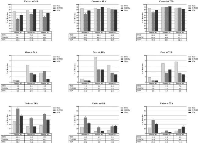
We obtained 54 mono-species and 459 dual-speciesCandida cultures (171 on BCG, 171 on CHROM and 171 on SDA plates), and the results of detectingCandida species are shown in
Fig 1(all species),S2 Fig(common species) andS3 Fig(uncommon species). The percentages of correct detections of the threeCandida species groups, compared to expected results, ranged with the BCG medium from 73.1 (24 h) to 90.1 (72 h), 73.3 (48/72 h) to 93.3 (72 h), and 64.8 (24 h) to 91.2 (72 h); with the CHROM medium from 44.4 (24 h) to 90.6 (72 h), 53.3 (24 h) to 93.3 (48/72 h), and 40.7 (24 h) to 87.9 (72 h); and with the SDA medium from 60.8 (24 h) to 92.4 (72 h), 86.7 (24/48/72 h) to 93.3 (24/48/72 h), and 52.7 (24 h) to 91.2 (72 h). The percent-ages of over detections ranged with the BCG medium from 0.6 (24 h) to 9.4 (48 h), 6.7 (24/48/ 72 h) to 20.0 (24/48/72 h), and 0.0 (24 h) to 7.7 (72 h); with the CHROM medium from 0.6 (24/48/72 h) to 4.7 (48 h), 0.0 (48/72 h) to 13.3 (48/72 h), and 0.0 (24 h) to 4.4 (24/72 h); and with the SDA medium from 0.0 (24 h) to 4.7 (48/72 h), 0.0 (24 h) to 13.3 (48/72 h), and 0.0 (24/48/72 h) to 6.6 (72 h). The percentages of under detections ranged with the BCG medium from 2.9 (72 h) to 26.3 (24 h), 0.0 (24/48/72 h) to 6.7 (24/48/72 h), and 1.1 (72 h) to 35.2 (24 h); with the CHROM medium from 5.8 (72 h) to 55.0 (24 h), 6.7 (48/72 h) to 40.0 (24 h), and 7.7
(72 h) to 59.3 (24 h); with the SDA medium from 2.9 (72 h) to 39.2 (24 h), 0.0 (24/48/72 h) to 6.7 (24 h), and 2.2 (72 h) to 47.3 (24 h).
As for all theCandida species observed by at least one reader, statistically significant differ-ences in the rates of correct or under detections did favour the BCG medium over the CHROM medium (24/48/72 h) and the SDA medium (24 h and 24/48/72 h, respectively) (Fig 1). As for the commonCandida species observed by at least one reader, statistically significant differences in the rates of correct or under detections did favour the BCG medium over the CHROM medium (24 h) (S2 Fig). As for the uncommonCandida species observed by at least one reader, statistically significant differences did favour the BCG medium over the CHROM medium in the rates of correct detections (24/48/72 h) and over both the CHROM and SDA media in the rates of under detections (24/48/72 h) (S3 Fig).
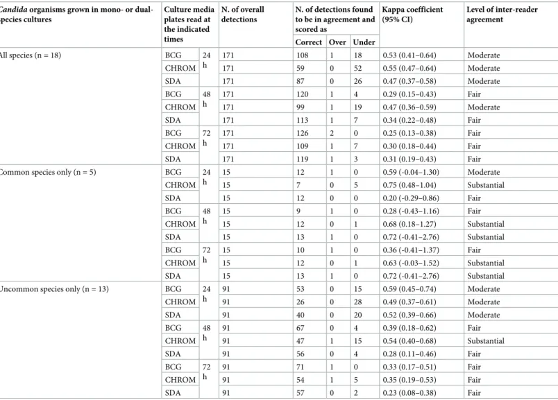
As shown inTable 1, we analysed detection results regarding inter-reader agreement. The levels of agreement for the 24-h readings of BCG, CHROM and SDA plates, for all or uncom-mon species, were at least moderate (kappa coefficient values, 0.41–0.60), whereas the levels of agreement for the 48-h and 72-h readings of BCG, CHROM and SDA plates were at least fair (kappa coefficient values, 0.21–0.40). Conversely, for common species, the levels of agreement for the 24-h/48-h/72-h readings of CHROM plates and for the 48-h/72-h readings of SDA plates were at least substantial (kappa coefficient values, 0.61–0.80). However, comparing the three readers with respect to the percentages of incorrect (over/under) detections for overall
Fig 1. Rates of correct, over or under detections by three readers for the overallCandida species grown as mono- (n = 18) or
dual-species cultures (n = 153) on the BCG (Candida bromcresol green), CHROM (chromogenic medium, i.e. Brilliance Candida agar) and SDA (Sabouraud dextrose agar) media. Asterisks indicate statistically significant differences between the rates of detections
obtained with the BCG medium and those of the CHROM or SDA media.
readings of BCG, CHROM and SDA plates revealed statistically significant differences between the readers only for CHROM and SDA plates (S2 Table).
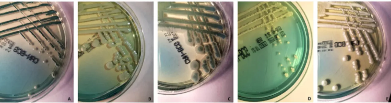
The BCG medium is similar to other selective and differential media for the primary iso-lation ofCandida species. It consists of peptone agar base supplemented with yeast extract (which is absent in the SDA) and dextrose to support growth [13]. However, bromcresol green helps to differentiate and identifyCandida species, because a change in the pH causes the medium to take on a yellow colour around theCandida colonies that ferment dextrose [13].Fig 2depicts the appearance on the BCG medium for the five common species of Can-dida tested by us. As it can see, C. albicans formed smooth, regular, matte, and white to dark-green colonies,C. glabrata smooth, circular, brilliant and white to pale-green colonies,
Table 1. Agreement by readers on the detection results ofCandida species cultured on three media plates that were obtained at 24, 48 or 72 h of incubation of the
plates.
Candida organisms grown in mono- or
dual-species cultures Culture media plates read at the indicated times N. of overall detections N. of detections found to be in agreement and scored as Kappa coefficient (95% CI) Level of inter-reader agreement
Correct Over Under
All species (n = 18) BCG 24 h 171 108 1 18 0.53 (0.41–0.64) Moderate CHROM 171 59 0 52 0.55 (0.47–0.64) Moderate SDA 171 87 0 26 0.47 (0.37–0.58) Moderate BCG 48 h 171 120 1 4 0.29 (0.15–0.43) Fair CHROM 171 99 1 19 0.47 (0.36–0.59) Moderate SDA 171 113 1 7 0.34 (0.22–0.48) Fair BCG 72 h 171 126 2 0 0.25 (0.13–0.38) Fair CHROM 171 109 1 7 0.30 (0.18–0.44) Fair SDA 171 119 1 3 0.31 (0.19–0.43) Fair
Common species only (n = 5) BCG 24
h 15 12 1 0 0.59 (-0.04–1.30) Moderate CHROM 15 7 0 5 0.75 (0.48–1.04) Substantial SDA 15 12 0 0 0.20 (-0.29–0.86) Fair BCG 48 h 15 9 1 0 0.28 (-0.43–1.16) Fair CHROM 15 12 0 1 0.68 (0.18–1.27) Substantial SDA 15 13 1 0 0.72 (-0.41–2.76) Substantial BCG 72 h 15 10 1 0 0.36 (-0.41–1.37) Fair CHROM 15 12 0 1 0.63 (-0.03–1.52) Substantial SDA 15 13 1 0 0.72 (-0.41–2.76) Substantial
Uncommon species only (n = 13) BCG 24
h 91 53 0 15 0.59 (0.45–0.74) Moderate CHROM 91 26 0 28 0.49 (0.37–0.61) Moderate SDA 91 40 0 20 0.52 (0.39–0.66) Moderate BCG 48 h 91 67 0 4 0.39 (0.18–0.62) Fair CHROM 91 47 1 15 0.54 (0.40–0.68) Substantial SDA 91 56 0 4 0.28 (0.11–0.46) Fair BCG 72 h 91 71 1 0 0.33 (0.17–0.51) Fair CHROM 91 54 1 5 0.35 (0.19–0.53) Fair SDA 91 57 0 2 0.23 (0.08–0.38) Fair
Mycological media used for mono- or dual-species cultures of all, common or uncommon species ofCandida were BCG (Candida bromcresol green), CHROM
(chromogenic medium, i.e. Brilliance Candida agar) and SDA (Sabouraud dextrose agar). We calculated the kappa coefficient with 95% confidence interval (95% CI) for the comparison among the rates of correct, over or under detections (according to the definitions specified in the text). With regard to the agreement by readers, we used values greater than zero to indicate none to slight (0.01–0.20), fair (0.21–0.40), moderate (0.41–0.60), substantial (0.61–0.80) or almost perfect (0.81–1.00) levels of agreement, and values lower than/equal to zero to indicate the absence of agreement
C. krusei rough, irregular, matte and green and white-edged colonies, C. parapsilosis rough, irregular, small and white colonies, andC. tropicalis smooth, regular, matte and white colonies.
Discussion
Apart from the overall slight superiority shown by the BCG medium, we noticed that 12 (66.7%) of 18 mono-species cultures at the 24-h readings had correct or over detections (i.e. cultures with �1 colony morphologies observed per plate) in the BCG plates as well in both the CHROM and SDA plates (S1 Table). The only exceptions wereC. albicans, C. incospicua, C. lusitaniae, C. pararugosa, C. pelliculosa and C. sorbosa. Interestingly, while detection of C. albicans in the CHROM plates occurred not prior to 48 h of incubation, over detection of C. glabrata occurred always in 100% of BCG plate readings, 55.6% of CHROM plate readings, and 88.9% of SDA plate readings. These results are consistent with those from some previous studies [12,18]. In one study, 132 (23%) of the 564C. albicans isolates recovered by routinely used media, did not grow on a chromogenic medium [18]. Another study showed the pre-sumptive identification of fiveCandida species (C. albicans, C. dubliniensis, C. krusei, C. tropi-calis and C. parapsilosis) on the CHROM medium (i.e. two additional species besides those identifiable by the medium) [12].
However, discrimination for severalCandida species, including C. glabrata, may be diffi-cult. Conversely, the bromcresol green, a non-toxic indicator contained in the BCG medium (i.e. a modified SDA), seems to aid primary isolation and detection ofCandida species from clinical samples based on dextrose fermentation [13]. While the medium colour around the colonies becomes yellow (usually within 72 h of incubation), theCandida species grown on the BCG medium produce convex to cone-shaped, smooth to rough colonies. Thus, the BCG medium would allow to easily revealing differences in colour (i.e. tonalities of yellow) as well in morphology (i.e. extents of roughness) (Fig 2). We noted that one reader differed from the two other readers with respect to the over detection at 24, 48 and 72 h mainly for the uncom-monCandida species (S3 Fig). Although we chose the three readers to represent a medium-to-high extent of expertise in medical mycology, it is plausible that subtle differences in their mycological skills may explain for the moderate or fair levels of agreement found across read-ers (Table 1).
Among mixed candidaemia episodes,C. albicans plus C. glabrata is usually the most fre-quent combination [5], but other combinations ofCandida species may be of great impor-tance. We noticed that 106 (69.3%), 60 (39.2%) and 78 (51.0%) of 153 dual-species cultures at
Fig 2. Appearance on the Candida bromcresol green (BCG) medium of fiveCandida isolates included in the study that belong to
(A)C. albicans, (B) C. glabrata, (C) C. krusei, (D) C. parapsilosis and (E) C. tropicalis. The isolates were seeded on the BCG plates and
incubated at 30˚C before the plates were imaged. (See the text for the detailed description of the isolates’ features).
the 24-h readings had correct or over detections (i.e. cultures with �2 colony morphologies observed per plate) in the BCG, CHROM or SDA plates, respectively (S1 Table). More interest-ingly, six cultures (C. albicans/C. parapsilosis, C. incospicua/C. pelliculosa, C. incospicua/C. sor-bosa, C. nivariensis/C. pelliculosa, C. pararugosa/C. sorbosa and C. sorbosa/C. tropicalis) in the CHROM medium and two cultures (C. auris/C. guilliermondii and C. auris/C. lusitaniae) in the SDA medium were always under detected with respect to one of the two species grown together. Although a mix of these species seems to be very uncommon, their incomplete detec-tion in candidaemia cases may have clinical repercussions, especially because of different anti-fungal susceptibility profiles exhibited by these species [5–8].
In conclusion, reading primary culture plates from patient samples in the clinical mycology laboratory remains somewhat subjective. ForCandida species, the existence of different mor-photypes, which underpins transitions between commensal and pathogenic cell types in the same species [19–21], complicates the situation. However, distinguishing as many as possible Candida colonies, which will likely correspond to different Candida species, in clinical sam-ples, is crucial in order to exploit the established, powerful MALDI-TOF MS capability of iden-tifying anyCandida organism to the species level (or even beyond). Therefore, using the BCG medium may represent an essential prerequisite for a specific and accurate diagnosis of the causative infection agent(s), especially in patients suffering from life-threatening candidiasis.
Supporting information
S1 Table. Results of the detections by three independent readers for mono-species or dual-speciesCandida cultures recorded at 24, 48 or 72 h of incubation of the BCG, CHROM or SDA culture plates.
(DOC)
S2 Table. Rates of incorrect (over/under) detections by three readers for the overall BCG, CHROM or SDA cultures ofCandida species. Bold indicates statistically significant
differ-ences in the comparisons between reader #1 and reader #2, reader #2 and reader #3, or reader #1 and reader #3.
(DOC)
S1 Fig. Scheme of the checkerboard-like dilution method used to obtain mono-species or dual-speciesCandida suspensions before spreading them on the mycological media under evaluation.
(DOC)
S2 Fig. Rates of correct, over or under detections by three readers for the commonCandida species grown as mono- (n = 5) or dual-species (n = 10) on the BCG (Candida bromcresol green), CHROM (chromogenic medium, i.e. Brilliance Candida agar) and SDA (Sabour-aud dextrose agar) media. Asterisks indicate statistically significant differences between the
rates of detections obtained with the BCG medium and those of the CHROM or SDA media. (JPG)
S3 Fig. Rates of correct, over or under detections by three readers for the uncommon Can-dida species grown as mono- (n = 13) or dual-species (n = 78) on the BCG (CanCan-dida brom-cresol green), CHROM (chromogenic medium, i.e. Brilliance Candida agar) and SDA (Sabouraud dextrose agar) media. Asterisks indicate statistically significant differences
between the rates of detections obtained with the BCG medium and those of the CHROM or SDA media.
Acknowledgments
We wish to thank Franziska Lohmeyer for her English language assistance.
Author Contributions
Conceptualization: Giulia De Angelis, Brunella Posteraro.
Data curation: Giulia De Angelis, Giulia Menchinelli, Riccardo Torelli, Elena De Carolis,
Patrizia Posteraro, Brunella Posteraro.
Formal analysis: Giulia Menchinelli, Riccardo Torelli, Elena De Carolis, Patrizia Posteraro. Investigation: Giulia Menchinelli, Riccardo Torelli, Elena De Carolis, Patrizia Posteraro. Methodology: Giulia Menchinelli, Brunella Posteraro.
Supervision: Maurizio Sanguinetti. Validation: Maurizio Sanguinetti.
Writing – original draft: Giulia De Angelis, Brunella Posteraro. Writing – review & editing: Brunella Posteraro.
References
1. Clark C, Drummond RA. The hidden cost of modern medical interventions: How medical advances have shaped the prevalence of human fungal disease. Pathogens 2019; 8:E45.https://doi.org/10.3390/ pathogens8020045PMID:30987351
2. Bongomin F, Gago S, Oladele RO, et al. Global and multi-national prevalence of fungal diseases-esti-mate precision. J Fungi (Basel) 2017; 3:E57.
3. Pappas PG, Lionakis MS, Arendrup MC, et al. Invasive candidiasis. Nat Rev Dis Primers 2018; 4:18026.https://doi.org/10.1038/nrdp.2018.26PMID:29749387
4. Guinea J. Global trends in the distribution of Candida species causing candidemia. Clin Microbiol Infect 2014; 20(Suppl 6):5–10.
5. Miceli MH, Dı´az JA, Lee SA. Emerging opportunistic yeast infections. Lancet Infect Dis 2011; 11:142– 51.https://doi.org/10.1016/S1473-3099(10)70218-8PMID:21272794
6. Marcos-Zambrano LJ, Puig-Asensio M, Pe´rez-Garcı´a F, et al. Candida guilliermondii complex is char-acterized by high antifungal resistance but low mortality in 22 cases of candidemia. Antimicrob Agents
Chemother 2017; 61:e00099–17.https://doi.org/10.1128/AAC.00099-17PMID:28438935
7. Pe´rez-Hansen A, Lass-Flo¨ rl C, Lackner M, et al. Antifungal susceptibility profiles of rare ascomycetous yeasts. J Antimicrob Chemother 2019; 74:2649–56.https://doi.org/10.1093/jac/dkz231PMID: 31203366
8. Lockhart SR. Candida auris and multidrug resistance: Defining the new normal. Fungal Genet Biol 2019; 131:103243.https://doi.org/10.1016/j.fgb.2019.103243PMID:31228646
9. Posteraro B, Spanu T, Fiori B, et al. Antifungal susceptibility profiles of bloodstream yeast isolates by Sensititre YeastOne over nine years at a large Italian teaching hospital. Antimicrob Agents Chemother 2015; 59:3944–55.https://doi.org/10.1128/AAC.00285-15PMID:25896705
10. Walsh TJ, McCarthy MW. The expanding use of matrix-assisted laser desorption/ionization-time of flight mass spectroscopy in the diagnosis of patients with mycotic diseases. Expert Rev Mol Diagn 2019; 19:241–8.https://doi.org/10.1080/14737159.2019.1574572PMID:30682890
11. Zhao L, de Hoog GS, Cornelissen A, et al. Prospective evaluation of the chromogenic medium Candi-Select 4 for differentiation and presumptive identification of non-Candida albicans Candida species.
Fungal Biol 2016; 120:173–8.https://doi.org/10.1016/j.funbio.2015.09.006PMID:26781374
12. Vecchione A, Florio W, Celandroni F, et al. Comparative evaluation of six chromogenic media for pre-sumptive yeast identification. J Clin Pathol 2017; 70:1074–8. https://doi.org/10.1136/jclinpath-2017-204396PMID:28663328
13. Zimbro MJ, Power DA. 2009. Difco & BBL Manual. Manual of Microbiological Culture Media. BD Diag-nostic Systems. 2ndedition. Becton, Dickinson and Company, Sparks, Maryland, USA.https://www. bd.com.
14. De Carolis E, Vella A, Vaccaro L, et al. Development and validation of an in-house database for matrix-assisted laser desorption ionization-time of flight mass spectrometry-based yeast identification using a fast protein extraction procedure. J Clin Microbiol 2014; 52:1453–8.https://doi.org/10.1128/JCM. 03355-13PMID:24554755
15. Larone DH (2011) Medically important fungi. A guide to identification. ASM Press, Washington DC, USA.
16. Lion T (2017) Human fungal pathogen identification. Methods in molecular biology, vol. 1508. Humana Press, New York, NY, USA.
17. Barnhart HX, Williamson JM. Weighted least-squares approach for comparing correlated kappa.
Bio-metrics 2002; 58:1012–9.https://doi.org/10.1111/j.0006-341x.2002.01012.xPMID:12495157
18. Murray CK, Beckius ML, Green JA, et al. Use of chromogenic medium in the isolation of yeasts from clinical specimens. J Med Microbiol 2005; 54:981–5.https://doi.org/10.1099/jmm.0.45942-0PMID: 16157554
19. Brockert PJ, Lachke SA, Srikantha T, et al. Phenotypic switching and mating type switching of Candida
glabrata at sites of colonization. Infect Immun 2003; 71:7109–18.https://doi.org/10.1128/IAI.71.12. 7109-7118.2003PMID:14638801
20. Priest SJ, Lorenz MC. Characterization of virulence-related phenotypes in Candida species of the CUG Clade. Eukaryot Cell 2015; 14:931–40.https://doi.org/10.1128/EC.00062-15PMID:26150417
21. Chowdhary A, Sharma C, Meis JF. Candida auris: a rapidly emerging cause of hospital-acquired multi-drug-resistant fungal infections globally. PLoS Pathog 2017; 13:e1006290.https://doi.org/10.1371/ journal.ppat.1006290PMID:28542486