2004;78:1761-1768
Ann Thorac Surg
C. Mineo
Eugenio Pompeo, Davide Mineo, Paola Rogliani, Alessandro F. Sabato and Tommaso
Nodules
Feasibility and Results of Awake Thoracoscopic Resection of Solitary Pulmonary
http://ats.ctsnetjournals.org/cgi/content/full/78/5/1761
located on the World Wide Web at:
The online version of this article, along with updated information and services, is
Print ISSN: 0003-4975; eISSN: 1552-6259.
Southern Thoracic Surgical Association. Copyright © 2004 by The Society of Thoracic Surgeons.
is the official journal of The Society of Thoracic Surgeons and the
The Annals of Thoracic Surgery
Resection of Solitary Pulmonary Nodules
Eugenio Pompeo,
MD,
Davide Mineo,
MD,
Paola Rogliani,
MD,
Alessandro F. Sabato,
MD,
and Tommaso C. Mineo,
MD
Division of Thoracic Surgery and Multidisciplinary Pulmonary Program, Policlinico Tor Vergata University, Rome, Italy
Background. General anesthesia with single-lung ven-tilation is considered mandatory for thoracoscopic pul-monary resection. We assessed in a randomized study the feasibility and results of awake thoracoscopic resection of solitary pulmonary nodules.
Methods. Between March 2001 and February 2003, 60 patients were randomized into two 30-patients arms: a general anesthesia arm entailing double-lumen intuba-tion and thoracic epidural anesthesia (control group); and an awake arm entailing sole thoracic epidural anesthesia at T4-T5 (awake group). Anesthesia time; operative time; global operating room time; patient satisfaction with the anesthesia and technical feasibility scored into 4 grades (from 1ⴝ poor to 4 ⴝ excellent); visual analog pain score (VAS), nursing care (number of patient calls per day), 24 hours changes in arterial oxygenation (⌬PaO2), and
hos-pital stay were assessed.
Results. There was no mortality. There was no difference
in technical feasibility between the groups although 2 patients in the awake group required conversion to thora-cotomy due to severe adhesions. Other 2 patients in each group required conversion due to unexpected lung cancer requiring lobectomy. Comparisons of awake versus control group results showed that in the awake group, anesthesia satisfaction score was greater (4 vs 3, p ⴝ 0.04), whereas ⌬PaO2(-3 mm Hg vs – 6.5 mm Hg, pⴝ 0.002); nursing care
(2.5 calls per day vs 4 calls per day, pⴝ 0.0001), and hospital stay (2 days vs 3 days, pⴝ 0.02) were significantly reduced. Conclusions. In our study, awake thoracoscopic resec-tion of solitary pulmonary nodules proved safely feasi-ble. It resulted in better patient satisfaction, less nursing care and shorter in-hospital stay than procedures per-formed under general anesthesia.
(Ann Thorac Surg 2004;78:1761– 8) © 2004 by The Society of Thoracic Surgeons
A
solitary pulmonary nodule is an approximately round lesion that is less than 3 cm in diameter and is completely surrounded by pulmonary parenchyma, without other abnormalities. It is noted on 0.09% to 0.20% of all chest radiographs [1, 2]. Infectious granulomas and hamartomas account for the most common benign diag-nosis but 40% up to 50% of indeterminate pulmonary nodules prove to be malignant [3].Surgical lung biopsy by video-assisted thoracic surgery (VATS) has demonstrated proven accuracy and has be-come an increasingly accepted approach for manage-ment of solitary pulmonary nodules [3– 6]. General anes-thesia with one-lung ventilation, is deemed mandatory for pulmonary VATS resection although several adverse effects can derive from this type of anesthesia including the development of atelectasis in both the dependent and nondependent lung, and the so called ventilator-induced lung injury [7].
We hypothesized that to avoid general anesthesia-related risks, awake VATS resection of peripheral solitary pulmonary nodules could be performed under sole
tho-racic epidural anesthesia (TEA). We reasoned that avoid-ance of general anesthesia and double-lumen intubation could result in shorter in-operating room time, and faster recovery eventually leading to a shorter hospital stay.
For this reason, having assessed feasibility in a short pilot study, we undertaken a randomized trial comparing results of awake VATS resection of solitary pulmonary nodules versus those of a control group operated under general anesthesia with one-lung ventilation.
Material and Methods
The study was started in March 2001 and was closed in February 2003. Written informed consent was obtained from all patients who took part and the study was approved by the institutional review board of the Tor Vergata University. Sixty patients with undetermined solitary pulmonary nodule, who met the entry criteria, were randomized by computer into two groups: 30 pa-tients underwent VATS resection of the pulmonary nod-ule under conventional general anesthesia with one-lung ventilation. Thirty patients underwent awake VATS re-section under sole thoracic epidural anesthesia (TEA). All patients underwent preoperative fiberoptic bronchos-copy, high resolution computed tomography and pulmo-nary function testing. To be eligible patients should have a solitary pulmonary nodule localized in the peripheral Accepted for publication May 4, 2004.
Presented at the Fortieth Annual Meeting of The Society of Thoracic Surgeons, San Antonio, TX, Jan 26 –28, 2004.
Address reprint requests to Dr Pompeo, Cattedra di Chirurgia Toracica, Università Tor Vergata, Policlinico Tor Vergata, V.le Oxford, 81, 00133 Rome, Italy; e-mail: [email protected].
GENERAL
one-third of the lung and measuring less than 3 cm at the high resolution computed tomography. In addition, to avoid the risk of suboptimal management of malignant lesions, only patients with radiologically benign nodules, discovered within 1 month from enrollment, no signs of enlarged lymphnodes, unremarkable fiberoptic bron-choscopy findings, and no history of malignancy were considered eligible. Other inclusion criteria were the absence of radiologic signs of pleural scarring in the hemithorax targeted for lung resection, no clinical signs of active infectious disease, less than 80 years old, ASA score equal to or less than 3, and the absence of severe emphysema requiring lung volume reduction surgery.
Contraindications for TEA included unfavorable anat-omy, previous surgery of the cervical or upper thoracic spine, compromised coagulation (thromboplastin time⬍ 80%, prothrombin time ⬎ 40 seconds, or platelets ⬍ 100/nl) or bleeding disorder.
Evaluated variables included anesthesia time calcu-lated as the time required for performing each type of anesthesia; operative time calculated as the time com-prised between skin incision and completion of skin closure; global operating room time that was the overall time spent by the patient into the operating room and included anesthesia time, operative time and the time required for weaning and achievement of stable clinical condition deemed necessary to allow the patient to be transferred from the recovery room to the ward. Finally, the patient was asked to express his satisfaction with the anesthesia type while the surgeon himself judged the technical feasibility of each procedure. For each of these factors 4 possible scores were possible (4⫽ excellent, 3 ⫽ good, 2⫽ satisfactory, 1 ⫽ unsatisfactory). Nursing care was estimated and scored by recording the number of patient calls for nursing care and was deemed a measure reflecting the overall patient discomfort and the degree of limitation in his autonomy during simple daily life activities. Postoperative pain was assessed by a standard visual analog pain scale (VAS).
Both nursing care need and VAS assessment were performed 24 hours after the procedure or just before discharge if hospital stay lasted 1 day only. In addition, preoperative to postoperative (day one) changes in arte-rial oxygenation (⌬PaO2), postoperative complications,
and hospital stay were also recorded in all patients. Criteria for discharge were standardized and discharge was performed in uneventful courses, 1 hour after
re-moval of the chest tube. The chest tube was removed after that a chest roentgenogram demonstrated complete re-expansion of the lung and 24 hours after that any air leak stopped and drainage fluid did not exceed 150 mL.
Surgical Technique
All the procedures were performed with the patient placed in full lateral decubitus position and through a three-flexible-thoracoscopic-trocars access. Localization of the nodule was performed by digital and instrumental palpation without additional adjuvant methods. To facil-itate identification of smallest nodules, a custom made ring forcep was used.
Excision of the nodule was performed by staple wedge resection (Fig 1). Dependent pleural drainage was ob-tained with a single 28F chest tube placed to –20 cm H2O suction at the conclusion of each procedure. All patients in the general anesthesia group were extubated in the operating room.
Anesthesia
The objective of TEA was to achieve somatosensory and motor block at the T1 to T8 level. The maximum permis-sible block level was C6, which was monitored by the development of Horner’s syndrome. One of the major objectives was to achieve motor block of the intercostal muscles while preserving diaphragmatic respiration. The thoracic epidural catheter was inserted at the T4 level after that patients were premedicated orally with 7.5 mg midazolam. In the operating room, patients received a continuous infusion of ropivacaine 0.5% and sufentanil 1.66 g/mL into the epidural space. Sensory level was tested every 5 minutes with warm-cold discrimination. Thus sensory block was achieved between the neck and the abdomen, including the arms.
During the procedure patients breathed O2through a ventimask as required to keep oxygen saturation above 90%. Whenever somatosensory block was judged unsat-isfactory by the patient, additive anesthesia was provided by local injection of a 50% mixture of ropivacaine (7.5%) and bupivacaine (2%). During wound closure, the TEA anesthetic regimen was changed to ropivacaine 0.16% and sufentanil 1 g/mL at 2 to 5 mL/hour to provide postoperative analgesia. All patients received lactated Ringer’s solution at 12 mL · Kg-1 · h-1. The epidural catheter was removed 24 hours after surgery. Patients undergoing general anesthesia with single-lung ventila-tion had a thoracic epidural catheter inserted between T5 and T8 with a continuous infusion of ropivacaine started during surgery and continued for the first postoperative 24 hours to assure postoperative analgesia. General an-esthesia was induced with intravenous propofol (1.5 to 2 mg/Kg), fentanyl (0.1 mg), and vecuronium (0.1 mg/Kg), and maintained using a continuous infusion of propofol (4 to 8 mg · Kg), fentanyl and vecuronium. A left-sided double-lumen tube was placed under fiberoptic control. In both groups usual monitoring was used. Whenever conversion to thoracotomy was required in patients un-dergoing awake VATS, this was performed after switch-ing to general anesthesia and sswitch-ingle-lung ventilation. In
Abbreviations and Acronyms
ASA ⫽ American Society of Anesthesiology COPD⫽ chronic obstructive pulmonary disease FEV1 ⫽ forced expiratory volume in 1 second
FVC ⫽ forced vital capacity PaO2 ⫽ arterial oxygen tension
TEA ⫽ thoracic epidural anesthesia VATS ⫽ video-assisted thoracic surgery VAS ⫽ visual analog pain scale
1762 POMPEO ET AL Ann Thorac Surg
AWAKE VATS FOR PULMONARY NODULES 2004;78:1761– 8
GENERAL
these cases, intubation with single-lumen tube and bron-chial blocker was performed under bronchoscopic guid-ance without changing the patient position.
Postoperatively, in the awake group, liquids infusion was stopped immediately and drinking, meal intake and ambulation were started on the same day of surgery. In the general anesthesia group, liquids infusion (1500 mL/24 hours) continued up to the morning after surgery when drinking, meal intake and ambulation were re-sumed. Urinary catheterization was not performed un-less urinary retention did occur.
Study Design and Statistical Methods
The primary outcome measure chosen was hospital stay while secondary outcome measures were global operat-ing room time, anesthesia satisfaction score, technical feasibility score,⌬PaO2, and nursing care requirement.
Given that in an historical cohort undergoing VATS resection of pulmonary nodules at our institution, 15% of the patients were discharged within the first 48 hours, trial size was originally determined by the number of patients necessary to yield, by a nonparametric two-sided test, with an␣ error of 0.05, an increment of at least 10% of discharges within the first 48 postoperative hours. Group descriptive statistics are presented as median with interquartile range. Due to the small cohort, the
nonpara-metric Mann-Whitney test was used to compare un-paired data while relationship between variables was assessed by the Spearman correlation test. Proportions were compared by the two tailed Fisher’s exact test and C.I. 95% was calculated by the Ciba-Geigy Scientific Tables [8].
Results
During the study period, 12 of 72 eligible patients refused to accept the randomization and were excluded from the analysis. Of these, 8 patients asked to undergo the awake procedure while 4 preferred general anesthesia.Table 1 shows that the two study groups were well matched in terms of base line characteristics and measures. Overall, the nodules were localized in the right upper, middle or right lower lobe in 22, 7, and 10 instances, respectively; in the left upper or lower lobe in 11 and 10 instances, respectively.
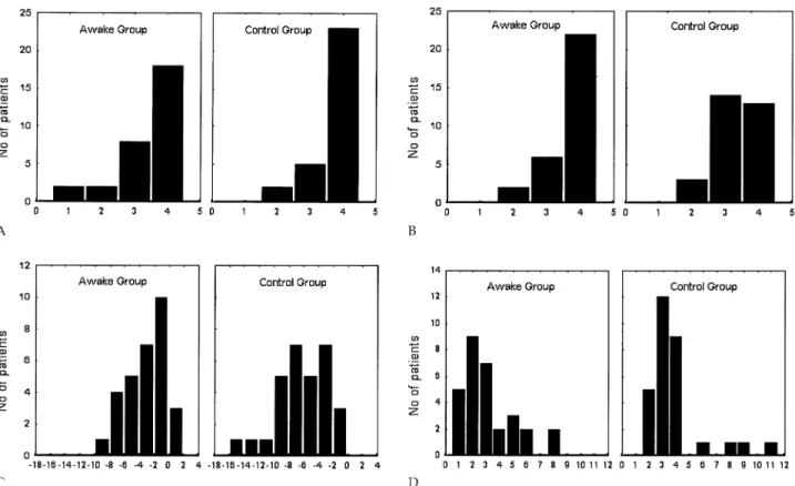
Perioperative and postoperative results in the study groups are illustrated inTable 2. There was no significant difference in median operative time between the groups while anesthesia time and global in-operating room time were significantly longer in the control group. Technical feasibility demonstrated that the procedure was easily accomplished in both groups (Fig 2A) although 2 patients Fig 1. Surgical details of awake VATS resection of pulmonary nodules. Patient positioning and thoracoscopic access (1); lysis of filmy adhe-sions is not compromised during awake VATS (2); custom-shaped ring forceps with a filled-in ring to facilitate palpation of small nodules (3); and staple resection of a nodule with the ventilating lung (4). (VATS⫽ video-assisted thoracic-surgery.)
GENERAL
in the awake group required conversion to thoracotomy and general anesthesia due to severe fibrous adhesions. Anesthesia satisfaction score was better in the awake group than in the control group with 22 patients versus 13 patients, respectively, having a score of 4 (p⫽ 0.03; Fig 2B).
Pathologic examination of benign nodules is detailed inTable 3. Despite radiologic evidence of benignity was required for eligibility, an unexpected malignant lesion was found in 9 patients (15%). Of these, 4 patients in the
awake group had nonsmall-cell lung cancer, 3 adenocar-cinomas, and 1 large-cell carcinoma. In the general an-esthesia group, 3 patients had nonsmall-cell lung carci-noma of the adenocarcicarci-noma type and 2 had pulmonary metastases from seminoma and renal-cell carcinoma, respectively. Among patients with carcinoma, 2 patients in each group underwent lobectomy through thoracot-omy after conversion to general anesthesia and single-lung ventilation, while other 3 patients with severe chronic obstructive pulmonary disease who would have
Table 2. Results in the Study Groups Variable Awake Group Quartile Range Control Group Quartile Range Intergroup p Value Nodule characteristics Size (cm) 1.75 1.1–2.3 1.75 1.5–2.1 0.95 Lung (Right:Left) 21–9 — 18–12 — 0.59 Benign:Malignant 26–4 — 25–5 — 0.73 Thoracotomy needed (n) 4 — 2 — 0.67
Anesthesia time (min) 17.5 10–22 36 33–40 ⬍0.00001
Operating time (min) 24 22–32 24 21–28 0.87
Global in-operating room time (min) 65 56–72 100 95–110 ⬍0.00001
Patient anesthesia satisfaction score 4 4–4 3 3–4 0.04
Technical feasibility score 4 3–4 4 3.5–4 0.57
⌬PaO2(mm Hg)* ⫺3 0.5–5 ⫺6.5 3.5–9 0.002
Pain (VAS score)* 2.5 2–3 3 2–3.5 0.19
Nursing Care (No of calls/day)* 2.5 2–3 4 3–5 0.0001
Mortality (n) 0 — 0 — 1.0
Morbidity (n) 1 — 3 — 0.61
Hospital stay 2 2–3 3 3–4 0.02
* Assessed 24 hours after surgery.
Table 1. Baseline Characteristics and Measures in the Study Groups Variable Awake Group Quartile Range Control Group Quartile Range Intergroup p Value Age (y) 60 45–68 63.5 55–68 0.4 Male:Female (n) 20:10 — 21:9 — NS Tobacco use (n) Current 22 — 24 — 0.76 Former 3 — 2 — 0.67 Never 5 — 4 — 0.73 ASA score 1 1–2 1 1–2 1.0
Home oxygen use (n) 3 — 2 — 0.67
Corticosteroid use (n) 3 — 2 — 0.67 Comorbidities COPD (n) 9 — 10 — 1.0 Cardiopathy (n) 8 — 6 — 0.76 Arteriopathy (n) 2 — 1 — 1.0 Diabetes (n) 3 — 3 — 0.61 Pulmonary function FEV1 (% predicted) 87.5 82–100 90.5 74–102 0.8 FVC (% predicted) 92 78–101 91 79–101 0.73 PaO2(mm Hg) 89 80–98 85.5 80–93 0.3
ASA ⫽ American Society of Anesthesiology; COPD ⫽ chronic obstructive pulmonary disease; FEV1 ⫽ forced expiratory volume in 1
second; FVC⫽ forced vital capacity; PaO2⫽ arterial oxygen tension.
1764 POMPEO ET AL Ann Thorac Surg
AWAKE VATS FOR PULMONARY NODULES 2004;78:1761– 8
GENERAL
not tolerated a lobectomy underwent sole wedge resection.
On postoperative day 1, the absolute decrease in PaO2 was greater in the control group than in the awake group with 23 versus 12 patients, respectively, having a⌬PaO2
greater than or equal to 3 mm Hg (p⫽ 0.008;Fig 2C). There was no mortality in either group while the only meaningful complication regarded prolonged air leaks, which occurred in 1 patient of the awake group and in 3 of the control group. In the awake group, no patient complained of fever or nausea postoperatively while 2 patients had urinary retention, which required tempo-rary urinary catheterization. Nursing care was signifi-cantly less frequently required in the awake group than in the control group with 15 patients versus 23 patients,
respectively, with more than 3 nurse calls per day (p⫽ 0.001). Overall, nursing care showed a direct relationship with VAS (R⫽ 0.39, p ⫽ 0.001).
Median hospital stay was significantly shorter in the awake group than in the control group (Fig 2D) with 14 patients or 46.7% (95% confidence interval [95% CI] ⫽ 28.3 to 65.7) versus 5 patients or 16.6% (95% CI⫽ 5.9 to 35.8) with an hospital stay of 2 days or less (p⫽ 0.025;Fig 2D). The difference in median hospital stay remained significant even after exclusion of the 4 patients with prolonged air leaks (p⫽ 0.04).
Comment
Selected controlled ventilation came with the introduc-tion of the double-lumen endobronchial tube introduced by Zavod in 1940 [9] and refined by Carlens in 1949 [10]. It proved to be a revolutionary advance in thoracic surgery and is now the standard type of anesthesia for both open and VATS pulmonary resections. However, despite some indisputable and well-known advantages, several adverse effects can derive from this type of anesthesia including an increased risk of pneumonia, impaired cardiac performance, and neuromuscular prob-lems relating to sedation and muscle relaxants. More-over, during general anesthesia, mechanical ventilation can cause damage known as ventilator-induced lung injury. This type of injury include airway pressure-induced injury, lung inflation-pressure-induced injury, injury due Fig 2. Categorized istograms of (A) technical feasibility score, (B) anesthesia satisfaction score, (C) changes in arterial oxygenation (mm Hg), and (D) hospital stay in the study groups.
Table 3. Benign Diagnoses Diagnosis Awake Group Control Group Total Inflammatory granuloma 9 12 21 Hamartoma 7 6 13 Scar nodule 5 4 9 Tuberculoma 2 2 4 Abscess 1 0 1 Carcinoid 1 0 1 Sarcoidosis 0 1 1 Pulmonary thromboembolysm 1 0 1 GENERAL THORACIC
to cyclic opening and closing of small airways/lung units, and release of a variety of proinflammatory mediators [7]. Yet, atelectasis in the dependent lung is a regular finding during one-lung ventilation with muscle paralysis [11]. Moreover, prolonged collapse of the nondependent lung during one-lung ventilation can delay its complete re-expansion after weaning leading to the development of further atelectasic areas in the nondependent lung.
Theoretically, these adverse effects could be avoided by using TEA in an awake patient. Successful awake major surgery under TEA including aortic aneurysm repair [12], and coronary artery bypass grafting [13] has been recently reported. Furthermore, TEA has been an-ecdotally employed for thoracoscopic management of secondary pneumothorax [14].
The major finding of this study is that awake VATS resection of solitary pulmonary nodules was easily and safely performed under TEA since the procedure could be completed in 26 of 28 patients in the awake arm with no mortality and negligible morbidity. These results contradict the accepted assumption that the main pre-requisite for allowing successful thoracoscopic lung sur-gery is general anesthesia with one-lung ventilation. No particular training has been necessary to accomplish awake pulmonary resection and the only two failures were due to the presence of extensive and dense pleural adhesions that thus represented the only contraindica-tion for the procedure. In addicontraindica-tion, awake pulmonary resection was easily accepted and well tolerated by the patients as confirmed by the high anesthesia satisfaction score in this group, which was significantly greater than in the control group.
Avoidance of general anesthesia reflected in a faster recovery with immediate return to many daily life activ-ities including drinking, eating and walking. As a result, the hospital stay was significantly shorter than in the control group.
One of our major concern related to the risk that operating on a ventilating lung would have rendered surgical maneuvers more difficult due to the lung move-ments and the lack of a sufficient “operating space”. Instead, we have early noticed that the open pneumotho-rax created after trocars’ insertion, produced a satisfac-tory lung collapse which did not hamper surgical maneuvers.
A further noteworthy finding is that despite radiologic features of benignity was a selection criteria, in 15% of our operated patients pathologic examination disclosed a malignant lesion. This result emphasizes that particularly in patients at increased risk for lung cancer, strict radio-logic surveillance and early removal of solitary pulmo-nary nodules should be pursued.
Preoperative and intraoperative nodule localization techniques include computed tomography dye injection [15, 16], intraoperative endoscopic ultrasonography [17] and preoperative computed tomography guided hook-wire localization [18]. In our series, nodules as small as 4 mm in size were identified without the need of additional methods.
In a recent study from Ueda and colleagues [19],
significant preoperative factors related to recovery after VATS pulmonary resections included age, breathless-ness, performance status, radiologic emphysema and partial pressure of arterial oxygen less than 80 mm Hg.
In our study including only wedge resections, the overall postoperative impairment of oxygenation was less pronounced than in the Ueda series. Nonetheless, we have observed not only that arterial oxygenation worsened during the first postoperative day but also that this impairment was greater in the general anesthesia group than in the awake group. We hypothesize that avoiding general anesthesia resulted in a faster recovery as indicated also by the significantly shorter hospital stay we observed in the awake group. One criticism might be addressed to the finding that despite the use of TEA, mean hospital stay still averaged 2 days, which reduces the cost-effectiveness of this method in comparison with nonsurgical alternative diagnostic methods. However, in our series, the patient discharge was strictly related to the criteria applied for chest tube removal, which so far remain an open matter for discussion. Russo and cowork-ers [20] reported that removal of the chest tube within 90 minutes after thoracoscopic wedge resection or biopsy resulted in a complication rate that compared that of a control group undergoing traditional chest tube manage-ment. Ueda and colleagues [19] reported that even though the average pleural drainage during the last day before removal of the chest tube was 168 ml, no patient developed collection of pleural effusion. This led the authors to conclude that the amount of pleural drainage does not represent a limiting factor for early removal of the chest tube if there is no hemorrhage or air leak. In accordance with this assumption, Chang and coworkers [21] recently reported on an outpatient thoracoscopic lung biopsy experience in which 59 out of 62 operated patients were discharged within 23 hours after the ation. Since in the Chang series all patients were oper-ated through general anesthesia and single-lung ventila-tion, we are tempted to predict that using awake VATS resection with more liberal criteria for chest tube removal will implement early discharge in future outpatient series.
Despite the satisfactory results achieved in this study are encouraging, we acknowledge that some criticisms can be raised against awake VATS.
First, the open pneumothorax created to perform an awake lung resection can determine a mediastinal move-ment with compression of the dependent lung eventually resulting in a functional compromise. On the other hand, in an awake patient, the maintained diaphragmatic mo-tion decreases the detrimental effect of the abdominal pressure, which leads the paralyzed diaphragm to com-press the dependent lung during general anesthesia.
A further concern relates to the patient participation in operating room conversations when unexpected lung cancer is encountered. We believe that in these delicated instances special care must be devoted by the operating team in reassuring the patient while explaining the reasons that require conversion to general anesthesia and thoracotomy.
1766 POMPEO ET AL Ann Thorac Surg
AWAKE VATS FOR PULMONARY NODULES 2004;78:1761– 8
GENERAL
In conclusion, this randomized study suggests that VATS resection of solitary pulmonary nodules was easily and safely performed under TEA in awake patients. Yet, this approach proved superior to conventional VATS resection under general anesthesia with one-lung venti-lation in terms of global in-operating room time, postop-erative recovery, need of nurse care and overall hospital stay. If confirmed by future studies, these results could lead to advocate an earlier VATS resection of peripheral solitary pulmonary nodules to reduce the risk of delaying a diagnosis of unexpected pulmonary malignancy.
This study has been carried out within the Research Fellowship Program Dottorato di Ricerca in Tecnologie e Terapie Avanzate in
Chirurgia, awarded by Tor Vergata University, and was
sup-ported partially by grant COFIN #9906274194 to 06 of the MURST and by Single Project 2002 CNR CU0100935CT26 by the National Council of the Research.
References
1. Holin SN, Dwork RE, Glaser S, Rickli AE, Stocklen JB. Solitary pulmonary nodules found in a community-wide chest roentgenographic survey. Am Tuberc Pulm Dis 1959; 79:427–39.
2. Swensen SJ, Silverstein MD, Edell ES, et al. Solitary pulmo-nary nodules: clinical prediction model versus physicians. Mayo Clin Proc 1999;74:319 –29.
3. Hazelrigg SR, Magee MJ, Cetindag IB. Video-assisted tho-racic surgery for diagnosis of the solitary lung nodule. Chest Surg Clin North Am 1998;8:763–74.
4. Mitruka S, Landreneau RJ, Fetterman LS, et al. Diagnosing the indeterminate pulmonary nodule: percutaneous biopsy versus thoracoscopy. Surgery 1995;118:676 – 84.
5. Allen MS, Deschamps C, Lee RE, Trastek VF, Daly RC, Pairolero PC. Video-assisted thoracoscopic stapled wedge excision for indeterminate pulmonary nodules. J Thorac Cardiovasc Surg 1993;106:1048 –52.
6. Murasugi M, Onuki T, Ikeda T, Kanzaki M, Nitta S. The role of video-assisted thoracoscopic surgery in the diagnosis of the small peripheral pulmonary nodule. Surg Endosc 2001; 15:734 – 6.
7. Whitehead T, Slutsky AS. The pulmonary physician in critical care. 7: Ventilator induced lung injury. Thorax 2002; 57:635– 42.
8. Lentner C. Tavole Scientifiche Geigy. Ciba Geigy Ediz VIII ediz. 1984:89 –102.
9. Zavod WA. Bronchospirography 1. Description of the cath-eter and the technique of intubation. J Thorac Surg 1940;10: 27–31.
10. Björk VO, Carlens E. The prevention of spread during pulmonary resection by use of a double-lumen catheter. J Thorac Surg 1950;20:151–7.
11. Tokics L, Hedenstierna G, Svensson L, et al. V/Q distribution and correlation to atelectasis in anesthetize paralyzed hu-mans. J Appl Physiol 1996;81:1822–33.
12. McGregor WE, Koler AJ, Labat GC, Perni V, Hirko MK, Rubin JR. Awake aortic aneurysm repair in patients with severe pulmonary disease. Am J Surg 1999;178:121– 4. 13. Aybek T, Kessler P, Dogan S, et al. Awake coronary artery
bypass grafting: utopia or reality? Ann Thorac Surg 2003;75: 1165–70.
14. Mukaida T, Andou A, Aoe M, Shimizu N. Thoracoscopic operation for secondary pneumothorax under local and epidural anesthesia in high-risk patients. Ann Thorac Surg 1998;65:924 – 6.
15. Asamura H, Kondo H, Naruke T, Tsuchiya R, Kaneko M, Suemasu K. Computed tomography-guided coil injection and thoracoscopic pulmonary resection under roentgeno-graphic fluoroscopy. Ann Thorac Surg 1994;58:1542– 44. 16. Wicky S, Mayor B, Cuttat JF, Schnyder P. CT-guided
local-ization of pulmonary nodules with metilene blue injections for thoracoscopic resections. Chest 1994;106:1326 – 8. 17. Shennib H, Bret P. Intraoperative transthoracic localization
of occult lung lesion. Ann Thorac Surg 1993;55:67–9. 18. Partik BL, Müller MR, Breitenseher M, et al. Using a
dedi-cated lung-marker system for localization of pulmonary nodules before thoracoscopic surgery. AJR 2003;180:805–9. 19. Ueda K, Kaneda Y, Sakano H, Tanaka T, Li T, Hamano K.
Obstacles for shortening hospitalization after video-assisted pulmonary resection for lung cancer. Ann Thorac Surg 2003;76:1816 –20.
20. Russo L, Wiechmann RJ, Magovern JA, et al. Early chest tube removal after video-assisted thoracoscopic wedge resection of the lung. Ann Thorac Surg 1998;66:1751– 4.
21. Chang AC, Yee J, Orringer MB, Iannettoni MD. Diagnostic thoracoscopic lung biopsy: an outpatient experience. Ann Thorac Surg 2002;74:1942–7.
DISCUSSION
DR KENNETH M. STEINGLASS(New York, NY): That was an intriguing series. I have a question. Four of your patients in the local anesthesia group required conversion. They were in the lateral thoracotomy position. Could you just give us some practical information on what happened in your operating room with your anesthesiologists placing double-lumen tubes in the lateral position, or did you turn the patients back into a supine position? Give us an idea of how that worked.
DR POMPEO: Thank you for the question. For patients in the awake group who needed the conversion to thoracotomy, we used a Univent without changing the position of the patient. The intubation was performed under fiberoptic con-trol with a Univent tube without changing the position of the patient.
DR PAUL VAN SCHIL (Antwerp, Belgium): Regarding your preoperative work-up, did you include results of PET scanning to decide which nodules should be treated and which not? Would you consider resecting nodules that do not show up on PET scan?
DR POMPEO: Well, actually we do not use the PET scan in Rome. We have no PET scan at our hospital. We don’t use it for studying pulmonary nodules before surgery. I know it is a promising diagnostic tool and will surely improve the possibility of avoiding resection for many patients. For the moment, we didn’t use it.
DR DANIEL L. MILLER(Atlanta, GA): Again, I agree that it’s very intriguing, but it looks to me like 90% of your patients had benign nodules, so why were you even doing the procedure to begin with? And, secondly, your hospital stay of 48 hours as your
GENERAL
norm, most people here, if we do a wedge excision thoracoscopi-cally, they are usually out the next day, or the majority of them, you know, 100% by the second day. So by cutting it down 1 day, you know, I don’t know if you’re really making a big difference. And the big thing, too, is why do it at all?
DR POMPEO:Thank you for the question. They are important points. There are many patients with benign lesions since we have excluded those with clearly malignant lesions for ethical problems. It was ethical problems at the beginning of the study to avoid suboptimal management of clearly malignant lesions. The patient could choose to undergo surgery thoracoscopically or follow a wait-and-see policy. This is the reason why there are so many benign lesions in this series.
Regarding the hospital stay, you said that the hospital stay is actually long. We have read an interesting study from the University of Michigan Medical Center where 60% of the pa-tients were discharged within 8 hours after the biopsy by VATS. This is interesting and the possibility of discharging your patient depends on the policy you apply to remove your chest tube, of course. In this study we were quite conservative, but I am confident that in future studies, if we will decide to remove our chest tubes earlier, maybe a possibility to perform such proce-dures under TEA will even improve the rate of patients who will be discharged earlier.
DR MILLER:Also, I’m not familiar with your country’s ethics in regards to studies. Is there an institutional review board that evaluated this study before it was done? How was that handled within your institution?
DR POMPEO:Yes, there is an ethical committee that is review-ing our studies and then is decidreview-ing if we can do that.
DR RICHARD H. FEINS (Rochester, NY): Certainly Dan has historical precedence for doing thoracoscopy under local anesthesia.
DR MILLER: It’s taking it from the pleuroscopy to wedge excision. It’s very intriguing, but as we have all learned over the years, I think there are new technologies, that we have to, you know, jump on board for those things.
DR RAJA FLORES(New York, NY): I can try to convince myself that this approach is justified in sick patients where one is trying to avoid general anesthesia. This would include patients who had poor PFTs, COPD, etc. Therefore, one is faced with a pneumothorax in an awake patient with compromised lung function. I’m curious about your physiologic effects. Did you have problems oxygenation and ventilation in patients like that or did you just perform this procedure on patients who were healthy? What’s the effect of intraoperative pneumothorax on awake, spontaneously breathing patient with poor PFTs and COPD?
DR POMPEO:The point is very important. I can answer you maybe in one way. I can say that we are performing lung-volume reduction surgery in awake patients under TEA and I hope we will publish our results in the near future.
DR STEVEN R. DEMEESTER(Los Angeles, CA): Did you have much trouble with patients coughing or moving around during the procedure?
DR POMPEO:Sometimes, yes. You actually can have coughing when there is a deeper or larger lesion. Maybe when cartilagi-nous bronchioles are touched, sometimes you can have cough-ing, but it is a temporary finding that can resolve rapidly.
1768 POMPEO ET AL Ann Thorac Surg
AWAKE VATS FOR PULMONARY NODULES 2004;78:1761– 8
GENERAL