ABSTRACT
Aim According to recent literature, the accuracy of digital
impression can be compared with traditional impressions for most indications. However, little is known about their suitability in digitizing edentulous jaws in view of mobile prosthetic rehabilitation. The aim of this study was to compare
in vivo an intra-oral scanner with conventional impression in
case of maxillary edentulous jaws.
Material and methods Four (1 male, 3 female) subjects who
had no previous experience with either conventional or digital impression participated in this study. Digital impression were taken using an intra-oral scanner. After that conventional impressions of maxillary edentulous jaws were taken with an irreversible hydrocolloid impression material. Then all IOSs datasets were loaded in a three-dimensional evaluation software (3DReshaper 2017, Hexagon), where they were superimposed on the model obtained using conventional impression and compared.
Results The mean value of difference between the two
impression techniques ranged from 219 to 347 μm. The comparison of models obtained with the two techniques showed that the compression given by the impression material on the peripheral areas, such as oral vestibule and soft palate, determined the most important differences recorded.
Conclusion Digitizing edentulous jaws with the use of IOS
appeared to be feasible in vivo, although peripheral tissue were not effectively reproduced. On the basis of the results of this study, the authors could not recommend the use of IOS for digitization of edentulous jaws in vivo in view of mobile prosthetic rehabilitation, until it will be found a way to give a selective pressure in peripheral areas as occurs during edging of impression tray.
with conventional impression of edentulous maxilla
in vivo. A preliminary study
L.F. D’ARienzo
1, A. D’ARienzo
2, A. BoRRACChini
1inTRoDUCTion
The success in mobile prosthesis is strictly linked to several factors, including the retention of the prosthetic device. Currently mobile prostheses are fabricated according to the traditional technique, that includes two impressions: the first to obtain an individual impression tray and the second one to build the body of the prosthesis.
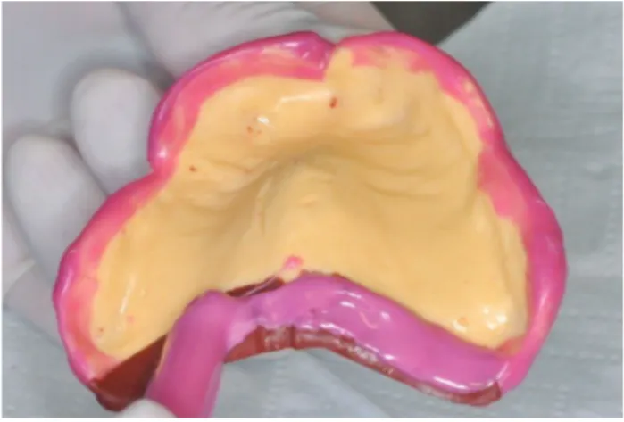
To obtain an acceptable retention it is important, in the second impression, to register the muscles’s function and to edge the impression tray in the peripheral areas (namely oral vestibule and soft palate) with the purpose of giving a selective pressure in order to obtain a particular effect, also known as “suction effect” (Fig. 1). Problems observed with conventional impressions include soft-tissue management, improper impression
1 Department of Prosthodontics and Dental Biomaterials, School of Dental Medicine, University of Siena, Italy 2 Private Practice, Florence, Italy
TO cITe THIS ARTIcle
D’Arienzo lF, D’Arienzo A, Borracchini A. comparison of the suitability of intra-oral scanning with conventional impression of edentulous maxilla in vivo. A preliminary study. J Osseointegr 2018;10(4):115-120.
DOI10.23805 /JO.2018.10.04.02
KeywORDS Digital dentistry; Intraoral scanner; edentulous jaws; mobile prosthesis, complete denture.
FIG. 1 The conventional impression tray also records peripheral areas such as oral vestibule and soft palate.
tray selection, separation of impression material from the impression tray, distortion of conventional impressions before pouring and storage of conventional impressions for potential remaking of casts (1, 2). Furthermore traditional impression has been perceived by patients as an uncomfortable treatment experience (3, 4), in particular for sensitive subjects, such as children and patients with a strong gag reflex (5). In addition, it can be difficult with some impression materials, like for example polysulphides, to remove any excesses from adjacent surfaces or clothes.
In recent years there has been an increased interest in digital technology that has invested several industries worldwide from military to aviation and also to the health care field, including dentistry. In particular, in the latter, the interest on intra-oral scanners has been growing every year and new devices are continuously launched.
The optical impression with intra-oral scanners (IOS) has a whole range of advantages and solves some problems of conventional impression (6). In fact digital impression allows better communication with dental technicians and with patients (7) and gives the possibility of complete digitalization of the CAD/ CAM process with the logical and direct access to the subtractive and additive technologies. Furthermore it solves the patient's discomfort due to retching and unpleasant taste of the impression material (7, 8, 9), eliminates potential allergies to impression materials. It is technically easier for the dentist (10, 11, 12) and especially young clinicians tends to prefer it compared to the conventional impression (13).
Nevertheless digital systems have some disadvantages compared to traditional impression systems, for example it can be difficult to detect deep marginal lines of prepared teeth and/or in case of bleeding (14, 15), it is difficult to impress in a correct way mobile tissues, intra-oral scan tend to modify the impression by reducing trueness when we impress all the jaw, there is a learning curve (13) and there are purchasing and managing costs (16).
However, despite the enhancement that digital impression made, conventional impression remains the most used technique in dentistry, and in particular in prosthesis, both for fixed and mobile prosthesis. As regards the second one, little is know about their feasibility and applicability in digitizing edentulous
jaws. To date, to our knowledge, there is only one study that assessed the accuracy of intra-oral scanner in edentulous jaws (17).
Premise that it is possible to evaluate the accuracy of digital impression exclusively with in vitro studies, using a tabletop reference scanner with high level of accuracy, there’s an important limitation in case of edentulous jaws. In fact edentulous jaws represent a tissue-based clinical situation involving several mobile zones (such as areas of the oral vestibule) and smooth-surface textures covered entirely by saliva that cannot be compared with a gypsum model. So results obtained in this study could not be easily applied in everyday work.
Therefore, the aim of the present study is not to evaluate the accuracy of intra-oral scanners, but rather to evaluate the suitability of using one of the most accurate intra-oral scanners (Trios, 3Shape, Copenhagen, Denmark) (18, 19, 20) to digitize edentulous jaws in vivo and to verify, on the obtained data sets, if it is possible to replace the traditional approach with the digital one in the perspective of mobile prosthetic rehabilitation (complete denture) (Fig. 2).
MATeRiALS AnD MeThoDS
Patients selection
The present pilot study was conducted on totally edentulous patients who needed a complete prosthetic rehabilitation on the upper jaw and were not seeking oral implant therapy.
After receiving clear information about the purpose of the study all patients provided written, informed consent.
The following inclusion criteria were used: agreement with informed consent; male/female aged at least 18 years; good health conditions; fully edentulous in the maxilla, regardless of the conditions in the lower arch; a history of edentulism of at least 3 months.
The following exclusion criteria were adopted: subjects unlikely to be able to comply with the study procedures, as judged by clinicians; systemic diseases or severe medical complications; disabilities; pregnancy; presence of chronic lesion of the maxillary mucosa, such as decubitus lesion, and bone neoformation, such as torus. In total, four patients were recruited and participated in the study (mean age 72.5 years, range 68–78 years; one FIG. 2
Digitization of edentulous jaws.
male, three females). The enrollment was conducted in a private dental office located in Florence, Italy.
Clinical procedures
The patients were treated by two clinicians, the first with expertise in the fields of conventional impression and the second one expert in digital impression. For each patient two impression of the upper arch were taken, one for each clinician. The first impression was taken by the doctor expert in digital impression with an intra-oral scanner (Trios3®); the second impression was taken by the doctor with expertise in the field of conventional impression using an irreversible hydrocolloid impression material (Hydrogum 5, Zhermack) with Schreinemakers impression tray. Impressions were taken always with this sequence in order to not condition the tissues with the impression material before using the IOS.
The intraoral scanner used in this study is one of the most accurate according to the recent literature (18, 19, 20). Trios 3® was launched in 2015 in three different versions: a trolley version with a touch-screen, a version incorporated into the dental treatment unit, and a USB version. We used the last one, plugged via a USB port to a computer with great performance. The Trios scanner (3Shape, Copenhagen, Denmark) is a powder-free, powerful and extremely fast structured light scanner. It works under the principle of confocal microscopy and ultrafast optical scanning, so it continuously captures 2D images from different positions to create a 3D surface and it produces high-quality color images. Therefore, shade matching is automated with this system, which also takes digital photographs, thus allowing the acquisition of high-definition photos for documentation or communication purposes. Unwanted objects (tongue, cheeks or lips) are detected automatically and digitally removed from the digital impression in real time. With the IOSs, the scanning process for the maxillary jaw followed a special pattern, starting at the distobuccal areas of the first quadrant, at the level of the tuber, following the crest to the opposite side, passing through the retroincisor papilla, and finally closing the palatal gaps by moving the scanner head in a zigzag movement over the palate. Finished the scan, it was possible to cut areas of no interest. In fact the acquisition software of Trios 3® used in the present study has further automatic artifacts elimination, an advanced cutting function, combined with smart blocking functions available for
surfaces. The time required for the optical impression of the upper arch was an average of 3 minutes and 6 seconds (respectively: 2.29, 3.11, 3.40 and 3.45), while the total number of 3D images was on average of 1244 (respectively: 1109, 1199, 1241, 1427).
Once the digital impression phase was completed, the traditional one was undertaken. For this purpose, a metal impression tray, suitable for the negative detection of totally edentulous ridges, called Schreinemakers impression tray, was used. The selection of the correct size of the impression tray was made using the appropriate plastic compass, which for the upper arch registers the distance between the vestibular contours of the two maxillary tuberoses. Having chosen the impression tray of the dimension immediately exceeding the recorded distance, both were edged, with an extremely soft material (Cera Azzurrina Morbidissima, Industrie Zingardi, Italy), designed to be applied on the edges of the impression tray in order to extend the extension and to create stops where necessary. So each impression tray was loaded with alginate (Hydrogum 5, Zhermack), suitable for the detection of preliminary impressions in total and partial mobile prostheses. The impression was taken and the patient was then dismissed. In the laboratory, preliminary gypsum models (Type III, Elite Model, Zhermack) were obtained and all the four models were scanned with a laboratory scanner (3Shape D1000).
At this point, both for the intra-oral scanner and for the laboratory scanner, the scanning software saved the data automatically to the STL file format (STereo Lithography interface format). We then used 3-D evaluation software (3DReshaper 2017, Hexagon) to remove artifacts from the visualized data sets and to crop them proximal of the vestibule. Data sets were superimposed by using the best-fit algorithm of the software, and the software automatically performed overall 3-D comparisons (x, y, z coordinates). Finally, the data sets were visually inspected and an analysis was conducted.
ReSULTS
Digitizing edentulous jaw was possible using the IOS of the 3Shape and the differences recorded between the two impression techniques in the four patients are
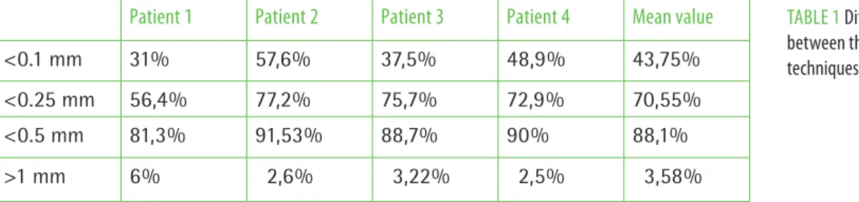
TABle 1
Differences recorded
between the two impression techniques.Patient 1 Patient 2 Patient 3 Patient 4 Mean value
<0.1 mm 31% 57,6% 37,5% 48,9% 43,75%
<0.25 mm 56,4% 77,2% 75,7% 72,9% 70,55%
<0.5 mm 81,3% 91,53% 88,7% 90% 88,1%
shown in Table 1, from which it is possible to infer that on average 10% of the arch recorded differences of more than 500 microns. However, since no similar study is available, it is not possible to specify whether these results are to be considered or not acceptable.
As regards the mean value of differences between the two impression techniques, they ranged from 219 to 347 μm, respectively for each patient: 219, 239, 246 and 347 μm.
The visual analysis of superimposed data sets revealed the greatest deviations in the peripheral areas such as oral vestibule and soft palate, where the impression material showed the maximum compression.
DiSCUSSion
Digital revolution is deeply changing the dental profession, through the launch of multiple devices, like software, CBCT, additive and subtractive manufacturing technologies (21, 22). In fact, nowadays the treatment plan is established by associating clinical evaluations with virtual ones, thanks to image acquisition systems, such as intra-oral scanner (23, 24), photos (25), face scanner (26), and cone beam computed tomography (27). A whole range of therapies (surgical, prosthetic and orthodontic) can be easily planned in virtual and applied in real thanks to 3D modeling and processing software (software for guided implant surgery, for orthodontic dental movement and prosthetic CAD software). For example, with the use of appropriate software of guided surgery the doctor is able to plan the best implant’s position in view of prosthetic rehabilitations and then switch from virtual to real and insert implants in the position that had been previously decided thanks to surgical templates obtained with 3D printers. Finally, using new materials (28), it is possible to fabricate the prosthetic restorations and in other cases also orthodontic devices, through the use of CAM technologies.
Certainly, today, the most attractive device is the IOS, as justified by the large number of studies that investigate its accuracy. Despite their use entails significant advantages for the clinician and for the patient, the diffusion of this device in the daily workflow is still limited although some clinicians and dental operators affirm that IOS are rapidly supplanting traditional impressions (with trays and materials).
The clinical study we discussed here compares the optical impression with the traditional one in an edentulous arch. In fact almost all studies in this regard are in vitro studies, due to the need to have a reference measurement, which can only be obtained with a specific device (laboratory scanner) that can not be used in vivo. Furthermore, available literature focuses on 3-D and linear measurements of impressions or casts of dentate jaws or implanted jaws and, to our knowledge, there is
only one study in which the accuracy of digitization of edentulous jaws with IOSs was evaluated (17). In this
in vitro study the authors scanned two representative
edentulous jaw models with a powerful (manufacturer’s specifications: accurate to within < 20 micrometers) industrial laser scanner (Activity 101, smart optics Sensortechnik, Bochum, Germany), in order to obtain a platform for reference measurements, and then with four different IOSs (Cerec AC Bluecam®, Zfx IntraScan®, Itero® and Lava COS®). The intraoral scans (five per maxillary model and five per mandibular model) were imported into software for superimposition of 3D surface models and then compared with values obtained with the reference scanner in terms of trueness and precision. Mean trueness values ranged from 44.1 to 591.8 micrometers and mean precision values ranged from 21.6 to 698.0 μm. At the end of the study, the distance errors were the smallest and most consistent for the Lava COS®, whereas they were the largest and least consistent for the Cerec Bluecam®. The authors concluded that digitizing edentulous jaw models with the use of IOSs appears to be feasible, although the accuracy of the intra-oral scanners differs significantly, and that further enhancements are necessary to recommend these IOSs for this particular indication (edentulous jaws). Nevertheless the results of this study could not be easily applied in everyday work because the features of a plaster model differs in important way from the edentulous jaw, for example for the presence of mobile areas, a limit that is less evident in case of in
vitro studies that analyze the accuracy of scans of tooth
or abutments. So, although the results of this in vitro study is very promising, there are some limitations when we scan the oral cavity.
Scanning in the mouth may double the error compared to scanning a model due to the different environment (29). In fact, the optical impression involves the management of soft tissues and the elimination of oral fluids (blood, saliva, crevicular fluid) exactly like the traditional impression. Another difference between in vivo and in
vitro scanning is the stability of the scanning surface, in
fact the shape of the mucosa can change depending on the jaw movements, which can complicate the scanning procedure (30). So, in particular for the lower arch, it is difficult to obtain repeatability of intraoral scans for full-arch impressions, also because the different intraoral acquisition strategies can influence the final result (31).
Therefore, the difficulty of translating in vivo the data obtained in vitro prompted us to perform an in vivo study.
So it is important to underline that the aim of the present study is not the search of accuracy, in terms of trueness and precision, of the Trios intra-oral scanner. The aim of the present in vivo study is to compare two different impression techniques with the purpose of evaluating the existing differences and analyze if the optical one
can be taken into consideration as an alternative to the conventional impression in view of mobile prosthetic rehabilitation, starting from the assumption that the areas in which it would be desirable to have the fewest differences are represented by those that fall within the definition of “peripheral seal”. This structure plays an important role in the retention of the prosthetic device, for this reason the actual trends forecast two appointment to impress the arch. In the first appointment the preliminary impression is made, which has as fundamental requirement to be overextended. On the model obtained from this impression, an individual impression tray will be then built up, thanks to which the final impression, also know as “functional impression”, will be executed. The retention of the mobile prosthesis is strictly linked to the “functional impression”, which should exert a selective pressure on peripheral areas and be mucostatic in the central structures.
To analyze the differences between the two impression techniques it has been used a dedicated software for superimposition of the resultant STL datasets, that represent an efficient technique to measure and compare differences at the microscopic level (32). The superimposition of STL datasets can be performed by “best-fit algorithm”, “least squares method” and the “zero method”. In the present study the first one was used, that it is one of the most common methodologies used to investigate the accuracy of impression (33). The mean value of difference between the two impression techniques ranged from 219 to 347 μm, and on average only 10% of the arch recorded differences exceeding 500 microns.
However, what we are interested in is another result. In fact, we focalized our attention in the research of that areas in which the impression material gives more compression than the intra-oral scanner. As it was possible to expect the hydrocolloid gives in all the
cases more compression in the peripheral areas, whereas it is not possible to register the same result in other places, so in general in almost the whole jaw covered by keratinized tissue.
In Figure 3 it is possible to deduce where the impression material gives more compression than the intra-oral scanner (areas colored in blue and red) and the differences recorded are somewhere also bigger than 500 micron (areas depicted in blue), in particular in areas such as soft palate and buccal vestibule, less in labial vestibule.
Furthermore, it is important to remember that these differences were recorded between IOS and preliminary impression (using an irreversible hydrocolloid). So, supposedly, these differences would have been even greater if IOS and functional impressions were compared.
ConCLUSion
Although the results of our study show that digitization of edentulous jaw was feasible with the use of the Trios scanner, the high levels of difference in compression in the peripheral sealing zone lead us to conclude that the use of optical scanning can be considered valid only to replace the preliminary impression, so to obtain a model thanks to which it is possible to build an individual impression tray. In fact, in view of mobile prosthetic rehabilitation, it is fundamental to exert a selective pressure in peripheral areas, that is currently not possible without the functional impression.
ReCoMMenDATionS
Assuming that a good fitting prosthesis is fundamental to avoid complications and assure the longevity of the construction, the literature must focus on the research of an alternative to the functional impression. Otherwise it can be possible to obtain a mobile prosthesis with a total digital workflow, but the retention of devices obtained with intra-oral scans will surely be lower than that obtained with conventional techniques.
In our opinion, a good solution could be the use of contact scanner, so scanner that can physically probe the surface of the jaw and exert a selective pressure on tissues, although it must be remembered that the physical contact with the probe can somehow modify tissues in a way not desirable. Furthermore contact scanners exist only for laboratory and they are slow and expensive.
Acknowledgments
The authors received no financial support.
1) L.F. D’Arienzo contributed to conception, design, data analysis, drafted and critically revised the manuscript; A. D’Arienzo contributed to conception and data FIG. 3
Areas where the impression material gives more compression than
the intra-oral scanner (in blue and red), there are also differences bigger than 500 micron (blue).
analysis, critically revised the manuscript; A. Borracchini contributed to conception and design, critically revised the manuscript.
All authors gave final approval.
2) The authors declare no potential conflicts of interest with respect to the authorship and/or publication of this article.
3) The present study was presented in the form of a poster in the “collegio docenti” (2018) held in Rome. Here it was awarded among more than 500 works with the “mention of honor".
BiBLioGRAFiA
1. christensen GJ. Impressions are changing: deciding on conventional, digital or digital plus in-office milling. JADA 2009;140(10): 1301-1304.
2. christensen GJ. will digital impressions eliminate the current problems with conventional impressions? JADA 2008;139(6):761-763.
3. Burhardt l, livas c, Kerdijk w, van der Meer wJ, Ren y. Treatment comfort, time perception, and preference for conventional and digital impression techniques: A comparative study in young patients. Am J Orthod Dentofacial Orthop 2016 Aug;150(2):261-7.
4. Hacker T, Heydecke G, Reissmann DR. Impact of procedures during prosthodontic treatment on patients' perceived burdens. J Dent 2015;43:51-7.
5. Dergović N, Stosić Z. Gagging reflex and vomiting in prosthetic dentistry. Stomatol Glas Srb 1971 Aug-Oct;18(4):268-72.
6. Zimmermann M, Mehl A, Mörmann wH, Reich S. Intraoral scanning systems. - a current overview. Int J comput Dent 2015;18(2):101–29. 7. Goracci c, Franchi l, Vichi A, Ferrari M. Accuracy, reliability, and efficiency
of intraoral scanners for full-arch impressions: a systematic review of the clinical evidence. eur J Orthod 2016;38(4):422–428.
8. yuzbasioglu e, Kurt H, Turunc R, Bilir H. comparison of digital and conventional impression techniques: evaluation of patients' perception, treatment comfort, effectiveness and clinical outcomes. BMc Oral Health 2014;14((10):7.
9. wismeijer D, Mans R, van Genuchten M, Reijers HA. Patients' preferences when comparing analogue implant impressions using a polyether impression material versus digital impressions (Intraoral Scan) of dental implants. clin Oral Implants Res 2014;25(10):1113–8.
10. Joda T, lenherr P, Dedem P, Kovaltschuk I, Bragger U, Zitzmann NU. Time efficiency, difficulty, and operator's preference comparing digital and conventional implant impressions: a randomized controlled trial. clin Oral Implants Res 2016 Sep 5.
11. Park HR, Park JM, chun yS, lee KN, Kim M. changes in views on digital intraoral scanners among dental hygienists after training in digital impression taking. BMc Oral Health 2015;15(1):7.
12. Joda T, Bragger U. Time-efficiency analysis comparing digital and conventional workflows for implant crowns: a prospective clinical crossover trial. Int J Oral Maxillofac Implants 2015;30((5):1047–53.
13. lee SJ, Macarthur RX 4th, Gallucci GO. An evaluation of student and clinician perception of digital and conventional implant impressions. J Prosthet Dent 2013 Nov;110(5):420-3.
14. Martin cB, chalmers eV, McIntyre GT, cochrane H, Mossey PA. Orthodontic scanners: what's available? J Orthod 2015; 42(2):136–143.
15. Mandelli F, Ferrini F, Gastaldi G, Gherlone e, Ferrari M. Improvement of a Digital Impression with conventional Materials: Overcoming Intraoral Scanner limitations. Int J Prosthodont 2017;30(4):373–376.
16. Mangano F, Gandolfi A, luongo G, logozzo S. Intraoral scanners in dentistry: a review of the current literature. BMc Oral Health 2017 Dec 12;17(1):149. 17. Patzelt SB, Vonau S, Stampf S, Att w. Assessing the feasibility and accuracy
of digitizing edentulous jaws. J Am Dent Assoc 2013 Aug;144(8):914-20. 18. Vandeweghe S, Vervack V, Dierens M, De Bruyn H. Accuracy of digital
impressions of multiple dental implants: an in vitro study. clin Oral Implants Res 2017 Jun;28(6):648-653.
19. Imburgia M, logozzo S, Hauschild U, Veronesi G, Mangano c, Mangano FG. Accuracy of four intraoral scanners in oral implantology: a comparative in vitro study. BMc Oral Health 2017 Jun 2;17(1):92.
20. Patzelt SB. evaluation of the Accuracy of Six Intraoral Scanning Devices: An in-¬vitro Investigation ADA 2015.
21. van Noort R. The future of dental devices is digital. Dent Mater. 2012;28(1):3–12. 31.
22. Mangano F. Digital Dentistry: The Revolution has Begun. Open Dent J 2018 Jan 31;12:59-60.
23. Joda T, Zarone F, Ferrari M. The complete digital workflow in fixed prosthodontics: a systematic review. BMc Oral Health 2017 Sep 19;17(1):124. 24. Aragón Ml, Pontes lF, Bichara lM, Flores-Mir c, Normando D. Validity and reliability of intraoral scanners compared to conventional gypsum models measurements: a systematic review. eur J Orthod 2016 Aug;38(4):429-34. 25. Zimmermann M, Mehl A. Virtual smile design systems: a current review. Int
J comput Dent 2015;18(4):303-17.
26. Joda T, Brägger U, Gallucci G. Systematic literature review of digital three-dimensional superimposition techniques to create virtual dental patients. Int J Oral Maxillofac Implants 2015;30(2):330–7.
27. Benavides e, Rios HF, Ganz SD, An cH, Resnik R, Reardon GT, Feldman SJ, Mah JK, Hatcher D, Kim MJ, Sohn DS, Palti A, Perel Ml, Judy Kw, Misch ce, wang Hl. Use of cone beam computed tomography in implant dentistry: the International congress of Oral Implantologists consensus report. Implant Dent 2012 Apr;21(2):78-86.
28. Zarone F, Ferrari M, Mangano FG, leone R, Sorrentino R. “Digitally Oriented Materials”: Focus on lithium Disilicate ceramics. Int J Dent 2016;2016:9840594.
29. Flugge, T.V., Schlager, S., Nelson, K., Nahles, S. & Metzger, M.c. Precision of intraoral digital dental impressions with itero and extraoral digitization with the itero and a model scanner. Am J Orthod Dentofacial Orthop 2013 Sep;144(3):471-8.
30. Andriessen FS, Rijkens DR, van der Meer wJ, wismeijer Dw. Applicability and accuracy of an intraoral scanner for scanning multiple implants in edentulous mandibles: a pilot study. J Prosthet Dent 2014 Mar;111(3):186-94.
31. ender A, Mehl A. Influence of scanning strategies on the accuracy of digital intraoral scanning systems. Internat J computerized Dentistry 2013; 16:11- 21.
32. ender A, Mehl A. Accuracy of complete-arch dental impressions: a new method of measuring trueness and precision. J Prosthetic Dentistry 2013; 109:121- 8.4.
33. Güth JF, Keul c, Stimmelmayr M, Beuer F, edelhoff D. Accuracy of digital models obtained by direct and indirect data capturing. clinical Oral Investigations 2013; 17:1201-8.