1655
Identification of a New Locus for Medullary Cystic Disease, on
Chromosome 16p12
Francesco Scolari,
1Daniela Puzzer,
4Antonio Amoroso,
4Gianluca Caridi,
5Gian Marco Ghiggeri,
5Rosario Maiorca,
1Paolo Aridon,
2Maurizio De Fusco,
2Andrea Ballabio,
2,3and Giorgio Casari
21Division and Chair of Nephrology, Spedali Civili and University of Brescia, Brescia,2Telethon Institute of Genetics and Medicine and 3Universita Vita-Salute, San Raffaele Biomedical Science Park, Milan,4Genetics Service, Istituto per l’Infanzia Burlo Garofolo and University of Trieste, Trieste, and5Laboratory of Nephrology, G. Gaslini Institute, Genoa, Italy
Summary
Autosomal dominant medullary cystic disease
(ADMCKD) is an interstitial nephropathy that has mor-phologic and clinical features similar to autosomal re-cessive nephronophthisis. The typical renal dysfunction associated with ADMCKD results mainly from a defect in urinary concentration ability, although results of uri-nalysis are normal. Recently, a locus on chromosome 1 was associated with ADMCKD, in DNA from two large Cypriot families, and genetic heterogeneity was inferred. We describe the genomewide linkage mapping of a new locus for medullary cystic disease, ADMCKD2, on chro-mosome 16p12 in a four-generation Italian pedigree. The family with ADMCKD2 fulfills the typical diag-nostic criteria of ADMCKD, complicated by hyperuri-cemia and gouty arthritis. Marker D16S3036 shows a maximum two-point LOD score of 3.68, and the defined critical region spans 10.5 cM, between D16S500 and SCNN1B1–2. Candidate genes included in the critical region are discussed.
Introduction
Autosomal dominant medullary cystic disease (ADMCKD; MIM 174000) is a renal disorder charac-terized by structural defects in the renal tubules, which lead to a reduction of urine concentrating ability and a decrease in sodium conservation. The disease progresses toward renal failure, which generally requires dialysis Received October 26, 1998; accepted for publication March 16, 1999; electronically published April 23, 1999.
Address for correspondence and reprints: Dr. Giorgio Casari, Tel-ethon Institute of Genetics and Medicine, San Raffaele Biomedical Science Park, Via Olgettina 58, 20132 Milan, Italy. E-mail: casari @tigem.it
q 1999 by The American Society of Human Genetics. All rights reserved. 0002-9297/99/6406-0018$02.00
or transplantation in patients aged !50 years. The
dis-ease is, however, heterogeneous in several clinical as-pects, including age at onset, rate of progression, and clinical appearance. This variability contributes to the diagnosis of ADMCKD being either rare or confused with other clinical entities. ADMCKD is usually diag-nosed in the third or fourth decade of life; however, cases of children with the disease, with slower progression toward renal failure, are not uncommon (Gardner 1971). Renal symptoms are mainly defects in urinary concentration ability with normal results of urinalysis.
The presence of small cysts in renal medulla is a central feature of the disease, although the cysts may be absent in the early stages. Discovery of these medullary cysts may also be confounded by the presence of cysts ac-quired as a result of chronic renal failure, in some pa-tients. In the most frequent presentation of ADMCKD, which is characterized by tubulointerstitial fibrosis and thickening of the tubular basement membrane, renal pathologic findings are not specific and are similar to those found in other pathological entities such as juve-nile-onset nephronophthisis (NPH; MIM 256100). The major difference between ADMCKD and NPH is the relative inheritance pattern, which is recessive in the lat-ter, with typical onset of renal symptoms in early child-hood and rapid evolution toward renal failure in the second decade of life (Hildebrandt et al. 1992; Neumann et al. 1997). Recent advances in the molecular genetics of NPH have helped differentiate between the two en-tities (Hildebrandt et al. 1997; Saunier et al. 1997; Chris-todoulou et al. 1998; Fuchshuber et al. 1998; Scolari et al. 1998; Stavrou et al. 1998).
A clear-cut definition for diagnosis of ADMCKD was needed for further differentiation among ADMCKD phenotypes. This led us to define several criteria for the diagnosis of ADMCKD (Scolari et al. 1998; see Meth-ods), on the basis of family inheritance and clinical and pathological characteristics, including associated symp-toms such as hyperuricemia. Recently, genetic linkage of ADMCKD to the ADMCKD1 locus was established on chromosome 1 (Christodoulou et al. 1998). In the
pre-Figure 1 Pedigree of the family with ADMCKD2. Haplotypes for markers D16S405, D16S500, D16S3017, and markers D16S3036, D16S3041, SCNN1B1–2, and D16S420, are shown from top to bottom. The disease-associated haplotype is boxed. Second-generation inferred haplotypes are shown between brackets.
sent article, we report genomewide linkage analysis of DNA from a large family and identification of a new locus associated with ADMCKD, thus showing the ge-netic heterogeneity of this clinically homogeneous renal disorder.
Subjects and Methods
Subjects and Diagnostic Criteria
The four-generation Italian family, which included 10 members (4 men and 6 women) affected by ADMCKD and diagnosed as indicated below, is depicted in figure 1. Four affected members (III-3, III-6, III-9, and IV-6) were given a clinical and histological diagnosis of ADMCKD; three members (II-3, III-4, and IV-1) had only a clinical diagnosis of ADMCKD; and three de-ceased relatives (I-1, II-1, and II-4) had a diagnosis of chronic nephritis listed on their death certificates. Five affected family members (II-3, III-3, III-6, III-9, and IV-6) had hyperuricemia (that required specific treatment with allopurinol) associated with ADMCKD; in two subjects (II-3 and III-9), hyperuricemia was complicated
by gouty arthritis. The age at diagnosis of ADMCKD ranged from 16 to 54 years (mean age, 31.5 years). Renal cysts were documented by sonogram in one subject (II-3). Twelve available members (1, 2, 5, 7, III-8, III-10, III-11, III-12, IV-2, IV-3, IV-4, and IV-5) were screened and proved to be negative for the disease (see table 1).
Diagnostic criteria were as follows: (1) autosomal dominant inheritance; (2) defective urine concentration with polyuria, isosthenuria, and relatively normal uri-nalysis results; (3) normal- or small-sized kidneys with occasional small medullary cysts; and (4) renal histologic findings characterized by tubular-interstitial fibrosis with infiltrates, tubular atrophy, and thickening of the tubular basement membrane with periodic acid Schiff–positive material. Available family members were considered healthy if they had results showing normal renal func-tion, negative renal sonography, negative urinalysis, and urinary osmolality 1800 mOsm/l, after overnight
de-hydration. In subjects with chronic renal insufficiency, hyperuricemia was defined as an elevation of plasma urate levels disproportionate to the degree of renal
in-Table 1
Clinical, Biochemical, and Histological Findings of ADMCKD2 Family
Subject Diagnosis Age at Diagnosis_Age at Screening (years) Morning Urinary Osmolarity (mOsm
H2O/kg) Renal Ultrasound
Renal Histology Serum Creatinine/ Uric Acid (mg/dl)
II-3 ADMCKD 54_ 300 Small kidneys with cysts ND 2/12.0
III-1 NO _44 860 Negative ND .8/4.7
III-2 NO _43 900 Negative ND .9/ND
III-3 ADMCKD 18_ 300 Small kidneys w/o cysts TIN 1.5/9.6
III-4 ADMCKD 25_ 330 Small kidneys w/o cysts ND 1.6/5.9
III-5 NO _36 861 Negative ND .8/ND
III-6 ADMCKD 30_ 315 Small kidneys w/o cysts TIN 1.3/9.2
III-7 NO _45 900 Negative ND .9/4.5
III-8 NO _44 895 Negative ND .7/4.2
III-9 ADMCKD 29_ 295 Small kidneys w/o cysts TIN 1.7/12
III-10 NO _38 910 Negative ND .97/2.8
III-11 NO _37 805 Negative ND .8/ND
III-12 NO _35 900 Negative ND .9/3.1
IV-1 ADMCKD 16_ 310 Small kidneys w/o cysts ND 1.6/5.5
IV-2 NO _23 1054 Negative ND .7/2.8
IV-3 NO _21 1183 Negative ND .8/
IV-4 NO _22 857 Negative ND .7/2.3
IV-5 NO _10 812 Negative ND .6/ND
IV-6 ADMCKD 18_ 330 Normal kidneys w/o cysts TIN 1.5/9.5
NOTE.—NO5 normal phenotype, w/o 5 without, ND 5 not done, and TIN 5 Tubulo-interstitial nephritis. sufficiency in subjects with chronic renal insufficiency
(Murray and Goldberg 1975). Linkage Analysis
After informed consent, a 10-ml venous blood sample was obtained from six affecteds and seven healthy rel-atives. DNA was extracted from peripheral blood lym-phocytes as described elsewhere (Sambrook et al. 1989). A genomewide microsatellite screening was done with 358 highly polymorphic fluorescent markers (Gyapay et al. 1994; Dib et al. 1996). This set of markers, which are formatted and properly fluorescence-labeled to allow efficient multiplexing on an automated sequencer, covers all the autosomal chromosomes at an average reciprocal distance of∼10 cM.
The DNA from six affected members was subjected to genomewide screening, and this preliminary set of linkage data was analyzed with the MLINK program of LINKAGE (Lathrop et al. 1984). Selected chromosomal regions were further investigated with additional mark-ers mapping telomeric and centromeric to the candidate loci. For linkage calculations, ADMCKD was modeled as an autosomal dominant trait with two liability classes for age-dependent penetrance: 0.5 for age!15 years and
0.9 for age115 years. The mutation rate was assumed
to be 1025. Recombination frequencies (v) were
consid-ered to be equal between male and female subjects. Marker alleles were assumed to be equifrequent in the preliminary screening, whereas published allelic fre-quencies were used for candidate loci calculations.
Mul-tipoint linkage analysis was done by LINKMAP (La-throp et al. 1984), with the same parameters.
Results
The loci responsible for autosomal recessive NPH1 (Hildebrandt et al. 1997) and the ADMCKD1 form (Christodoulou et al. 1998) were excluded from linkage with the family in our previous study (Scolari et al. 1998; G. Casari, unpublished material). We therefore consid-ered this pedigree for genomewide linkage mapping. We tested the probability of detecting significant linkage by simulation analysis of 400 replicate pedigrees by using SLINK (Weeks et al. 1990), considering the phenotype with a frequency of 1 # 1025. The maximum pairwise LOD score obtained from 400 simulated pedigrees was 4.01 at v5 .02.A genomewide search was performed on all affected pedigree members. The selected loci sug-gestive of linkage (LOD score 11.0) were further
char-acterized by testing all pedigree members’ DNA and by locally increasing marker density.
The chromosome 16p locus, preliminarily identified by the fluorescent markers D16S407 and D16S405 (LOD scores of 1.36 and 2.10 at v5 .1 and .05, re-spectively), was confirmed with flanking markers. As reported in table 2, marker D16S3036 achieves a max-imum value of 3.68 with no recombination within the disease locus. A 10.5-cM critical region between markers D16S500 and SCNN1B1–2 (a polymorphic dinucleotide repeat intragenic to the beta subunit of the epithelial
Table 2
Two-Point LOD Score Values for 16p13–12 Markers
MARKER LOD SCORE ATv5 .00 .01 .05 .10 .20 .30 .40 D16S423 25.98 21.43 2.54 2.14 .15 .15 .06 D16S407 23.29 .69 1.27 1.36 1.15 .73 .24 D16S3114 .00 .02 .09 .13 .16 .14 .09 D16S405 22.28 1.66 2.10 2.06 1.64 1.02 .34 D16S500 21.91 2.02 2.43 2.35 1.87 1.20 .47 D16S3017 2.71 2.66 2.45 2.19 1.64 1.04 .41 D16S3036 3.69 3.62 3.36 3.01 2.27 1.46 .59 D16S3041 1.32 1.30 1.21 1.08 .83 .56 .28 SCNN1B1-2 22.97 2.30 .29 .45 .43 .26 .07 D16S420 24.38 2.38 .35 .62 .71 .51 .19
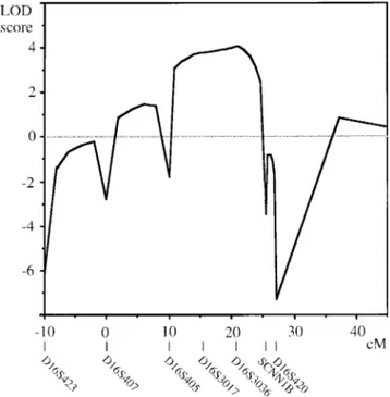
Figure 2 Multipoint linkage graph of the region surrounding the ADMCKD2 locus.
sodium channel; Shimkets et al. 1994) was defined by recombinant pedigree members. A common haplotype (6_6_4) for markers D16S3017, D16S3036, and D16S3041 is shared by all affected individuals (fig. 1); individual IV-5 is a carrier of the disease-associated hap-lotype but does not show the ADMCKD phenotype, probably because of his young age (10 years), which is much less than the age at onset among the other affected family members (aged 16–20 years; table 1). Data from subject III-4 was observed to be a double recombinant for marker D16S3036 (or for markers D16S3017 and D16S3036, because D16S3017 is not informative) in the unaffected maternal haplotype.
The multipoint linkage graph (fig. 2) shows a multi-point LOD score of 4.18 around D16S3036, thus iden-tifying a new locus for ADMCKD that we named “ADMCKD2.” Several partially characterized tran-scripts (GeneMap98), and some known genes, map to this critical region, including a phosphodiesterase gene (Bolger et al. 1993), the multidrug resistance-associated protein (Cole et al. 1992), the alanine aminotransferase (Sohocki et al. 1997), the ubiquinol-cytochrome c re-ductase complex core protein 2 precursor (Hosokawa et al. 1989), the major Yo paraneoplastic antigen (Fa-thallah Shaykh et al. 1991), the mu-crystallin (Kim et al. 1992), and the uromodulin/Tamm-Horsfall glyco-protein (Pennica et al. 1987). The genes coding for the beta and gamma subunits of the kidney epithelial sodium channel (SCNN1B and SCNN1G; Voilley et al. 1995) are both localized to a 400-kb area in 16p12 at the centromeric boundary of the critical region and have been excluded from linkage to ADMCKD2 by the in-tragenic marker SCNN1B1–2.
Discussion
ADMCKD has been considered a rare disease, on oc-casion, because of a paucity of symptoms at early stages and aspecific clinical and pathological features once re-nal failure has developed. Furthermore, because of sev-eral clinical and pathological similarities, ADMCKD has
been easily confused with juvenile NPH. Advances in the molecular genetics of renal cyst diseases have con-tributed to a definite differentiation between these two clinical entities. Recently, linkage analysis and positional cloning approaches to recessive juvenile NPH have con-tributed to the identification of a candidate gene map-ping to chromosome 2q13, which is deleted in165% of
patients with NPH1 (Hildebrandt et al. 1997).
An autosomal dominant form of ADMCKD has been mapped to chromosome 1q21 in DNA from members of two large Cypriot families affected with renal cysts, hyperuricemia, and gout (Stavrou et al. 1998). No ob-vious candidate genes have been proposed. The identi-fication of a common haplotype in both families with ADMCKD is probably due to a founder effect, fre-quently observed in small island communities. In the present article, we show evidence of linkage of ADMCKD2 associated with chromosome 16p12 in DNA from a large Italian family. In addition to the typ-ical renal cysts, affected members show hyperuricemia and gout. These symptoms are also found in the Cypriot families and seem to identify a more homogeneous sub-group of ADMCKD. Despite symptom and clinical ho-mogeneity, the data in the present article show genetic heterogeneity.
The 10.5-cM critical region of ADMCKD2 is dense with transcripts (see Results), and the considerable size of this genomic area renders a positional cloning ap-proach difficult. Nonetheless, the localization of the uro-modulin (also known as “Tamm-Horsfall protein”) gene
to the ADMCKD2 critical region (Pook et al. 1993) is interesting. Uromodulin is expressed mainly in the kid-ney, where it is localized to the epithelial cells of the thick ascending limb (TAL) of the Henle loops (Allen and Tischer 1976), and has been functionally associated with the water nonpermeability of the TAL (Sikri et al. 1978), a function that is altered in ADMCKD. Further-more, abnormal peritubular and interstitial deposits of uromodulin have been considered characteristic of NPH and tubulointerstitial inflammatory disease (Resnick et al. 1978; Zanger et al. 1978). However, DNA from ad-ditional pedigrees will need to be analyzed to refine the linkage mapping of the ADMCKD2 locus and to suit-ably narrow the candidate region for a positional cloning approach. The identification of the new ADMCKD2 lo-cus and evidence of genetic heterogeneity, notwithstand-ing the shared common phenotype, will further contrib-ute to the understanding of cystic disease pathogenesis.
Acknowledgments
We would like to thank the subjects involved in this study for their cooperation and Ms. M. Smith for help in preparation of the manuscript. We also thank Dr. S. Carrabino for geno-typing assistance. This work was supported by the Italian Tel-ethon Foundation (Grant E770 to TelTel-ethon Institute of Ge-netics and Medicine and G.M.G.) and by Health Ministry grant P.R.C. 51/98.
Electronic-Database Information
Accession numbers and URLs for data in this article are as follows:
GeneMap98, http://www.ncbi.nlm.nih.gov/genemap98/ (a new gene map of the human genome)
Online Mendelian Inheritance in Man (OMIM), http://www .ncbi.nlm.nih.gov/Omim (for ADMCKD [MIM 174000] and NPH [MIM 256100])
References
Allen F, Tischer CC (1976) Morphology of the ascending thick limb of Henle. Kidney Int 9:8–22
Bolger G, Michaeli T, Martins T, St. John T, Steiner B, Rodgers L, Riggs M, et al (1993) A family of human phosphodies-terases homologous to the dunce learning and memory gene product of Drosophila melanogaster are potential targets for antidepressant drugs. Mol Cell Biol 13:6558–6571 Christodoulou K, Tsingis M, Stavrou C, Eleftheriou A,
Pa-papavlou P, Patsalis PC, Ioannou P, et al (1998) Chromo-some 1 localization of a gene for autosomal dominant med-ullary cystic kidney disease. Hum Mol Genet 7:905–911 Cole SP, Bhardwaj G, Gerlach JH, Mackie JE, Grant CE,
Almquist KC, Stewart AJ, et al (1992) Overexpression of a transporter gene in a multidrug-resistant human lung cancer cell line. Science 258:1650–1654
Dib C, Faure´ S, Fizames C, Samson D, Drouot N, Vignal A, Millasseau P, et al (1996) A comprehensive genetic map of
the human genome based on 5,264 microsatellites. Nature 380:152–154
Fathallah Shaykh H, Wolf S, Wong E, Posner JB, Furneaux HM (1991) Cloning of a leucine-zipper protein recognized by the sera of patients with antibody-associated paraneo-plastic cerebellar degeneration. Proc Natl Acad Sci USA 88: 3451–3454
Fuchshuber A, Deltas CC, Berthold S, Stavrou C, Vollmer M, Burton C, Feest T, et al (1998) Autosomal dominant med-ullary cystic kidney disease: evidence of gene locus hetero-geneity. Nephrol Dial Transplant 13:1955–1957
Gardner KD Jr (1971) Evolution of clinical signs in adult-onset cystic disease of the renal medulla. Ann Intern Med 74: 47–54
Gyapay G, Morissette J, Vignal A, Dib C, Fizames C, Millas-seau P, Marc S, et al (1994) The 1993–94 Ge´ne´thon human genetic linkage map. Nat Genet 7:246–339
Hildebrandt F, Otto E, Rensing C, Nothwang HG, Vollmer M, Adolphs J, Hanusch H, et al (1997) A novel gene en-coding an SH3 domain protein is mutated in nephronoph-thisis type 1. Nat Genet 17:149–153
Hildebrandt F, Waldherr R, Kutt R, Brandis M (1992) The nephronophthisis complex: clinical and genetic aspects. Clin Invest 70:802–808
Hosokawa Y, Suzuki H, Toda H, Nishikimi M, Ozawa T (1989) Complementary DNA encoding core protein II of human mitochondrial cytochrome bc1 complex: substantial diversity in deduced primary structure from its yeast coun-terpart. J Biol Chem 264:13483–13488
Kim RY, Gasser R, Wistow GJ (1992) mu-crystallin is a mam-malian homologue of agrobacterium ornithine cyclodeam-inase and is expressed in human retina. Proc Natl Acad Sci USA 89:9292–9296
Lathrop GM, Lalouel JM, Julier C, Ott J (1984) Strategies for multilocus linkage analysis in humans. Proc Natl Acad Sci USA 81:3443–3446
Murray T, Goldberg M (1975) Chronic interstitial nephritis: etiologic factors. Ann Intern Med 82:453
Neumann HP, Zauner I, Strahm B, Bender BU, Schollmeyer P, Blum U, Rohrbach R, et al (1997) Late occurrence of cysts in autosomal dominant medullary cystic kidney disease. Ne-phrol Dial Transplant 12:1242–1246
Pennica D, Kohr WJ, Kuang WJ, Glaister D, Aggarwal BB, Chen EY, Goeddel DV (1987) Identification of human uro-modulin as the Tamm-Horsfall urinary glycoprotein. Science 236:83–88
Pook MA, Jeremiah S, Scheinman SJ, Povey S, Thakker RV (1993) Localization of the Tamm-Horsfall glycoprotein (uromodulin) gene to chromosome 16p12.3–16p13.11. Ann Hum Genet 57:285–290
Resnick JS, Sisson S, Vernier RL (1978) Tamm-Horsfall protein abnormal localization in renal disease. Lab Invest 38: 550–555
Sambrook J, Fritsch EF, Maniatis T (1989) Molecular cloning: a laboratory manual. Cold Spring Harbor Laboratory Press, Cold Spring Harbor, NY
Saunier S, Calado J, Heilig R, Silbermann F, Benessy F, Morin G, Konrad M, et al (1997) A novel gene that encodes a protein with a putative src homology 3 domain is a can-didate gene for familial juvenile nephronophthisis. Hum Mol Genet 6:2317–2323
Scolari F, Ghiggeri GM, Casari G, Amoroso A, Puzzer D, Car-idi G, Valzorio B, et al (1998) Autosomal dominant med-ullary cystic disease: a disorder with variable clinical pictures and absence of linkage with the NPH1 locus. Nephrol Dial Transplant 13:2536–2546
Shimkets RA, Warnock DG, Bositis CM, Nelson-Williams C, Hansson JH, Schambelan M, Gill JRJ, et al (1994) Liddle’s syndrome: heritable human hypertension caused by muta-tions in the beta subunit of the epithelial sodium channel. Cell 79:407–414.
Sikri KL, Foster CL, Bloomfield FJ, Marshall RD (1978) Co-localization by immunofluorescence and by light- and elec-tron-microscopic immunoperoxidase techniques of Tamm-Horsfall glycoprotein in adult hamster kidney. Biochem J 181:525–532
Sohocki MM, Sullivan LS, Harrison WR, Sodergren EJ, Elder FF, Weinstock G, Tanase S, et al (1997) Human glutamate pyruvate transaminase (GPT): localization to 8q24.3, cDNA
and genomic sequences, and polymorphic sites. Genomics 40:247–252
Stavrou C, Pierides A, Zouvani I, Kyriacou K, Antignac C, Neophytou P, Christodoulou K, et al (1998) Medullary cys-tic kidney disease with hyperuricemia and gout in a large Cypriot family: no allelism with nephronophthisis type 1. Am J Med Genet 77:149–154
Voilley N, Bassilana F, Mignon C, Merscher S, Mattei MG, Carle GF, Lazdunski M, et al (1995) Cloning, chromosomal localization, and physical linkage of the beta and gamma subunits (SCNN1B and SCNN1G) of the human epithelial amiloride-sensitive sodium channel. Genomics 28:560–565 Weeks DE, Ott J, Lathrop GM (1990) SLINK: a general sim-ulation program for linkage analysis. Am J Hum Genet Suppl 47:A204
Zanger RA, Cotran RS, Hoyer JR (1978) Pathologic locali-zation of Tamm-Horsfall protein in interstitial deposit in renal disease. Lab Invest 38:52–57