Fiona Cuskin,
Harry J. Gilbert,
Carme Rovira,
*
Ethan D. Goddard-Borger,
*
Spencer J. Williams,
*
,‡and Gideon J. Davies
*
,††York Structural Biology Laboratory, Department of Chemistry, University of York, Heslington, YO10 5DD, U.K.
‡School of Chemistry and Bio21 Molecular Science and Biotechnology Institute and∥Department of Medical Biology, University of Melbourne, Parkville, Victoria 3010, Australia
§ACRF Chemical Biology Division, The Walter and Eliza Hall Institute of Medical Research, Parkville, Victoria 3010, Australia ⊥Departament de Química Inorgànica i Orgànica (Secció de Química Orgànica) & IQTCUB, Universitat de Barcelona, 08028
Barcelona, Spain
#Institute for Cell and Molecular Biosciences, The Medical School, Newcastle University, Framlington Place, Newcastle upon Tyne NE2 4HH, U.K.
∇Institució Catalana de Recerca i Estudis Avançats (ICREA), 08020 Barcelona, Spain
*
S Supporting InformationABSTRACT: The enzymatic cleavage of β-1,4-mannans is achieved by endo-β-1,4-mannanases, enzymes involved in germination of seeds and microbial hemicellulose degradation, and which have increasing industrial and consumer product applications. β-Mannanases occur in a range of families of the CAZy sequence-based glycoside hydrolase (GH) classification scheme including families 5, 26, and 113. In this work we reveal that β-mannanases of the newly described GH family 134 differ from other mannanase families in both their mechanism and tertiary structure. A representative GH family 134 endo- β-1,4-mannanase from a Streptomyces sp. displays a fold closely related to that of hen egg white lysozyme but acts with inversion of stereochemistry. A Michaelis complex with mannopentaose, and a product complex with mannotriose, reveal ligands with pyranose rings distorted in an unusual inverted chair conformation. Ab initio quantum mechanics/ molecular mechanics metadynamics quantified the energetically accessible ring conformations and provided evidence in support of a1C4→3H4‡→3S1conformational
itinerary along the reaction coordinate. This work, in concert with that on GH family 124
cellulases, reveals how the lysozyme fold can be co-opted to catalyze the hydrolysis of different polysaccharides in a mechanistically distinct manner.
■
INTRODUCTIONβ-1,4-Mannans are a diverse group of polysaccharides present within the plant cell wall and endosperm.1Their degradation is
catalyzed by β-1,4-mannanases and β-mannosidases, and
assorted auxiliary enzymes, which act in concert to deconstruct theβ-1,4-mannan backbone to liberate, ultimately,D-mannose.2
β-Mannanases are of increasing industrial significance in the food, detergent, and biofuels industries,3 which process β-mannan-rich plant material, and, in the oil and gas industries, which deployβ-1,4-mannan materials to modulate fluid rheology in fracking processes.4,5Nature has evolved a range of elegant strategies to overcome the stereochemical challenges associated with catalyzing mannosyl transfer,6,7 and which continue to inspire methods in organic synthesis for nucleophilic substitution at the anomeric center of mannose.8−10
endo-β-1,4-Mannanase activity was originally reported for three sequence-based glycoside hydrolase (GH) families of the
CAZy classification scheme; GH families 5, 26, and 113 (see
www.cazy.org;www.cazypedia.org).11These families belong to clan GH-A and all share a (β/α)8-barrel protein fold, similar
catalytic machinery, and a stereochemically retaining double displacement mechanism involving a covalent glycosyl-enzyme intermediate.12−14GH family 5, 26, and 113β-mannanases,14 along with GH family 2 and 130 β-mannosidases, operate through conserved1S
5→ B2,5‡→OS2conformational itineraries
for the glycosylation half-reaction;15,16distortion of the pyranose ring to a1S
5conformation in the Michaelis complex provides an
unfettered trajectory for approach of the catalytic nucleophile (Figure S1). Recently, a new family of fungal and bacterial endo-β-1,4-mannanases was reported, GH family 134.17
Here, we delineate the mechanism, substrate preference, and tertiary
Received: August 18, 2016
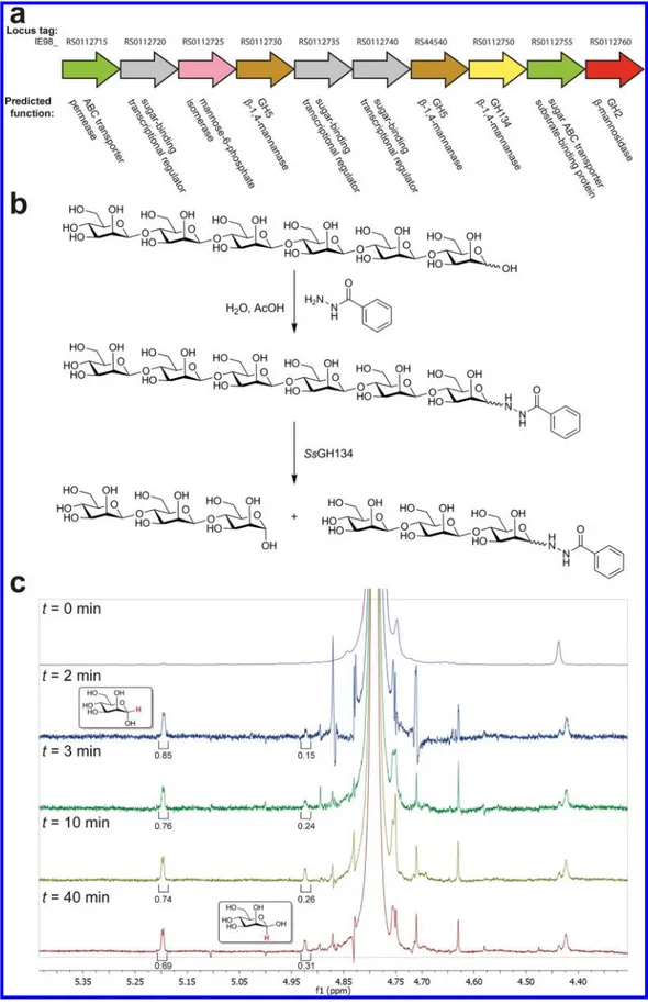
Figure 1.Family GH134β-mannanases act with inversion of anomeric stereochemistry. (a) Genomic context of SsGH134 within the genome of Streptomyces sp. NRRL B-24484. (b) Summary of derivatization and stereochemistry determination:β-1,4-mannohexaose (M6) was converted to the glycosyl hydrazide and then purified. The mannohexaosyl benzoylhydrazide was digested with SsGH134 and the stereochemical outcome was determined. (c) M6-benzoyl hydrazide was incubated with SsGH134 and the stereochemistry of the reaction was monitored by1H NMR spectroscopy. At t = 0 there are no anomeric protons corresponding to the hemiacetal. At t = 2 min, two new peaks, assigned as H1α and H1β of M3 at δ 5.2 and 4.92 ppm, respectively. Mutarotation is at equilibrium by t = 40 min.
β-mannanases, one GH134 β-mannanase, sugar ABC transporters, and a mannose-6-phosphate isomerase (Figure 1a). Hypothetical protein WP_030268297.1, the putative GH134 β-mannanase, possesses a signal peptide and CBM10 domain (carbohydrate binding modules, CBMs, reviewed in ref 18) attached through a short GlySer linker to the GH134 catalytic domain (Figure S2a). A gene encoding the catalytic domain, hereafter termed SsGH134, was synthesized and expressed in Escherichia coli with an N-terminal hexahistidine-tagged maltose binding protein (MBP) fusion partner (Figure S2b). This fusion protein was assayed for activity on β-1,4-oligomannosides, revealing an ability to cleave mannopentaose (M5), to yield mannotriose (M3) and mannobiose (M2), and to cleave mannohexaose (M6), to give predominantly M3, with smaller amounts of mannotetraose (M4) and M2, with optimal activity (kcat/KM2.7× 105M−1min−1) toward M6 at pH 5 and a strong
preference for unsubstituted linearβ-mannans over gluco- and galactomannans (Figure S3); a specificity rarely seen for β-mannanases. To define the cleavage site within these repeating oligosaccharides, enzymatic hydrolysis was performed in 18 O-labeled water, which resulted in incorporation of18O into the
nonreducing end fragment.19−21Mass spectrometric analysis of the products formed revealed that M5 is cleaved to provide M2
and M3 + 2 quasi-molecular ions, demonstrating 18O
incorporation into the latter fragment, and consistent with hydrolysis of M5 across the−3 → +2 subsites (Figure S4).22 Similar analysis of the products formed from cleavage of M6 revealed a preferred cleavage of the hexasaccharide across the−3 → + 3 subsites, as well as some cleavage across the −4 → +2 subsites (Figure S5).
We assessed the stereochemistry of substrate cleavage using
1H NMR spectroscopy. Owing to spectral overlap of the
substrate reducing end anomeric protons with those of the newly
the three-dimensional structure of SsGH134. After removal of the MBP fusion partner, SsGH134 crystals were obtained that diffracted to atomic resolution (approximately 1 Å), allowing the native structure to be solved by a novel“no-prior knowledge” molecular replacement method23using only an isolated standard helix as the search model (Tables 1 and S1). The SsGH134 structure revealed a mixed α-helix/β-sheet fold with a strong superficial resemblance to family GH19 chitinases, GH22 C-type lysozymes, GH23 G-type lysozymes, and GH124 cellulases (Figures 2a,3a−d, andS8).
Structural similarity searches using PDBeFold25revealed that SsGH134 is most similar to the GH22 C-type lysozyme family, albeit with a relatively low Z score of 3.3 reflecting 88 Cα carbons overlapping with an rmsd of approximately 2.6 Å (Figure 3a). The GH22 C-type lysozyme family is exemplified by hen egg white lysozyme (HEWL),26the archetypal glycoside hydrolase that performs catalysis with net retention of anomeric configuration, via the formation and subsequent hydrolysis of a covalent intermediate bonded to D52 and with E35 acting as a general acid/base.27 In contrast, SsGH134 performs catalysis with inversion of anomeric configuration, presumably through the direct attack of water in a single displacement mechanism, assisted by residues acting as a general acid and general base. Comparison of the location of the catalytic residues of the SsGH134 E45Q structure in complex with M5 (vide infra) with that of a chitohexaose complex of HEWL28 reveals that the general acid/base of HEWL, E35, overlays with E45 (Q in the mutated variant) of SsGH134, whereas the nucleophile D52 of HEWL overlays with N65 of SsGH134. As Asn is a poor nucleophile, this alternative residue in SsGH134 is consistent with its opposite stereochemical outcome versus HEWL (Figure 3e). In the case of the inverting chitinase A of family GH19,29the general acid E61 of chitinase A overlays with E45 in SsGH134, and the putative nucleophilic water coordinated by N65 and D57 in SsGH134 is located 4.3 Å away from an equivalent water in chitinase A held by E70 and S102. For the inverting G-type lysozyme,30the essential catalytic residue E73 overlays with E45 of SsGH134. Finally, the catalytic general acid in Clostridium thermocellum CtCel124A overlays with E45 of SsGH134.31
In order to probe the molecular basis of catalysis, and confirm catalytic acid and base assignments inferred from the overlaps described above, the structure of SsGH134 in complex with M3 was solved at a resolution of 1.2 Å (Figure 4a,Table 1). M3 binds in the−3 → −1 subsites32with the−1 subsite occupied by an α-mannose group. This represents a product complex, with the stereochemistry of theα-mannose group matching that formed through catalysis with inversion of anomeric configuration. Remarkably, the−1 subsite mannose in the product complex was in a ring-flipped1C
4conformation, hinting at a novel
conforma-tional itinerary for GH family 134. The location of E45 is Table 1. 3-D Structure Data forSsGH134
native
SsGH134-mannotriose
SsGH134 E45Q-mannopentaose
PDB Code 5JTS 5JU9 5JUG
space group P212121 P212121 P212121 resolution (Å) 34 (1.09) 45 (1.18) 45 (0.96) Rmerge 0.066 (0.86) 0.056 (0.56) 0.054 (0.52) I/σI 12.2 (1.8) 16.6 (2.4) 12.4 (1.1) completeness (%) 99.9 (99.9) 97.5 (80.2) 84.1 (29.2)a rmsd bond lengths (Å) 0.014 0.012 0.012 rmsd bond angles (deg) 1.56 1.57 1.67
aThe low completeness for the outer shell is due to the integration of data into the corners of the square detector. Data in the penultimate shell are 99% complete.
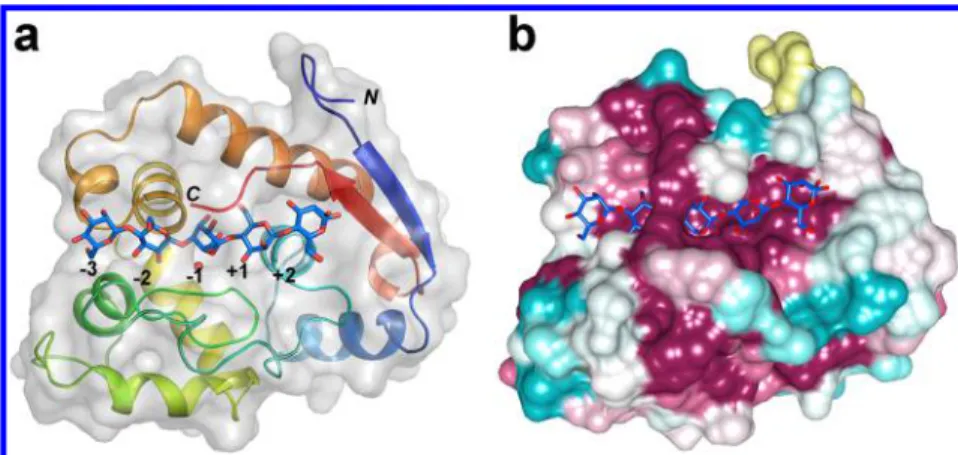
Figure 2.Family GH134β-mannanases adopt a lysozyme-like fold. SsGH134 E45Q variant in complex with β-1,4-mannopentaose. (a) Overview of complex rainbow-colored from the N- to C-terminus. (b) Surface representation of complex colored by sequence conservation according to the sequence alignment shown inFigure S9(cyan = variable, burgundy = conserved). Thefigure was prepared using the CONSURF server.24
Figure 3.Pairwise comparison of the fold, active site clefts, and key catalytic residues of SsGH134 and representative glycoside hydrolases with lysozyme folds. SsGH134 E45Q (PDB 5JUG, light green) with M5 bound (blue) overlaid with (a) retaining GH22 family HEWL (PDB 2WAR, yellow) with chitopentaose bound (plum), (b) inverting GH19 family chitinase A (PDB 3WH1, pink) with chitobiose and chitotriose bound (magenta), (c) inverting GH124 family cellulose CtCel124 (PDB 2XQO, lavender) with two molecules of cellotriose bound (chocolate), (d) inverting GH23 G-type lysozyme (PDB 3GXR, dark green) with chitobiose and chitotriose bound (brown). (e) Close up overlay of the catalytic residues of SsGH134 (light green), HEWL (yellow), chitinase A (pink), CtCel124 (lavender), and G-type lysozyme (dark green).
consistent with its role as a general acid catalyst (as expected from the overlaps above), and the spatial location of D57 implicates it as a general base, positioned to deprotonate a water nucleophile in a single displacement inverting mechanism. These structure-based assignments were supported by mutagenesis: the D57N and E45Q variants were inactive against M5 and M6.
A “Michaelis” complex was obtained by soaking M5 into crystals of the catalytically inactive mutant SsGH134-E45Q. Clear electron density at 0.96 Å resolution revealed M5 bound from the−3 → +2 subsites (Figure 4b), consistent with the18
O-water incorporation experiments. With the exception of the−1 subsite, discussed below, all the remaining sugar rings are in ground state4C
1conformations. Residues lying within a major
loop region (44−72) form contacts with the substrate down the length of the active site, and a contiguous stretch of residues within the C-terminal segment (168−173) seals the active center from−2 to +2. Notably, the C-terminal carboxylate of I173 is hydrogen bonded to O6 of the−2 mannosyl unit and forms a salt-bridge with the highly conserved K59 (Figure 4c, residues highlighted in green). This interaction presumably locks the carboxylate in place and completes an active center tunnel now reminiscent of processive glycoside hydrolases (Figure 2b). Completion of the tunnel occurs only in the Michaelis complex (when the positive subsites are occupied) as the C-terminal segment is disordered in both “apo” and M3 product-bound forms. Alignment of SsGH134 with other family member sequences in the NCBI nonredundant database reveals a highly conserved C-terminus ending with an isoleucine (Figure S9). Two exceptions are the putative proteins KFZ17922.1 and KFZ19809.1 from Pseudogymnoascus sp. (VKM F-4519), which are fused to a predicted GH27α-galactosidase linked through the
GH134 C-terminus and which we postulate may act on galactomannan, a β-1,4-mannan polysaccharide modified with α-1,6-galactosyl residues. This suggests a role for the C-terminal Ile in distinguishingβ-1,4-mannans lacking a substituent at the −2 mannose from more complex mannans (Figure 4c). In support of this hypothesis, it is notable that SsGH134 is only weakly active on carob galactomannan and konjac glucomannan, but is unusual in its ability to process highly purified β-1,4-mannan and ivory nut β-1,4-mannan extensively (Figure S3). The structure of the Michaelis complex contains a well-ordered water molecule positioned to act as a nucleophile and coordinated to the general base D57, as well as K59 and N65 on theα-face of the mannoside in the−1 subsite. The side chain carbonyl oxygen of Q45 is 3.4 Å away from the leaving-group oxygen, consistent with the assignment of E45 as the general acid. Notably, the −1 mannoside resides in a ringflipped1C
4conformation, similar to
that seen in the SsGH134-M3 product complex, with the leaving group C1−O oriented axially, and the nucleophilic water positioned for in-line nucleophilic attack with a water−C1−O1 angle of 160°. The structures of the Michaelis and product complexes are suggestive of a Southern hemisphere1C4→3H4‡
→3S
1conformational itinerary along the reaction coordinate,
with the product relaxing to a Michaelis-mimicking 1C4 conformation (possibly via a 3H4 conformation) after the reaction is complete. Such a conformational itinerary has never been reported for aβ-mannosidase or β-mannanase; however, a reversed Southern hemisphere3S
1→3H4‡→1C4conformational
itinerary has been assigned to invertingα-mannosidases from family GH47.34−36
The unusual conformations observed in the Michaelis and product complexes prompted study of the conformational Figure 4.Complexes of (a) wild-type SsGH134 with M3 and (b) E45Q variant with M5 showing the proposed general acid (E45/Q45) suitably positioned near thefissile glycosidic bond, and on the opposing side the proposed general base D57, coordinating the nucleophilic water molecule. Electron density meshes areσA-weighted 2F0− Fccontoured at 1σ (0.58 and 0.47 electrons per Å3, respectively), assembled using CCP4mg.33(c) Cartoon showing major protein substrate interactions for the SsGH134−mannopentaose complex. Interactions are shown for the model of the SsGH134 bound to theα-anomer of β-1,4-mannopentaose. The red spheres denote water molecules.
coordinate by classical molecular dynamics (MD) and QM/MM metadynamics.37Three collective variables (CV), including all bonds to be broken/cleaved by the enzyme, were considered.
CV1accounts for the nucleophilic attack of the catalytic water molecule; CV2accounts for proton transfer between D57 and the
water molecule; and CV3accounts for the transfer of the E45 proton to the glycosidic oxygen atom (Figure S11). The shape of the reconstructed free energy surface, projected onto CV1/CV2 (Figure 5a), is indicative of a concerted one-step reaction. The reaction free energy barrier (17 kcal mol−1) is commensurate with the measured reaction rate.17
Representative states along the lowest free energy pathway are shown inFigure 5b. The −1 sugar at the reactants state (R) adopts a conformation intermediate between1C
4and3E. The
reaction commences with elongation of the glycosidic bond simultaneous with the transfer of the carboxylic hydrogen atom of the acid residue to the glycosidic oxygen. At the transition state (TS), the −1 mannopyranose ring distorts from 1C
4/3E to 3H
4/3E (Figures 5andS12), a conformation compatible with the
requirement of an oxocarbenium ion like TS.6,7At this stage, the E45 proton is already transferred, the glycosidic bond is completely broken (3.3 Å;Table S2), and the bond between the nucleophilic water and the anomeric carbon is partially formed (2.0 Å;Table S2). Proton transfer from the water to D57 then takes place, while the −1 sugar changes to a 3,OB/3S
1
conformation (P) (Figures 5andS12). Thereafter, the anomeric OH loses its interaction with D57 (transition from P to P′, ΔG‡= 2 kcal mol−1), and the −1 mannopyranose spontaneously undergoes relaxation to a 1C
4 conformation (see also Figure S13), matching the conformation observed in the product complex of SsGH134 with M3 (Figure 4a). It is expected that once the product exits the active site it relaxes to its most stable
4C
1conformation in solution.
36
The computed mechanism can be considered an electrophilic migration of the anomeric carbon from the departing sugar residue to the nucleophilic water, assisted by E45 as general acid and D57 as general base (Figure 5).
■
CONCLUDING REMARKSThe present work reveals the emergence of a new enzyme activity from the lysozyme fold, a fold known since thefirst-determined X-ray structure of an enzyme, that of HEWL.26 This work provides an elegant example of how tertiary structures can be co-opted during evolution to bind different substrates and process them using entirely different mechanisms. While the majority of mannoside hydrolases appear to act through mechanisms involving a transition state in a B2,5 conformation (
β-mannosidases/mannanases: GH2, 5, 26, 113, 130; α-mannosi-dases/mannanases: GH38, 76, 92)7theβ-mannanases of family GH134 stand with theα-mannosidases of family GH47 as GHs operating through a 3H4 transition state. Collectively these
groups illustrate two distinct solutions to the problem of nucleophilic attack on aβ-mannopyranoside residue.
■
ASSOCIATED CONTENT*
S Supporting InformationThe Supporting Information is available free of charge on the
ACS Publications websiteat DOI:10.1021/acscentsci.6b00232. Supplementaryfigures (Figures S1−S13), Supplementary Tables (Tables S1 and S2), all experimental details, X-ray data collection, processing and refinement statistics (PDF)
Accession Codes
The coordinates and structure factors have been deposited in the Protein Data Bank (accession codes 5JTS, 5JU9, 5JUG). Figure 5.QM/MM metadynamics simulations of the mechanism of
SsGH134. (a) Free energy landscape of the SsGH134-catalyzed reaction obtained by QM/MM metadynamics. (b) Reaction coordinate for SsGH134 inverting endo-β-1,4-mannanase. R denotes reactants (Michaelis complex), TS the transition state, and P the products. From P to P′, a solvent water molecule enters the active site and fills the space previously occupied by the anomeric hydroxyl group of the mannose in the−1 subsite. Hydrogen atoms have been omitted for clarity, except those of the catalytic water and the carboxylate group of E45.
Foundation and VESKI with additional support from the Australian Cancer Research Foundation and Victorian State Government Operational Infrastructure Support, NHMRC IRIISS Grant 9000220, the Spanish Ministry of Economy and Competitiveness (MINECO, CTQ2014-55174-P) and the Generalitat de Catalunya (2014SGR-987). G.J.D. is supported by the Royal Society. We acknowledge the staff of the Diamond Light Source (UK) for provision of I02 beamline facilities (Proposal Number mx-9948), and the support, technical expertise, and assistance provided by the Barcelona Super-computing Center: Centro Nacional de Supercomputación (BSC−CNS). L.R. acknowledges a Ph.D. fellowship from the University of Barcelona (APIF-UB).
■
REFERENCES(1) Rodríguez-Gacio, M. d. C.; Iglesias-Fernández, R.; Carbonero, P.; Matilla, Á. J. Softening-up mannan-rich cell walls. J. Exp. Bot. 2012, 63, 3976−3988.
(2) Malgas, S.; van Dyk, J. S.; Pletschke, B. I. A review of the enzymatic hydrolysis of mannans and synergistic interactions between β-mannanase, β-mannosidase and α-galactosidase. World J. Microbiol. Biotechnol. 2015, 31, 1167−1175.
(3) Dhawan, S.; Kaur, J. Microbial mannanases: an overview of production and applications. Crit. Rev. Biotechnol. 2007, 27, 197−216.
(4) Moreira, L. R.; Filho, E. X. An overview of mannan structure and mannan-degrading enzyme systems. Appl. Microbiol. Biotechnol. 2008, 79, 165−178.
(5) van Zyl, W. H.; Rose, S. H.; Trollope, K.; Görgens, J. F. Fungal β-mannanases: Mannan hydrolysis, heterologous production and biotechnological applications. Process Biochem. 2010, 45, 1203−1213.
(6) Davies, G. J.; Planas, A.; Rovira, C. Conformational analyses of the reaction coordinate of glycosidases. Acc. Chem. Res. 2012, 45, 308−316. (7) Speciale, G.; Thompson, A. J.; Davies, G. J.; Williams, S. J. Dissecting conformational contributions to glycosidase catalysis and inhibition. Curr. Opin. Struct. Biol. 2014, 28, 1−13.
(8) Crich, D. Mechanism of a chemical glycosylation reaction. Acc. Chem. Res. 2010, 43, 1144−1153.
(9) Crich, D.; Chandrasekera, N. S. Mechanism of 4,6-O-benzylidene-directedβ-mannosylation as determined by α-deuterium kinetic isotope effects. Angew. Chem., Int. Ed. 2004, 43, 5386−5389.
(10) Huang, M.; Garrett, G. E.; Birlirakis, N.; Bohe, L.; Pratt, D. A.; Crich, D. Dissecting the mechanisms of a class of chemical glycosylation using primary13C kinetic isotope effects. Nat. Chem. 2012, 4, 663−667. (11) Lombard, V.; Golaconda Ramulu, H.; Drula, E.; Coutinho, P. M.; Henrissat, B. The carbohydrate-active enzymes database (CAZy) in 2013. Nucleic Acids Res. 2014, 42, D490−495.
(12) Davies, G.; Henrissat, B. Structures and mechanisms of glycosyl hydrolases. Structure 1995, 3, 853−859.
(13) Vocadlo, D. J.; Davies, G. J. Mechanistic insights into glycosidase chemistry. Curr. Opin. Chem. Biol. 2008, 12, 539−555.
(14) Williams, R. J.; Iglesias-Fernandez, J.; Stepper, J.; Jackson, A.; Thompson, A. J.; Lowe, E. C.; White, J. M.; Gilbert, H. J.; Rovira, C.; Davies, G. J.; Williams, S. J. Combined inhibitor free-energy landscape
T.; Takaya, N.; Kobayashi, T.; Kato, M. Novel β-1,4-mannanase belonging to a new glycoside hydrolase family in Aspergillus nidulans. J. Biol. Chem. 2015, 290, 27914−27927.
(18) Boraston, A. B.; Bolam, D. N.; Gilbert, H. J.; Davies, G. J. Carbohydrate-binding modules: fine-tuning polysaccharide recognition. Biochem. J. 2004, 382, 769−781.
(19) Couturier, M.; Roussel, A.; Rosengren, A.; Leone, P.; Stålbrand, H.; Berrin, J.-G. Structural and biochemical analyses of glycoside hydrolase families 5 and 26β-1,4-mannanases from Podospora anserina reveal differences upon manno-oligosaccharide catalysis. J. Biol. Chem. 2013, 288, 14624−14635.
(20) McGregor, N.; Morar, M.; Fenger, T. H.; Stogios, P.; Lenfant, N.; Yin, V.; Xu, X.; Evdokimova, E.; Cui, H.; Henrissat, B.; Savchenko, A.; Brumer, H. Structure-function analysis of a mixed-linkageβ-glucanase/ xyloglucanase from the key ruminal bacteroidetes Prevotella bryantii B14. J. Biol. Chem. 2016, 291, 1175−1197.
(21) Schagerlof, H.; Nilsson, C.; Gorton, L.; Tjerneld, F.; Stalbrand, H.; Cohen, A. Use of 18O water and ESI-MS detection in subsite characterisation and investigation of the hydrolytic action of an endoglucanase. Anal. Bioanal. Chem. 2009, 394, 1977−1984.
(22) Davies, G. J.; Wilson, K. S.; Henrissat, B. Nomenclature for sugar-binding subsites in glycosyl hydrolases. Biochem. J. 1997, 321, 557−559. (23) Greive, S. J.; Fung, H. K.; Chechik, M.; Jenkins, H. T.; Weitzel, S. E.; Aguiar, P. M.; Brentnall, A. S.; Glousieau, M.; Gladyshev, G. V.; Potts, J. R.; Antson, A. A. DNA recognition for virus assembly through multiple sequence-independent interactions with a helix-turn-helix motif. Nucleic Acids Res. 2016, 44, 776−789.
(24) Celniker, G.; Nimrod, G.; Ashkenazy, H.; Glaser, F.; Martz, E.; Mayrose, I.; Pupko, T.; Ben-Tal, N. ConSurf: Using evolutionary data to raise testable hypotheses about protein function. Isr. J. Chem. 2013, 53, 199−206.
(25) Krissinel, E.; Henrick, K. Secondary-structure matching (SSM), a new tool for fast protein structure alignment in three dimensions. Acta Crystallogr., Sect. D: Biol. Crystallogr. 2004, 60, 2256−2268.
(26) Blake, C. C. F.; Koenig, D. F.; Mair, G. A.; North, A. C. T.; Phillips, D. C.; Sarma, V. R. Structure of hen egg-white lysozyme. Nature 1965, 206, 757−761.
(27) Vocadlo, D. J.; Davies, G. J.; Laine, R.; Withers, S. G. Catalysis by hen egg-white lysozyme proceeds via a covalent intermediate. Nature 2001, 412, 835−838.
(28) Davies, G. J.; Withers, S. G.; Vocadlo, D. J. The chitopentaose complex of a mutant hen egg-white lysozyme displays no distortion of the− 1 sugar away from a4C
1chair conformation. Aust. J. Chem. 2009, 62, 528−532.
(29) Ohnuma, T.; Umemoto, N.; Nagata, T.; Shinya, S.; Numata, T.; Taira, T.; Fukamizo, T. Crystal structure of a″loopless″ GH19 Chitinase in complex with chitin tetrasaccharide spanning the catalytic center. Biochim. Biophys. Acta, Proteins Proteomics 2014, 1844, 793−802.
(30) Helland, R.; Larsen, R. L.; Finstad, S.; Kyomuhendo, P.; Larsen, A. N. Crystal structures of g-type lysozyme from Atlantic cod shed new light on substrate binding and the catalytic mechanism. Cell. Mol. Life Sci. 2009, 66, 2585−2598.
(31) Bras, J. L.; Cartmell, A.; Carvalho, A. L.; Verze, G.; Bayer, E. A.; Vazana, Y.; Correia, M. A.; Prates, J. A.; Ratnaparkhe, S.; Boraston, A. B.; Romao, M. J.; Fontes, C. M.; Gilbert, H. J. Structural insights into a
unique cellulase fold and mechanism of cellulose hydrolysis. Proc. Natl. Acad. Sci. U. S. A. 2011, 108, 5237−5242.
(32) Davies, G. J.; Wilson, K. S.; Henrissat, B. Nomenclature for sugar-binding subsites in glycosyl hydrolases. Biochem. J. 1997, 321, 557−559. (33) Winn, M. D.; Ballard, C. C.; Cowtan, K. D.; Dodson, E. J.; Emsley, P.; Evans, P. R.; Keegan, R. M.; Krissinel, E. B.; Leslie, A. G. W.; McCoy, A.; McNicholas, S. J.; Murshudov, G. N.; Pannu, N. S.; Potterton, E. A.; Powell, H. R.; Read, R. J.; Vagin, A.; Wilson, K. S. Overview of the CCP4 suite and current developments. Acta Crystallogr., Sect. D: Biol. Crystallogr. 2011, 67, 235−242.
(34) Karaveg, K.; Siriwardena, A.; Tempel, W.; Liu, Z. J.; Glushka, J.; Wang, B. C.; Moremen, K. W. Mechanism of class 1 (glycosylhydrolase family 47) α-mannosidases involved in N-glycan processing and endoplasmic reticulum quality control. J. Biol. Chem. 2005, 280, 16197−16207.
(35) Vallée, F.; Karaveg, K.; Herscovics, A.; Moremen, K. W.; Howell, P. L. Structural basis for catalysis and inhibition of N-glycan processing class Iα-1,2-mannosidases. J. Biol. Chem. 2000, 275, 41287−41298.
(36) Thompson, A. J.; Dabin, J.; Iglesias-Fernandez, J.; Ardevol, A.; Dinev, Z.; Williams, S. J.; Bande, O.; Siriwardena, A.; Moreland, C.; Hu, T. C.; Smith, D. K.; Gilbert, H. J.; Rovira, C.; Davies, G. J. The reaction coordinate of a bacterial GH47α-mannosidase: A combined quantum mechanical and structural approach. Angew. Chem., Int. Ed. 2012, 51, 10997−11001.
(37) Iannuzzi, M.; Laio, A.; Parrinello, M. Efficient exploration of reactive potential energy surfaces using Car-Parrinello molecular dynamics. Phys. Rev. Lett. 2003, 90, 238302.