Altered CSNK1E, FABP4 and NEFH protein levels in the dorsolateral
prefrontal cortex in schizophrenia
Raquel Pinacho
a, Núria Villalmanzo
a, J. Javier Meana
b, Isidre Ferrer
c, Adriana Berengueras
d, Josep M. Haro
a,
Judit Villén
e, Belén Ramos
a,⁎
a
Unitat de recerca, Parc Sanitari Sant Joan de Déu, Fundació Sant Joan de Déu, Universitat de Barcelona, Centro de Investigación Biomédica en Red de Salud Mental, CIBERSAM. Dr. Antoni Pujadas, 42, Sant Boi de Llobregat, 08830 Barcelona, Spain
b
Departamento de Farmacología, Universidad del País Vasco/Euskal Herriko Unibertsitatea UPV/EHU, Instituto BioCruces, Centro de Investigación Biomédica en Red de Salud Mental, CIBERSAM, Bº Sarriena s/n, 48940 Leioa, Bizkaia, Spain
cInstituto de Neuropatología, IDIBELL-Hospital Universitari de Bellvitge, Universitat de Barcelona, Centro de Investigación Biomédica en Red para enfermedades neurodegenerativas, CIBERNED, Feixa Llarga s/n, Hospitalet de LLobregat, 08907 Barcelona, Spain
d
Banc de Teixits Neurologics, Parc Sanitari Sant Joan de Déu, Centro de Investigación Biomédica en Red de Salud Mental, CIBERSAM, Dr. Antoni Pujadas, 42, Sant Boi de Llobregat, 08830 Barcelona, Spain
e
Genome Sciences Department, School of Medicine, University of Washington, 3720 15th Ave NE, Seattle 98195, WA, USA
a b s t r a c t
a r t i c l e i n f o
Article history:
Received 24 October 2015
Received in revised form 15 March 2016 Accepted 27 April 2016
Available online xxxx
Schizophrenia constitutes a complex disease. Negative and cognitive symptoms are enduring and debilitating components of the disorder, highly associated to disability and burden. Disrupted neurotransmission circuits in dorsolateral prefrontal cortex (DLPFC) have been related to these symptoms. To identify candidates altered in schizophrenia, we performed a pilot proteomic analysis on postmortem human DLPFC tissue from patients with schizophrenia (n = 4) and control (n = 4) subjects in a pool design using differential isotope peptide label-ling followed by liquid chromatography tandem mass spectrometry (LC-MS/MS). We quantified 1315 proteins with two or more unique peptides, 116 of which showed altered changes. Of these altered proteins, we selected four with potential roles on cell signaling, neuronal development and synapse functioning for further validation: casein kinase I isoform epsilon (CSNK1E), fatty acid-binding protein 4 (FABP4), neurofilament triplet H protein (NEFH), and retinal dehydrogenase 1 (ALDH1A1). Immunoblot validation confirmed our proteomic findings of these proteins being decreased in abundance in the schizophrenia samples. Additionally, we conducted immuno-blot validation of these candidates on an independent sample cohort comprising 23 patients with chronic schizo-phrenia and 23 matched controls. In this second cohort, CSNK1E, FABP4 and NEFH were reduced in the schizophrenia group while ALDH1A1 did not significantly change. This study provides evidence indicating these proteins are decreased in schizophrenia: CSNK1E, involved in circadian molecular clock signaling, FABP4 with possible implication in synapse functioning, and NEFH, important for cytoarchitecture organization. Hence, thesefindings suggest the possible implication of these proteins in the cognitive and/or negative symp-toms in schizophrenia.
© 2016 Elsevier B.V. All rights reserved. Keywords:
Schizophrenia
Postmortem dorsolateral prefrontal cortex Proteomics
CSNK1E FABP4 NEFH
1. Introduction
Schizophrenia is a complex disorder in which genetic and environ-mental factors are proposed to interact and contribute to the emergence of the disease. These factors may converge and impact upon the same physiopathological pathways in the brain, affecting neural microcircuit-ry (Harrison and Weinberger, 2005; Sullivan, 2012).
Negative symptoms (e.g. lack of volition, poor social functioning, and blunted affect) and cognitive impairments (e.g. deficits in executive functions and working memory) are core symptoms of schizophrenia, and are the most resilient to currently available treatments (Gold,
2004; Millan et al., 2014; Stahl and Buckley, 2007). The dorsolateral prefrontal cortex (DLPFC) is involved in both cognitive deficits (Frith and Dolan, 1996; Lewis and Moghaddam, 2006; Teffer and Semendeferi, 2012) and negative symptoms (Semkovska et al., 2001; Toda and Abi-Dargham, 2007). A dysfunction in this region has been widely described in functional and structural imaging studies and in many molecular reports (English et al., 2011; Goldstein et al., 1999; Konradi, 2005; Wong and Van Tol, 2003). Several neurotransmitter systems have been implicated in this dysfunction. Hypodopaminergic activity has been associated with cognitive impairments and negative symptoms (Kienast and Heinz, 2006; Toda and Abi-Dargham, 2007). Excitatory glutamatergic and inhibitory GABAergic neurotransmission systems have also been implicated in these symptoms in schizophrenia (Krystal et al., 1994; Lewis and Moghaddam, 2006; Moghaddam and
Schizophrenia Research xxx (2016) xxx–xxx
⁎ Corresponding author.
E-mail address:[email protected](B. Ramos).
http://dx.doi.org/10.1016/j.schres.2016.04.050
0920-9964/© 2016 Elsevier B.V. All rights reserved.
Contents lists available atScienceDirect
Schizophrenia Research
Javitt, 2012). However, an integrative understanding of common molec-ular pathways affected by these systems is just starting to be unveiled. Previous proteomic and transcriptomic screenings have reported mi-tochondrial function, cytoskeleton formation, and oligodendrocytes to be consistently altered in the DLPFC in schizophrenia (English et al., 2011; Konradi, 2005; Martins-de-Souza et al., 2010a; Martins-de-Souza et al., 2009a). These approaches are useful tools that help to provide an
overall picture of altered common functions and pathways in human tissues. However, there is still missing information of altered proteins in schizophrenia, which could potentially be obtained using alternative proteomic approaches.
Here, we designed a pilot quantitative proteomic analysis using differential isotope peptide labelling followed by liquid chromatogra-phy fractionation and tandem mass spectrometry (LC-MS/MS) in grey
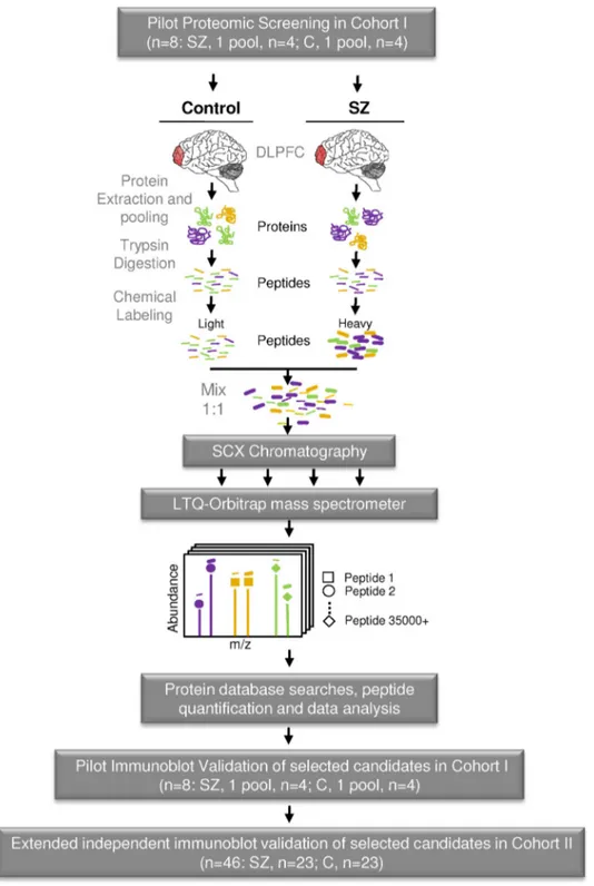
Fig. 1. Experimental strategy for a pilot large scale quantitative proteomic analysis to identify differentially expressed proteins in schizophrenia. Protein lysates from the postmortem dorsolateral prefrontal cortex (DLPFC) of controls (n = 4) and schizophrenia patients (SZ, n = 4) from cohort I (Table 1) were processed as depicted. Samples from the same group were pooled and digested with trypsin. The resultant peptides were labelled with either hydrogen (light peptides, control) or deuterium (heavy peptides, schizophrenia) isotopes through a reductive dimethylation reaction. Then, differentially labelled peptides were mixed 1:1, separated by SCX chromatography and analyzed by LC-MS/MS on a hybrid linear ion trap-Orbitrap mass spectrometer for identification and relative quantification of pair peptide sequences. Subsequently, protein database searches, peptide quantification and data analysis were performed as described in the experimental procedures section. A panel of 4 candidates from significantly regulated proteins was selected for further validation by immunoblot:first, in a pilot validation in the pooled samples of cohort I (Table 1, n = 8, 4 samples per group) and then, in an extended independent validation in a larger cohort II in individual samples (Table 1; n = 46; 23 samples per group).
2 R. Pinacho et al. / Schizophrenia Research xxx (2016) xxx–xxx
Please cite this article as: Pinacho, R., et al., Altered CSNK1E, FABP4 and NEFH protein levels in the dorsolateral prefrontal cortex in schizophrenia, Schizophr. Res. (2016),http://dx.doi.org/10.1016/j.schres.2016.04.050
matter DLPFC samples from four pooled schizophrenia and four pooled control individuals with the end goal to discover possible common altered proteins across patients with schizophrenia for further valida-tion of selected candidates in a larger cohort of individual samples (Fig. 1). Thus, after the initial proteomic screen, 23 samples per group (control and SZ groups) were used in this study for independent validation of three candidate proteins by immunoblot in individual patients. Our validation was focused on novel altered protein isoforms in schizophrenia with a plausible role on cell signaling, neuronal development and synapse functioning.
2. Materials and methods 2.1. Brain tissue samples
For the pilot proteomic analysis, we used postmortem human brain tissue from the DLPFC of patients with schizophrenia (1 pool composed of 4 SZ patients) and control subjects with no history of psychiatric episodes (1 pool composed of 4 control individuals) from the UPV/EHU brain collection (see more details in Supplementary material and
Table 1). Samples were obtained at autopsy by forensic pathologists under research policies with postmortem samples. All deaths were subjected to retrospective analysis for previous medical diagnosis. Subjects with antemortem criteria for paranoid schizophrenia according to the Diagnostic and Statistical Manual of Mental Disorders (DSM-IV) that died by suicide were matched to control subjects who died by accidental causes in a paired design, based on gender, age, and postmortem delay (PMD). Toxicological screening for antipsychotics, antidepressants, and other drugs was performed at the National
Institute of Toxicology, Madrid, Spain. We further validated the candi-dates identified in the quantitative proteomic assay in an independent set of postmortem human DLPFC of patients with chronic schizophrenia (n = 23) and control individuals with no history of psychiatric episodes (n = 23) from the collection of neurologic tissues of Parc Sanitari Sant Joan de Déu (Roca et al., 2008) and the Institute of Neuropathology Brain Bank (HUB-ICO-IDIBELL Biobank) (Table1). All SZ patients were institutionalized donors with a long duration of the illness (Table 1) who had no history of neurological episodes. The study was approved by the Institutional Ethics Committee of Parc Sanitari Sant Joan de Déu. We matched schizophrenia and control groups by gender (only male patients were included), age, postmortem delay and pH. Experienced clinical examiners interviewed each donor antemortem to confirm schizophrenia diagnosis according to the Diagnostic and Statistical Manual of Mental Disorders (DSM-IV) and International Classification of Diseases 10 (ICD-10) criteria. All deaths were due to natural causes. Neuropathologists from the Institute of Neuropathology Brain Bank (HUB-ICO-IDIBELL Biobank) examined the contralateral hemisphere for signs of neurodegenerative disorders in both schizo-phrenia patients and control. 76.1% of both schizoschizo-phrenia and control groups showed low degree of Alzheimer disease-related changes (Stage≤ III, Braak and Braak scale (Braak et al., 2006; Braak and Braak, 1991)). The last daily chlorpromazine equivalent dose for the antipsy-chotic treatment of patients was calculated based on the electronic records of last drug prescriptions administered up to death as described previously (Gardner et al., 2010) (Table 1). Patients and controls were chosen among the collected brains on the basis, whenever possible, of the following criteria: (a) negative medical information on the presence of neurological disorders or drug abuse, (b) accidental or natural cause
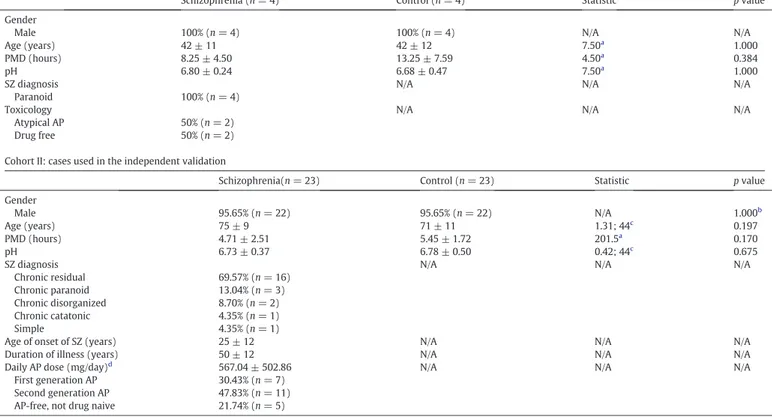
Table 1
Demographic, clinical and tissue related features of cases. Cohort I: cases used in the pilot proteomic analysis
Schizophrenia (n = 4) Control (n = 4) Statistic p value
Gender
Male 100% (n = 4) 100% (n = 4) N/A N/A
Age (years) 42 ± 11 42 ± 12 7.50a
1.000
PMD (hours) 8.25 ± 4.50 13.25 ± 7.59 4.50a 0.384
pH 6.80 ± 0.24 6.68 ± 0.47 7.50a
1.000
SZ diagnosis N/A N/A N/A
Paranoid 100% (n = 4)
Toxicology N/A N/A N/A
Atypical AP 50% (n = 2)
Drug free 50% (n = 2)
Cohort II: cases used in the independent validation
Schizophrenia(n = 23) Control (n = 23) Statistic p value
Gender Male 95.65% (n = 22) 95.65% (n = 22) N/A 1.000b Age (years) 75 ± 9 71 ± 11 1.31; 44c 0.197 PMD (hours) 4.71 ± 2.51 5.45 ± 1.72 201.5a 0.170 pH 6.73 ± 0.37 6.78 ± 0.50 0.42; 44c 0.675
SZ diagnosis N/A N/A N/A
Chronic residual 69.57% (n = 16)
Chronic paranoid 13.04% (n = 3)
Chronic disorganized 8.70% (n = 2)
Chronic catatonic 4.35% (n = 1)
Simple 4.35% (n = 1)
Age of onset of SZ (years) 25 ± 12 N/A N/A N/A
Duration of illness (years) 50 ± 12 N/A N/A N/A
Daily AP dose (mg/day)d 567.04 ± 502.86 N/A N/A N/A
First generation AP 30.43% (n = 7)
Second generation AP 47.83% (n = 11)
AP-free, not drug naive 21.74% (n = 5)
Mean ± standard deviation or relative frequency are shown for each variable; PMD, postmortem delay; SZ, schizophrenia; AP, antipsychotics; N/A, not applicable aMann-Whitney U is shown for non-parametric variables.
b
Frequencies were analyzed by Fisher's exact test. c
T-statistic and degrees of freedom are shown for parametric variables. d
of death that does not compromise the integrity of the region of interest, and (c) brain pH higher than 6. Samples were codified by the brain bank staff according to data protection procedures.
2.2. Protein extraction
Specimens of the DLPFC (Brodmann area 9), extending from the pial surface to white matter and only including grey matter were dissected from coronal slabs stored at−80 °C using a standard human brain atlas (Mai et al., 1997). Due to collection methods in each institution, left dorsolateral prefrontal cortex from schizophrenia patients was paired with the contralateral hemisphere from controls. Protein extracts were prepared from tissue samples using NP40 lysis buffer as described previously (Pinacho et al., 2011). Protein concentration was determined by Bradford assay (Biorad, Hercules, CA, USA).
2.3. Mass spectrometry analysis
500μg of total protein extracts from control and schizophrenia ly-sates (one pool per group composed of four samples, 125μg of protein per sample) were each reduced with 5 mM dithiothreitol at 56 °C for 30 min in 50 mM Tris pH 8, alkylated with 15 mM iodoacetamide in the dark at room temperature for 30 min and quenched with additional 5 mM dithiothreitol for 15 min. Each extract was digested with 5 ng/μL trypsin in 50 mM Tris, 1 mM CaCl2pH 8 at 37 °C for 16 h. Peptides were
desalted by reversed-phase in a Sep-Pak tC18cartridge (100 mg, Waters
Associates, Milford, MA, USA). Peptide mixtures were resuspended in 500μL of 1 M HEPES pH 7.5 and subjected to a reductive dimethylation reaction as described previously (Khidekel et al., 2007). The light and heavy dimethylated peptide solutions were mixed 1:1. Peptides were desalted by reversed-phase in a Sep-Pak tC18cartridge and subjected
to strong cation exchange chromatography on a polysulfoethyl A col-umn. Twelve fractions were collected over 48-min in a gradient of KCl in 5 mM potassium phosphate, 30% ACN, and dried by vacuum centrifu-gation. Peptide fractions were resuspended in 1 mL 0.1% trifluoroacetic acid, desalted by reversed-phase in a Sep-Pak tC18cartridge and dried
by vacuum centrifugation. Peptides were resuspended in 5% ACN and 4% formic acid for LC-MS/MS analysis. Each peptide fraction was separated by reverse phase chromatography on a capillary C18column
and analyzed online on a hybrid linear ion trap Orbitrap (LTQ-Orbitrap XL, Thermo Fisher Scientific, San Jose, CA, USA) mass spectrometer. For each cycle, one full MS scan acquired at high mass resolution (AGC target = 1 × 10⁶, maximum ion injection time = 1000 ms) in the
Orbitrap analyser was followed by 10 MS/MS spectra on the linear ion trap (AGC target = 5 × 10³, maximum ion injection time = 120 ms) for the ten most abundant precursor ions. Fragmented precursor ions were dynamically excluded from further selection for 35 s. Ions were also excluded if their charge was eitherb2 or unassigned. All spectra were acquired in centroid mode.
2.4. Protein database searches, peptide quantification and data analysis Rawfiles were converted to mzXML format using ReadW version 4.3.1 using default parameters. MS/MS spectra were searched against a concatenated target-decoy IPI human protein database (version 3.20, n = 61.225 target sequences) using the Sequest algo-rithm. Search parameters included fully tryptic enzyme specificity with up to two missed cleavages permitted, mass tolerance of 50 ppm for the precursor and 1 Da for fragments ions,fixed modifi-cations of carboxamidomethylation on cysteines (+ 57.02146) and dimethylation on lysines and peptide N-termini (+ 28.03130), and as variable modifications methionine oxidation (+ 15.99491) and the difference between heavy (6 deuterium) and light dimethyl on lysines and peptide N-termini (+ 6.03766). Peptide matches were filtered to b1% false-discovery rate using the target-decoy database strategy. Peptides matching to multiple proteins were arbitrarily
assigned to the proteinfirst listed in the database. Peptides were quantified using in-house software by peak-area integration, and heavy/light peptide ratios were calculated. Among the set of inde-pendent measurements retained for each protein, the median of the log2heavy/light ratio of all peptides of the same protein was
used to determine the protein ratio, and the standard deviation (SD) was calculated (Supplementary Data 1). Quality cutoffs were as described previously (Baek et al., 2008).
2.5. Immunoblotting
Validation of candidate proteins was approached in two steps. Afirst pilot validation was performed in the same pooled protein lysates ana-lyzed in the proteomic analysis (a control pool and a schizophrenia pool each comprising equal amounts of protein lysates from four different samples). This was followed by an extensive validation in an indepen-dent cohort of 46 samples. Both cohorts are described inSection 2.1. In both validation steps 50μg of total protein lysates were resolved by SDS-PAGE electrophoresis and immunoblotted with polyclonal anti-body against FABP4 (ab23693, Abcam, Cambridge, UK); NEFH (ab40796, Abcam); and monoclonal antibodies against CSNK1E (ab82426, Abcam); ALDH1A1 (ab52492, Abcam)α-tubulin (T6199, Sigma-Aldrich, St Louis, MO, USA);β-actin (A5316, Sigma-Aldrich) and glyceraldehyde-3-phosphate dehydrogenase (GAPDH) (MAB374, Millipore-Chemicon). All proteins were detected by a unique band at the predicted molecular weight. Densitometric quantification of candidate proteins was performed using Quantity One software (BioRad). Values were normalized to the geometric mean of α-tubulin,β-actin and GAPDH, and a control reference sample.
2.6. Data and statistical analysis
In the pilot protein analysis, a quality cutoff for protein determina-tions was set to≥2 peptide sequences and to ≥4 spectral counts for proteins with unique peptide quantifications. Then, the log2of the
median of the heavy/light ratio for each protein was transformed to a z-score for asymmetrical standard deviations of the main distribution as described previously (Graumann et al., 2008). A significance value for each protein ratio was calculated from the complementary error function for the normalized distribution of the z-scores (Graumann et al., 2008), which provides an indicator of the chance that a given protein is altered in this pilot analysis. Higher chance to be altered is provided for proteins located at the end of the tails in the normalized distribution with lower significance values. Correction of significance values for multiple testing in the quantified protein data set was performed following the Benjamini and Hochberg method (Benjamini and Hochberg, 1995). A False Discovery Rate (FDR) was computed for all the significance values and FDR threshold was set to 0.01.
Normal distribution of the variables was determined by D'Agostino & Pearson test. Demographic and tissue-related features of the samples were compared between schizophrenia and control conditions by Fisher exact test for qualitative variables, by Student t-test for parametric quantitative variables, and by Mann-Whitney U test for quantitative non-parametric variables. Differences of the protein levels between schizophrenia and control groups were performed by one-tailed unpaired Student's t-test based on the results already provided by the pilot proteomic analysis indicating the expected direction of change. Grubbs test was used to detect outliers. Spearman or Pearson correlation analyses were carried out to detect association of our molecular measures with other clinical, demographic and tissue related variables (age, postmortem delay, pH, daily chlorpromazine equivalent dose and duration of illness). Statistical analysis was performed with GraphPad Prism version 5.00, with significance level set to 0.05.
4 R. Pinacho et al. / Schizophrenia Research xxx (2016) xxx–xxx
Please cite this article as: Pinacho, R., et al., Altered CSNK1E, FABP4 and NEFH protein levels in the dorsolateral prefrontal cortex in schizophrenia, Schizophr. Res. (2016),http://dx.doi.org/10.1016/j.schres.2016.04.050
Table 2
Top twenty up and down regulated proteins in the pilot proteomic analysis of the postmortem dorsolateral prefrontal cortex in schizophrenia. Acc.
number Gene symbol
Protein description Log2ratio H/L Ratio
H/L
RBC? Biological function Previously reported in the dlPFC in SZ Norm
median
SD Norm median
Q9Y6C7 LOH3CR2A Loss of heterozygosity 3 chromosomal region 2 gene A protein −5.50 0.29 0.02 No Unknown
Q6FGZ8 TUBB TUBB protein (fragment)a −5.25 0.72 0.03 No Cell growth/maintenance ▼English et al. (2009),▲Behan et al.
(2008)
Q5VWI4 NRAP Nebulin-related anchoring protein −4.90 2.18 0.03 No Cell growth/maintenance XP_372916 LOC391352 Predicted: similar to peptidylprolyl isomerase A isoform 1 −4.36 1.95 0.05 No Unknown
Q569K3 LOC644936 Actin/actin-like family protein −3.80 1.31 0.07 No Cell comunication/signal transduction Q5VVH4 PHIP Pleckstrin homology domain interacting protein −3.74 1.94 0.07 No Cell comunication/signal transduction P11473 VDR Vitamin D3 receptor −3.54 1.63 0.09 No Regulation of gene expression, epigenetic
P22061-2 PCMT1 Isoform 2 of Protein-L-isoaspartate(D-aspartate) O-methyltransferase −2.00 0.96 0.25 No Protein metabolism ▲English et al. (2009); ▲Martins-de-Souza et al. (2009b)
P49674 CSNK1E Casein kinase I isoform epsilon −1.66 0.04 0.32 No Cell comunication/signal transduction (▲CSNK2A1)Martins-de-Souza et al. (2009b)
P11217 PYGM Glycogen phosphorylase, muscle form −1.12 0.31 0.46 No Metabolism/energy pathways Q96DZ9-2 CMTM5 Isoform 2 of CKLF-like MARVEL transmembrane domain-containing
protein 5
−1.07 0.72 0.48 No Unknown
P02689 PMP2 Myelin P2 protein −0.96 0.21 0.51 No Transport
Q96NS9 MAP4 CDNA FLJ30134fis, clone BRACE1000187, weakly similar to microtubule-associated protein 4
−0.96 0.56 0.51 No Cell growth/maintenance (▲MAP6)Martins-de-Souza et al. (2009b)
P49753-1 ACOT2 Isoform 1 of acyl-coenzyme A thioesterase 2 −0.92 0.26 0.53 No Metabolism/energy pathways Q8NF17 IGHM FLJ00385 protein (fragment) −0.91 0.29 0.53 No Immune response
P12036 NEFH Neurofilament triplet H protein −0.85 0.32 0.55 No Cell growth/maintenance
P15090 FABP4 Fatty acid-binding protein, adipocyte −0.85 0.01 0.56 No Cell comunication/signal transduction
P00352 ALDH1A1 Retinal dehydrogenase 1 −0.77 0.39 0.59 Yes Aldehyde metabolism ▼Prabakaran et al. (2004); (▲ALDH4A1)
Wesseling et al. (2013)
Q96IX5 USMG5 Up-regulated during skeletal muscle growth protein 5 −0.74 0.11 0.60 No Unknown
P63000-1 RAC1 Isoform A of Ras-related C3 botulinum toxin substrate 1 precursor −0.73 0.03 0.60 No Cell comunication/signal transduction O96000 NDUFB10 NADH dehydrogenase [ubiquinone] 1 beta subcomplex subunit 10 0.68 0.25 1.61 No Metabolism/energy pathways Q02410 APBA1 Amyloid beta A4 precursor protein-binding family A member 1 0.70 0.19 1.62 No Cell comunication/signal transduction O94772 LY6H Lymphocyte antigen Ly-6H precursor 0.70 0.00 1.63 No Immune response
P63027 VAMP2 Vesicle-associated membrane protein 20.70 0.16 1.63 No Transport
P00403 MT-CO2 Cytochrome c oxidase subunit 2 0.71 0.13 1.64 No Metabolism/energy pathways Q15836 VAMP3 Vesicle-associated membrane protein 3 0.73 0.25 1.66 Yes Transport
Q2TBE9 OAT Phospholysine phosphohistidine inorganic pyrophosphate phosphatase 0.74 0.24 1.66 No Unknown
P55087-1 AQP4 Isoform 2 of aquaporin-4 0.75 0.42 1.68 No Transport ▲Chan et al. (2011)
Q9H5G0 ISOC2 Isochorismatase domain-containing protein 2, mitochondrial 0.78 0.36 1.72 No Metabolism/energy pathways Q8NBS8 CAMKV CDNA FLJ90813fis, clone Y79AA1000967, weakly similar to
calcium/calmodulin-dependent protein kinase type I
0.78 0.13 1.72 No Unknown Q12904 NPNT Multisynthetase complex auxiliary component p43 0.79 0.32 1.73 No Unknown
P35232 PHB Prohibitin 0.82 0.34 1.76 No Cell comunication/signal transduction ▲Behan et al. (2008);▼Smalla et al. (2008)
Q99623 PHB2 Prohibitin-2 0.83 0.00 1.78 No Regulation of nucleobase, nucleoside, nucleotide and nucleic acid metabolism
(▲PHB)Behan et al. (2008); (▼PHB)
Smalla et al. (2008)
P00167-2 CYB5A Isoform 2 of cytochrome b5 0.86 0.16 1.82 No Metabolism/energy pathways P10636-6 MAPT Isoform tau-D of microtubule-associated protein tau 0.86 0.55 1.82 No Cell growth/maintenance
P62158 CALM1 Calmodulin 1.13 0.84 2.20 No Cell comunication/signal transduction ▼Novikova et al. (2006)
Q5UE58 CLSTN1 Calsyntenin 1 isoform 2 1.22 0.89 2.33 No Cell comunication/signal transduction Q8N163-2 KIAA1967 Isoform 2 of protein KIAA1967 1.66 0.49 3.16 No Cell comunication/signal transduction Q6ZS99 ARMC9 CDNA FLJ45706fis, clone FEBRA2028457, highly similar to nucleolin 3.08 1.75 8.43 No Unknown
Q8WZ42-2 TTN Isoform 2 of titin 8.83 3.75 456.17 No Transport
Access number from Uniprot database Uniprot; H/L ratio between heavy (schizophrenia) and light (control) peptide areas; RBC, red blood cell protein. Significance is the complementary error function for z-scores values of the protein distribution as described in the methods section. Selected candidates for validation are shown in bold. Related proteins previously reported to be altered in this brain area in SZ are indicated in brackets.
a 5 R. Pi na ch o et a l. / Sch iz op h re n ia Re se ar ch xx x (2 0 1 6 ) xx x– xx x e a se ci te th is ar ti cl e a s: P in a ch o ,R. ,e t a l., A lte red CS NK 1 E ,F AB P 4 an d N EF H p ro te in le ve ls in th e d o rso la te ra lp re fr o n ta lc o rte x in sc h iz o p h ren ia, hi zo p h r. Re s. (2 0 1 6 ), ht tp :/ /d x .do i.or g /1 0 .1 01 6/ j. sc h re s.2 01 6. 04 .0 5 0
3. Results
3.1. Proteomic analysis of postmortem DLPFC from schizophrenia patients and controls
To identify protein changes related to schizophrenia, we performed a pilot proteomic analysis in pools of DLPFC protein extracts from four male patients with schizophrenia and four control individuals matched for gender, age and postmortem delay. No differences were observed between schizophrenia and control groups for any demographic- or tissue-related variables (Table 1). Tryptic peptides from protein extracts were subjected to a reductive dimethylation reaction for both pools. Schizophrenia peptides were labelled heavy and control peptides were labelled light, providing a difference of 6.0377 Da for the same peptide sequence in the MS spectrum (Fig. 1). Labelled heavy and light peptides were mixed equally in a 1:1 weight proportion, separated by SCX and analyzed by LC-MS/MS (Fig. 1). We quantified 36,226 peptides corresponding to 2115 proteins with adequate quantification quality. 58% of proteins were identified with 2 or more peptides and 33% with four or more peptides (Fig. S1A). 1315 proteins were quantified with two or more unique peptide sequences and four or
more spectral counts) (Supplementary Data 1 and Fig. S1B). The distri-bution of protein H/L ratios shows that the majority of proteins of the DLPFC proteome were not altered in schizophrenia (Fig. S1C and S1D). We identified 116 proteins (9%) with a false discovery rate accep-tance of 1%, of which 60 were down-regulated and 56 up-regulated (Supplementary Data 2) By comparing our results with previous prote-omic studies of the DLPFC, we found that 22% of the altered proteins in our list (26 out of 116 proteins) had been previously described for the same isoform reported here and/or a closely related protein (Behan et al., 2008; Chan et al., 2011; English et al., 2009; English et al., 2011; Johnston-Wilson et al., 2000; Martins-de-Souza et al., 2009a; Martins-de-Souza et al., 2009b; Novikova et al., 2006; Pennington et al., 2008; Prabakaran et al., 2004; Smalla et al., 2008; Wesseling et al., 2013) (Supplementary Data 2). Moreover, we restricted our can-didate list to the top-20 upregulated and the top-20 downregulated proteins (Table 2). We further classified the altered proteins according to their biological function using the Human Protein Reference Database (HPRD-http://www.hprd.org) and we compared them to the non-regulated proteome. Similar biological functions were found in both data sets and no biological function was enriched (Fig. S1E). The most prevalent functions were cell communication and signaling pathways,
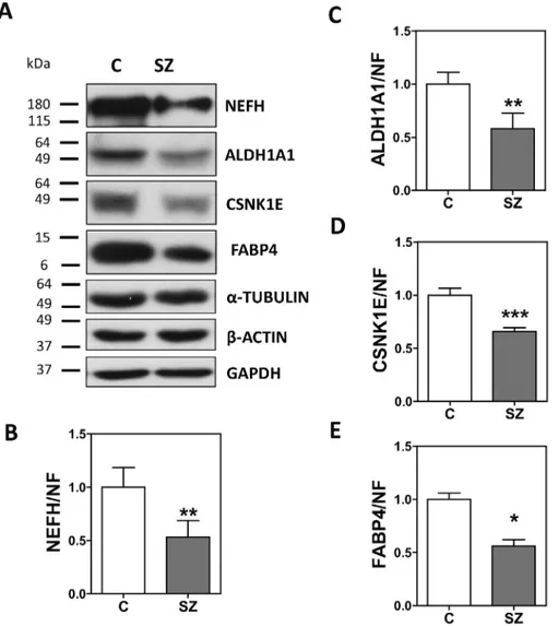
Fig. 2. Immunoblot analysis of CSNK1E, FABP4, NEFH and ALDH1A1 in the brain samples used for the proteomic analysis. Pooled protein extracts from samples of the postmortem DLPFC of controls (C, n = 4), and schizophrenia (SZ, n = 4) patients from the UPV/EHU brain collection were analyzed by immunoblotting for CSNK1E, FABP4, NEFH, ALDH1A1, andα-tubulin (TUB), β-actin (ACT) and GAPDH. Protein levels for each hit were quantified by densitometry and normalized to the geometrical mean of α-tubulin, β-actin and GAPDH values, and to the reference control sample. Images show representative immunoblots of a pool of controls (left band, C) and a pool of schizophrenia (right band, SZ) patients. Analysis was performed in duplicate. Bars represent mean ± standard deviation of the analysis of duplicates in two independent dissections, except for FABP4, which validated only in the original dissection used in the proteomic assay. Statistical analysis was performed using t-test. Statistical analysis was performed using t-test for independent samples. (n.s.-not significant, *p b 0.05, **pb 0.01, ***p b 0.001).
6 R. Pinacho et al. / Schizophrenia Research xxx (2016) xxx–xxx
Please cite this article as: Pinacho, R., et al., Altered CSNK1E, FABP4 and NEFH protein levels in the dorsolateral prefrontal cortex in schizophrenia, Schizophr. Res. (2016),http://dx.doi.org/10.1016/j.schres.2016.04.050
metabolism and energy pathways and cell growth and maintenance (Fig. S1E).
3.2. Validation of hit protein changes in DLPFC schizophrenia samples
From the significantly altered and top-20 upregulated and top-20 downregulated proteins (Table 2), 4 hit candidates were selected for further validation by immunoblot due to their protein function on cell signaling, neuronal development and synapse functioning: casein ki-nase I isoform epsilon (CSNK1E), fatty acid-binding protein 4 (FABP4), neurofilament triplet H protein (NEFH), and retinal dehydrogenase 1 (ALDH1A1).
In afirst phase, we analyzed the protein levels of CSNK1E, FABP4, NEFH and ALDH1A1 in pooled samples from the same cohort analyzed by proteomics. All candidates were referenced to the geometrical mean ofα-tubulin, β-actin and GAPDH levels in each pool, which showed no differences between the two pools (Fig. S2). We observed that NEFH, CSNK1E, FABP4, and ALDH1A1 protein levels were signi fi-cantly decreased in schizophrenia (Fig. 2). To validate these proteins as potentially decreased proteins in DLPFC in schizophrenia, we further characterized NEFH, CSKN1E, FABP4, and ALDH1A1 protein levels by immunoblot in an independent cohort of 23 male elderly chronic
schizophrenia patients and 23 matched controls (Table 1). We found that the following protein levels were significantly reduced in the schizophrenia group: NEFH [t = 1.917, df = 44; p = 0.0308; fold change (FC) ± SEM: control (C) = 1.000 ± 0.1449, SZ = 0.6856 ± 0.0768], CSNK1E [t = 1.942, df = 44; p = 0.0293; FC ± SEM: C = 1.000 ± 0.0511, SZ = 0.8773 ± 0.0372] and FABP4 [t = 1.693, df = 42; p = 0.0489; FC ± SEM: C = 1.000 ± 0.2052, SZ = 0.6050 ± 0.1110] (Fig. 3). However, ALDH1A1 was not significantly altered in this cohort (t = 1.036, df = 43; p = 0.1530; FC ± SEM: C = 1.000 ± 0.1752, SZ = 1.268 ± 0.1898) (Fig. 3). This protein is the only candidate that was previously reported to be expressed in the red cell blood pro-teome (Pasini et al., 2006) (Supplementary Data 2), which could be a confounding factor for ALDH1A1, but not for the other candidates not expressed in red blood cells. Further, we have analyzed the influence of other demographic, clinical and tissue-related variables (Table 3). FABP4 and CSNK1E showed significant correlations with age (Spearman's r =−0.313, p = 0.039) and PMD (Spearman's r = 0.334, p = 0.023), respectively. However, there are no significant differences in age and PMD between groups (Table 1). NEFH protein levels inversely correlated with the chlorpromazine equivalent dose (Pearson's r =−0.418, p = 0.047 n = 23), indicating that NEFH may be influenced by antipsychotic treatments. FABP4 protein levels nega-tively correlated with the duration of the illness (FABP4: Spearman's r =−0.540, p = 0.009, n = 22), suggesting that this protein may be changing with the progression of the disease.
4. Discussion
This study reveals novel altered proteins in the DLPFC in schizophre-nia including NEFH, CSNK1E, and FABP4. A decrease in the abundance of these proteins may be associated with cognitive and/or negative symptoms of schizophrenia.
4.1. Neurofilament triplet H protein
NEFH is a component of the neurofilament intermediate proteins found in the cytoskeleton of mature neurons that regulates axon caliber (Lee and Cleveland, 1996). There are previous reports of altered protein levels of NEFH, neurofilament triplet light protein (NEFL) and neurofil-ament triplet medium protein (NEFM) in schizophrenia (English et al., 2009; English et al., 2011; Focking et al., 2011; Martins-de-Souza et al.,
Fig. 3. CSNK1E, FABP4 and NEFH are reduced in the DLPFC of chronic schizophrenia. Protein extracts from samples of the postmortem dorsolateral prefrontal cortex of control individuals (C, n = 23), and schizophrenia patients (SZ, n = 23) from the collection of neurologic tissues of Parc Sanitari Sant Joan de Déu (Roca et al., 2008) and the Institute of Neuropathology Brain Bank (HUB-ICO-IDIBELL Biobank) were analyzed by immunoblot for the same proteins as inFig. 2and quantified by densitometry. (A) Protein levels for each protein were normalized to the geometrical mean of α-tubulin,β-actin and GAPDH values, and to a reference healthy control sample. Bars represent mean ± standard error of the mean for each group. Statistical analysis was performed using t-test for independent samples. (n.s.-not significant, *p b 0.05). (B) Representative Western blot images for the indicated proteins in 5 control individuals and 5 patients with schizophrenia.
Table 3
Association analysis of other variables in the independent validation cohort.
Age PMD pH r r r SZ-C (n = 46) CSNK1E −0.086a 0.334a⁎ −0.097a FABP4† −0.313a⁎ 0.010a 0.035a NEFH† 0.105a 0.030b 0.094b
Daily AP dose‡ Duration of illness
r r SZ (n = 23) CSNK1E 0.132b 0.015a FABP4† 0.077a −0.540a⁎⁎ NEFH −0.418b⁎ −0.156a
PMD, postmortem delay; SZ, schizophrenia; C, control; AP, antipsychotic. Significant associations are indicated in bold.
a
r, Spearman's correlation for non-parametric variables. b
r', Pearson's r for parametric variables.
† An outlier was detected for NEFH, and two outliers for FABP4 and therefore excluded from the analysis (NEFH: C, n = 22, SZ, n = 23; FABP4: C, n = 22, SZ group, n = 22).
‡ Last chlorpromazine equivalent dose was calculated based on the electronic records of drug prescriptions of the patients.
⁎ p b 0.05. ⁎⁎ p b 0.01.
2010a; Martins-de-Souza et al., 2010b; Martins-de-Souza et al., 2010c). In addition, a role for NEFM and NEFL in connectivity functions in schizophrenia has also been suggested (English et al., 2011), with an im-pact on synapses and plasticity, core features of the disorder (Friston, 1999; Harrison and Weinberger, 2005). Here we describe for thefirst time a reduction in NEFH protein levels in grey matter in schizophrenia patients. Therefore, a dysregulation of NEFH protein levels together with the other neurofilament proteins is likely to have a role in the connectivity deficits present in this disorder, participating in the neuropathology of schizophrenia.
4.2. Casein kinase I isoform epsilon
We describe a reduction in CSNK1E protein levels in the postmortem DLPFC in schizophrenia. CSNK1E is a member of the clock gene family that regulates signal transduction pathways related to the circadian molecular clock (Ko and Takahashi, 2006). Sleep and circadian rhythms abnormalities are often a co-morbidity in schizophrenia, suggesting common brain mechanisms (Klingaman et al., 2015; Pritchett et al., 2012). Cognitive impairments occur in sleep and circadian rhythm disruption as well as in schizophrenia (Pritchett et al., 2012). This together with our finding that CSNK1E expression is reduced in schizophrenia suggests that the molecular clock pathway in the prefrontal cortex may have a role in cognitive deficits in both disorders. More work is needed to investigate this possibility.
4.3. Fatty acid-binding protein 4
FABP4 is a member of the FABP family of proteins whose primary role is facilitating the uptake and intracellular transport of hydrophobic fatty acids and so participating in energy metabolism, signaling path-ways and regulation of transcription (Chmurzynska, 2006). Although FABP4 is typically expressed in adipocytes and macrophages, we have detected it in human postmortem brain by mass spectrometry and an isoform-specific antibody, in line with previous reports (Anderson et al., 2011; Chaerkady et al., 2011). We found that FABP4 protein levels are decreased in schizophrenia patients. Notably, a recent study reported a reduction in FABP4 expression in the scalp hair follicle in patients with schizophrenia in an attempt to provide more accessible biomarkers for this disorder (Maekawa et al., 2015). This report also analyzed FABP4 gene expression in the brains but found no changes in Brodmann area 46. Interestingly, here we have found that FABP4 protein levels are reduced in Brodmann area 9, further suggesting a role for FABP4 in the pathology of schizophrenia. Despite the role of FABP4 in brain function is unknown, it has been reported that other FABP proteins such as FABP3, FABP5 and FABP7 play important roles in brain development (Liu et al., 2010). These additional FABP proteins facilitate the cellular functions of long chain polyunsaturated fatty acids (PUFAs), which have also been linked to schizophrenia (Freeman, 2000; Maekawa et al., 2011). PUFAs are essential for the nor-mal development of the brain (Basak et al., 2013; Neuringer et al., 1988; Wainwright, 2002), participate in synaptic vesicle trafficking (Ben Gedalya et al., 2009), and have been related to altered dopamine vesicle density in rat frontal cortex (Zimmer et al., 2000) and behavioral disturbances (Bourre et al., 1989; Yoshida et al., 1997). Therefore, we suggest that a decrease in FABP4 in schizophrenia could be limiting vesicle formation in the presynaptic terminal. However, further studies will be needed to confirm this mechanism.
4.4. Retinal dehydrogenase 1
ALDH1A1 appeared to be altered in the pool of the smaller cohort but this was not replicated in the larger cohort used in this study, com-prising tissue from older individuals. This could be a consequence of ALDH1A1 being part of the red blood cell proteome (Pasini et al., 2006) and therefore a false positive or may be the result of an alteration
present in only one patient. However, this protein has been previously described as being reduced in white matter in the DLPFC in schizophre-nia (Prabakaran et al., 2004) and regulated by psychotropic drugs in the context of Parkinson Disease (Lauterbach, 2012). The closely related protein ALDH1A2 has also been associated with schizophrenia previ-ously (Wan et al., 2009). Therefore, further studies should be conducted to determine whether this factor is associated with the pathophysiology of schizophrenia.
4.5. Limitations
The use of human postmortem brain constitutes a useful tool to dissect the molecular mechanisms disrupted in psychiatric disorders, but has limitations. Therefore, although potentially interesting, the molecularfindings of this study should be further interrogated for functional validity using orthogonal techniques. First, our pilot proteo-mic analysis used pooled samples. This type of design is a useful approach for rapidly detecting common altered pathways (Behan et al., 2008; Martins-de-Souza et al., 2009a; Martins-de-Souza et al., 2009b); however, it does not allow for the control of inter-individual variations, which, as noted for ALDH1A1, could account for modi fica-tions in the results. A cautious interpretation of this panel of altered protein should be considered. Further validation in individual samples will be needed for this list of possible altered candidates. Second, the possible effect of laterality in our sample cannot be ruled out, since only contralateral prefrontal cortex was available for the non-psychiatric control group. Left schizophrenia prefrontal cortex was compared to right control prefrontal cortex. Further analyses should be performed to explore the laterality effects on the abundance of these proteins in the prefrontal cortex and their potential as biomarkers. To investigative their possible role as biomarkers it would be of great interest to extend the study of these candidates to tissues that can be studied with less invasive approaches such as cerebrospinalfluid (CSF) and peripheral blood cells. Third, the patients in our second validation cohort had long-lasting and heterogeneous antipsychotic medications. To control for this variable, we have used the last daily chlorpromazine equivalent dose in a bivariate analysis. We found that NEFH significantly correlated with the antipsychotic dose suggesting that the reduction of NEFH observed in this study could be the result of the antipsychotic treatments. Further pharmacological studies in cellular and animal models, as well as in drug naive patients, will help to clarify the possible influence of antipsychotic treatments in these candidate proteins. Fourth, we had elderly patients and matched con-trols in this study. Fifth, the study only included men. Further studies in a younger cohort with equal representation of both genders and if possible drug naive patients would be of interest. Finally, we were not able to validate thefindings for ALDH1A1 in the larger cohort. This protein has also been described in the red cell proteome (Pasini et al., 2006). This raised the question whether some changes may be related to the presence of blood in the samples. A careful post-hoc analysis of candidates expressed in red blood cells should be taken in postmortem tissue analysis to address this potential confound. Despite these limita-tions,findings from this study may contribute towards a better under-standing of the molecular mechanisms that underlie schizophrenia.
4.6. Conclusions
Ourfindings in the DLPFC in schizophrenia provide evidence of altered proteins involved in synaptic function (FABP4), cytoarchitecture organization (NEFH), and circadian molecular clock signaling (CSNK1E), which may be contributing to the cognitive and/or negative symptoms in this disorder. Moreover, FABP4, CSNK1E and NEFH could become potentially useful biomarkers for schizophrenia in the future.
Supplementary data to this article can be found online athttp://dx. doi.org/10.1016/j.schres.2016.04.050.
8 R. Pinacho et al. / Schizophrenia Research xxx (2016) xxx–xxx
Please cite this article as: Pinacho, R., et al., Altered CSNK1E, FABP4 and NEFH protein levels in the dorsolateral prefrontal cortex in schizophrenia, Schizophr. Res. (2016),http://dx.doi.org/10.1016/j.schres.2016.04.050
Role of funding source
The funding bodies had no role in study design, in collection, analyses and interpreta-tion of the data, in the writing of the article, or in the decision to submit the paper for publication.
Contributors
Author RP performed human postmortem tissue dissection and validation expression measurements, analyzed validation results, performed the statistical analysis, and co-wrote thefirst draft of the manuscript. Author NV carried out the postmortem tissue processing and did expression measurements. Author JJM contributed to the discussion of the results. IF carried out the histological analysis and contributed to the discussion of the results. AB contributed to the samples clinical database. Author JMH designed and implemented the clinical protocol and contributed to the discussion of the results. Author JV co-designed and implemented the proteomic analysis, discussed results and contribut-ed to manuscript writing. Author BR designcontribut-ed the study, performcontribut-ed the processing of human postmortem tissue samples in the proteomic analysis, analyzed the proteomic results, supervised the protein expression analysis, and co-wrote thefirst draft of the manuscript. All authors contributed to and approved thefinal manuscript.
Conflict of interest
All authors declare that they have no conflicts of interest. Acknowledgements
These studies were supported by Predoctoral Fellowship Program from ISCIII (PFIS) FI10/00177 to R.P. and the Centro de Investigación Biomédica en Red de Salud Mental (CIBERSAM) (CB/07/09/0028) to B.R. and J.M.H. Financial support of this work comes from Marie Curie Program IRG RTD REG/T.2 (2007)D/530573 to B.R., Plan Nacional de Investigación BFU2008-01103 (MCINN) to B.R. and SAF2009-08460 to J.J.M. The authors thank the donors and their families for the donation of their brains; the collaboration of the staff members of the Basque Institute of Legal Medicine for their help.
References
Anderson, G.D., Farin, F.M., Bammler, T.K., Beyer, R.P., Swan, A.A., Wilkerson, H.W., Kantor, E.D., Hoane, M.R., 2011.The effect of progesterone dose on gene expression after traumatic brain injury. J. Neurotrauma 28 (9), 1827–1843.
Baek, D., Villen, J., Shin, C., Camargo, F.D., Gygi, S.P., Bartel, D.P., 2008.The impact of microRNAs on protein output. Nature 455 (7209), 64–71.
Basak, S., Das, M.K., Duttaroy, A.K., 2013.Fatty acid-induced angiogenesis infirst trimester placental trophoblast cells: possible roles of cellular fatty acid-binding proteins. Life Sci. 93 (21), 755–762.
Behan, A.T., Byrne, C., Dunn, M.J., Cagney, G., Cotter, D.R., 2008.Proteomic analysis of membrane microdomain-associated proteins in the dorsolateral prefrontal cortex in schizophrenia and bipolar disorder reveals alterations in LAMP, STXBP1 and BASP1 protein expression. Mol. Psychiatry 14 (6), 601–613.
Ben Gedalya, T., Loeb, V., Israeli, E., Altschuler, Y., Selkoe, D.J., Sharon, R., 2009. Alpha-synuclein and polyunsaturated fatty acids promote clathrin-mediated endocytosis and synaptic vesicle recycling. Traffic 10 (2), 218–234.
Benjamini, Y., Hochberg, Y., 1995.Controlling the false discovery rate: a practical and powerful approach to multiple testing. J. R. Stat. Soc. Ser. B 57, 289–300.
Bourre, J.M., Francois, M., Youyou, A., Dumont, O., Piciotti, M., Pascal, G., Durand, G., 1989.
The effects of dietary alpha-linolenic acid on the composition of nerve membranes, enzymatic activity, amplitude of electrophysiological parameters, resistance to poisons and performance of learning tasks in rats. J. Nutr. 119 (12), 1880–1892.
Braak, H., Braak, E., 1991.Neuropathological stageing of Alzheimer-related changes. Acta Neuropathol. 82 (4), 239–259.
Braak, H., Alafuzoff, I., Arzberger, T., Kretzschmar, H., Del Tredici, K., 2006.Staging of Alzheimer disease-associated neurofibrillary pathology using paraffin sections and immunocytochemistry. Acta Neuropathol. 112 (4), 389–404.
Chaerkady, R., Letzen, B., Renuse, S., Sahasrabuddhe, N.A., Kumar, P., All, A.H., Thakor, N.V., Delanghe, B., Gearhart, J.D., Pandey, A., Kerr, C.L., 2011.Quantitative temporal prote-omic analysis of human embryonic stem cell differentiation into oligodendrocyte progenitor cells. Proteomics 11 (20), 4007–4020.
Chan, M.K., Tsang, T.M., Harris, L.W., Guest, P.C., Holmes, E., Bahn, S., 2011.Evidence for disease and antipsychotic medication effects in post-mortem brain from schizophre-nia patients. Mol. Psychiatry 16 (12), 1189–1202.
Chmurzynska, A., 2006.The multigene family of fatty acid-binding proteins (FABPs): function, structure and polymorphism. J. Appl. Genet. 47 (1), 39–48.
English, J.A., Dicker, P., Focking, M., Dunn, M.J., Cotter, D.R., 2009.2-D DIGE analysis implicates cytoskeletal abnormalities in psychiatric disease. Proteomics 9 (12), 3368–3382.
English, J.A., Pennington, K., Dunn, M.J., Cotter, D.R., 2011.The Neuroproteomics of Schizo-phrenia. Biol. Psychiatry 69 (2), 163–172.
Focking, M., Dicker, P., English, J.A., Schubert, K.O., Dunn, M.J., Cotter, D.R., 2011.Common proteomic changes in the hippocampus in schizophrenia and bipolar disorder and particular evidence for involvement of cornu ammonis regions 2 and 3. Arch. Gen. Psychiatry 68 (5), 477–488.
Freeman, M.P., 2000.Omega-3 fatty acids in psychiatry: a review. Ann. Clin. Psychiatry 12 (3), 159–165.
Friston, K.J., 1999.Schizophrenia and the disconnection hypothesis. Acta Psychiatr. Scand. Suppl. 395, 68–79.
Frith, C., Dolan, R., 1996.The role of the prefrontal cortex in higher cognitive functions. Brain Res. Cogn. Brain Res. 5 (1–2), 175–181.
Gardner, D.M., Murphy, A.L., O'Donnell, H., Centorrino, F., Baldessarini, R.J., 2010. Interna-tional consensus study of antipsychotic dosing. Am. J. Psychiatry 167 (6), 686–693.
Gold, J.M., 2004.Cognitive deficits as treatment targets in schizophrenia. Schizophr. Res. 72 (1), 21–28.
Goldstein, J.M., Goodman, J.M., Seidman, L.J., Kennedy, D.N., Makris, N., Lee, H., Tourville, J., Caviness Jr., V.S., Faraone, S.V., Tsuang, M.T., 1999.Cortical abnormalities in schizo-phrenia identified by structural magnetic resonance imaging. Arch. Gen. Psychiatry 56 (6), 537–547.
Graumann, J., Hubner, N.C., Kim, J.B., Ko, K., Moser, M., Kumar, C., Cox, J., Scholer, H., Mann, M., 2008.Stable isotope labeling by amino acids in cell culture (SILAC) and proteome quantitation of mouse embryonic stem cells to a depth of 5,111 proteins. Mol. Cell. Proteomics 7 (4), 672–683.
Harrison, P.J., Weinberger, D.R., 2005.Schizophrenia genes, gene expression, and neuro-pathology: on the matter of their convergence. Mol. Psychiatry 10 (1), 40–68.
Johnston-Wilson, N.L., Sims, C.D., Hofmann, J.P., Anderson, L., Shore, A.D., Torrey, E.F., Yolken, R.H., 2000.Disease-specific alterations in frontal cortex brain proteins in schizophrenia, bipolar disorder, and major depressive disorder. The Stanley Neuropa-thology Consortium. Mol. Psychiatry 5 (2), 142–149.
Khidekel, N., Ficarro, S.B., Clark, P.M., Bryan, M.C., Swaney, D.L., Rexach, J.E., Sun, Y.E., Coon, J.J., Peters, E.C., Hsieh-Wilson, L.C., 2007.Probing the dynamics of O-GlcNAc glycosylation in the brain using quantitative proteomics. Nat. Chem. Biol. 3 (6), 339–348.
Kienast, T., Heinz, A., 2006.Dopamine and the diseased brain. CNS Neurol. Disord. Drug Targets 5 (1), 109–131.
Klingaman, E.A., Palmer-Bacon, J., Bennett, M.E., Rowland, L.M., 2015.Sleep disorders among people with schizophrenia: emerging research. Curr. Psychiatry Rep. 17 (10), 616.
Ko, C.H., Takahashi, J.S., 2006.Molecular components of the mammalian circadian clock. Hum. Mol. Genet. 15 (2), R271–R277.
Konradi, C., 2005.Gene expression microarray studies in polygenic psychiatric disorders: applications and data analysis. Brain Res. Brain Res. Rev. 50 (1), 142–155.
Krystal, J.H., Karper, L.P., Seibyl, J.P., Freeman, G.K., Delaney, R., Bremner, J.D., Heninger, G.R., Bowers Jr., M.B., Charney, D.S., 1994.Subanesthetic effects of the noncompetitive NMDA antagonist, ketamine, in humans. Psychotomimetic, perceptual, cognitive, and neuroendocrine responses. Arch. Gen. Psychiatry 51 (3), 199–214.
Lauterbach, E.C., 2012. Psychotropics regulate Skp1a, Aldh1a1, and Hspa8 transcription—potential to delay Parkinson's disease. Prog. Neuro-Psychopharmacol. Biol. Psychiatry 40, 236–239.
Lee, M.K., Cleveland, D.W., 1996.Neuronal intermediatefilaments. Annu. Rev. Neurosci. 19, 187–217.
Lewis, D.A., Moghaddam, B., 2006.Cognitive dysfunction in schizophrenia: convergence of gamma-aminobutyric acid and glutamate alterations. Arch. Neurol. 63 (10), 1372–1376.
Liu, R.Z., Mita, R., Beaulieu, M., Gao, Z., Godbout, R., 2010.Fatty acid binding proteins in brain development and disease. Int. J. Dev. Biol. 54 (8–9), 1229–1239.
Maekawa, M., Owada, Y., Yoshikawa, T., 2011.Role of polyunsaturated fatty acids and fatty acid binding protein in the pathogenesis of schizophrenia. Curr. Pharm. Des. 17 (2), 168–175.
Maekawa, M., Yamada, K., Toyoshima, M., Ohnishi, T., Iwayama, Y., Shimamoto, C., Toyota, T., Nozaki, Y., Balan, S., Matsuzaki, H., Iwata, Y., Suzuki, K., Miyashita, M., Kikuchi, M., Kato, M., Okada, Y., Akamatsu, W., Mori, N., Owada, Y., Itokawa, M., Okano, H., Yoshikawa, T., 2015.Utility of scalp hair follicles as a novel source of biomarker genes for psychiatric illnesses. Biol. Psychiatry 78 (2), 116–125.
Mai, J.K., Assheuer, J., Paxinos, G., 1997.Atlas of the Human Brain. Academic Press, San Diego.
Martins-de-Souza, D., Gattaz, W.F., Schmitt, A., Maccarrone, G., Hunyadi-Gulyas, E., Eberlin, M.N., Souza, G.H., Marangoni, S., Novello, J.C., Turck, C.W., Dias-Neto, E., 2009a.Proteomic analysis of dorsolateral prefrontal cortex indicates the involvement of cytoskeleton, oligodendrocyte, energy metabolism and new potential markers in schizophrenia. J. Psychiatr. Res. 43 (11), 978–986.
Martins-de-Souza, D., Gattaz, W.F., Schmitt, A., Rewerts, C., Maccarrone, G., Dias-Neto, E., Turck, C.W., 2009b.Prefrontal cortex shotgun proteome analysis reveals altered calci-um homeostasis and immune system imbalance in schizophrenia. Eur. Arch. Psychi-atry Clin. Neurosci. 259 (3), 151–163.
Martins-de-Souza, D., Dias-Neto, E., Schmitt, A., Falkai, P., Gormanns, P., Maccarrone, G., Turck, C.W., Gattaz, W.F., 2010a.Proteome analysis of schizophrenia brain tissue. World J. Biol. Psychiatry 11 (2), 110–120.
Martins-de-Souza, D., Maccarrone, G., Wobrock, T., Zerr, I., Gormanns, P., Reckow, S., Falkai, P., Schmitt, A., Turck, C.W., 2010b.Proteome analysis of the thalamus and cerebrospinal fluid reveals glycolysis dysfunction and potential biomarkers candidates for schizophrenia. J. Psychiatr. Res. 44 (16), 1176–1189.
Martins-de-Souza, D., Schmitt, A., Roder, R., Lebar, M., Schneider-Axmann, T., Falkai, P., Turck, C.W., 2010c.Sex-specific proteome differences in the anterior cingulate cortex of schizophrenia. J. Psychiatr. Res. 44 (14), 989–991.
Millan, M.J., Fone, K., Steckler, T., Horan, W.P., 2014.Negative symptoms of schizophrenia: clinical characteristics, pathophysiological substrates, experimental models and prospects for improved treatment. Eur. Neuropsychopharmacol. 24 (5), 645–692.
Moghaddam, B., Javitt, D., 2012.From revolution to evolution: the glutamate hypothesis of schizophrenia and its implication for treatment. Neuropsychopharmacology 37 (1), 4–15.
Neuringer, M., Anderson, G.J., Connor, W.E., 1988.The essentiality of n-3 fatty acids for the development and function of the retina and brain. Annu. Rev. Nutr. 8, 517–541.
Novikova, S.I., He, F., Cutrufello, N.J., Lidow, M.S., 2006.Identification of protein biomarkers for schizophrenia and bipolar disorder in the postmortem prefrontal
cortex using SELDI-TOF-MS ProteinChip profiling combined with MALDI-TOF-PSD-MS analysis. Neurobiol. Dis. 23 (1), 61–76.
Pasini, E.M., Kirkegaard, M., Mortensen, P., Lutz, H.U., Thomas, A.W., Mann, M., 2006. In-depth analysis of the membrane and cytosolic proteome of red blood cells. Blood 108 (3), 791–801.
Pennington, K., Beasley, C.L., Dicker, P., Fagan, A., English, J., Pariante, C.M., Wait, R., Dunn, M.J., Cotter, D.R., 2008.Prominent synaptic and metabolic abnormalities revealed by proteomic analysis of the dorsolateral prefrontal cortex in schizophrenia and bipolar disorder. Mol. Psychiatry 13 (12), 1102–1117.
Pinacho, R., Villalmanzo, N., Lalonde, J., Haro, J.M., Meana, J.J., Gill, G., Ramos, B., 2011.The transcription factor SP4 is reduced in postmortem cerebellum of bipolar disorder subjects: control by depolarization and lithium. Bipolar Disord. 13 (5–6), 474–485.
Prabakaran, S., Swatton, J.E., Ryan, M.M., Huffaker, S.J., Huang, J.T., Griffin, J.L., Wayland, M., Freeman, T., Dudbridge, F., Lilley, K.S., Karp, N.A., Hester, S., Tkachev, D., Mimmack, M.L., Yolken, R.H., Webster, M.J., Torrey, E.F., Bahn, S., 2004.Mitochondrial dysfunc-tion in schizophrenia: evidence for compromised brain metabolism and oxidative stress. Mol. Psychiatry 9 (7), 684–697 (643).
Pritchett, D., Wulff, K., Oliver, P.L., Bannerman, D.M., Davies, K.E., Harrison, P.J., Peirson, S.N., Foster, R.G., 2012.Evaluating the links between schizophrenia and sleep and cir-cadian rhythm disruption. J. Neural Transm. 119 (10), 1061–1075.
Roca, M., Escanilla, A., Monje, A., Baño, V., Planchat, L., Costa, J., Haro, J., 2008.Banco de tejidos neurológicos de Sant Joan de Déu-Serveis de Salut Mental para la investigación de las enfermedades mentales. La importancia de un programa de donación en vida. Psiquiatría Biológica 15 (3), 73–79.
Semkovska, M., Bedard, M.A., Stip, E., 2001.Hypofrontality and negative symptoms in schizophrenia: synthesis of anatomic and neuropsychological knowledge and ecolog-ical perspectives. L'Encéphale 27 (5), 405–415.
Smalla, K.H., Mikhaylova, M., Sahin, J., Bernstein, H.G., Bogerts, B., Schmitt, A., van der Schors, R., Smit, A.B., Li, K.W., Gundelfinger, E.D., Kreutz, M.R., 2008.A comparison of the synaptic proteome in human chronic schizophrenia and rat ketamine
psychosis suggest that prohibitin is involved in the synaptic pathology of schizophre-nia. Mol. Psychiatry 13 (9), 878–896.
Stahl, S.M., Buckley, P.F., 2007.Negative symptoms of schizophrenia: a problem that will not go away. Acta Psychiatr. Scand. 115 (1), 4–11.
Sullivan, P.F., 2012.Puzzling over schizophrenia: schizophrenia as a pathway disease. Nat. Med. 18 (2), 210–211.
Teffer, K., Semendeferi, K., 2012.Human prefrontal cortex: evolution, development, and pathology. Prog. Brain Res. 195, 191–218.
Toda, M., Abi-Dargham, A., 2007.Dopamine hypothesis of schizophrenia: making sense of it all. Curr. Psychiatry Rep. 9 (4), 329–336.
Wainwright, P.E., 2002.Dietary essential fatty acids and brain function: a developmental perspective on mechanisms. Proc. Nutr. Soc. 61 (1), 61–69.
Wan, C., Shi, Y., Zhao, X., Tang, W., Zhang, M., Ji, B., Zhu, H., Xu, Y., Li, H., Feng, G., He, L., 2009.Positive association between ALDH1A2 and schizophrenia in the Chinese pop-ulation. Prog. Neuro-Psychopharmacol. Biol. Psychiatry 33 (8), 1491–1495.
Wesseling, H., Chan, M.K., Tsang, T.M., Ernst, A., Peters, F., Guest, P.C., Holmes, E., Bahn, S., 2013.A combined metabonomic and proteomic approach identifies frontal cortex changes in a chronic phencyclidine rat model in relation to human schizophrenia brain pathology. Neuropsychopharmacology 38 (12), 2532–2544.
Wong, A.H., Van Tol, H.H., 2003.Schizophrenia: from phenomenology to neurobiology. Neurosci. Biobehav. Rev. 27 (3), 269–306.
Yoshida, S., Yasuda, A., Kawazato, H., Sakai, K., Shimada, T., Takeshita, M., Yuasa, S., Kobayashi, T., Watanabe, S., Okuyama, H., 1997.Synaptic vesicle ultrastructural changes in the rat hippocampus induced by a combination of alpha-linolenate deficiency and a learning task. J. Neurochem. 68 (3), 1261–1268.
Zimmer, L., Delpal, S., Guilloteau, D., Aioun, J., Durand, G., Chalon, S., 2000.Chronic n-3 polyunsaturated fatty acid deficiency alters dopamine vesicle density in the rat frontal cortex. Neurosci. Lett. 284 (1–2), 25–28.
10 R. Pinacho et al. / Schizophrenia Research xxx (2016) xxx–xxx
Please cite this article as: Pinacho, R., et al., Altered CSNK1E, FABP4 and NEFH protein levels in the dorsolateral prefrontal cortex in schizophrenia, Schizophr. Res. (2016),http://dx.doi.org/10.1016/j.schres.2016.04.050