é
ergia M
Revista
Al
xico
Clínica A.C. Original articleThe role of the fern test in the treatment of rhinitis
El papel de la prueba de “helecho” en el tratamiento
de la rinitis
Matteo Gelardi,1 Giuseppe Porro,1 Nicola Quaranta,1 Brigida Sterlicchio,1 Michela Silvestri,2 Giorgio Ciprandi3
1University of Bari Aldo Moro, Department of Basic Medical Scien-ce, Neuroscience and Sensory Organs, Bari, Italy
2Istituto Giannina Gaslini, Genoa, Italy 3Ospedale Policlinico San Martino, Genoa, Italy
How to cite this article: Gelardi M, Porro G, Quaranta N, Sterlicchio B, Silvestri M, Ciprandi G.The role of the fern test in the treatment of rhinitis. Rev Alerg Mex. 2019;66(2):184-191
ORCID
Matteo Gelardi, 0000-0003-4406-0008; Giuseppe Porro, 0000-0003-2860-7608; Nicola Quaranta,
0000-0001-6214-2336; Brigida Sterlicchio, 0000-0002-6606-4811; Michela Silvestri, 0000-0003-4804-2421; Giorgio Ciprandi, 0000-0001-7016-8421
Abstract
Background: The fern test is a method for assessing the characteristics of the nasal section in the treatment of patients with mucous dysfunction of the airway.
Objective: The aim of this study was to investigate the role of the fern test in patients with rhinitis and to assess the classifi cation of each type of rhinitis (types I-IV) in clinical practice.
Methods: A cross-sectional study, which included consecutive patients from a third level Rhinology Unit, worked with 182 patients with rhinitis and 30 healthy subjects as control. The patients were subdivided according to their type of rhinitis: allergic rhinitis (59), infectious rhinitis (32), polyps (31), NARES (Non-allergic rhinitis with eosinophilia syndrome) (30) and NARNE (non-allergic rhinitis with neutrophils) (30).
Results: The control subjects had only type I or II rhinitis, whereas patients with rhinitis usually showed type III or IV. Allergic rhinitis and nasal polyps had the most serious deterioration according to the fern test (type IV).
Conclusions: The fern test is effective for assessing mucus alterations in patients with rhinitis and it could be included as a new parameter in the study of rhinitis as a potential biomarker of the function of damaged epithelial cells.
Keywords: Rhinitis; Fern test; Infl ammation of the mucosa
Correspondence: Giorgio Ciprandi. [email protected]
Received: 2018-08-28 Accepted: 2019-01-24 DOI: 10.29262/ram.v66i2.544
Background
Mucus is a complex, viscoelastic, and adherent se-cretion that is produced and secreted by specialized goblet cells and mucous cells in the columnar epi-thelia that line the lumen of all the mucosa that are exposed to the external environment.1,2 In particular,
mucus covers the inner linings of organs of the re-spiratory tract, the gastrointestinal tract, the repro-ductive tract, and the ocular surface. The concept of mucus is very old as its existence was already known from the time of the Ancient Greeks: phlegm was one of 4 humors. Mucus is widely produced in both kingdoms, plant and animal.3
From a physiological point of view, mucus car-ries out many protective functions for the underlying epithelia, such as mucosal lubrication, which is use-ful for material transport and cellular hydration of epithelial cells, mainly in the respiratory tract, eyes and mouth, which are exposed directly to the drying evaporative effects of air, and it provides a barrier against noxious agents and exposure to pathogens
by trapping them.4 It also works as a cleansing
trans-port, in which the external particles that are trapped in the mucus layer can be eliminated from organ cavities by cilia-facilitated expulsion of the mucus layer5 and provides a selectively permeable gel layer
for the diffusion, exchange and absorption of gases (eyes and lungs) and nutrients (gastrointestinal tract) with the underlying epithelium.6
The rheology of nasal mucus deserves prop-er attention in clinical practice.7,8 From a chemical
point of view, mucus should be seen as an integrat-ed structure of biopolymers. The physical charac-teristics of mucus are complex, for example; it is a non-Newtonian fl uid, and its physical characteris-tics range from a viscous liquid to an elastic solid. Mucus can be measured considering its consistency by viscosity (resistance to fl ow) and elasticity (stiff-ness). It must be noted that impaired mucus rheolo-gy may have signifi cance on its functions, including lubrication, barrier, and defense against infective agents and noxious substances.8
Resumen
Antecedentes: La prueba de “helecho” es un método que sirve para evaluar las características de la secreción nasal en el tratamiento de pacientes con disfunción de la mocosa de la vía aérea.
Objetivo: El objetivo del presente estudio fue investigar el papel de la prueba de helecho en pacientes con rinitis y evaluar la clasifi cación de cada tipo de rinitis (tipos I a IV) en la práctica clínica.
Métodos: Estudio transversal en el que se incluyeron pacientes consecutivos de una unidad de rinología de tercer nivel. Se incluyeron 182 pacientes con rinitis y 30 sujetos sanos como controles. Los pacientes se subdividieron según el tipo de rinitis: alérgica (59), infecciosa (32), pólipos (31), rinitis eosinofílica no alérgica (30) y rinitis no alérgica con neutrófi los (30).
Resultados: Los sujetos control solo presentaron rinitis tipo I o II, mientras que los pacientes con rinitis generalmente mostraban tipo III o IV. La rinitis alérgica y los pólipos nasales tuvieron el deterioro más grave según la prueba de helecho (tipo IV).
Conclusiones: La prueba de helecho es efectiva para evaluar las alteraciones del moco en pacientes con rinitis y podría incluirse como un nuevo parámetro en el estudio de la rinitis como biomarcador potencial de la función de las células epiteliales dañadas.
Palabras clave: Rinitis; Prueba de "helecho"; Infl amación de la mucosa
Abbreviations and acronyms
AR, allergic rhinitis
MGG, May-Grünwald-Giemsa
NARES, non-allergic rhinitis with eosinophilia NARNE, non-allergic rhinitis with neutrop hils
Mucus is a thixotropic gel that is able to proper-ly respond to shear stress. This ability depends on its composition. The components of mucus are mucins, DNA fragments, lipids, ions, proteins, cells, cellu-lar debris, and water.9,10 In this regard, mucins are
the most important component of the mucus as it is formed by cross-linked, bundled, and entangled mu-cin fi bers which are produced and secreted by both goblet cells and sero-mucinous glands.
The bio-rheological properties of the nasal mu-cus are the cohesive forces, the load-bearing capaci-ty, the spinnabilicapaci-ty, the thixotropy, the creep, and the adhesiveness. This complexity may easily explain how disorders could signifi cantly affect the compo-sition and function of nasal mucus. Infections, in-fl ammatory disorders (including allergic and non-al-lergic infi ltrates), polyps, drugs, and primary mucus defects may signifi cantly alter the composition and function of mucus.
The properties of mucus may be assessed by using sophisticated tests, but a simple way could be represented by the nasal cytology. Long time ago, Papanicolaou proved that some cervical mucus that was spread on a slide and left to dry was able to crystallize with a characteristic shape, like an “ar-borization”: a fern-like reaction.11 He brilliantly
con-ceived that this phenomenon could depend on the ovulation time as a refl ection of the estrogenic activ-ity that was able to affect the characteristics of the mucus. Further on, it was proven, by inducing the mucus-fern phenomenon,12,13 that the alterations of
mucus, depending on the cycle of sexual hormones, occurred also in other mucus-secreting surfaces of the body; including the nose, salivary glands, and lacrimal glands. Abou-Shabanah and Plotz proved that the fern reaction depended on electrolyte, pro-tein and/or saccharide concentrations.14 So, the fern
test was used to assess the characteristics of mucus in several disorders. Rolando et al. defi ned a scoring system to assess the fern test15 and reported fern
im-pairment in different disorders at an ocular level.16,17
These outcomes were confi rmed by further studies that were always conducted at an ocular level,18,19 so
the fern test is commonly used in clinical practice in an ophthalmological setting. However, only another study considered the fern phenomenon in the nose: it compared nasal and cervical mucus. The authors concluded that mucus is an ancestral biological component, but they did not speculate about nasal
disorders.20 Thus, we aimed to investigate the role
of the fern test in patients with rhinitis in clinical practice.
Methods
Globally, 182 consecutive patients (102 males and 80 females, mean age: 43.15 ± 19.03 years) with rhinitis were visited at the Rhinology Unit because of their intensive treatment and they were included in this study.
The inclusion criteria were: • Adult men and women.
• Documented diagnosis of rhinitis, including al-lergic rhinitis, infectious rhinitis, non-alal-lergic rhinitis (NARES or NARNE), or nasal polyps. • Presence of nasal symptoms.
The exclusion criteria were:
• Concomitant comorbidities and chronic illness-es that could interfere in the interpretation of the fi ndings.
• Current treatments able to interfere in the inter-pretation of the fi ndings. In addition, 30 healthy subjects were included in the study.
The Review Board approved the procedure and a written informed consent was given by all participants.
A nasal endoscopy was carried out with a 3.4 mm diameter fl exible fi beroscope (Vision-Sciences®
ENT-2000) to assess the nasal cavities.
Nasal cytology involves: sampling, processing and microscope reading. The sampling requires the collection of cells from the surface of nasal mucosa, which is done by a sterile disposable curette. Sam-ples should be collected from the middle portion of the inferior turbinate. The procedure is performed under anterior rhinoscopy, with an appropriate light source, and the procedure is completely painless. The obtained sample is smeared immediately on a glass slide and it is air-dried. The sample staining is carried out by using the common May-Grünwald-Giemsa (MGG) staining because of its ability to correctly identify the infl ammatory nasal cells. The traditional MGG staining procedure requires about 30 minutes. The stained sample is read at optical microscopy, with a 1000x objective with oil immersion. Fifty is
the minimum number of fi elds in order to identify a suffi cient number of cells. The count of each cell type was expressed by a semi-quantitative grading as it was described previously.21 The detection of
eo-sinophils, mast cells, bacteria or fungal ifae clearly identifi es a pathological condition. In nasal cytolo-gy, bacterial infectious rhinitis is usually character-ized by the presence of many neutrophils, with intra and extracellular bacteria that can be easily identi-fi ed at optical microscopy. Lymphocytes and mac-rophage can accompany the neutrophilic infi ltrate.22
Allergic rhinitis (AR) is always characterized by the presence of infl ammatory cells (eosinophils, mast cells, neutrophils, and lymphocytes) in the nose. Non-allergic infl ammatory rhinitis is defi ned by the presence of infl ammatory cells in the nasal muco-sa and by the absence of allergic sensitization. The best known and fi rst described non-allergic rhinitis was NARES: the presence of eosinophils is not only predominant but also massively present with an ex-pression that is even higher than in seasonal rhinitis. Another important type of infl ammatory non-aller-gic rhinitis is NARNE; which is characterized by a massive presence of neutrophils without concomi-tant bacterial colonization.23 Nasal polyps may show
an infl ammatory pattern, as previously reported.24
For the fern test, nasal specimens were taken, as well as the traditional nasal cytology. The
collect-ed smears were put on microscope slides and left to dry at room temperature for 10 minutes. They were examined under a phase contrast microscope at 100× and 400× magnifi cation and they were classifi ed from type I to type IV according to the Rolando scoring.25
In type I (normal pattern), the fern pattern is uniform with closely branching arborization. In type II, the ferns are well distinguished but with less branching. In type III, there are scattered ferns with rare branch-ing, and, in type IV, no fern pattern is seen.
Statistical analysis
Descriptive statistics were performed and reported in terms of absolute frequencies or percentages for qualitative data and in terms of medians with fi rst and third quartiles (1q-3q) or means with standard deviation for quantitative data. The comparison of frequency distributions was made by means of the chi-square test or the Fisher’s exact test in case of expected frequencies < 5. All tests were two-tailed, and p-values < 0.05 have been considered statisti-cally signifi cant. The analyses were performed using the GraphPad Prism software, GraphPad Software Inc, CA, USA
Results
Table 1 reports demographic and clinical character-istics of control and patients with different types of
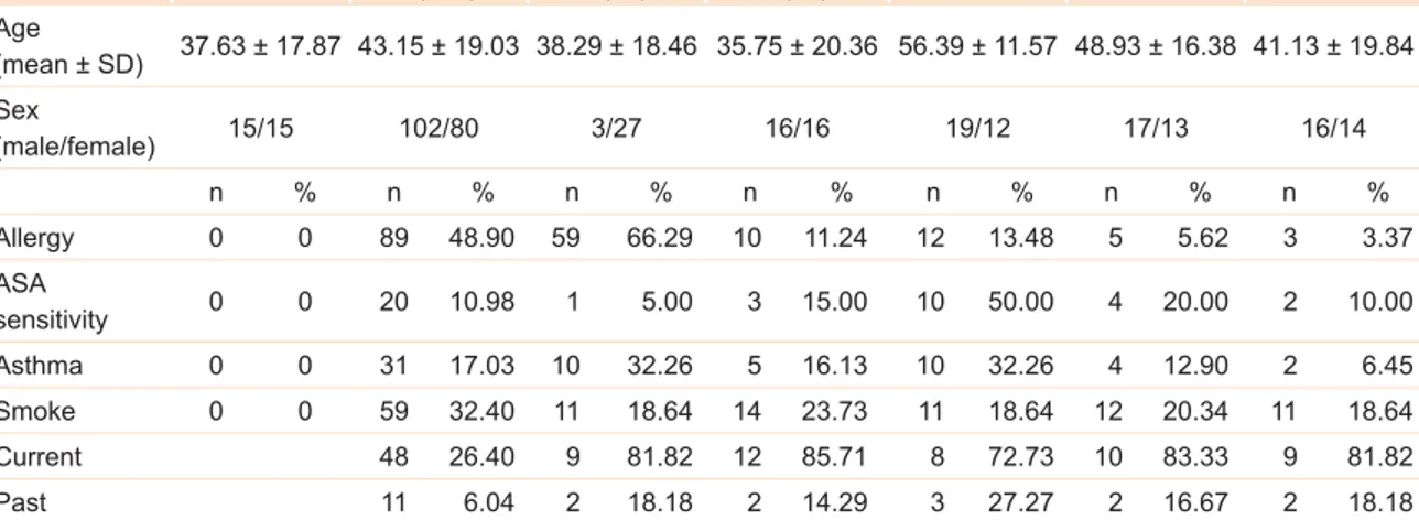
Table 1. Demographic and clinical characteristics of controls and patients with rhinitis Controls (30) Patients with rhinitis (182) Allergic rhinitis (59) Infectious rhinitis (32) Nasal polyps (31) NARES (30) NARNE (30) Age (mean ± SD) 37.63 ± 17.87 43.15 ± 19.03 38.29 ± 18.46 35.75 ± 20.36 56.39 ± 11.57 48.93 ± 16.38 41.13 ± 19.84 Sex (male/female) 15/15 102/80 3/27 16/16 19/12 17/13 16/14 n % n % n % n % n % n % n % Allergy 0 0 89 48.90 59 66.29 10 11.24 12 13.48 5 5.62 3 3.37 ASA sensitivity 0 0 20 10.98 1 5.00 3 15.00 10 50.00 4 20.00 2 10.00 Asthma 0 0 31 17.03 10 32.26 5 16.13 10 32.26 4 12.90 2 6.45 Smoke 0 0 59 32.40 11 18.64 14 23.73 11 18.64 12 20.34 11 18.64 Current 48 26.40 9 81.82 12 85.71 8 72.73 10 83.33 9 81.82 Past 11 6.04 2 18.18 2 14.29 3 27.27 2 16.67 2 18.18
NP = nasal polyps, NARES = non-allergic rhinitis with eosinophils, NARNE = non-allergic rhinitis with neutrophils, SD = standard deviation, ASA = acetylsalicylic acid
rhinitis. Allergic rhinitis was the most frequently di-agnosed type of rhinitis (59 out of 182); all the other types of rhinitis i.e. infectious, polyposis, NARNE, NARES were found almost equally among patients. Allergy was a common comorbidity, ASA sensi-tivity was more frequently present in patients with nasal polyps, asthma was more commonly reported in patients with allergic rhinitis and nasal polyps, and smoking was reported in about 1/3 of patients.
Considering nasal specimens, we found that type II was the most common one in control subjects (76.67 %), whereas type III (59.34 %), followed by type IV (28.02 %), was the most frequently observed among patients with rhinitis (fi gure 1 and table 2). It is worthy of note that no control subject had a type III or type IV fern pattern. The frequency of differ-ent fern patterns was signifi cantly differdiffer-ent among patients with different types of rhinitis and control subjects (p = 0.0066).
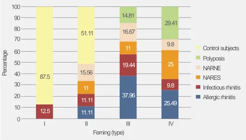
In type I fern patterns, only 12.5 % of nasal specimens were collected from patients with rhini-tis (i.e. infectious); all the other were isolated from control subjects (87.5 %) (fi gure 2). More than half of the nasal specimens characterized by type II fern pattern were collected from control subjects, where-as the remaining specimens were recovered from pa-tients with allergic rhinitis, infectious rhinitis,
NA-RES or NARNE (fi gure 2). Allergic rhinitis was the most frequent type of rhinitis (37.96 %), followed by infectious (19.44 %) associated to a type III fern pattern. Differently, considering type IV, about 30 % and 25 % of this pattern was found in patients with nasal polyps or in patients with allergic rhinitis, re-spectively (fi gure 2).
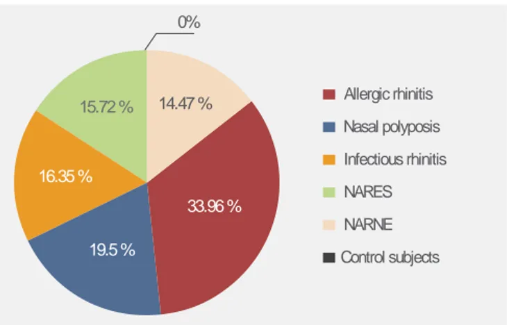
We also observed that the type of rhinitis that was most commonly found in the most “pathologic” fern patterns, i.e. type III or type IV, was allergic rhi-nitis, which affected about 1/3 of the patients whose specimens had type III or type IV fern patterns. The other 2/3 were almost equally distributed among the other types of rhinitis (fi gure 3).
In order to verify possible differences in fern patterns between control subjects and patients with rhinitis, we analyzed each type of rhinitis separate-ly. Patients with infectious rhinitis, with NARNE or with allergic rhinitis had a type III fern pattern more frequently, whereas control subjects had a type II pattern more frequently (p < 0.0001, each com-parison), (data not shown). In patients with NARES or nasal polyps, type III and type IV were the most frequent patterns whereas, in control subjects, type II was predominant (p < 0.0001, each comparison).
No statistically signifi cant difference in fern patterns was found in patients with different
de-Figure 1. The four grades of nasal ferning taken at 100x magnifi cation, classi-fi ed from type I to type IV according to the Rolando scoring.25 Panel A, type I (normal pattern): the fern-ing pattern is uniform with closely branching arboriza-tion. Panel B, type II: the ferns are well distinguished but with less branching. Panel C, type III: there are scattered ferns with rare branching. Panel D, type IV: no ferning pattern is seen. Light microscope at 400× magnifi cation.
grees of nasal eosinophilia or neutrophilia (data not shown).
Discussion
The defi nition of rhinitis assumes the concept of an infl ammatory reaction; the characteristics of nasal infl ammation, mainly regarding the peculiar cellular infi ltrate, allow the differentiation of several types of rhinitis. However, a common denominator in infl am-matory disorders is the presence of impaired mucus. Altered mucus is per se a pathogenic factor that in-duces a vicious circle that involves a deterioration of infl ammatory disorders.
Nasal cytology is commonly used in clinical practice to defi ne the type of rhinitis, to quantify
the cellular infi ltrate, and to monitor infl ammatory change over time.21 Traditionally, the nasal
cytolo-gy assessment considers cellular presence and mor-phology, bacteria, and biofi lm. However, mucus is a component that is neglected in the treatment of nasal disorders. A possible explanation is the complexity of diagnostic tests able to assess the characteristics of mucus.
In this regard, the fern test could be a very simple and useful method to investigate mucus in patients with rhinitis. The current study, conducted in a real-world setting, such as a Rhinology Unit, proved that rhinitis is associated with impaired fern patterns. In particular, healthy subjects have only type I or II fern patterns and never III or IV. On the
Table 2. Frequency of different ferning Types within each rhinitis group
Study groups Disease
Ferning type I II III IV n % n % n % n % Patients with rhinitis 1 0.55 22 12..09 108 59.34 51 28.02 Allergic rhinitis 0 0 5 8.47 41 69.49 13 22.03 Infectious rhinitis 1 3.13 5 15.63 21 65.63 5 15.63 NARES 0 0 5 16.67 12 40 13 43.33 NARNE 0 0 7 23.33 18 60 5 16.67 nasal polyps 0 0 0 0 16 51.61 15 48.39 Control subjects 7 23.33 23 76.67 0 0 0 0
NARES = non-allergic rhinitis with eosinophils, NARNE = non-allergic rhinitis with neutrophils.
Figure 2. Frequency of different rhinitis within each ferning type. 100 90 80 70 60 50 40 30 20 10 0 Control subjects Polyposis NARNE NARES Infectious rhinitis Allergic rhinitis 87.5 12.5 11.11 11.11 11 15.56 51.11 14.81 16.67 11 19.44 37.96 25.49 9.8 9.8 29.41 25 I II III IV Ferning (type) Percentage
other hand, patients with rhinitis frequently present type III or IV fern patterns. Allergic rhinitis and na-sal polyps are most frequently associated with type IV fern patterns. This fi nding could depend on more intense infl ammation, even though a clear-cut rela-tionship between cellular infi ltrate and fern pattern was not reported.
The most relevant outcome of the present study is the feasibility of the fern test in the treatment of patients with rhinitis. The fern test may be consid-ered a reliable tool for assessing the characteristics of the mucus, and its fi ndings have clinical rele-vance. It is conceivable that impaired fern patterns, such as type III and IV, could be the expression of damaged epithelial cells, mainly concerning mu-ciparous cells. In fact, patients with rhinitis usually present altered fern patterns. In other words, type I (and type II) fern patterns could mean the wellness of the epithelium.
However, the present study has some limita-tions, including the cross-sectional design, the lack of mediator’s assessment, and the lack of assessment of clinical symptoms. Therefore, other studies should be designed to fulfi ll these unmet needs. In particu-lar, it is necessary to correlate the severity of nasal disorders with the type of fern patterns. If a strong re-lationship between the type of fern pattern and severe symptoms and/or infl ammatory aspects exists, this test could be fruitful in phenotyping patients with rhinitis. Therefore, the fern test could be considered a potential biomarker that is useful for defi ning epi-thelial damage in nasal disorders. This issue should be properly investigated in further studies.
In conclusion, the fern test could be considered a fruitful method to assess mucus alterations in pa-tients with rhinitis and it could be included as a new parameter in the treatment of rhinitis as a potential biomarker of damaged epithelial cell function.
References
1. Bansil R, Turner BS. The biology of mucus: composition, synthesis and organization. Adv Drug Deliv Rev. 2018;124:3-15. DOI: 10.1016/j.addr.2017.09.023
2. Taherali F, Varum F, Basit AW. A slippery slope: on the origin, role and physiology of mucus. Adv Drug Deliv Rev. 2018;124:16-33. DOI: 10.1016/j.addr.2017.10.014
3. Lang T, Klasson S, Larsson E, Johansson ME, Hansson GC, Samuelsson T. Searching the evolutionary origin of epithelial mucus protein components-mucins and FCGBP. Mol Biol Evol. 2016;33(8):1921-1936. DOI: 10.1093/molbev/msw066
4. Cone RA. Barrier properties of mucus. Adv Drug Deliv Rev. 2009;61(2):75-85. DOI: 10.1016/j. addr.2008.09.008
5. Silberberg A. On mucociliary transport. Biorheology. 1990;27(3-4):295-307. DOI: 10.3233/BIR-1990-273-408 33.96 % 19.5 % 16.35 % 15.72 % 14.47 % 0% Allergic rhinitis Nasal polyposis Infectious rhinitis NARES NARNE Control subjects
Figure 3. Frequency of different rhinitis in fern-ing type III or IV.
6. Neutra MR, Forstner JF. Gastrointestinal mucus: synthesis, secretion, and function. In: Johnson LR (editor). Physiology of the gastrointestinal tract. EEUU: Raven Press; 1987.
7. Quraishi MS, Jones NS, Mason J. The rheology of nasal mucus: a review. Clin Otolaryngol Allied Sci. 1998;23(5):403-413. DOI: 10.1046/j.1365-2273.1998.00172.x
8. Lai SK, Wang YY, Wirtz D, Hanes J. Micro- and macrorheology of mucus. Adv Drug Deliv Rev. 2009;61(2):86-100. DOI: 10.1016/j.addr.2008.09.012
9. Carlstedt I, Sheehan JK. Structure and macromolecular properties of cervical mucus glycoproteins. Symp Soc Exp Biol. 1989;43:289-316.
10. Thornton DJ, Sheehan JK. From mucins to mucus: toward a more coherent understanding of this essential barrier. Proc Am Thorac Soc. 2004;1(1):54-61. DOI: 10.1513/pats.2306016
11. Papanicolaou GN. Mucus test. Anat Rec. 1945;91:293.
12. Roland M. A simple test for the determination of ovulation, estrogen activity, and early pregnancy using the cervical mucus secretion; a preliminary report. Am J Obstet Gynecol. 1952;63(1):81-89. DOI: 10.1016/S0002-9378(16)38983-9
13. Zondek B, Rozin S. Cervical mucus arborization; its use in the determination of corpus luteum function. Obstet Gynecol. 1954;3(5):463-470. DOI: 10.1097/00006254-195508000-00049
14. Abou-Shabanah EH, Plotz EJ. A biochemical study of the cervical and nasal mucus fern phenomenon. Am J Obs Gynecol. 1957;74(3):559-568. DOI: 10.1016/0002-9378(57)90508-2
15. Rolando M. Tear mucus ferning test in normal and keratoconjunctivitis sicca eyes. Chibret Int J Ophthalmol. 1984;2:33-41.
16. Rolando M, Baldi F, Zingirian M. The effect of hyperosmolarity on tear mucus ferning. Fortschr Ophthalmol. 1986;83(6):644-646.
17. Rolando M, Baldi F, Calabria G. Tear mucus crystallization in children with cystic fi brosis. Ophthalmologica. 1988;197(4):202-206. DOI: 10.1159/000309944
18. Kalayci D, Kiper N, Ozcelik U, Gocmen A, Hasiripi H. Clinical status, ocular surface changes and tear ferning in patients with cystic fi brosis. Acta Ophthalmol Scand. 1996;74(6):563-565. DOI: 10.1111/j.1600-0420.1996.tb00735.x
19. Horwath J, Ettinger K, Bachernegg M, Bodner E, Schmut O. Ocular Ferning Test - effect of temperature and humidity on tear Ferning patterns. Ophthalmologica. 2001;215(2):102-107. DOI: 10.1159/000050838
20. Lavaud MC, Trouillas J. The mucus: a medium of life. Gynecol Obstet Fertil. 2012;40(1):19-23. DOI: 10.1016/j.gyobfe.2011.07.031
21. Gelardi M. Atlas of nasal cytology. Second edition. Milan, Italy: Edi Ermes; 2012.
22. Gelardi M, Fiorella ML, Russo C, Fiorella R, Ciprandi G. Role of nasal cytology. Int J Immunopathol Pharmacol. 2010;23(Suppl 1):45-49.
23. Gelardi M, Maselli-Del-Giudice A, Fiorella ML, Fiorella R, Russo C, Soleti P, et al. Non-allergic rhinitis with eosinophils and mast cells constitutes a new severe nasal disorder. Int J Immunopathol Pharmacol. 2008;21(2):325-331. DOI: 10.1177/039463200802100209
24. Gelardi M, Iannuzzi L, De-Giosa M, Taliente S, DeCandia N, Quaranta N,et al. Non-surgical management of chronic rhinosinusitis with 1 nasal polyps based on clinical cytological grading: a precision medicine-based approach. Acta Otorhinolaryngol Ital. 2017;37(1):38-45. DOI: 10.14639/0392-100X-1417
25. Rolando M. Tear mucus ferning test in normal and keratoconjunctivitis sicca eyes. Chibret Int J Ophthalmol. 1984;2:32-41.