REVIEW
Myiasis in domestic cats: a global review
Marco Pezzi
1*, Teresa Bonacci
2, Marilena Leis
1, Elisabetta Mamolini
1, Maria Gabriella Marchetti
1,
Stjepan Krčmar
3, Milvia Chicca
1, Carlo Nicola Francesco Del Zingaro
4, Michel J. Faucheux
5and Chiara Scapoli
1Abstract
Myiasis is an infestation caused by larvae of Diptera in humans and other vertebrates. In domestic cats, Felis silvestris
catus L. (Carnivora: Felidae), four dipteran families have been reported as agents of obligatory and facultative myiasis:
Oestridae, Calliphoridae, Sarcophagidae and Muscidae. Among agents of obligatory myiasis, the most frequent genus is Cuterebra Clark (Oestridae) and the most frequent species is Cochliomyia hominivorax (Coquerel) (Calliphoridae). Among the agents of facultative myiasis, the most frequent species is Lucilia sericata (Meigen) (Calliphoridae). A survey of myiasis in cats reported in literature shows that the cases are distributed worldwide and linked to the geographical range of the dipteran species. Factors favouring the occurrence of myiasis in cats are prowling in infested areas, poor hygiene conditions due to diseases and/or neglect, and wounds inflicted during territorial or reproductive competi-tion. The aim of the review is to provide an extended survey of literature on myiasis in cats, as general information and possible development of guidelines for veterinarians, entomologists and other researchers interested in the field. Keywords: Domestic cat, Calliphoridae, Literature review, Muscidae, Myiasis, Oestridae, Sarcophagidae
© The Author(s) 2019. This article is distributed under the terms of the Creative Commons Attribution 4.0 International License (http://creat iveco mmons .org/licen ses/by/4.0/), which permits unrestricted use, distribution, and reproduction in any medium, provided you give appropriate credit to the original author(s) and the source, provide a link to the Creative Commons license, and indicate if changes were made. The Creative Commons Public Domain Dedication waiver (http://creat iveco mmons .org/ publi cdoma in/zero/1.0/) applies to the data made available in this article, unless otherwise stated.
Background
Diseases caused by insect larvae invading the body of animals were initially named “scolechiasis” [1] without any distinction of insect species, from the historical term “scolex” indicating a larva. In 1840, Reverend Frederick W. Hope introduced a new name for this type of disease based on the insect order of larvae infesting the human body: “myiasis” for Diptera, “chantariasis” for Coleoptera and “scholechiasis” for Lepidoptera [2]. Later, Profes-sor Walter S. Patton extended the term “myiasis”, defin-ing it as “the condition or conditions, resultdefin-ing from the invasion of tissues and organs of man and other animals by all stages of the Diptera” [3]. According to this state-ment, myiasis is not only caused by larvae but also by all dipteran life stages. In his monography “The flies that cause myiasis in man”, Professor Maurice T. James criti-cized the definition of Patton as “unnecessary” and “con-fusing”, “since … it is the larva that is the active stage in relation to myiasis” [4]. The current and widely accepted definition of myiasis is that by Professor Fritz K. Zumpt,
formulated “as the infestation of live human and verte-brate animals with dipterous larvae, which, at least for a certain period, feed on the host’s dead or living tissue, liquid body-substances, or ingested food”. The key crite-rion to define myiasis is therefore the fact that the dip-teran larva must complete its normal development or a part of it in a vertebrate body [5].
Myiasis can be classified based on host-parasite rela-tionships, that is whether the dipteran species cause obligatory or facultative myiasis. The species causing obligatory myiasis require a living host for their develop-ment, while those causing facultative only occasionally lay eggs or larvae on living hosts and usually develop on decaying matter. Facultative myiasis can be classified as primary, secondary and tertiary, based on the ability of the species to initiate the myiasis (primary) or to super-impose to pre-existing myiasis (secondary and tertiary). Myiasis can also be classified based on anatomical locali-zation of the larva (or larvae) in the host, either external or internal: auricular, cutaneous, gastrointestinal, oph-thalmic, oral and urogenital [6–8]. Besides these types, myiasis involving structures of the nervous system will be considered as “neurological” in this review and those involving structures of the respiratory system will be con-sidered as “respiratory”.
Open Access
*Correspondence: [email protected]
1 Department of Life Sciences and Biotechnology, University of Ferrara, Via Luigi Borsari 46, 44121 Ferrara, Italy
This review summarizes the worldwide reported cases of myiasis in domestic cats (hereafter cats), Felis silves-tris catus L. (Carnivora: Felidae), the dipteran species involved and their ecological and biological features. The parasitological, epidemiological and risk factors will also be examined in detail, in order to provide information and suitable guidelines for veterinarians, entomologists and other researchers interested in the field.
Bibliographic methods
More than 7000 published references were examined for this study. The initial set of publications on myia-sis in cats were obtained by PubMed indexed literature and the search was extended through web engines. The search had no time and language limits. For each publica-tion obtained via web or interlibrary services or by direct contacts with the authors, the reference list was checked for further cases of myiasis in cats. Publications not spe-cifically reporting myiasis on cats in their titles were also investigated, examining the abstracts or the entire pub-lication for useful information. All pubpub-lications report-ing myiasis on cats were classified accordreport-ing to infestreport-ing taxon, type of myiasis, number of cases, sex, age and country. The literature data were then described accord-ing to the Diptera family and to the genera or species agents of obligatory and facultative myiasis.
Literature data
The first cases of myiasis in cats reported in scientific journals appeared around the end of the 19th century in the USA [9, 10]. A case of cutaneous myiasis in a foot of a cat from Texas, due to a fighting wound in which numer-ous “screw-worms” developed, was reported in a letter by Professor G. W. Curtis to the distinguished entomologist, Professor Herbert Osborn, dated 15 December 1888; the agent involved was presumably Cochliomyia sp. (Diptera: Calliphoridae) [10].
A case of myiasis in a cat from Iowa by Cuterebra sp. was later reported by Osborn in 1892 [11], but the origi-nal work could not be retrieved. A case of “warble” fly on the belly of a cat from North Carolina was described in the extracts from correspondence of the journal “Insect Life”; the editors suggested it could be due to a species of the genus Cuterebra [9]. Another two cases of myiasis (“bots”) were reported in the same journal in the notes from correspondents, the first one in the eye and back of a cat in New York State and the second one in the neck of a kitten in Missouri; in both cases no identification of the
agents was provided [9]. From 1894 onwards, the cases
of myiasis in cats were described as caused by species of the families Oestridae, Calliphoridae, Sarcophagidae and Muscidae (Tables 1, 2, 3, 4, 5 and 6). The cases of myiasis
are therefore reported according to the dipteran family involved.
Oestridae
The Oestridae is a large dipteran family in which all spe-cies are obligate parasites of wild and domestic animals and of humans. It is divided into four subfamilies: Cuter-ebrinae (New World skin bot flies), Oestrinae (nose bot flies), Gasterophilinae (stomach bot flies) and Hypo-dermatinae (Old World skin bot flies) [6, 7]. The cases of myiasis in cats reported as caused by species of the Oestridae are indicated in Table 2, almost all belonging to the Cuterebrinae and Oestrinae. Within the subfam-ily Cuterebrinae, species of the genus Cuterebra and Der-matobia hominis Linnaeus Jr. in Pallas, a species of the
monotypic Dermatobia, both from the New World [6,
12] have been reported as agents of myiasis in cats. The species of genus Cuterebra exhibit a marked host specifity: some species preferentially attack rodents such as voles, Microtus spp., woodrats, Neotoma spp., and deer mice, Peromyscus spp. (Cricetidae), and lagomorphs such as hares, Lepus spp., and cottontail rabbits, Sylvilagus spp. (Leporidae) [7]. They may also develop in atypical
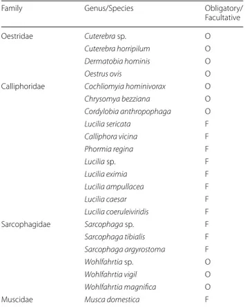
Table 1 Genera and species of Diptera involved in myiasis in cats
Abbreviations: O, obligatory; F, facultative
Family Genus/Species Obligatory/
Facultative
Oestridae Cuterebra sp. O
Cuterebra horripilum O
Dermatobia hominis O
Oestrus ovis O
Calliphoridae Cochliomyia hominivorax O
Chrysomya bezziana O Cordylobia anthropophaga O Lucilia sericata F Calliphora vicina F Phormia regina F Lucilia sp. F Lucilia eximia F Lucilia ampullacea F Lucilia caesar F Lucilia coeruleiviridis F Sarcophagidae Sarcophaga sp. F Sarcophaga tibialis F Sarcophaga argyrostoma F Wohlfahrtia sp. O Wohlfahrtia vigil O Wohlfahrtia magnifica O
rodent hosts, such as black rats, Rattus rattus (L.) (Muri-dae) [13], and European rabbits, Oryctolagus cuniculus (L.) (Leporidae) [14]. Besides cats (Table 2), other atypi-cal mammalian hosts may be affected, among which are red kangaroos, Macropus rufus Desmarest and Bennett’s
wallabies, Macropus rufogriseus fruticus (Ogilby) (Dipro-todontia: Macropodidae) [15], cattle, Bos taurus L. [16] and Günther’s dik-diks, Madoqua guentheri Thomas [15] (Artiodactyla: Bovidae), domestic pigs, Sus scrofa domesticus Erxleben (Artiodactyla: Suidae) [16], horses,
Table 2 Cases of myiasis in cats caused by taxa of the family Oestridae
Abbreviations: neu, neurological; ophth, ophthalmic; cut, cutaneous; resp, respiratory tract; unk, unknown; A, adult; K, kitten; w, weeks; y, years
Taxon Type of myiasis No. of cases Sex Age Country References
Cuterebra sp. neu 1 unk unk USA [62]
neu 1 ♂ A USA [54]
neu 1 ♀ K USA [64]
neu 2 ♀ A USA [43]
neu 3 2♀; 1♂ 1K; 2A USA [65]
neu 11 6♀; 5♂ A USA [44]
neu 9 5♀; 4♂ 7A; 2unk USA [45]
neu 1 ♀ A USA [47]
neu 1 ♀ A USA [50]
ophth 1 unk 8w to 2y USA [42]
ophth 1 ♀ A USA [56]
ophth 1 ♀ A USA [46]
ophth 1 ♀ A USA [48]
ophth 1 ♀ A USA [73]
oral 1 unk unk USA [63]
oral 1 unk unk USA [51]
resp 1 unk unk USA [11]
resp 1 ♀ K USA [61]
resp 2 ♂ 1A; 1K USA [57]
resp 1 ♂ A USA [59]
resp 2 1♀; 1♂ A USA [60]
resp 1 ♂ A USA [74]
resp 1 ♂ A Canada [66]
resp 1 unk unk USA [51]
cut 1 unk K USA [39]
cut 2 unk unk USA [11]
cut 1 unk unk USA [41]
cut 6 unk 8w to 2y USA [42]
cut 20 unk unk USA [51]
cut 1 unk K USA [49]
aur 1 ♂ A USA [58]
neu-cut 1 ♀ K USA [65]
neu-cut 1 ♀ A USA [45]
unk 1 unk unk USA [40]
C. horripilum cut 2 unk K USA [20]
Dermatobia hominis cut 1 unk A Brazil [78]
cut 1 unk unk Venezuela [79]
cut 1 ♀ A Brazil [80]
Oestrus ovis resp 1 ♂ A Australia [81]
Cuterebrinae unk 2 unk 1K; 1A USA [19]
Oestridae cut 1 unk K USA [169]
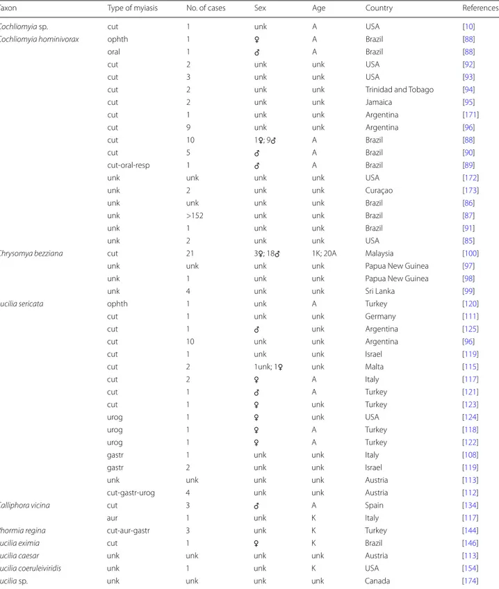
Table 3 Cases of myiasis in cats caused by taxa of the family Calliphoridae
Abbreviations: urog, urogenital; gastr, gastrointestinal; aur, auricular; ophth, ophthalmic; cut, cutaneous; resp, respiratory tract; unk, unknown; A, adult; K, kitten
Taxon Type of myiasis No. of cases Sex Age Country References
Cochliomyia sp. cut 1 unk A USA [10]
Cochliomyia hominivorax ophth 1 ♀ A Brazil [88]
oral 1 ♂ A Brazil [88]
cut 2 unk unk USA [92]
cut 3 unk unk USA [93]
cut 2 unk unk Trinidad and Tobago [94]
cut 2 unk unk Jamaica [95]
cut 1 unk unk Argentina [171]
cut 9 unk unk Argentina [96]
cut 10 1♀; 9♂ A Brazil [88]
cut 5 ♂ A Brazil [90]
cut-oral-resp 1 ♂ A Brazil [89]
unk unk unk unk USA [172]
unk 2 unk unk Curaçao [173]
unk unk unk unk Brazil [86]
unk >152 unk unk Brazil [87]
unk 1 unk unk Brazil [91]
unk 2 unk unk USA [85]
Chrysomya bezziana cut 21 3♀; 18♂ 1K; 20A Malaysia [100]
unk unk unk unk Papua New Guinea [97]
unk 1 unk unk Papua New Guinea [98]
unk 4 unk unk Sri Lanka [99]
Lucilia sericata ophth 1 unk A Turkey [120]
cut 1 unk unk Germany [111]
cut 1 ♂ unk Argentina [125]
cut 10 unk unk Argentina [96]
cut 1 unk unk Israel [119]
cut 2 1unk; 1♀ unk Malta [115]
cut 2 ♀ A Italy [117]
cut 1 ♂ A Turkey [121]
cut 1 ♀ unk Turkey [123]
urog 1 ♀ unk USA [124]
urog 1 ♀ A Turkey [118]
urog 1 ♀ A Turkey [122]
gastr 1 unk unk Italy [108]
gastr 2 unk unk Israel [119]
unk unk unk unk Austria [113]
cut-gastr-urog 4 unk unk Austria [112]
Calliphora vicina cut 3 ♂ A Spain [134]
aur 1 unk K Italy [117]
Phormia regina cut-aur-gastr 3 unk K Turkey [144]
Lucilia eximia cut 1 ♀ K Brazil [146]
Lucilia caesar unk unk unk unk Austria [113]
Lucilia coeruleiviridis unk 1 unk K USA [154]
Equus ferus caballus L. [17] and mules, Equus asinus L. × E. ferus caballus [16] (Perissodactyla: Equidae), domestic dogs, Canis lupus familiaris L. (Carnivora: Canidae) [18–25], snow leopards, Panthera uncia (Schre-ber) (Carnivora: Felidae) [26], raccoons, Procyon lotor
L. (Carnivora: Procyonidae) [27] and primates such as
ring-tailed lemurs, Lemur catta L. (Lemuridae) [28] and humans [29–36]. Myiasis caused by species of the genus Cuterebra has also been reported in non-mammalian species such as reptiles [37] and birds [38].
In cats, Cuterebra spp. have been reported to cause neurological, ophthalmic, oral, respiratory and furun-cular (cutaneous) myiasis (Table 2). Infestations were detected from June to September [39–51]. Emerging of adults occurred from late spring, followed by mating, oviposition and egg development [52]. The common ways to enter the host are natural body openings or wounds [53]. Species have been clearly determined only in one case, probably because the authors typically focused on clinical symptoms, treatments and outcomes of infesta-tions: Cuterebra horripilum Clark was reported in a case involving two kittens in the USA [20]. In another case involving an adult male, the author mentioned the spe-cies as Cuterebra americana (Fabricius) or C. horrip-ilum [54]. In another recent case of myiasis in a kitten the author reported that it was “most likely” Cuterebra abdominalis Swenk [49]. The recent availability of molec-ular techniques such as DNA barcoding may provide a precise identification of the species, as for other Diptera responsible for myiasis [55]. All cases of myiasis in cats by Cuterebra spp. have been reported in North America. The US states where these cases were reported and/or the cats were clinically treated are the following, accord-ing to the region. Midwest: Illinois [56], Indiana [41, 57],
Table 4 Cases of myiasis in cats caused by association of species of Diptera
Abbreviations: ophth, ophthalmic; cut, cutaneous; unk, unknown; A, adult; K, kitten
Species involved Type of myiasis No. of cases Sex Age Country Reference
Lucilia sericata and Lucilia ampullacea unk 1 unk unk Austria [114]
L. sericata and Sarcophaga tibialis cut 1 unk A Italy [116]
L. sericata and Calliphora vicina ophth 1 unk K Italy [117]
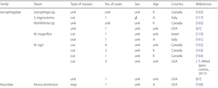
Table 5 Cases of myiasis in cats caused by taxa of the families Sarcophagidae and Muscidae
Abbreviations: cut, cutaneous; resp, respiratory tract; unk, unknown; A, adult; K, kitten
Family Taxon Type of myiasis No. of cases Sex Age Country References
Sarcophagidae Sarcophaga sp. unk unk unk K Canada [165]
S. argyrostoma cut 1 ♂ A Italy [117]
Wohlfahrtia sp. unk unk unk K Canada [165]
unk 1 unk unk USA [67]
W. magnifica cut 1 unk unk Israel [119]
oral 1 unk A Italy [161]
W. vigil cut 4 unk unk Canada [162]
cut 2 unk K Canada [163]
cut 1 unk K Canada [164]
cut 3 unk unk USA J. T. Alfred
(pers. comm., 2017)
unk 1 unk unk USA [67]
Muscidae Musca domestica resp 1 unk A USA [168]
Table 6 Cases of myiasis in cats caused by undetermined taxa of Diptera
Abbreviations: ophth, ophthalmic; cut, cutaneous; unk, unknown; A, adult; K, kitten
Type of myiasis No. of cases Sex Age Country References
ophth 1 ♂ A USA [175]
ophth 2 ♀ A USA [176]
ophth 2 1♀; 1♂ 1A; 1 unk USA [177]
cut 2 unk 1K; 1 unk USA [9]
cut 2 unk unk USA [92]
Iowa [11], Michigan [51, 58, 59], Missouri [60], Ohio [48] and Wisconsin [46, 49]; Northwest: Connecticut [39,
40], Massachusetts [61] and New York [44, 45, 47, 62]; Southwest: North Carolina [63]; South: Alabama [64,
65], Georgia [43, 50, 65], Maryland [54], Tennessee [11], Texas [42] and Washington D.C. [11]. Recently, a case of respiratory tract myiasis with an infestation of pharynx caused by Cuterebra sp. was reported in the province of Ontario, Canada, together with two presumptive cases of neurological myiasis in which, however, no dipteran larva was found [66].
Species of the genus Cuterebra usually lays eggs on grasses along narrow trails or paths used by the typical host [52]; thus cats and other atypical hosts may become parasitized. While strolling, the cat sniffs the ground or brushes the contaminated area, accidentally collecting eggs or larvae [29, 50, 67]. Contacts with the parasites are more frequent in adults, while in kitten infestations are related to the daily activities of the mother, which may collect the parasites in her fur and carry them to the lit-ter [67]. The consequences of myiasis by Cuterebra spp. depend on the site of infestation.
Cuterebra spp. larvae have been found inside the cat skin, causing “subcutaneous abscesses” [41], “lesions” [49] and “seeping wounds” [42]. The ability to use skin wounds or lacerations as a way to enter the host has been reported for Cuterebra latifrons Coquillett [68] and Cuterebra fontinella Clark [69]. Experimental infes-tations in rats by Cuterebra tenebrosa Coquillett were obtained by placing first-stage larvae on fur [70] and in humans by Cuterebra spp. larvae placed on intact skin [71], but these results were not confirmed for larvae of Cuterebra lepivora Coquillett in rabbits [72]. In cats, lar-vae of Cuterebra spp. were detected in the skin of head, neck, shoulder, thorax, abdomen and sides [11, 20, 42,
49, 51]. In all these cases, the only method to remove the parasite was mechanical extraction by surgery.
When the larva migrates to the brain, the invariable outcome is death [43, 64, 65] either natural or induced by euthanasia practiced by veterinarians [43, 45, 47, 50, 54]. The neurological signs depend on the area of the brain infested [67] and include ataxia, blindness, circling, head tilt, lethargy, seizures, hysteria and convulsion [43–45,
47, 50, 64, 65]. In some cases, aggressive behaviours such as clawing, irritability to noise and hissing often mislead veterinarians to diagnose rabies and therefore imme-diately euthanize the cat [43, 50]. Indeed, most cases of brain migration of Cuterebra spp. have been correctly diagnosed only after necroscopy and the commonly reported clinical signs before the onset of neurological symptoms are respiratory problems, probably due to lar-val migration in the respiratory tract [43–45, 47, 50, 64,
65].
Among the five reported cases of ophthalmomyiasis by Cuterebra spp. in cats, one was external [42] and the other four were internal: in three cases the larva was in the anterior chamber of the eye [46, 56, 73] and in one case in the posterior chamber [48]. When the larva was in the anterior chamber, it was found dead [46, 56, 73], presumably because of the unsuitable site of migration [73]. The dead larva may have caused an intense inflam-matory reaction [56]. The larvae in the anterior cham-ber were removed by surgery with survival of cats in all three cases, but blindness occurred in two cases by reti-nal degeneration [46] and corneal edema [56]. When the larva migrated to the posterior chamber, it caused coagu-lation necrosis, haemorrhagic lesions to retina and opti-cal nerve and anterior uveitis. The systemic conditions of the patient worsened so the cat had to be euthanized [48].
Other sites where larvae of Cuterebra spp. have been found are the oral cavity, including mandible [51], soft palate [63] and pharynx [57], and the respiratory tract, such as nasal cavity [11, 51, 61] and trachea [59, 60, 74]. When the larva infested the oral cavity, the reported clin-ical signs were lethargy, listlessness and anorexia [57] and when it infested the nasal cavity, the signs were sneezing and nasal discharge [61]. If the larva reached the phar-ynx, the signs were dyspnea and caked blood on nostrils [57], and if it reached the trachea, they were dyspnea, subcutaneous emphysema, cyanosis, stridor, coughing, sneezing, inappetence, lethargy and fever [59, 60, 74].
When the above symptoms are detected in late summer or early autumn, the veterinarian should consider the possibility of an oral or respiratory infestation by Cuter-ebra spp. [60]. In cases of oral and respiratory myiasis by Cuterebra spp., the only attempts to remove the infesting larvae were by surgery [57, 60, 61, 63, 74], successful in all cases except one, where the larva was extracted from the soft palate and the cat died for debilitation and serious respiratory problems [63]. A case of tracheal infestation by Cuterebra sp. was described as a post-mortem diag-nosis in which death was probably due to tissue damages caused by migration and anaphylaxis [59].
Another species of the subfamily Cuterebrinae reported as an agent of obligatory furuncular myiasis in cats is D. hominis (Table 2), a common fly species in cen-tral and southern America [75]. The adult female exhib-its a special behaviour to ensure the contact between the eggs and the host: it usually captures other zoophilic insects or arthropods and sticks her eggs to their abdo-mens. When the zoophilic carrier settles on the host, the larvae of D. hominis hatch and penetrate the host skin [7], gaining access to the dermis. Each larva forms a furuncular lesion [8]. This species is a very serious cattle pest in tropical America and is known to parasitize man,
dogs and other mammals [4]. In travellers returning from tropical America it should be considered for differential diagnosis of furuncular lesions [8]. Cats are reported as possible hosts of D. hominis together with other domestic and wild animals [10, 76, 77], but the first documented case of myiasis by D. hominis in cats was reported in 1998 in Rio de Janeiro (Brazil) in a cat showing cutane-ous boils on a paw and among toes [78]. Two other cases
were recently reported, one in Venezuela [79] and the
other in Brazil, in which the cat showed three cutaneous boils on the cranioventral region of the neck [80]. The infestation by D. hominis is less aggressive in comparison to that by Cochliomyia hominivorax (Coquerel) (Dip-tera: Calliphoridae), another common agent of obligatory myiasis in Brazil, and may be considered as benign, not only because the infestation is limited to the host skin without migrations inside the body, but also because of “feline self-grooming behavior” which increases the probability of removal of D. hominis larvae. However, if the cat is clinically debilitated and can not perform the self-grooming behaviour, infestation may occur [80].
The only species of the subfamily Oestrinae reported as an agent of myiasis in cats is Oestrus ovis (L.), which deposits first stage larvae in nostrils of animal and human hosts [6]. In cats, only one case of nasal infestation by O. ovis was described in New South Wales (Australia), with a positive outcome probably due to early diagnosis and intervention. The first-instar larva was mechanically removed by repeated washing with saline solution. The predisposing risk factor for the cat was living in a sheep-grazing area [81].
Calliphoridae
The family Calliphoridae (“blow flies”) includes over 1000 species distributed worldwide. From a medical and vet-erinary point of view, many saprophagous species of this family may cause facultative myiasis [7] and thus have forensic relevance [82, 83]. A small number of Calliphori-dae are characterized by obligatory myiasis [7]. Those reported in cats are Co. hominivorax, Chrysomya bez-ziana Villeneuve and Cordylobia anthropophaga (Blan-chard) (Table 3).
The first two species, respectively called “New World screw-worm” and “Old World screw-worm”, are econom-ically relevant and occupy the same ecological niche in their respective ranges [6, 7]. All warm-blooded animals, either domestic and wild, and also humans may become
hosts for these two species [84], whose females are
attracted by open wounds or by mucosae of natural body openings [6]. The species Co. hominivorax was originally
distributed from southern USA to South America [84].
From 1952 to 1982, campaigns of dispersal of sterile flies succeeded to eradicate the parasite from the USA, Mexico
and several Caribbean islands. In 1987 an autochthonous spreading of Co. hominivorax was reported in Libya, probably originating from livestock imported from Cen-tral America. In this outbreak, sheep, goats, cattle, cam-els and more than 200 humans were infested, but in 1991 eradication was achieved by an international campaign of sterile fly release [84]. In 2016 an outbreak of Co. homini-vorax occurred in the Florida Keys, with confirmed cases involving ten Key deers, Odocoileus virginianus clavium Barbour & G. M. Allen (Artiodactyla: Cervidae), one rac-coon, three domestic dogs, one pet pig and two cats. The outbreak was ended in 2017 by sterile fly release [85]. In cats, Co. hominivorax has been historically detected in the USA, Curaçao, Jamaica, Trinidad and Tobago, Bra-zil and Argentina (Table 3). The country with the highest number of reported cases is Brazil, with reports from Rio de Janeiro [86–90] and the Federal District [91]. In Rio de Janeiro, this infestation has been reported as “quite com-mon” in cats [90], infesting wounds [88–90, 92–96] and ears, oral and nasal cavities [88, 89]. Concerning wounds, infestation of Co. hominivorax have been reported in cats with surgery wounds due to castration [95] or cryosur-gery [90], or with traumatic wounds due to bites of hae-matophagous bats [94], and territorial or reproductive fights among males [88, 90].
Infestations by Co. hominivorax have also been reported in the umbilical region of kittens [94]. Myiasis by Co. hominivorax may be fatal, especially in stray cats for which diagnosis and treatment are difficult due to feral behaviour [88, 90].
The area of distribution of the Old World screw-worm, Ch. bezziana, is Southeast Asia, New Guinea and Africa [7]. Contrary to its American counterpart, the number of cases of myiasis in cats reported for this species are few, located in Papua New Guinea [97, 98], Sri Lanka [99] and Malaysia [100]. Cats have been also reported as possible hosts for Ch. bezziana in the Sultanate of Oman [101]. In the Sri Lanka survey, among 299 cases of myiasis in domestic dogs and cats by Ch. bezziana, four concerned
cats [99]. The authors did not advance any hypothesis
about the high prevalence of myiasis in dogs in compari-son to cats, an interesting point that would deserve fur-ther investigation.
The “tumbu fly”, Cor. anthropophaga, is an agent of obligatory cutaneous (furuncular) myiasis, widely distrib-uted in the sub-Saharan region [6], although at least one autochtonous case has been recently reported in Saudi Arabia [102]. This species, with rodents as natural hosts, has secondarily adapted to other wild and domestic ani-mals and also to humans [7]. Cats have been historically mentioned as hosts of the tumbu fly [5, 103] but accord-ing to scientific literature there are no specific reports of myiasis caused by this species in cats.
Species of the Calliphoridae reported as agents of fac-ultative myiasis in cats are Lucilia sericata (Meigen), Calliphora vicina Robineau-Desvoidy, Phormia regina Meigen, Lucilia caesar (L.), Lucilia eximia (Wiedemann), Lucilia ampullacea Villeneuve and Lucilia coeruleiviridis Macquart (Tables 3, 4).
Lucilia sericata has a nearly cosmopolitan distribu-tion and is a common necrophagous fly in temperate regions of the northern hemisphere [83]. Together with Ca. vicina and L. illustris, this species is characteristic for urban and suburban areas in Europe [104] and in north-ern Europe is the primary agent of cutaneous myiasis [105]. Adults thrive on carrions in bright sunshine [106] but the female searches shaded areas of the dead body to lay eggs [83]. Larvae generally feed on dead tissues but are also attracted by wounds and lesions [5], thus they are used in a kind of biotherapy known as “maggot therapy” [107]. They are well-known agents of facultative myia-sis in domestic mammals and humans [5, 108–110]. The reported cases of myiasis caused by L. sericata in cats occurred in southern and central Europe [108, 111–117], the Middle East [118–123], North America [124] and South America [96, 125]. This species has been found in skin lesions [96, 111, 112, 115–117, 119, 121, 123, 125] caused by traumas such as falls [115], dog bites [116] and car accidents [117], decubitus ulcers and fur soiled by urine and faeces due to neglected conditions [115], old age [117] or postpartum lesions [123]. Lucilia seri-cata has been reported as agent of urogenital myiasis in three female cats, the first two bearing dead fetuses [118,
122] and the third one with postpartum problems [124]. In males, myiasis of prepuces contaminated by urine and smegma have also been described [112]. Gastrointestinal myiasis by L. sericata have been reported as localized in the rectal region [108] and around and inside the anal region [119]. Myiasis in the perineal region associated with diarrhea due to feline infectious peritonitis have also been reported [112]. Cases of ophtalmic myiasis by L. sericata have been described in a stray cat [120] and in an abandoned kitten, in association with Ca. vicina [117]. A case of myiasis in a cat by L. sericata in association with Lucilia ampullacea Villeneuve (Diptera: Calliphoridae) was reported in Austria [114]. Four cases of myiasis by L. sericata have been reported in Italy: the first three were traumatic and one of them was in association with Sar-cophaga tibialis Macquart [116, 117]; the fourth one was ophthalmic and in association with Ca. vicina [117].
Calliphora vicina, common in the northern hemisphere [5], is a polyphagous synanthropic species [106, 126] with forensic relevance [127]. When causing myiasis, the spe-cies is characterized by a remarkable voracity and by the tendency to invade deep tissues [128]. The species has
been reported as agent of myiasis in humans [109] and
other mammals recently including the common noctule, Nyctalus noctula Schreber (Chiroptera: Vespertilionidae) and the crested porcupine, Hystrix cristata L. (Roden-tia: Hystricidae) [129–131], and in non-mammal species such as the Hermann’s turtle, Testudo hermanni Gmelin (Testudines: Testudinidae) [132] and the Eastern imperial eagle, Aquila heliaca Savigny (Accipitriformes: Accipitri-dae) [133]. This species has been reported in Spain as an agent of cutaneous myiasis in three neutered cats, obese for excessive and inappropriate food [134]. Obesity pre-vented the cats from normal grooming, so they had painful, putrid-smelling ulcers on thighs and tails. The accumulation of anal sac secretions in the perineal region not only attracted females of Ca. vicina but also pro-vided a suitable substrate for egg laying [134]. Two other cases of myiasis by Ca. vicina were recently reported in orphaned kitten: the first, auricular, involved a newborn still bound to the placenta, while the second, as previ-ously mentioned, ophthalmic and in association with L. sericata [117].
Phormia regina, distributed in the Holartic region [83], is common in early spring when temperatures are cool and less abundant in summer [107]. The larvae are sap-rophagous and breed in large numbers on dead bodies [4], thus this species is considered of forensic relevance [82, 135]. Adults of P. regina are also attracted by animal feces [136] including those of cats [137]. This fly has been historically reported as the most important secondary species invading wounds in myiasis caused by Co. homi-nivorax [92]. It has been reported as agent of myiasis in cattle [138] and humans, infesting wounds in individu-als destituted, neglected or with psychiatric problems [139–142] or causing gastrointestinal [143] and auricular myiasis [140]. Cases of myiasis by P. regina were reported in Turkey in the auricular, anal and umbilical regions of three newborn kittens; the multiple site infestation occurred because the kittens had been neglected [144].
Lucilia eximia, distributed in the New World [83] is considered a significant indicator of an outdoor foren-sic scene [145]. In cats, the species has been reported in Brazil as causing abdominal and urogenital myiasis in absence of detectable wounds, but probably attracted by body weakness and skin desquamation [146]. Recently, a similar infestation by L. eximia in absence of detectable skin wounds was reported in a domestic dog [147]; prob-ably in this case the predisposing factors were obesity and grooming negligence.
Lucilia ampullacea, distributed in the Palaeartic region and India [148], has been reported as agent of myiasis in non-mammal [149], mammal and human hosts [114, 150,
151]. The only reported case of myiasis by L. ampullacea, previously mentioned, occurred in Austria and in asso-ciation with L. sericata [114].
Lucilia caesar is widely distributed in the Palaeartic region [126]. This species is a “sheep blowfly” in northern Europe [129] and has also been reported as an agent of wound myiasis in humans [152, 153]. There is only one report of myiasis in cats by L. caesar, which occurred in Austria [113]. No details were mentioned in this report about the type of myiasis and the number of individuals affected.
Lucilia coeruleiviridis, a Nearctic specis, is a very com-mon in southern USA [83]. There is a historical report about an initial myiasis in a cat involving this fly in Vir-ginia (USA) [154]. The host was a kitten in neglected con-ditions, weak and ill but without wounds; some females of L. coeruleiviridis females had laid eggs on its fur [154]. To date, this is the only report involving potential myiasis by L. coeruleiviridis in cats.
Sarcophagidae
This large and widely distributed family includes 3079 [155] larviparous species which can be kleptoparasites, predators, parasitoids, saprophagous and/or agents of
myiasis [156]. Some species are forensically relevant
[157]. The most important genera of Sarcophagidae
causing myiasis are Wohlfahrtia Brauer & von Bergen-stamm and Sarcophaga Meigen (Tables 4, 5). The genus Wohlfahrtia includes species causing common obligatory
myiasis in warm-blooded vertebrates [158]. The genus
Sarcophaga includes species responsible for facultative myiasis in vertebrates, but is considered less relevant from a medical and veterinary point of view [158].
Concerning Wohlfahrtia, the species reported as agents of myiasis in cats are Wohlfahrtia magnifica (Schiner) and Wohlfahrtia vigil (Walker). Wohlfahrtia magnifica is a Palaearctic species [156] infesting wounds or body openings [6, 105, 159]; it is considered the most relevant agent of obligatory myiasis in Europe, Russia, Asia Minor and North Africa, and frequently parasitizes humans
[160]. Only two cases of myiasis by W. magnifica have
been reported in cats. The first one involved a cutane-ous traumatic myiasis in Israel [119]. The second case was reported in Italy and involved a young stray cat; the larvae of W. magnifica caused a respiratory tract and oral myiasis with heavy infestation of nose, palate and tongue [161]. Wohlfahrtia vigil is common in Nearctic, Palaearctic and Oriental regions [156]. Females usually deposit larvae on the intact skin [4], causing cutaneous furuncular myiasis with lesions containing one to five larvae [6]. Cases of myiasis in cats by W. vigil have been
reported since 1935 [162] in Ontario (Canada), mostly
located on the unfurred regions of the head. Two other
cases were reported in Alberta (Canada) [163], one in
British Columbia (Canada) [164], and nine in the USA,
one in Colorado [67] and other eight cases in Colorado, Kansas, Nebraska and Utah reported by Dr J. T. Alfred (National Veterinary Services Laboratories, USA; 2017, personal communication). In the latter cases the infesta-tions were found on chest, tail, sides, hind legs, abdomen and dorsum, all regions covered by fur (J. T. Alfred, 2017, personal communication); this suggests that larvae of W. vigil may reach skin even through the fur. Two other cases of myiasis in cats by Wohlfahrtia sp. were reported without identification of the species: the first occurred in Alberta (Canada) and, although unclearly described, apparently was in association with Sarcophaga sp. [165], and the second occurred in Colorado (USA) [67].
Only two species of the genus Sarcophaga, S. tibi-alis and Sarcophaga argyrostoma (Robineau-Desvoidy) have been reported as agent of facultative myiasis in cats [116, 117]. Both species are synanthropic, widely distrib-uted [156] and known since 1913 [128] and 1941 [166], respectively, as agents of myiasis in humans. Moreover, S. argyrostoma has been reported in Serbia as an agent of traumatic myiasis in sheep in association with W. mag-nifica [167]. The reported cases of myiasis in cats by these two species occurred recently in Italy and involved wounds respectively caused by a dog bite and a road acci-dent [116, 117]. As previously mentioned, the first case was in association with L. sericata [116].
Muscidae
The only reported case of myiasis in a cat involving a member of the family Muscidae was a respiratory tract myiasis caused by a larva of the cosmopolitan species Musca domestica L. (Diptera: Muscidae) [168] in the tra-cheal lumen (Table 5). After euthanasia, the larva was found inside a nodule in the tracheal epithelium. The authors advanced the hypothesis that the cat became infested by contact with organic matter containing the larvae [168].
Conclusions
The dipteran species reported as the most frequent agent of obligatory myiasis is Co. hominivorax, while that reported as the most frequent agent of facultative myiasis is L. sericata, both belonging to the family Cal-liphoridae. Another six species from this family have been reported as agents of myiasis in cats, thus calli-phorids appear prominent as causes of this infestation. Concerning the Oestridae, most cases of myiasis in cats have been reported as caused by Cuterebra spp., without a precise identification of the species. The other dipteran families reported as agents of myiasis in cats are the Sar-cophagidae and Muscidae. Based on literature data, the factors increasing the risk of myiasis in cats are related
to the biology of the agent: when the female lays eggs on grass or wet soil, the cat may be infested while crossing or prowling the infested area, or by ingesting contaminated organic matter. When eggs or larvae are laid directly on the host, the cat may be infested when grooming is not correctly performed, for example when the animal is sick, obese or neglected, or in the absence of parental care. Concerning the anatomical localization of myiasis, the cutaneous type is the most frequent, often connected to open wounds caused by accidents, bites and competitive fighting, or, in old and neglected individuals affected by decubitus ulcers. Other risk factors for urogenital myia-sis in females are complications during pregnancy or delivery. Conditions of neglect are a key factor in devel-opment of myiasis in stray cats or abandoned kitten. A general survey of myiasis in cats, one of the most com-mon and cherished domestic animals, for the first time gathering widely scattered literature data, could be inter-esting for veterinarians and animal caretakers. It would be interesting also for entomologists studying the biolog-ical and ecologbiolog-ical features of Diptera causing this infes-tation, which could be invalidating and even lethal. Cases of myiasis in cats may appear rare probably because there is also a lack of awareness by cat owners, who underes-timate the danger of these infestations. Thus veterinar-ians should be alerted about the relevance to report these cases, in order to adapt local practices or guidelines to the different distribution of these economically impor-tant and often devastating parasites. It would be also use-ful to extend the survey on myiasis to other household pets, for comparative and epidemiological studies. The literature data presented in this review could also be use-ful for the development of guidelines and to promote a fruitful collaboration among veterinarians, entomologists and researchers interested in the field.
Acknowledgements
The authors wish to thank Dr Egizia Zironi and Dr Maria Silvia Bellotti, librarians of the University Library Network (University of Ferrara, Ferrara, Italy), and Dr Michele Donegatti of the University Library (University of Ferrara, Ferrara, Italy) for their special and valuable assistance in providing hard-to-find references. Thanks are also due to Dr Jeffery T. Alfred, M.S., from the Parasitology section of the National Veterinary Services Laboratories, US Department of Agricul-ture’s Animal and Plant Health Inspection Service, for his special and valuable assistance in providing data on myiasis by Wohlfahrtia vigil in cats in the USA. The authors are also grateful to Mr Michele Mantovani for valuable technical assistance. The manuscript is dedicated to the loving memory of Mr Romano Ancarani, grandfather of MP. “People die only when we forget them” (Isabel Allende).
Authors’ contributions
MP conceived the idea for the review, conducted the literature search, analysed the data, wrote the first draft and revised the manuscript. MC provided help in the literature search, data analysis and manuscript drafting. TB provided help in data analysis and revised the manuscript. MGM revised the anatomical data and the draft of the manuscript. ML provided funds for the study and revised the manuscript. EM, SK, CNFD, MJF and CS provided help in the draft and critically reviewed the manuscript. All authors read and approved the final manuscript.
Funding
Funding for this study was provided by “Consorzio Futuro in Ricerca” (Ferrara, Italy), code n. LEIS34/16, assigned to Marilena Leis.
Availability of data and materials
The data supporting the results and conclusions of this article are included within the article. The raw datasets are available from the corresponding author upon reasonable request.
Ethics approval and consent to participate
Not applicable.
Consent for publication
Not applicable.
Competing interests
The authors declare that they have no competing interests.
Author details
1 Department of Life Sciences and Biotechnology, University of Ferrara, Via Luigi Borsari 46, 44121 Ferrara, Italy. 2 Department of Biology, Ecology and Earth Science, University of Calabria, Via P. Bucci, Arcavacata di Rende, 87036 Cosenza, Italy. 3 Department of Biology, Josip Juraj Strossmayer Univer-sity of Osijek, Cara Hadrijana 8/A, HR-31000 Osijek, Croatia. 4 Veterinary Clinic, Via Reale 124, Alfonsine, 48011 Ravenna, Italy. 5 Laboratoire d’Endocrinologie des Insectes Sociaux, Université de Nantes, 2 rue de la Houssinière, B. P. 92208, 44322 Nantes Cedex 03, France.
Received: 2 March 2019 Accepted: 19 July 2019
References
1. Kirby W, Spence W. An introduction to entomology: or elements of the natural history of insects: with plates, vol. 1. 3rd ed. London: Richard and Arthur Taylor Printers; 1818.
2. Hope FW. On insects and their larvae occasionally found in the human body. Trans Roy Ent Soc London. 1840;2:256.
3. Patton WS. Notes on the myiasis-producing Diptera of man and ani-mals. Bull Entomol Res. 1921;12:239–61.
4. James MT. The flies that cause myiasis in man. Washington, DC: United States Department of Agriculture Miscellaneous Publications; 1947. 5. Zumpt F. Myiasis in man and animals in the world. London: Butterworth
& Co; 1965.
6. Hall MJR, Smith KGC. Diptera causing myiasis in man. In: Lane RP, Cross-key RW, editors. Medical insects and arachnids. London: Chapman and Hall; 1993. p. 429–69.
7. Scholl PJ, Catts EP, Mullen GR. Myiasis (Muscoidea, Oestroidea). In: Mullen G, Durden L, editors. Medical and veterinary entomology. San Diego: Elsevier; 2009. p. 309–38.
8. Francesconi F, Lupi O. Myiasis. Clin Microbiol Rev. 2012;25:79–105. 9. Riley CV, Howard LO. Insect life, vol. 6. Washington: Government
Print-ing Office; 1894.
10. Osborn H. Insects affecting domestic animals: an account of the spe-cies of importance in North America, with mention of related forms occurring on other animals. Washington: Government Printing Office; 1896.
11. Hall MC. Cuterebra larvae from cats, with a list of those recorded from other hosts. J Am Vet Med Assoc. 1921;59:480–4.
12. Sabrosky CW. North American species of Cuterebra, the rabbit and rodent bot flies (Diptera: Cuterebridae). Maryland: Library of Congress Thomas Say Foundation Monographs—Entomological Society of America; 1986.
13. Kaufman PE, Wood LA. Discovery and successful development of
Cuter-ebra americana (Diptera: Oestridae) from an atypical host, Rattus rattus
(Rodentia: Muridae), in Florida, USA. Fla Entomol. 2015;98:349–51. 14. Sutherland M, Higbie CT, Crossland NA, Espinheira F, Evans D, Brines
CM, et al. Aberrant migration of Cuterebra larvae in 2 domestic rabbits (Oryctolagus cuniculus). J Exot Pet Med. 2017;26:57–62.
15. Suedmeyer WK, Catts EP, Greiner E. Cuterebra myiasis in a group of red kangaroos (Megaleia rufa), a Bennett’s wallaby (Macropus rufogriseus
fruticus), and a Gunther’s dik dik (Maloqua guentheri smithi). J Zoo Wildl
Med. 2000;31:124–8.
16. Knipling EF, Bruce WG. Three unusual host records for cuterebrine larvae (Diptera: Oestridae). Entomol News. 1937;48:156–8. 17. Slansky F. Cuterebrosis in a Florida horse: first equid record for North
America. Fla Entomol. 2007;90:795–7.
18. French C. Cuterebra emasculator in dog. J Comp Med Vet Arch New York. 1893;14:379.
19. Hall MC. The occurrence of cuterebrid larvae in dogs and cats, and the possible modes of infection. J Econ Entomol. 1925;18:331–4. 20. Dalmat HT. A contribution to the knowledge of the rodent warble flies
(Cuterebridae). J Parasitol. 1943;29:311–8.
21. MacDonald JM, Delahunta A, Georgi J. Cuterebra encephalitis in a dog. Cornell Vet. 1976;66:372–80.
22. Sartin EA, Hendrix CM, Dillehay DL, Nicholls B. Cerebral cuterebrosis in a dog. J Am Vet Med Assoc. 1986;189:1338–9.
23. Crumley WR, Rankin AJ, Dryden MW. Ophthalmomyiasis externa in a puppy due to Cuterebra infestation. J Am Anim Hosp Assoc. 2011;47:e150–5.
24. Yates JL, Stroup ST. Endoscopic removal of a nasal Cuterebra larva from a puppy. Compend Contin Educ Vet. 2011;33:E1–5.
25. Edelmann ML, Lucio-Forster A, Kern TJ, Bowman DD, Ledbetter EC. Ophthalmomyiasis interna anterior in a dog: keratotomy and extraction of a Cuterebra sp. larva. Vet Ophthalmol. 2014;17:448–53.
26. Ryan JA, Roudebush P, Shores JA. Laryngeal obstruction associated with cuterebrosis in a snow leopard (Felis uncia). J Zoo Wildl Med. 1990;21:351–2.
27. Slansky F, Huckabee J. First records of rodent-infesting Cuterebra bot flies parasitizing raccoons (Procyon lotor) in North America. J Parasitol. 2006;92:1369–73.
28. Tuten HC, Miller HC, Ellis AE. Cuterebrid myiasis (Diptera: Oestridae) in captive ring-tailed lemurs (Lemur catta) at a South Carolina zoo. J Zoo Wildl Med. 2011;42:504–7.
29. Baird JK, Baird CR, Sabrosky CW. North American cuterebrid myiasis. Report of seventeen new infections of human beings and review of the disease. J Am Acad Dermatol. 1989;21:763–72.
30. Rao R, Nosanchuk JS, Mackenzie R. Cutaneous myiasis acquired in New York State. Pediatrics. 1997;99:601–2.
31. Engelbrecht NE, Yeatts RP, Slansky F. Palpebral myiasis causing preseptal cellulitis. Arch Ophthalmol. 1998;116:684.
32. Cornet M, Florent M, Lefebvre A, Wertheimer C, Perez-Eid C, Bangs MJ, et al. Tracheopulmonary myiasis caused by a mature third-instar
Cuter-ebra larva: case report and review. J Clin Microbiol. 2003;41:5810–2.
33. Safdar N, Young DK, Andes D. Autochthonous furuncular myiasis in the United States: case report and literature review. Clin Infect Dis. 2003;36:e73–80.
34. Slinger R, Scholten T. Facial furuncle on 3-year-old boy camping in Ontario. CMAJ. 2003;168:1159.
35. Delshad E, Rubin AI, Almeida L, Niedt GW. Cuterebra cutaneous myiasis: case report and world literature review. Int J Dermatol. 2008;47:363–6. 36. MacFadden DR, Waller B, Wizen G, Boggild AK. Imported and locally
acquired human myiasis in Canada: a report of two cases. CMAJ. 2015;187:272–5.
37. Garrigues RM. A Cuterebra (Diptera: Cuterebridae) infestation in the Grand Canyon Rattlesnake, Crotalus viridis abyssus, with a list of those recorded from other hosts. Trans Kan Acad Sci. 1964;67:689–92. 38. Artmann JW. Cuterebra parasitism of an American woodcock. J Parasitol.
1975;61:65.
39. Britton WE. Forty-fourth annual report of the Connecticut Agricultural Experiment Station. New Haven: The Wilson H. Lee Company; 1921. 40. Britton WE. Connecticut state entomologist thirty-eight report. New
Haven: Connecticut Agricultural Experiment Station Bulletin; 1939. p. 428.
41. Bequaert J. Cuterebra larvae in a domestic cat in Indiana (Diptera). Bull Brooklyn Entomol Soc. 1947;41:154.
42. Fischer K. Cuterebra larvae in domestic cats. Vet Med Sm Anim Clin. 1983;78:1231–3.
43. Cook JR, Levesque DC, Nuehring LP. Intracranial cuterebral myiasis causing acute lateralizing meningoencephalitis in two cats. J Am Anim Hosp Assoc. 1985;21:279–84.
44. Glass EN, Cornetta AM, deLahunta A, Center SA, Kent M. Clinical and clinicopathologic features in 11 cats with Cuterebra larvae myiasis of the central nervous system. J Vet Intern Med. 1998;12:365–8.
45. Williams KJ, Summers BA, de Lahunta A. Cerebrospinal cuterebriasis in cats and its association with feline ischemic encephalopathy. Vet Pathol. 1998;35:330–43.
46. Harris BP, Miller PE, Bloss JR, Pellitteri PJ. Ophthalmomyiasis interna anterior associated with Cuterebra spp in a cat. J Am Vet Med Assoc. 2000;216:352–5.
47. King JM. Cuterebra species infection in a cat. Vet Med. 2000;95:291. 48. Wyman M, Starkey R, Weisbrode S, Filko D, Grandstaff R, Ferrebee E.
Ophthalmomyiasis (interna posterior) of the posterior segment and central nervous system myiasis: Cuterebra spp. in a cat. Vet Ophthalmol. 2005;8:77–80.
49. Slansky F. Feline cuterebrosis caused by a lagomorph-infesting
Cuter-ebra spp. larva. J Parasitol. 2007;93:959–61.
50. Rissi DR, Howerth EW. Pathology in practice. J Am Vet Med Assoc. 2013;243:493–5.
51. Rutland BE, Byl KM, Hydeskov HB, Miniter B, Johnson CA. Systemic manifestations of Cuterebra infection in dogs and cats: 42 cases (2000–2014). J Am Vet Med Assoc. 2017;251:1432–8.
52. Catts EP. Biology of new world bot flies: Cuterebridae. Annu Rev Ento-mol. 1982;27:313–38.
53. Slansky F. Insect/mammal associations: effects of cuterebrid bot fly parasites on their hosts. Annu Rev Entomol. 2007;52:17–36.
54. Hatziolos BC. Cuterebra larva in the brain of a cat. J Am Vet Med Assoc. 1966;148:787–93.
55. Zammarchi L, Giorni A, Gabrielli S, Strohmeyer M, Cancrini G, Bartoloni A. Human oestriasis acquired in Florence and review on human myiasis in Italy. Parasitol Res. 2014;113:2379–85.
56. Johnson BW, Helper LC, Szajerski ME. Intraocular Cuterebra in a cat. J Am Vet Med Assoc. 1988;193:829–30.
57. Kazacos KR, Bright RM, Johnson KE, Anderson KL, Dan Cantwell H.
Cuterebra sp. as a cause of pharyngeal myiasis in cats. J Am Anim Hosp
Assoc. 1980;16:773–6.
58. Bennett RA, Lowrie CT, Bell TG. An intracranial Cuterebra sp. larva in cat. Calif Vet. 1985;39:13–4.
59. Fitzgerald SD, Johnson CA, Peck EJ. A fatal case of intrathoracic cutereb-riasis in a cat. J Am Anim Hosp Assoc. 1996;32:353–7.
60. Dvorak LD, Bay JD, Crouch DT, Corwin RM. Successful treatment of intratracheal cuterebrosis in two cats. J Am Anim Hosp Assoc. 2000;36:304–8.
61. Wolf AM. Cuterebra larva in the nasal passage of a kitten. Feline Pract. 1979;9:25–6.
62. Stunkard HW, Landers EJ. A Cuterebra larva (diptera) from the epidural space of a cat. J Parasitol. 1956;42:432–4.
63. Thirloway L. Aberrant migration of a Cuterebra larva in a cat. Vet Med Small Anim Clin. 1982;77:619–920.
64. McKenzie BE, Lyles DI, Clinkscales JA. Intracerebral migration of
Cuter-ebra larva in a kitten. J Am Vet Med Assoc. 1978;172:173–5.
65. Hendrix CM, DiPinto MN, Cox NR, Sartin EA, Clemons-Chevis CL. Aber-rant intracranial myiasis caused by larval Cuterebra infection. Comp Cont Ed Pract Vet. 1989;11:550–62.
66. James FMK, Poma R. Neurological manifestations of feline cuterebriasis. Can Vet J. 2010;51:213–5.
67. Bowman DD, Hendrix CM, Lindsay DS, Barr SC. Feline clinical parasitol-ogy. Ames: The Iowa State University Press; 2002.
68. Catts EP. Biology of a California rodent bot fly Cuterebra latifrons Coquil-lett (Diptera: Cuterebridae). J Med Entomol. 1967;4:87–101.
69. Gingrich RE. Migration behavior of the larval rodent bot fly
Cuter-ebra fontinella in normal and resistant mice. Folia Entomol Mex.
1975;33:67–9.
70. Moilliet TK. Some preliminary observations on the life-history of
Cuter-ebra tenebrosa Coquillett. Proc Ent Soc Br Columbia. 1950;46:1–3.
71. Penner LR. Concerning a rabbit cuterebrid, the larvae of which may penetrate the human skin. J Kans Entomol. 1958;31:67–71.
72. Ryckman RE, Lindt CC. Cuterebra lepivora reared from Sylvilagus
audubo-nii sanctidiegi in San Bernardino County, California. J Econ Entomol.
1954;47:1146–8.
73. Stiles J, Rankin A. Ophthalmomyiasis interna anterior in a cat: surgical resolution. Vet Ophthalmol. 2006;9:165–8.
74. Bordelon JT, Newcomb BT, Rochat MC. Surgical removal of a Cuterebra larva from the cervical trachea of a cat. J Am Anim Hosp Assoc. 2009;45:52–4.
75. Leclercq M. Myiase cutanée furonculoïde humaine par Dermatobia
hominis (Linnaeus Jr., 1781) (Diptera: Cuterebridae) une observation en
Belgique. Bull Annls Soc R Belge Ent. 1995;31:327–34.
76. Baldassarre T. Contributo all’ophthalmomyiasis: localizzazione palpe-brale di larva di Dermatobia cyaniventris, Macquart, 1840. Boll Ocul. 1924;3:663–82.
77. Zumpt F. Diptera parasitic on vertebrates in Africa south of the Sahara and in South America, and their medical significance. In: Meggers BJ, Ayensu ES, Duckworth WD, editors. Tropical forest ecosystems in Africa and South America: a comparative review. Washington: Smithsonian Institution Press; 1973. p. 197–205.
78. da Silva VP Jr, de Souza AL, Moya Borja GE. Ocorrência do berne,
Derma-tobia hominis (Diptera: Cuterebridae) em varios hospedeiros, no Rio de
Janeiro, Brasil. Parasitol Día. 1998;22:97–101.
79. Marcial T, Moissant de Roman E, Vivas Pivat I. Estudio retrospectivo de doscientos casos de miiasis presentados en el hospital de pequeños animales “Dr. Daniel Cabello Mariani”. Facultad de Ciencias Veterinarias, Universidad Central de Venezuela durante los años 1996 a 1999. Rev Fac Cs Vets UCV. 2003;44:87–95.
80. Verocai GG, Fernandes JI, Correia TR, de Souza CP, Melo RM, Scott FB. Furuncular myiasis caused by the human bot-fly Dermatobia hominis in a domestic cat from Brazil. J Feline Med Surg. 2010;12:491–3. 81. Webb SM, Grillo VL. Nasal myiasis in a cat caused by larvae of the nasal
bot fly, Oestrus ovis. Aust Vet J. 2010;88:455–7.
82. Leclercq M, Verstraeten C. Entomologie et médicine légale. L’entomofaune des cadavres humains: sa succession par son inter-prétation, ses résultats, ses perspectives. J Med Lég Droit Médical. 1993;36:205–22.
83. Byrd JH, Castner JL. Forensic entomology: the utility of arthropods in legal investigations. Boca Raton: CRC Press; 2001.
84. Spradbery JP. Screw-worm fly: a tale of two species. Agric Zoo Rev. 1994;6:1–62.
85. United States Department of Agriculture. Final Report for the APHIS Veterinary Services Response to the 2016-2017 Outbreak of New World Screwworm (NWS) in Florida, May 30, 2017. Public Version; 2017. https ://www.aphis .usda.gov/anima l_healt h/emerg ency_manag ement / downl oads/publi c-nws-usdaa phis-final -repor t.pdf. Accessed 3 May 2019.
86. Cramer-Ribeiro BC, Sanavria A, de Oliveira MQ, de Souza FS, Cardoso PG, Rocco FS. Inquérito sobre os casos de miíases por C. hominivorax e D.
hominis em cães e gatos atendidos no centro do município do Rio de
Janeiro no ano 2000. Anais da XI JINC. 2001;11:145–8.
87. Cramer-Ribeiro BC, Sanavria A, de Oliveira MQ, de Souza FS, Rocco FS, Cardoso PG. Inquérito sobre os casos de miíase por Cochliomyia
homi-nivorax em gatos das zonas norte, sul e oeste e do centro do município
do Rio de Janeiro no ano 2000. Braz J Vet Res Anim Sci. 2002;39:165–70. 88. Mendes-de-Almeida F, Labarthe N, Guerrero J, Landau-Remy G,
Rodri-gues DP, Borja GE, et al. Cochliomyia hominivorax myiasis in a colony of stray cats (Felis catus Linnaeus, 1758) in Rio de Janeiro. RJ. Vet Parasitol. 2007;146:376–8.
89. Marotta Ribeiro C, Scherer PO, Sanavria A. Miíase interna oro-nasal e cutânea por Cochliomyia hominivorax (Coquerel, 1858) em felino (Felis
catus)—relato de caso. Rev Bras Med Vet. 2011;33:137–41.
90. de Souza CP, Verocai GG, Ramadinha RH. Myiasis caused by the New World screwworm fly Cochliomyia hominivorax (Diptera: Calliphoridae) in cats from Brazil: report of five cases. J Feline Med Surg. 2010;12:166–8. 91. Cansi ER, Demo C. Ocorrência de miíases em animais de companhia no
Distrito Federal, Brasil. Acta Sci Vet. 2011;39:1–5.
92. Knipling EF, Rainwater HT. Species and incidence of dipterous larvae concerned in wound myiasis. J Parasitol. 1937;23:451–5.
93. Brennan JM. The incidence and importance of Cochliomyia americana and other wound-invading species. J Econ Entomol. 1938;31:646–9.
94. Rawlins SC, Alexander FC, Moe V, Caesar E, Moll K, Applewhaite L. Screwworm (Diptera: Calliphoridae) myiasis in the southern Caribbean, and proposals for its management. J Econ Entomol. 1983;76:1106–11. 95. Rawlins SC, Chen Sang J. Screwworm myiasis in Jamaica and proposals
for its eradication. Trop Pest Manag. 1984;30:125–9.
96. Vignau ML, Arias DO. Myiasis cutáneo-ulcerosas en pequeños animales. Parasitol Día. 1997;21:36–9.
97. Anderson JL. Animal health picture of the territory of Papua and New Guinea. Papua New Guinea Agr. 1960;13:52–8.
98. Spradbery JP. Chrysomya bezziana infestation. 2015. http://www.cabi. org/isc/datas heet/12099 4#20136 90004 8. Accessed 3 Jul 2018. 99. Bandara WRUA, Karunaratne WAIP, Fuward RM, Dangolla A, Yasakeerthi
ADH. Myiasis in dogs and cats treated in two veterinary clinics in perad-eniya, Sri Lanka. J Entomol Zool Stud. 2016;4:211–5.
100. Han HS, Toh PY, Yoong HB, Loh HM, Tan LL, Ng YY. Canine and feline cutaneous screw-worm myiasis in Malaysia: clinical aspects in 76 cases. Vet Dermatol. 2018;29:442-e148.
101. Spradbery JP, Khanfar KA, Harpham D. Myiasis in the sultanate of Oman. Vet Rec. 1992;131:76–7.
102. Afifi MA, Jiman-Fatani AA, Alsiny FI, Anshasi WS. A new focus of autoch-thonous transmission of Cordylobia anthropophaga in Saudi Arabia. J Microsc Ultrastruct. 2015;3:82–5.
103. Blacklock B, Thompson MG. A study of the tumbu-fly, Cordylobia
anthropophaga Grünberg, in Sierra Leone. Ann Trop Med Parasitol.
1923;17:443–510.
104. Hwang C, Turner BD. Spatial and temporal variability of necrophagous Diptera from urban to rural areas. Med Vet Entomol. 2005;19:379–91. 105. Colebrook E, Wall R. Ectoparasites of livestock in Europe and the
Medi-terranean region. Vet Parasitol. 2004;120:251–74.
106. Fischer OA. Blowflies of the genera Calliphora, Lucilia and
Proto-phormia (Diptera, Calliphoridae) in south-Moravian urban and rural
areas with respect to Lucilia bufonivora Moniez, 1876. Acta Vet Brno. 2000;69:225–31.
107. Robinson WH. Urban insects and arachnids: a handbook of urban entomology. Cambridge: Cambridge University Press; 2005.
108. Principato M, Cioffi A. Notes on the incidence of the Lucilia genus (Dip-tera: Calliphoridae) in Umbria, Central Italy. A case of myiasis by Lucilia
ampullacea (Villen 1922) in Testudo graeca. In: Proceedings of the 20th
International Congress of Entomology, 25–31 August 1996, Florence, Italy. p. 769.
109. Singh A, Singh Z. Incidence of myiasis among humans - a review. Parasitol Res. 2015;114:3183–99.
110. Bonacci T, Brandmayr P. Primi dati sui ditteri che causano miasi canine in Calabria. In: Proceedings of the 25th National Italian Congress of Entomology, 20–24 June 2016, Padua, Italy, p. 315.
111. Ribbeck R, Schroder E, Schumann H. Lucilia-sericata-Larven als Erreger von Wundmyiasis bei Hund und Katze. Mh Vet Med. 1979;34:383–4. 112. Hinaidy HK, Frey H. Weitere Fakultativmyiasis-Fälle bei Wirbeltieren in
Österreich. Wien Tierärztl Monat. 1984;71:237–8.
113. Supperer R, Hinaidy HK. Ein Beitrag zum Parasitenbefall der Hunde und Katzen in Österreich. Dtsch Tierarztl Wochenschr. 1986;93:383–6. 114. Hinaidy HK, Frey H. Neue myiasis-fälle bei tieren in Österreich. Mitt
Österr Ges Tropenmed Parasitol. 1990;12:111–20.
115. Gatt P, Zammit T. First record of Oestrus ovis Linnaeus, 1758 from Malta, and case reports of myiasis from the Maltese Islands (Diptera: Brachyc-era). Bull Entomol Soc Malta. 2008;1:5–10.
116. Pezzi M, Whitmore D, Chicca M, Lanfredi M, Leis M. Traumatic myiasis caused by an association of Sarcophaga tibialis (Diptera: Sarcophagi-dae) and Lucilia sericata (Diptera: CalliphoriSarcophagi-dae) in a domestic cat in Italy. Korean J Parasitol. 2015;53:471–5.
117. Pezzi M, Whitmore D, Bonacci T, Del Zingaro CNF, Chicca M, Lanfredi M, et al. Facultative myiasis of domestic cats by Sarcophaga argyrostoma (Diptera: Sarcophagidae), Calliphora vicina and Lucilia sericata (Diptera: Calliphoridae) in northern Italy. Parasitol Res. 2017;116:2869–72. 118. Yücel Ş, Çiçek H, Kar S, Eser M. Genital myiasis in a cat. Türkiye Parazitol
Derg. 2008;32:241–3.
119. Schnur HJ, Zivotofsky D, Wilamowski A. Myiasis in domestic animals in Israel. Vet Parasitol. 2009;161:352–5.
120. Eren H, Aypak S, Ural K, Seven F. Traumatic myiasis in a dog and ocular myiasis in a cat cases due to Lucilia sericata (Diptera: Calliphoridae) larvae. Kafkas Univ Vet Fak Derg. 2010;16:883–6.