doi: 10.3389/fncel.2018.00487 Edited by: Marco Martina, Northwestern University, United States Reviewed by: Scott Michael Wilson, University of Alabama at Birmingham, United States Hermona Soreq, Hebrew University of Jerusalem, Israel *Correspondence: Giuseppina Amadoro [email protected]
†These authors have contributed
equally to this work
Received: 27 September 2018 Accepted: 29 November 2018 Published: 13 December 2018 Citation: Latina V, Caioli S, Zona C, Ciotti MT, Borreca A, Calissano P and Amadoro G (2018) NGF-Dependent Changes in Ubiquitin Homeostasis Trigger Early Cholinergic Degeneration in Cellular and Animal AD-Model. Front. Cell. Neurosci. 12:487. doi: 10.3389/fncel.2018.00487
NGF-Dependent Changes in
Ubiquitin Homeostasis Trigger Early
Cholinergic Degeneration in Cellular
and Animal AD-Model
Valentina Latina1, Silvia Caioli2, Cristina Zona2,3, Maria Teresa Ciotti1, Antonella Borreca4, Pietro Calissano1†and Giuseppina Amadoro1,5*†
1European Brain Research Institute, Rome, Italy,2IRCCS Santa Lucia Foundation, Rome, Italy,3Department of Systems
Medicine, University of Rome Tor Vergata, Rome, Italy,4Institute of Cellular Biology and Neurobiology – National Research
Council, Rome, Italy,5Institute of Translational Pharmacology – National Research Council, Rome, Italy
Basal forebrain cholinergic neurons (BFCNs) depend on nerve growth factor (NGF) for their survival/differentiation and innervate cortical and hippocampal regions involved in memory/learning processes. Cholinergic hypofunction and/or degeneration early occurs at prodromal stages of Alzheimer’s disease (AD) neuropathology in correlation with synaptic damages, cognitive decline and behavioral disability. Alteration(s) in ubiquitin-proteasome system (UPS) is also a pivotal AD hallmark but whether it plays a causative, or only a secondary role, in early synaptic failure associated with disease onset remains unclear. We previously reported that impairment of NGF/TrkA signaling pathway in cholinergic-enriched septo-hippocampal primary neurons triggers “dying-back” degenerative processes which occur prior to cell death in concomitance with loss of specific vesicle trafficking proteins, including synapsin I, SNAP-25 andα-synuclein, and with deficit in presynaptic excitatory neurotransmission. Here, we show that in this in vitro neuronal model: (i) UPS stimulation early occurs following neurotrophin starvation (−1 h up to −6 h); (ii) NGF controls the steady-state levels of these three presynaptic proteins by acting on coordinate mechanism(s) of dynamic ubiquitin-C-terminal hydrolase 1 (UCHL-1)-dependent (mono)ubiquitin turnover and UPS-mediated protein degradation. Importantly, changes in miniature excitatory post-synaptic currents (mEPSCs) frequency detected in −6 h NGF-deprived primary neurons are strongly reverted by acute inhibition of UPS and UCHL-1, indicating that NGF tightly controls in vitro the presynaptic efficacy via ubiquitination-mediated pathway(s). Finally, changes in synaptic ubiquitin and selective reduction of presynaptic markers are also found in vivo in cholinergic nerve terminals from hippocampi of transgenic Tg2576 AD mice, even from presymptomatic stages of neuropathology (1-month-old). By demonstrating a
crucial role of UPS in the dysregulation of NGF/TrkA signaling on properties of cholinergic synapses, these findings from two well-established cellular and animal AD models provide novel therapeutic targets to contrast early cognitive and synaptic dysfunction associated to selective degeneration of BFCNs occurring in incipient early/middle-stage of disease.
Keywords: Alzheimer’s disease, nerve growth factor, cholinergic synapse(s), ubiquitin-proteasome system, ubiquitin-C-terminal hydrolase 1, neurodegeneration
INTRODUCTION
Basal forebrain cholinergic neurons -whose axons project to cortical mantle and hippocampus where they form the largest part of cholinergic synapses (McKinney et al., 1983;Sofroniew et al., 1990) involved in the regulation of synaptic activity and modulation of memory and attention (Conner et al., 2003, 2005; Hasselmo and Giocomo, 2006)- critically depend on retrograde transport of target-derived NGF for their survival, neurite outgrowth, phenotypic expression and maintenance (Hefti and Weiner, 1986; Hartikka and Hefti, 1988b; Debeir et al., 1999;Cuello et al., 2007). Experimental and clinical studies have clearly demonstrated that the cholinergic hypofunction and/or denervation due to imbalance in NGF/TrkA signaling pathway are causally related to MCI at prodromal stages of AD neuropathology (Mufson et al., 2000, 2002, 2008; Salehi et al., 2003; Niewiadomska et al., 2011; Schliebs and Arendt, 2011; Hampel et al., 2018). NGF replacement therapy turned out to be an effective disease-modifying treatment to improve the cholinergic deficits in humans affected from mild AD and, then, to prevent and/or delay the cognitive deterioration of symptomology (Tuszynski et al., 2005; Mufson et al., 2008). In this context, constant adaptations in the delivery mode to CNS and in the drug-design pharmacological approach with the development of TrkA mimetics have recently allowed to achieve significant results in increasing the NGF bioavailability to target neurons and/or in reducing its potential indirect and unwanted side-effects (Mufson et al., 2008) with consequent long-term improvement of the cholinotrophic basal forebrain function affected in AD patients (Mufson et al., 2008). Beyond the decline of cholinergic activity, the deficits in distribution, production and function of NGF have also been proved to be causative for the abnormal processing of the APP and tau dysmetabolism associated in the CNS with the two prominent clinico-pathological AD hallmarks, i.e., the deposits of Aβ and the tau-positive NFT. Therefore, advancements in NGF basic research are highly relevant for potential therapeutic implications in the field of AD neurodegeneration mainly for the most
Abbreviations: Aβ, amyloid-β peptides; AD, Alzheimer’s disease; APP, amyloid precursor protein; BFCNs, basal forebrain cholinergic neurons; ChAT, choline acetyltransferase; CNS, central nervous system; DUBs, deubiquitylating enzymes; HMW, high molecular-weight; LTP, long-term potentiation; M1, muscarinic receptor 1; MCI, mild cognitive impairment; mEPSCs, miniature post synaptic currents; NFT, neurofibrillary tangles; NGF, nerve growth factor; NR1, N-methyl-D-aspartate (NMDA) receptor subunit 1; Tg, transgenic; TrkA, tropomyosin receptor kinase A; UCHL-1, C-terminal hydrolase 1; UPS, ubiquitin-proteasome system; vGAT, vesicular GABA transporter; vGLUT1, vesicular glutamate transporter 1; WT, wild-type.
common, sporadic, late-onset forms (Cattaneo and Calissano, 2012;Triaca and Calissano, 2016;Canu et al., 2017).
The vulnerable, AD-affected neuronal populations early undergo a classic “dying-back” pattern of atrophy leading to retrograde degeneration of neural networks (Griffin and Watson, 1988; Mandelkow et al., 2003; Roy et al., 2005;
Overk and Masliah, 2014) through which the loss in synaptic integrity and function(s) -the major biological correlate of cognitive impairment in AD (Terry et al., 1991; Selkoe, 2002; Coleman and Yao, 2003; Arendt, 2009)- long precedes somatic cell death (Campenot, 1982; Brady and Morfini, 2010). Compelling studies using in vitro compartmentalized chamber of sensory sympathetic neurons (Campenot, 1982) and NGF-deprived septo-hippocampal cholinergic-enriched primary neurons (Latina et al., 2017) support the pivotal role of deprivation from trophic support in triggering the “dying-back” axonal/synaptic pruning, in the absence of overt neuronal death. To this regard, the coordinated regulation between protein ubiquitylation and UPS-dependent degradation (Watts et al., 2003; Zhai et al., 2003; Hoopfer et al., 2006;Yaron and Schuldiner, 2016) has been indicated to play a physiopathological role in synapse(s) remodeling. Evidence has demonstrated that the proteasomal activity controls the formation/maintenance of synaptic connections and, then, the synaptic plasticity by locally regulating the abundance and/or distribution of different classes of pre- and postsynaptic proteins (Hegde, 2004;Patrick, 2006; Tai and Schuman, 2008; Segref and Hoppe, 2009). Furthermore, aberrations in the UPS have been implicated, directly or indirectly, in selective AD neuropathology featured by decreased synaptic density and subtle alterations in synaptic efficacy occurring prior to neuronal degeneration (Gadhave et al., 2016). Pharmacological and genetic inhibition of proteasomal function(s) has been shown to protect sympathetic neurons -such as superior ganglia, dorsal root ganglion neurons and retinal ganglion cells- followingin vitro NGF withdrawal (Sadoul et al., 1996;Zhai et al., 2003;MacInnis and Campenot, 2005) and to delay degeneration of crushed optic nervesin vivo (Zhai et al., 2003). Conversely, the stimulation of neuritogenesis and synaptic differentiation in NGF-exposed pheochromocytoma PC12 cell-line is accompanied by an upregulation of the endogenous rate of cellular ubiquitylation (Obin et al., 1999) with accumulation of ubiquitin-conjugated proteins and coincident reductions in levels of free monomer (Takada et al., 1994;Ohtani-Kaneko et al., 1996). Additional evidence pointing to a potential role of ubiquitin dyshomeostasis in triggering an AD-like “dying-back”-type neurodegenerative phenotype comes from UCHL-1 (PGP9.5),
a neuron-specific ubiquitin C-terminal hydrolase involved in disease pathogenesis (Pasinetti, 2001; Choi et al., 2004; Gong et al., 2006) which is enriched at nerve terminals (Liu et al., 2002) where it controls their physiopathological structural reshaping via ubiquitin recycling (Cartier et al., 2009). Spontaneousgad (gracile axonal dystrophy) mice carrying UCHL-1 deletion suffer an early (6 weeks old) retrograde synaptic/axonal degeneration with accumulation of APP/Aβ peptide(s) and ubiquitin into spheroids bodies (Ichihara et al., 1995; Saigoh et al., 1999) accompanied by impaired memory performance (Sakurai et al., 2008).
Altogether, these findings indicate a potential causal link between the dysregulation of ubiquitin homeostasis and/or of UPS enzymes activities and the early synaptic failure associated to alterations in NGF/TrkA signaling pathway in incipient AD neuropathology. However, whether alterations in UPS-dependent protein turnover actually mediate the NGF-induced changes in synaptic stability and function(s) in cholinergic-based cellular and animal AD models have not yet been investigated. Likewise, the time-window and the specific targets which are regulated by the UPS proteolysis at nerve cholinergic endings in response to NGF availability still remain to be investigated.
In this study, we explore whether changes in UPS activation underlie the neurotrophin-regulated functional elimination of synaptic contacts by turning to two well-established cellular and animal AD models such as cholinergic-enriched primary septo-hippocampal neurons- which undergo a “dying-back” degeneration following NGF withdrawal (Latina et al., 2017 )-and aging (huAPP695.K670N/M671L)2576 (Tg2576) transgenic mice- which display reduced expression of NGF and its cognate receptor(s) alongside spatial memory deficits associated with degeneration of hippocampal cholinergic synapses (Zhu et al., 2017).
MATERIALS AND METHODS
Reagents and Antibodies
MG132 (specific and cell-permeable proteasomal inhibitor,
Myung et al., 2001) and LDN-57444 (specific and cell-permeable UCHL-1 inhibitor, Cartier et al., 2009) were purchased from Sigma-Aldrich (St. Louis, MO, United States) and were dissolved in DMSO. Bortezomib (specific and cell-permeable proteasomal inhibitor,Buac et al., 2013) was purchased from Calbiochem and was dissolved in DMSO. Chloroquine (an autophagy inhibitor;
Klionsky et al., 2012) was purchased from Sigma-Aldrich and was dissolved in water. Z-VAD-FMK (carbobenzoxy-valyl-alanyl-aspartyl-[O-methyl]-fluoromethylketone) (cell-permeant pan caspase inhibitor) was purchased from Calbiochem and was dissolved in DMSO. NGF was purified from submaxillary glands (Bocchini and Angeletti, 1969). NGF from Xiamen Bioway (Biotech Co., Ltd., Xiamen, Fujian, China) was also used in the study.
Picrotoxin and strychnine were purchased from Sigma-Aldrich (St. Louis, MO, United States) and were dissolved in EtOH and water, respectively. Tetrodotoxin (TTX) was from Alomone Labs (Jerusalem, Israel) and was dissolved in
water. The drugs were diluted to their final concentration with the extracellular solution. MG132 (10µM) and LDN (2.5 µM) were added to the culture medium for 6 h prior the electrophysiological recordings.
The following antibodies were used: anti-ubiquitin antibody rabbit Z0458 Dako-Cytomation; anti-synapsin I antibody rabbit AB1543P Millipore; anti-SNAP25 antibody (clone SMI 81) mouse 836301 BioLegend; anti-α-synuclein antibody (clone 42) mouse 610786 BD Transduction Laboratories; anti-synaptophysin antibody (D-4) mouse sc-17750 Santa Cruz; anti-syntaxin 1 mouse S1172 Sigma-Aldrich; anti-TrkA antibody (763) rabbit sc-118 Santa Cruz; anti-choactase antibody (H-95) rabbit sc-20672 Santa Cruz; anti-mAChR M1 antibody (H120) rabbit sc-9106 Santa Cruz; anti-vGLUT1 antibody rabbit 135 302 Synaptic System; anti-vGAT antibody rabbit 131 003 Synaptic System; NMDAζ1 antibody (C-20) goat sc-1467 Santa Cruz; anti-β-Amyloid 1-16 antibody (clone 6E10) mouse Signet 932002; anti-APP 66-81 antibody (clone 22C11) mouse MAB348 Millipore; LC-3 pAb antibody rabbit PD014 MBL; anti-β-actin antibody mouse S3062 Sigma-Aldrich; anti-UCHL-1 (C-4) antibody mouse sc-271639 Santa Cruz; anti-mouse IgG (whole molecule)-Peroxidase antibody A4416 Sigma-Aldrich; anti-rabbit IgG (whole molecule)-Peroxidase antibody A9169 Sigma-Aldrich; donkey anti-goat IgG-HRP antibody sc2056 Santa Cruz.
Animals
All protocols (527/2017-PR) involving animals were performed in accordance with the guidelines established by the European Communities Council (Directive 2010/63/EU of 22 September 2010) regarding the care and use of animals for experimental procedures and with the Italian legislation on animal experimentation (Decreto L.vo 116/92).
Male mice overexpressing the APP695 fragment with the Swedish mutation (TgHuAPP695swe: Tg2576) in a hybrid genetic background (87% C57BL/6 × 12.5% SJL) were subsequently backcrossed to C57BL/6 × SJL F1 females. The offspring was genotyped to confirm the presence of human mutant APP DNA sequence by PCR. Each experiment was carried out in transgenic mice and WT mice of 1 and 9 months of age. Mice were maintained on a 12-h light/dark cycle with ad libitum access to food and water. All efforts were made to minimize the number of animals used and suffering.
Primary Cultures of Cholinergic-Enriched
Septo-hippocampal Neurons
Septal neurons were prepared from embryonic day 17/18 (E17/18) Wistar rats (Charles River Laboratories), as previously described (Hartikka and Hefti, 1988a,b) with some modifications. Briefly, embryos were surgically removed and septo-hippocampal areas were dissected from the cerebral tissue in ice-cold Hanks’ balanced salt solution (HBSS, Gibco, Life Technologies), freed of meninges, digested with 0.25% trypsin for 15 min at 37◦
C, dissociated by trituration and seeded as follows: 2 × 10⁶cells on poly-l-lysine (Sigma-Aldrich)-coated plates (BD
analyses and 10 × 104 cells on glass coverslips in 24-well plates (BD Falcon; 351147) for immunofluorescence analyses. The dissociated cells were plated in serum-free Neurobasal medium (Gibco, Life Technologies) supplemented with 0.2% B27 (Invitrogen Inc., Carlsbad, CA, United States) in the presence of NGF (100 ng/ml) for 10–12 days. One day after plating, cytosine arabinoside (5 mM) was added to inhibit glial proliferation. Cultures were kept at 37◦
C in a humidified incubator in a 5% CO2atmosphere without further medium changes until used for
experiments.
Drug Treatment
Treatment of primary neurons with UPS or UCHL-1 inhibitors was performed in conditioned medium without preincubation. When cultures were cotreated with proteasomal inhibitors and UCHL-1 inhibitors (i.e., MG132+LDN), both inhibitors were added simultaneously. All inhibitors were diluted in conditioned medium from a 1,000× stock in DMSO.
Assessment of Neuronal Viability
Cell viability (living/dead neurons) was quantified by DNA condensation-based assays with DAPI staining and by the MTT tetrazolium salt assay, as reported in Ramirez et al. (2010)and
Corsetti et al. (2015).
Immunofluorescence
Following treatment, septo-hippocampal cultures were washed twice with PBS and fixed in 4% (w/v) paraformaldehyde for 15 min at room temperature. Cells were permeabilized with 0.1% (v/v) Triton X-100/PBS pH 7.4 for 7 min at room temperature. Coverslips were saturated with 2% BSA and 10% normal goat serum (NGS) for 3 h followed by incubation overnight at 4◦
C in a humidified chamber with primary antibodies. Unbound antibody was removed by three washes and bound antibody was detected by incubation with donkey-anti-rabbit-488 IgG and donkey anti-mouse-555 IgG from Jackson ImmunoResearch (1:300), at room temperature for 30 min. Nuclei were stained with nuclear marker 4,6-diamidino-2-phenylindole dihydrochloride (DAPI; Sigma, St. Louis, MO, United States) 1:1000 in PBS for 5 min and samples were mounted on glass slides and coverslipped with antifade medium. Negative controls were performed by omitting either the primary or the secondary antibody. Images (60×) are representative of at least three independent experiments and were acquired with spinning disk system for fast fluorescence confocal microscopy, with led or laser light source – Crest Optics, (Crisel Instruments, Rome, Italy). Olympus Confocal Microscope Quantitative image analysis were performed by using Metamorph Research Imaging and ImageJ 1.4 software1.
Quantification of fluorescence images (from 10 different fields for a total of at least 50 neurons) was performed using ImageJ software. Fixed cells within an experiment were simultaneously stained and imaged with identical settings (exposure time and fluorescence light intensity were kept constant throughout acquisition). Selection of ROIs to acquire fluorescent images was
1http://imagej.nih.gov/ij/
performed either on the axonal or somal marker to avoid bias acquisition. A fixed threshold over the background was applied.
Protein Cellular Lysates Preparation
Total proteins from primary neuronal cultures were extracted by scraping the septo-hippocampal cells in ice-cold RIPA buffer (50 mM Tris-HCl pH 8, 150 mM NaCl, 1% NP40, 0.1% SDS, 5% sodium deoxycholate plus proteases inhibitor cocktail Aldrich, P8340) and phosphatase inhibitor cocktail (Sigma-Aldrich, P5726/P2850) for 30 min and centrifuged at 4◦
C for 20 min at 13,000 rpm. The amount of total protein was determined by Bradford assay (Protein Assay Dye Reagent Concentrate, Bio-Rad, Hercules, CA, United States).
Cell Lysis for Ubiquitin Blots
Ubiquitin analysis requires the use of deubiquitinase (DUB) inhibitor(s) (Gupta et al., 2017). Lysis without these inhibitors runs the risk of degradation of the substrates to be targeted. As such, N-ethylmaleimide (Sigma-Aldrich), a potent DUB inhibitor, was used in the lysis buffer. The lysis buffer was 1% (v/v) protease inhibitor cocktail, 1% (v/v) PMSF, 1% (v/v) leucine, and 50µM NEM in 1% RIPA buffer solution. The amount of total protein was determined by Bradford assay (Protein Assay Dye Reagent Concentrate, Bio-Rad, Hercules, CA, United States).
Crude Synaptosomal Fractionation
Synaptosome-enriched subcellular fractions -which are largely enriched in both pre- and postsynaptic proteins such as PSD95 and GluR2/3 (Cho et al., 1992; Wemmie et al., 2002 )-were prepared from brain hippocampi of WT and Tg2576 mice according to Amadoro et al. (2010). In brief, after a rapid remotion of the brains and dissection of hippocampi, tissues were homogenized in 2 ml of homogenization buffer (320 mM sucrose/4 mM Hepes, pH 7.4/1 mM EGTA/0.4 mM PMSF/plus proteases inhibitor cocktail (Sigma-Aldrich P8340) and phosphatase inhibitor cocktail (Sigma-Aldrich, Oakville, ON, Canada P5726/P2850) with 15 strokes of a glass Dounce tissue grinder (Wheaton). The homogenate was centrifuged at 1,000 × g for 10 min. The supernatant was collected and centrifuged at 12,000 ×g for 15 min, and the second pellet was resuspended in 2 ml of homogenization buffer and centrifuged at 13,000 ×g for 15 min. The resulting pellet was resuspended in 0.3 ml of homogenization buffer. The amount of total protein was determined by Bradford assay (Protein Assay Dye Reagent Concentrate, Bio-Rad, Hercules, CA, United States).
SDS-PAGE, Western Blot Analysis, and
Densitometry
Equal amounts of protein were separated by SDS-PAGE in 4–12% Bis-Tris gels (Invitrogen), transferred to nitrocellulose membranes (0.45 µm, GE healthcare, Little Chalfont, United Kingdom) and blocked in PBS-T containing 5% non-fat dried milk for 1 h at room temperature. The nitrocellulose filters with 0.1µM pores followed by cross-linking treatment with PFA 0.4% were used to increase the retention of low-molecular-weight monoubiquitin after transfer and before its detection (Emmerich
and Cohen, 2015). Proteins were visualized using appropriate primary antibodies. All primary antibodies were diluted in PBS-T and incubated with the nitrocellulose membrane overnight at 4◦
C. Incubation with secondary peroxidase coupled anti-mouse or anti-rabbit antibodies was performed by using the ECL system (Thermo Scientific SuperSignal West Pico, 34080; Amersham ECL Prime, RPN2232). Protein loading was monitored by normalization to β-actin. The films were digitized using a professional scanner and quantitative densitometric analysis (expressed in arbitrary units normalized on the expression of the housekeeping protein β-actin) was performed by using ImageJ (see footnote 1).
Detection of Polyubiquitinated
Synapsin I, SNAP25, and
α-Synuclein in
Primary Septo-Hippocampal Neurons
For polyubiquitinated protein pulldown, proteins (synapsin I, SNAP25, and α-synuclein) were enriched using a kit (Ubiquitin Enrichment Kit, Thermo Scientific), according to the manufacturer’s instructions followed by Western blot. The polyubiquitin Affinity Resin binds polymers of ubiquitin containing four or more ubiquitin subunits while monoubiquitinated proteins and short chain polymers (<4 ubiquitin monomers) are recovered in the flow-through. Proteins were extracted from primary septal neuronal cultures by homogenization in RIPA buffer (supplemented with 0.1 mM PMSF, 1 mMβ-glycerophosphate, 1 mM sodium orthovanadate, plus proteases and phosphatase inhibitor cocktail). Briefly, 300 µg of extracted proteins were incubated overnight with 20 µl of resin at 4◦
C on an end-over-end rotator. The next day the column was centrifuged to eliminate the flow-through and washed three times (10-min intervals each) in the wash buffer (20 mM Tris-HCl, pH 8.0, 150 mM NaCl, 0.1% Tween-20), and bound proteins were eluted with 75 µl SDS sample buffer and then subjected to SDS-PAGE. After boiling and a brief centrifugation, the eluate containing the ubiquitin-enriched fraction was separated by SDS-PAGE in 4–12% Bis-Tris gels (Invitrogen). To aid the transfer of higher molecular weight proteins, the gels were incubated with gel soaking buffer (63 mM Tris-HCl, pH 6.8, 2.3% SDS, 5.0%β-mercaptoethanol) for 30 min. After transfer, the membranes were analyzed by Western blotting by probing with anti-synapsin I, SNAP25 and α-synuclein antibodies to visualize high molecular weight protein(s) poly-ubiquitin conjugates and with an anti-pan ubiquitin antibody as control. The films were digitized using a professional scanner and quantitative densitometric analysis was performed by using ImageJ (see footnote 1).
Assessment of Chymotrypsin-Like 20S
Proteasome Activity
Most cells express the 26S proteasome, which is composed of a constitutive 20S (20S) catalytic core protease, capped by the 19S regulatory complex at each end. Constitutively expressed mammalian 20S proteasomes have three active subunits, beta1,
beta 2, and beta 5, possessing post-glutamyl peptide hydrolase-like (PGPH) (i.e., caspase-hydrolase-like, trypsin-hydrolase-like, and chymotrypsin-like activities, respectively). These subunits are responsible for cleaving proteins into short, 3–22 amino acid long, polypeptides (Navon and Ciechanover, 2009).
To measure chymotrypsin-like 20S proteasome activity in cultured septo-hippocampal neurons, cells were plated at a density of 2 × 10⁶cells onto poly-l-lysine coated 35 mm dishes and maintained in B27-supplemented Neurobasal medium for 10 days. After a time course of NGF deprivation, the cells were collected in cold PBS by scraping from dishes and centrifuged at 4◦
C for 10 min at 1,200 rpm. The pellets were washed twice in cold PBS, lysed in 0.5% NP40 for 30 min on ice and centrifuged at 4◦
C for 15 min at 13,000 rpm. The supernatant was collected and used to measure the proteasome activity with a Proteasome Activity Assay Kit (Abcam, Cambridge, United Kingdom) according to the manufactures’ protocol. The assay was based on detection of the fluorophore 7-amino-4-methylcoumarin (AMC) after cleavage from the labeled substrate. 20 µg total protein was incubated in the provided buffer with fluorophore-labeled peptide substrate (LLVY-7-amino-4-methylcoumarin [AMC]) for 120 min at 37◦
C. The free AMC fluorescence was measured using Ex/Em = 350/340 nm filter set in a microplate reader fluorometer (Victor) in the presence/absence of MG132 inhibitor (10µM) after 15 min at 37◦
C for 30–60 min. Assay buffer without lysate served as blank. The activity was linear with respect to the amount of protein (in the range of 20–200µg of protein). Lysates of cultures treated with MG132 showed <5% of the activity. Samples were assayed in quadruplicate. Data was calculated by plotting AMC standard curve serial dilutions and normalized by the protein concentrations as mean relative fluorescence units (RFU) (350/340 nm)/µg of total proteins and pmol AMC/µg of total proteins ± SEM (three replicates per experiment) and was representative of three independent experiments.
Electrophysiological Recordings
Whole cell patch-clamp recordings were performed from 10- to 12-day-oldin vitro (D.I.V.) primary septum neurons to study the excitatory neurotransmission under different culture conditions (Caioli et al., 2011). The recording pipettes were pulled from borosilicate glass with an outer diameter of 1.2 mm and had open tip resistances of 3–5 M. The internal solution for filling pipettes consisted of (in mM): 140 CsCl, 1 EGTA, 10 HEPES, 6D -glucose (pH 7.4 with CsOH). The standard extracellular solution consisted of (in mM): 130 NaCl, 3 KCl, 2 MgCl2, 1.5 CaCl2, 10
HEPES, 6D-glucose, and 10 tetraethyl-ammonium (TEA) Cl (pH 7.4 with NaOH).
To isolate the miniature excitatory post synaptic currents (mEPSCs), 0.5 µM tetrodotoxin (TTX), 5 µM strychnine, 100 µM picrotoxin were added in the bath solution, in order to block voltage-dependent Na+
currents, glycine and GABAA
receptors, respectively. Recordings were carried out for 5 min from each neuron and the last 2 min of each recording were analyzed.
In order to evaluate the effect of treatment with MG132 (10µM) and LDN (2.5 µM), drugs were added for 6 h to the
culture medium of primary cholinergic neurons immediately after NGF withdrawal.
Experiments were performed at room temperature (22–24◦
C). Recordings were made using a MultiClamp 700B amplifier (Axon CNS, Molecular Device). pCLAMP 9.2 software was utilized for the data acquisition system (Axon Instruments). The whole cell capacitance was assessed online using the Membrane test function of pClamp9.2. Current signals were sampled at 100 kHz and filtered at 3 kHz.
Data Analysis
Tissues from at least n = 3 animals per experimental group were analyzed. All experiments using primary neurons were performed at least three times independently, each in triplicate. Data was expressed as mean ± SEM. Statistically significant differences were calculated by unpaired independent (two-tailed) t-Student’s test for two-groups comparison and by one-way analysis of variance (ANOVA) followed by Bonferroni post hoc correction for multiple comparison among more than two groups, as indicated in the figure legends.p< 0.05 was accepted as statistically significant (∗
p< 0.05;∗∗
p< 0.01;∗∗∗
p< 0.0001). Analysis of biochemical results were performed by GraphPad Prism 6 software. For electrophysiological recordings, the 6.0.7 version of Mini Analysis Program (Synaptosoft Inc., Decatur, GA, United States) was used to analyze mEPSCs. mEPSCs were manually detected using an 8 pA threshold crossing algorithm. Frequency, event amplitude, kinetic characteristics (rise and decay time), and event area were compared between the different experimental conditions. Fitting and statistical analysis were performed using SPSS 17.0.0 for Windows (SPSS Inc., Chicago, IL, United States) and Origin 7.0 (Microcal Software, Northampton, MA, United States).
RESULTS
The Ubiquitin-Proteasome System (UPS)
Is Early Activated by NGF Withdrawal in
Cholinergic Septo-Hippocampal Neurons
It has been reported that local catabolism of only a few presynaptic proteins is mediated by 26S UPS (Speese et al., 2003; Yi and Ehlers, 2005; Segref and Hoppe, 2009; Bingol and Sheng, 2011; Franco et al., 2011; Na et al., 2012) and that the retrograde NGF/TrkA signaling is able to control the synapse(s) assembly independently of its ability to support de novo gene expression (Sharma et al., 2010). We have previously shown that NGF withdrawal inducesin vitro an early, selective and reversible structural and functional deterioration of cholinergic presynaptic terminals, just resembling the “dying-back”-like mechanism(s) of cell degeneration occurring in vivo into basal forebrain circuit at the onset of AD neuropathology. In NGF-responsive septal cholinergic-enriched primary neurons, the short-term (−1 h up to −6 h) removal of neurotrophin causes a rapid failure in excitatory neurotransmission which is causally correlated with a concomitant and progressive loss in three specific presynaptic vesicle-trafficking proteins,
such as synapsin I, SNAP25 and α-synuclein (Latina et al., 2017).
Therefore, in view of these findings and with the intent of exploring the molecular mechanism(s) whereby NGF starvation negatively impacts on the presynaptic maintenance and function(s) in thisin vitro cholinergic system, the catalytic activity of the UPS and the distribution of ubiquitin-conjugated protein forms were investigated by means of biochemical and morphological assays. To this aim, cultures grown continuously (10–12 D.I.V.) in 0.2% B27 media in the presence of exogenous NGF (100 ng/ml) from plating (t0)
were deprived of this trophic support for different periods of time (−1.5, −3, −6 h) and Western blotting was carried out on whole-cell lysates by probing with anti-pan ubiquitin antibody (Z0458 Dako) (Emmerich and Cohen, 2015). This polyclonal antibody is reported to detect both conjugated-ubiquitin and free conjugated-ubiquitin (Myeku and Figueiredo-Pereira, 2011; Noor et al., 2013; Emmerich and Cohen, 2015) with similar affinity for Lys48- and Lys63-ubiquitinated substrates (Myeku and Figueiredo-Pereira, 2011). Following short-term removal of neurotrophin, we detected a progressive decrease in immunoreactivity of polyubiquitin chains-conjugates (Figures 1A,B) whose degradation -especially of those formed with ubiquitin lysine 48 (K48) linkage- generally relies on the 26S proteasome (Thrower et al., 2000; Grice and Nathan, 2016) and is canonically used as an indirect cellular indicator of activation in proteasomal function(s) (Myeku et al., 2011). We also found out that significant loss of ubiquitin conjugated was inversely correlated with a parallel accumulation of free ubiquitin up to −6 h of neurotrophin withdrawal which is reminiscent of increased proteasomal degradative activity (Emmerich and Cohen, 2015) (Figures 1A,B and Supplementary Figure S1A). Importantly, densitometric quantification revealed that the median intensity of Ub-positive HMW-smeared signal significantly declined starting from 3 h (0.74 ± 0.061 arbitrary units A.U.); ∗
p < 0.05 t-Student’s test versus untreated controlt0) up to 6 h (0.34 ± 0.087 A.U.∗∗p< 0.01
t-Student’s test versus t0) of NGF starvation in concomitant
with progressive increase of immunoreactivity signal of monoubiquitin (∗∗
p < 0.01 t-Student’s test versus t0) and
within a frame-time which completely matched the concomitant and selective downregulation in the steady-state levels of presynaptic synapsin I, SNAP25 andα-synuclein (Latina et al., 2017).
Having established that the reduction in ubiquitin-conjugated proteins suggestive of a proteasomal stimulation was temporally correlated with the early loss of selected presynaptic markers evoked by NGF withdrawal, in order to examine more directly the effect of NGF removal on intracellular UPS-mediated degradation, we set out to monitor for a duration of 1.5, 3, and 6 h the chymotrypsin-like proteolytic activity of 20S subunit catalytic core, which is the main and most active proteasomal activity (Heinemeyer et al., 1991;Orlowski and Wilk, 2000). An efficient and sensitive fluorimetric assay was carried out on crude protein extracts from control and NGF-deprived cultures, in absence or in the presence of MG132, a potent, reversible and cell-permeant
FIGURE 1 | UPS activity is early stimulated by NGF starvation in septo-hippocampal primary neurons. (A,B) Western blotting analysis was carried out on equal amounts of total protein extract (20µg) from septal primary neurons cultured for 10–12 D.I.V. in defined medium supplemented with 0.2% B27, in the presence (t0)
or absence (–1.5, –3, –6 h) of exogenously added NGF (100 ng/ml). By probing with anti-pan ubiquitin antibody (Dako antibody Z0458), immunoblots showed the reduction of polyubiquitinated protein conjugates and, inversely, a parallel increase in the free monoubiquitin which are indicative of an accelerated UPS degradative function(s). Molecular weights are indicated on the right of the blots and expressed in kDa. Densitometric quantification of immunoreactivity levels was normalized by calculating the ratio of the intensity of HMW-smeared signal and monoubiquitin to that ofβ-actin which was used as loading control for each sample/lane. Values are mean ± standard error of the mean (SEM) of at least five independent experiments and are expressed with respect to control neurons (t0) at the respective
FIGURE 1 | Continued
experimental time-points (–1.5, –3, –6 h). Statistically significant differences were calculated by unpaired-two tailed t-Student’s test (∗p< 0.05,∗ ∗p< 0.01). (C,D)
Septal cholinergic-enriched cultures grown continuously in 0.2% B27 media in the presence of exogenous NGF (100 ng/ml) from plating were deprived of their trophic support for different periods of time (–1.5, –3, –6 h) and the chymotrypsin-like activity was evaluated by utilizing a Succ-LLVY-AMC-tagged substrate peptide which releases free, highly fluorescent AMC in the presence of proteolytic activity of UPS. The kinetics of fluorescence development at Ex/Em = 350/340 nm were measured in the presence/absence of MG132 proteasomal inhibitor which significantly suppressed proteasome activity at all analyzed time points. Data was calculated by plotting AMC standard curve serial dilutions and expressed as mean relative fluorescence units (RFU) (350/340 nm)/µg of total proteins and pmol AMC/µg of total proteins ± SEM (three replicates per experiment). Higher R.F.U. values indicated higher 20S proteasome activity. Statistically significant differences were calculated by unpaired-two tailed t-Student’s test (∗
p< 0.05 vs. control (t0). (E,F) Confocal microscopy analysis of immunofluorescence was carried out on
septo-hippocampal primary neurons cultured for 10–12 D.I.V. in defined medium supplemented with 0.2% B27 in the presence or absence of exogenously added NGF (100 ng/ml). Staining with anti-pan ubiquitin antibody (Dako antibody Z0458) (green channel) enabled to visualize the intracellular expression levels and the cellular distribution of ubiquitin in septal primary cultures undergoing NGF deprivation for different periods of time (–1.5, –3, –6 h). Nuclei (blue channel) were counterstained with DAPI. The cell bodies appeared round with healthy nuclei whereas axons started to degenerated distally following NGF starvation (“dying-back” phenomenon). Notice the general loss of ubiquitin immunolabeling and the shift from a more diffuse cytoplasmic/nuclear distribution to a more punctate neuritic pattern under the experimental conditions of prolonged NGF withdrawal. Arrows indicated signs of axonal degeneration (beading, varicosities) visible in NGF-deprived cultures. Graph represents the averaged fluorescence intensity ± SEM (n> 100 cells) of total ubiquitin-positive structures per 60 × image-field arbitrary unit (A.U.). Statistically significant differences were calculated by unpaired-two tailed t-Student’s test [∗ ∗p< 0.01 and∗ ∗ ∗p< 0.0001 vs. control (t
0)]. Images
were representative of at least three independent experiments. Scale bar: 10µM.
proteasomal inhibitor (Buac et al., 2013) included as positive control. The evaluation of proteasomal activity was performed by measuring the emission of aminomethylcoumarin (AMC) which was released after cleavage from the labeled succinyl-Leu-Leu-Val-Tyr-AMC (suc-LLVY-AMC) fluorogenic substrate. As shown in Figures 1C,D and in line with our Western blotting data (Figures 1A,B), the proteasomal degradative function(s) in extracts from NGF-deprived septal neurons increased in a time-dependent manner, peaking after 6 h of neurotrophin removal at value of 61.5% (93.83 ± 6.53 pmol AMC/µg of total proteins; ∗
p < 0.05 t-Student’s test versus t0) higher
than in untreated controls (57.76 ± 10.96 pmol AMC/µg of total proteins). Pre-treatment of primary cultures with MG132 (10 µM), completely abrogated the signal of the fluorophore AMC, clearly, thus indicating that the intracellular enzymatic activity experimentally assessed was specific and sensitive to proteasomal inhibition.
Finally, to examine whether NGF deprivation could also alter the subcellular distribution of ubiquitination pattern, immunofluorescence confocal microscopy studies followed by quantitative analysis were carried out in septo-hippocampal cultures from 1.5 to 6 h of neurotrophin removal. Interestingly, in control neurons (t0) the ubiquitin-immunoreactivity was
strongly detectable with diffuse staining throughout the cytoplasm and nuclei whereas neuritic processes appeared only slightly labeled or even negative (Figures 1E,F and
Supplementary Figure S1B). After 3 h of NGF withdrawal, a general reduction of the ubiquitin-immunopositive signal was evident in starving neurons along with a robust redistribution of its intensity from perikarya toward the proximal and distal neurite segments which appeared dystrophic, swollen and decorated with regular varicosities (“beads-on-string”) (arrows). In particular, the ubiquitin-positive labeling was no longer evenly distributed in the cell bodies but instead a more punctate staining -mainly localized to the periphery-was clearly detectable away from somatic compartment. This pathological in vitro feature resembles the classical “dying-back” axonopathy of the earliest stages of UPS-dependent Wallerian degeneration occurring in NGF-deprived peripheral
sympathetic neurons (Zhai et al., 2003;Korhonen and Lindholm, 2004; MacInnis and Campenot, 2005). Moreover, after 6 h of NGF-removal, the density of ubiquitin-conjugated structures was greatly reduced (approximately −70% ∗∗∗
p < 0.0001 t-Student’s test versus t0) and a more pronounced punctate,
dot-like staining was clearly appreciable throughout the entire culture.
Taken together, these findings strongly demonstrate that stimulation of UPS early takes place in cholinergic septo-hippocampal primary neurons following NGF withdrawal over a short period of time window (−1 h up to −6 h) overlapping the loss of selected presynaptic vesicle trafficking markers, such as synapsin I, SNAP25 andα-synuclein.
Pharmacological Suppression of UPS
Activity Attenuates in a Dose-Dependent
Manner the NGF-Dependent Reduction
of Presynaptic Synapsin I, SNAP25, and
α-Synuclein
To further explore the causal role of UPS in in vitro NGF-induced “dying-back” mechanism(s) of presynaptic terminals pruning, we set out to assess the effect of proteasomal inhibition on the steady-state levels of presynaptic synapsin I, SNAP25 and α-synuclein upon short-term neurotrophin removal. To this aim, septo-hippocampal cultures grown continuously (10– 12 D.I.V.) in 0.2% B27 media in the presence of exogenous NGF (100 ng/ml) from plating (t0) were deprived of this
trophic support for 6 h, in the absence or presence of increasing doses of MG132 inhibitor (2.5, 5, 10µM). Western blotting analysis was performed on total protein RIPA-buffer extracts from treated and control cultures by probing both with anti-pan ubiquitin antibody (Z0458 Dako) and with specific antibodies against the three presynaptic markers. As shown in Figures 2C,D, acute proteasomal inhibition in 6 h NGF-deprived septal neurons was able to significantly attenuate the reduction in the expression levels of synapsin I, SNAP25 andα-synuclein in a dose-dependent way (∗
p< 0.05,
∗∗
p < 0.01, ∗∗∗
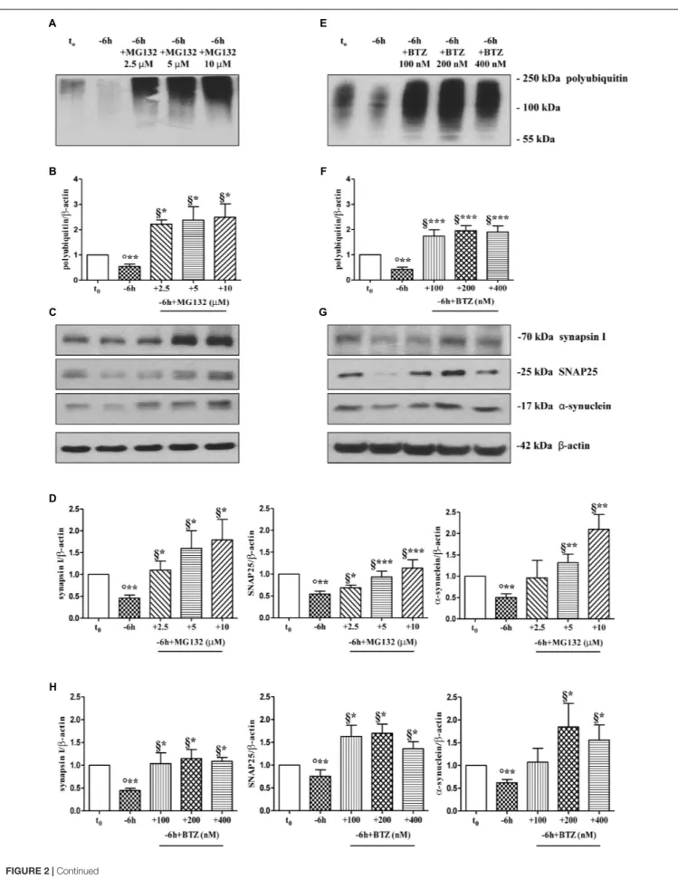
FIGURE 2 | Pharmacological blockage of UPS by MG132 and BTZ, two well-established selective and cell-permeable proteasomal inhibitors, prevents the decline in the steady-state levels of presynaptic markers synapsin I, SNAP25, andα-synuclein in NGF-deprived septo-hippocampal primary neurons. (A–H) Representative blots of detergent lysates from septal cholinergic-enriched cultures which were grown continuously (10–12 D.I.V.) in 0.2% B27 media in the presence of exogenous NGF (100 ng/ml) from plating (t0) and then deprived of their trophic support for 6 h (–6 h) in the absence or presence of increasing concentration of MG132 (2.5, 5,
10µM) and BTZ (100, 200, 400 nM) inhibitors. At time points indicated, cells were collected and equal amounts of total protein extracts (20 µg) were analyzed by SDS-PAGE by probing with anti-pan ubiquitin antibody (Dako antibody Z0458) (A-E for MG132 and BTZ, respectively) and specific antibodies against the indicated presynaptic markers (C-G for MG132 and BTZ, respectively). Densitometric quantification of immunoreactivity levels of ubiquitin (B-F for MG132 and BTZ, respectively) and synapsin I, SNAP25 andα-synuclein (D-H for MG132 and BTZ, respectively) was calculated by normalizing the signals of each band to correspondingβ-actin intensities on the same blots. Values were mean ± SEM of at least of five independent experiments and were expressed with respect to control neurons (t0). Statistically significant differences were calculated at the respective experimental points by unpaired-two tailed t-Student’s test [∗p< 0.05, ∗ ∗p< 0.01 and∗ ∗ ∗p< 0.0001 vs. t
0control neurons (◦) and vs. –6 h NGF deprivation (§)].
leading to the appearance of several low-molecular-weight ubiquitinated species only following higher time expositions of the blots. The concomitant and global accumulation of HMW polyubiquitinated conjugates (Figures 2A,B) confirmed the efficacy of the treatment with MG132, in line with previous findings (Leitch et al., 2001). Due to the evidence that MG132 is endowed with reactivity not only toward the active proteasomal subunits (β1, β2, and β5) but also against different types of proteases, including serine proteases and calpain (Myung et al., 2001), we further validated these results by incubation of cultures with bortezomib (BTZ) a more potent, specific and not-reversible proteasome-blocking drug (Buac et al., 2013). As shown in Figures 2G,H, treatment of cholinergic neurons undergoing 6 h of NGF withdrawal with increasing doses of BTZ (100, 200, 400 nM) yielded results comparable with those obtained with MG132, in agreement with inhibitor(s) ability in promoting the accumulation of HMW-smeared immunoreactivity signal (Figures 2E,F). Nonetheless, the strongest stabilizing effect of BTZ on the intracellular turnover of synapsin I (1.147 ± 0.198 A.U.), SNAP25 (1.699 ± 0.199 A.U.) and α-synuclein (1.84 ± 0.51 A.U.) in comparison to −6 h untreated cultures (0.6 ± 0.08 A.U.) was obtained at 200 nM concentration (∗
p< 0.05, t-Student’s test versus t = −6 h) because the highest concentration (400 nM) turned out to be less effective. This effect appeared to be specific for proteasome inhibitors because incubation of cultures with Z-VAD-fmk caspase inhibitor or pepstatin A serine protease inhibitor were ineffective (Supplementary
Figures S2A,B). No significant change on the basal levels of synapsin I, SNAP25 and α-synuclein was detected in control neurons upon incubation with proteasome inhibitors alone (data not shown). Likewise, under the same experimental conditions, the protein expression of other presynaptic and/or post-synaptic markers -such as syntaxin I, synaptophysin and NR1 that we previously showed not to be changed by NGF withdrawal in this in vitro neuronal model (Latina et al., 2017)- were contextually unaffected by MG132 and BTZ administration (Supplementary
Figure S2C). This evidence is in agreement with the findings that metabolic turnover of most synaptic proteins does not rely on the UPS but likely follows alternative intracellular pathways of degradation (Hakim et al., 2016). Furthermore, treatment of neuronal cultures with MG132 and BTZ were not per se neurotoxic, as assessed by MTT assay and quantification of DAPI-positive nuclear staining (Supplementary Figures S2D,E), consistent with previous report showing that>16 h of exposure
to proteasomal inhibitors is required to induce a noticeable in vitro cell death in primary neurons (Snider et al., 2002). Importantly, short term (−6 h) incubation with MG132 and BTZ did not significantly change the levels of free monoubiquitin (Supplementary Figures S3A,B) indicating that their inhibitory effects were directly mediated on proteolysis and not by indirect action on ubiquitination of target proteins. This is in line with the previous findings referring that only under long period of incubation time (>16 h) with proteasome inhibitors, ubiquitin is not recycled and the pool of monoubiquitin available for ubiquitination is reduced (Patnaik et al., 2000;
López et al., 2011).
Protein ubiquitination is not necessarily indicative of proteasomal degradation because monoubiquitination regulates the protein trafficking, involving endosomes, as well as other important cellular functions (Hicke, 2001). Thus, to ascertain that the three presynaptic proteins were actually polyubiquitinated in a NGF-dependent manner, we performed a pulldown analysis of polyubiquitinated-conjugates on whole-cell proteins lysates from septo-hippocampal cultures undergoing 6 h of neurotrophin withdrawal, in the absence or presence of MG132 inhibitor (10 µM). This assay protocol relies on a commercially available affinity resin (Thermo Scientific) which allows for the isolation of polyubiquitinated proteins whereas the monoubiquitinated species, or short chain polymers (<4 ubiquitin monomers), are removed being recovered in the flow-through. As shown in Figures 3A,B, after polyUb-enrichment partitioning and Western blotting analysis with anti-pan ubiquitin (Z0458 Dako) and anti-synapsin I antibodies, we found that the immunoreactivity of affinity-purified synapsin I declined in neuronal cultures following 6 h NGF starvation and that its levels dramatically increased upon pharmacological proteasomal blockade in a MG132-reversible manner. Importantly, this trend virtually mirrored the pattern detected in not-fractionated whole-cell lysates, demonstrating that NGF withdrawal actually stimulates the polyubiquitination of synapsin I and its degradation by the UPS under genuine cellular conditions. Moreover, after the fractionation procedure, we contextually detected a clear electrophoretic mobility shift in the signal of the heterogeneous HMW-smeared signal which plainly proved that the polyubiquitinated-enriched proteins were successfully recovered after affinity-based column elution, having an added mass of at least 32 kDa (corresponding to a chain of 4 × 8 kDa monomers of ubiquitin) compared to the mass of the non-modified ones from total lysates. Similar
results were found for α-synuclein and SNAP25 (data not shown).
A neuronal cross-talk between the ubiquitin-proteasome and autophagy-lysosome systems takes place intracellularly and the types of ubiquitin linkages are known to influence the final protein fate with polyubiquitylation (K48- and K-63-linked chains, respectively) which targets substrates for proteolysis along both degradative pathways (Korolchuk et al., 2010). In order to distinguish between these two alternative but not mutually exclusive possibilities, we investigated the effect of pharmacological treatment with chloroquine (CQ), an alkalizing reagent used to raise the lysosomal pH leading to inhibition of fusion of autophagosome with lysosome and, then, of lysosomal protein degradation. In contrast to the robust action of MG132 proteasomal inhibitor on the stability of presynaptic marker(s) (∗∗
p < 0.01, t-Student’s test versus t = −6 h), incubation of NGF-deprived septal neurons with 10 µM CQ failed to attenuate the NGF-induced downregulation in the steady-state levels of SNAP25 (p = 0.2762 versus t = −6 h) (Figures 3C,D). The efficacy of CQ treatment in blocking the autophagy-mediated degradation was confirmed by the increased conversion ratio LC3II/LC3I which is frequently used to monitor the endogenous autophagic flux in neurons (Kabeya et al., 2000; Klionsky et al., 2008, 2012; Viscomi et al., 2012). In addition, the weak and not-significant (p = 0.0934 versus t = −6 h) increase in immunoreactivity of polyubiquitin-conjugates detected by Dako anti-pan ubiquitin antibody after treatment with CQ indicates that, in this in vitro model, the K63-polyubiquitin linkages -which specifically target proteins to the endosomal– lysosomal system (Grice and Nathan, 2016)- contributed less than K48-polyubiquitin chains, which are the major signal for UPS-mediated degradation (Grice and Nathan, 2016), in controlling the intracellular protein turnover. On the other hand, in line with previous observations (Myeku and Figueiredo-Pereira, 2011), the minimal accumulation of polyubiquitinated proteins detected after CQ treatment was more likely to be due to weak proteasomal inhibition by the lysosomotropic agent (or by impaired flux through the UPS owing to substrate excess) because autophagy inhibition fails to elevate ubiquitin chains unless the UPS is affected. Finally, similar results were found for the other two analyzed presynaptic markers, synapsin I and α-synuclein (Supplementary Figures S4A,B).
Taken together, these in vitro results indicate that synapsin I, SNAP25 and α-synuclein are actually polyubiquitinated and degraded by UPS in septo-hippocampal primary neurons in response to short-term (−6 h) NGF withdrawal.
The Activity of Ubiquitin C-Terminal
Hydrolase L-1 (UCHL-1) Is Required for
the Downregulation of Presynaptic
Markers in NGF-Deprived Cholinergic
Septo-Hippocampal Neurons
Dynamic ubiquitylation is governed by the antagonistic intracellular actions of ubiquitin ligases and DUBs. Among
the DUBs, UCHL-1 is the most abundant, neuron-specific ubiquitin C-terminal hydrolase which regulates the structure and function(s) of synaptic terminals by controlling the local ubiquitin dynamics (Cartier et al., 2009). Relevantly, UCHL-1 changes are involved in the pathogenesis of AD (Pasinetti, 2001;Choi et al., 2004;Gong et al., 2006). UCHL-1 is endowed with both hydrolase activity, by removing ubiquitin molecules from the degraded proteins in order to be reused for de novo cycles of ubiquitination (Finley et al., 1989; Larsen et al., 1998), and with ubiquityl ligase activity, by linking ubiquitin molecules and generating polyubiquitin chains that tag the target proteins for UPS-mediated elimination (Liu et al., 2002). Therefore, to ascertain whether the UCHL-1 activity contributed, directly or indirectly, to the UPS-mediated clearance of selected presynaptic proteins, we evaluated the effects of changes in UCHL-1 dependent dynamics of the total (i.e., bound and monomeric/unconjugated) ubiquitin pool on the turnover of synapsin I, SNAP25 andα-synuclein under in vitro conditions of NGF starvation. To this aim, cultures grown continuously (10–12 D.I.V.) in 0.2% B27 media in the presence of exogenous NGF (100 ng/ml) from plating (t0) were deprived of this
trophic support for 6 h (−6 h) in the absence or presence of increasing doses (2.5, 5, 10 µM) of LDN, an inhibitor which selectively halts UCHL-1 without any effect on other UCH family members (Liu et al., 2003; Gong et al., 2006; Cartier et al., 2009). Western blotting on whole-cell lysates were carried out to check the protein expression of synapsin I, SNAP25 and α-synuclein, the pattern of polyubiquitinated-conjugates and the stability in the intracellular pool of free/monomeric ubiquitin. Pharmacological suppression of UCHL-1 (Figures 4C,D) with the lowest used concentration of LDN (2.5 µM) was effective in significantly rescuing the NGF-dependent drop in synapsin I (∗∗
p < 0.01 t-Student’s test versus t = −6 h), SNAP25 (∗
p < 0.05, t-Student’s test versus t = −6 h) and α-synuclein immunoreactivity (∗
p < 0.05, t-Student’s test versus t = −6 h), in line with drug ability (Figures 4A,B) of inducing a strong elevation in the Ub-positive HMW-smeared signal (∗
p < 0.05, t-Student’s test versus t = −6 h). Conversely, treatment with higher experimental doses of LDN (5, 10 µM) (Figures 5C,D) turned out to be unsuccessful in elevating the intracellular levels of synapsin I (p = 0.126, p = 0.671 t-Student’s test versus t = −6 h, respectively), SNAP25 (p = 0.857, p = 0.363 t-Student’s test versus t = −6 h, respectively) and α-synuclein (p = 0.205, p = 0.423 t-Student’s test versus t = −6 h, respectively) consistent with their more potent effect in lessening the free monomeric ubiquitin (∗∗
p < 0.01 t-Student’s test versus t = −6 h) than in causing the concomitant accumulation of polyubiquitin conjugates (∗
p< 0.05, t-Student’s test versus t = −6 h) (Figures 4C,D). These results are in agreement with previous findings showing that the homeostasis of unconjugated ubiquitin is per se crucial in maintaining the synapse structure and function(s) (Chen et al., 2011) and that synaptic-enriched UCHL-1 is able to associate with and inhibit the intracellular degradation of its monomeric free pool (Osaka et al., 2003). Furthermore, Western blotting analysis followed by densitometric quantification indicated that there was a trend for an increase of the intracellular abundance
FIGURE 3 | Inhibition of UPS -but not of autophagy- protects the presynaptic terminals of septo-hippocampal neurons from NGF withdrawal. (A,B) The intracellular levels of synapsin I-polyubiquitin conjugates were determined in control (t0) and 6 h NGF-deprived (–6 h) cultures in the absence or presence of MG132 (10µM),
following polyUb-pull down of total protein extracts by ubiquitin affinity beads and immunoblotting with anti-synapsin I and anti- pan ubiquitin (Dako antibody Z0458) antibodies. Ponceau staining was used to ascertain the equal protein loading of gels. Dotted boxes indicate the area used for quantification (B) Notice thatβ-actin signal was not recovered after column elution of polyUb-conjugates. Values were mean ± SEM of three independent experiments and were expressed with respect to control neurons (t0). Statistically significant differences were calculated at the respective experimental points by unpaired-two tailed t-Student’s test [∗p< 0.05, ∗ ∗p< 0.01 vs. t
0control neurons (◦) and vs. –6 h NGF deprivation (§)]. (C,D) Septal cholinergic-enriched cultures were grown continuously (10–12 D.I.V.) in 0.2%
B27 media in the presence of exogenous NGF (100 ng/ml) from plating (t0) and then deprived of their trophic support for 6 h (–6 h) in the absence or presence of CQ
(10µM) and MG132 (10 µM). Cell lysates were extracted and analyzed by Western blotting by probing for ubiquitin (Dako antibody Z0458), SNAP25, LC3 and β-actin. Representative blot showing the effect of drugs treatment on the intracellular levels of neuronal proteins (C) and relative densitometric quantification (D) were shown. Data were reported as mean ± SEM and expressed ratio of the value from control neurons (t0) Statistically significant differences were calculated at the
respective experimental points by unpaired-two tailed t-Student’s test [∗p< 0.05,∗ ∗p< 0.01 vs. t
0control neurons (◦) and vs. –6 h NGF deprivation (§)]. Only the
inhibition of proteasomal activity by MG132 led to significant accumulation of polyubiquitin-conjugates and increase in the expression levels of SNAP25 in 6 h NGF-deprived septo-hippocampal neurons. The increase in LC3-II/LC3-I ratio confirmed the efficacy of CQ treatment.
of UCHL1 in neuronal cultures during the time-dependent NGF removal (Supplementary Figures S5A,B). Finally, the co-incubation of −6 h NGF-deprived cholinergic neurons with lowest effective subtoxic concentrations of MG132 (2.5µM) and LDN (2.5 µM) displayed an additive effect in preventing the decline in SNAP25 immunoreactivity (∗∗
p < 0.01 t-Student’s test versust = −6 h), as shown in Western blotting analysis on total proteins extracts from co-treated cultures (Figures 5A,B). Consistent with our biochemical results, immunocytochemistry staining (Figures 5C,D) also showed a significant increase in the density of SNAP25- positive puncta in (MG132+LDN)-exposed cultures in comparison with its 6 h NGF-deprived counterpart (∗∗
p< 0.01 t-Student’s test versus t = −6 h). Similar results were found for the other two presynaptic proteins of interest, synapsin I and α-synuclein (Supplementary Figures
S6A,B).
These in vitro findings indicate that: (i) UCHL-1 is early involved in the “dying-back”-type degeneration occurring in septo-hippocampal primary neurons following alterations in NGF/TrkA signaling pathway and that its moderate inhibition is effective in blocking the selective loss ofα-synuclein, SNAP25 and synapsin I; (ii) NGF withdrawal orchestrates in thisin vitro model an early (−6 h) UPS-mediated destruction of these presynaptic markers by means of balanced fine-tuning in the dynamic UPS-mediated ubiquitylation/degradation and UCHL-1-dependent (mono)ubiquitin turnover.
The UPS Activity and UCHL-1-Dependent
(Mono)ubiquitin Homeostasis Support
the NGF-Dependent Modulation of
Excitatory Neurotransmission in
Cholinergic Septo-Hippocampal Neurons
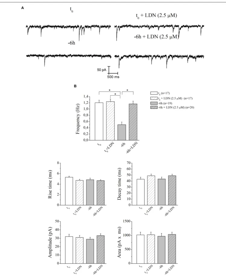
Nerve growth factor exerts a potent and specific stimulatory presynaptic action on cholinergic nerve terminals (Wu and Yeh, 2005; Huh et al., 2008) by increasing the frequency and not the amplitude of spontaneous mEPSCs. In this context, we have previously shown that the short-term (−6 h) interruption in NGF/TrkA signaling pathway in cholinergic septo-hippocampal primary neurons decreases the averaged frequency of mEPSCs by downregulating the steady-state levels of synapsin I, SNAP25, and α-synuclein (Latina et al., 2017) which critically control
the vesicular trafficking and neurotransmission at presynaptic terminals.
Therefore, having demonstrated an early (−6 h) and causal role of UPS-degradation/ubiquitination rate in controlling the NGF-dependent turnover of these three presynaptic markers, we further explored the physiologic relevance of acute perturbations of UPS activity in modulating the excitatory neurotransmission in this in vitro paradigm. To this aim, electrophysiological recordings of mEPSCs -which are known to rise from random release of presynaptic vesicles at nerve endings and to play important roles in maintaining functional connections of synaptic terminals (Caioli et al., 2011;Saba et al., 2016)- were carried out on cholinergic primary neurons undergoing short-term removal of exogenous NGF for 6 h (−6 h), in the presence or absence of MG132 (10µM) and LDN (2.5 µM). As shown in
Figures 6A,B, the mean of the mEPSCs frequency recorded at the holding potential −60 mV was significantly lower in −6 h NGF-deprived neurons (−6 h: 0.44 ± 0.04 Hz;n = 15) than in control ones (t0:0.99 ± 0.19 Hz;n = 11; p ≤ 0.02), in keeping
with our previous findings (Latina et al., 2017). Interestingly, acute inhibition of UPS by MG132 treatment (10 µM) was fully able to rescue the excitatory neurotransmission because the mEPSCs frequency of −6 h NGF-deprived neurons+MG132 was significantly higher than of −6 h untreated cultures (−6 h+MG132: 0.91 ± 0.1 Hz;n = 19; p ≤ 0.02 versus −6 h) by approximating the t0 control values (p > 0.05 versus t0).
These electrophysiological data tightly correlated with Western blotting analysis (Figure 2) showing the ability of MG132 in selectively preventing the NGF-induced downregulation of synapsin I, SNAP25, andα-synuclein, the three well-established presynaptic crucially involved in neurotransmitter exocytosis at nerve endings. On the contrary, no significant effect was detected in the mEPSCs frequency when MG132 was added to cultures in basal conditions (t0+MG132: 0.95 ± 0.14 Hz;n = 12;
p> 0.05 versus t0). Likewise, in line with previous data reporting
a specific effect of this neurotrophin on in vitro cholinergic neurons at the presynaptic level (Wu and Yeh, 2005;Huh et al., 2008; Latina et al., 2017), the mean amplitude as well as the mean area and the kinetic parameters of the mEPSCs recorded from cultures were not modified in any experimental condition (p> 0.05) neither by NGF deprivation alone nor by incubation of MG132.
FIGURE 4 | UCHL-1 activity is required to the NGF-dependent control of the synapsin I, SNAP25 andα-synuclein stability in septo-hippocampal primary neurons. (A–D) Septal cholinergic-enriched cultures were grown continuously (10–12 D.I.V.) in 0.2% B27 media in the presence of exogenous NGF (100 ng/ml) from plating (t0) and then deprived of their trophic support for 6 h (–6 h) in the absence or presence of increasing concentration of LDN (2.5, 5, 10µM), a selective and
cell-permeable UCHL-1 inhibitor. Representative Western Blotting of sample extracts analyzed for the expression levels of poly-/monoubiquitin (A), synapsin I, SNAP25 andα-synuclein (C) were shown. Cropped image was shown in (A). Bar graphs (B–D) showed the densitometric quantification of immunoreactivity levels normalized by calculating the ratio of the intensity of the signal for the protein of interest to that ofβ-actin which was used as loading control for each sample/lane. Values were mean ± SEM of at least of five independent experiments and were expressed with respect to control neurons (t0). Statistically significant differences
were calculated by unpaired-two tailed t-Student’s test [∗
p< 0.05,∗ ∗
FIGURE 5 | Co-treatment with MG132 and LDN has additive effect in preventing the presynaptic degeneration occurring in –6 h NGF-deprived cholinergic neurons. (A–D) Septal cholinergic-enriched cultures were grown continuously (10–12 D.I.V.) in 0.2% B27 media in the presence of exogenous NGF (100 ng/ml) from plating
FIGURE 5 | Continued
(t0) and then deprived of their trophic support for 6 h (–6 h) in the absence or presence of MG132 (2.5µM), LDN (2.5 µM) and MG132 (2.5 µM)+LDN (2.5 µM)
combination. Levels of SNAP25 andβ-actin were assessed by Western blotting analysis (A) and densitometric quantification (B) was calculated as ratio of the value from control neurons (t0) and reported as mean ± SEM. Statistically significant differences were calculated at the respective experimental points by unpaired-two
tailed t-Student’s test [∗
p< 0.05,∗ ∗
p< 0.01 vs. t0control neurons (◦) and vs. –6 h NGF deprivation (§)]. Representative immunofluorescence images (C) and bar
graph (D) showing the SNAP25 expression levels in cholinergic primary neurons following NGF withdrawal and inhibitors treatments were reported. Cultures were fixed, permeabilized and immunostained for SNAP25 (red channel) and nuclei were counterstained with DAPI (blue channel). Quantification of SNAP25-positive structures per 60 × image-field arbitrary unit (A.U.) was calculated as ratio of the value from control neurons (t0) and reported as mean ± SEM. Statistically
significant differences were calculated at the respective experimental points by unpaired-two tailed t-Student’s test [∗
p< 0.05,∗ ∗
p< 0.01 vs. t0control neurons (◦)
and vs. –6 h NGF deprivation (§)]. Images were representative of at least three independent experiments. Scale bar: 10µM.
Similar results (Figures 7A,B) were found after incubation of −6 h NGF-deprived septal cultures with UCHL-1 inhibitor LDN (2.5µM) (−6 h+LDN: 1.16 ± 0.09 Hz; n = 20; p < 0.001 versus −6 h,p> 0.05 versus t0;t0+LDN: 1.24 ± 0.07 Hz;n = 17; p> 0.05
versust0).
These results highlight the pivotal role of UPS and UCHL-1-dependent (mono)ubiquitin dynamics in mediating the NGF-induced modulatory actions on the presynaptic morphology and neurosecretory function(s) inin vitro cholinergic septal primary neurons.
Early Decline in Ubiquitination Pattern
and Loss of Specific Presynaptic
Proteins Also Occur in vivo, in
Hippocampal Synaptoneurosomes From
Aging AD Transgenic Mouse Model
Tg2576 (HuAPP695 SWE), in Association
With Selective Down-Regulation of
Cholinergic Markers
Reduction of ChAT- labeled nerve endings, deficiency in cholinergic transmission and spatial learning/memory ability along with downregulation in expression of NGF and its cognate receptors have been detected in aging Tg2576 animals (Apelt et al., 2002;Chauhan and Siegel, 2003;Klingner et al., 2003;Lüth et al., 2003;Simmons et al., 2014;Zhu et al., 2017, a transgenic AD mouse line carrying the mutated form of APP named AβPPSWE (K670N/M671L). In this well-established AD paradigm, memory deficits are just evident at 3 months of age and are progressive (Pompl et al., 1999; King and Arendash, 2002; Kobayashi and Chen, 2005) although insoluble Aβ aggregates start to raise from 6 months (Kawarabayashi et al., 2001) and Aβ-laden plaque deposition is frankly detectable only at 11 months (Hsiao et al., 1996;Irizarry et al., 1997).
In order to strengthen the pathological relevance of our in vitro findings in AD neurodegeneration, regional synaptosomes were isolated from young/presymptomatic (1-month-old) and middle-aged (9-months-old) Tg2576 mice and WT control counterparts. Since basal forebrain neurons are the main source of cholinergic innervation to the cortex and hippocampusin vivo (Niewiadomska et al., 2011), hippocampal pinched-off nerve terminals – which are known to represent mainly the presynaptic compartment obtained by fractionated brain homogenates (Gylys et al., 2004) – were evaluated for the abundance of synapsin I, SNAP25 andα-synuclein, the temporal
profile of ubiquitination and cholinergic markers expression. As shown in Figures 8C,D, the steady-state levels of synapsin I was significantly reduced in aging AD animals from both experimental groups (0.1662 ± 0.032 A.U. from 1-month-old Tg2576 mice; 0.1017 ± 0.027 A.U. from 9-months-old Tg2576 mice) in comparison with their age-matched non-transgenic littermates (0.5136 ± 0.096 A.U. from 1-month-old WT mice; 0.2952 ± 0.076 A.U. from 9-months-old WT mice, ∗
p< 0.05, t-Student’s test). A similar negative trend was found for SNAP25 andα-synuclein (data not shown). Relevantly and in line with the evidence that vesicle trafficking proteins are not equally affected in human AD brains (Sze et al., 2000;Honer, 2003;Reddy et al., 2005), no change was contextually detected up to 9 months in the expression rates of syntaxin-I (p = 0.5296 and 0.3838 for 1- and 9-months-old group versus age-matched WT mice, respectively) and synaptophysin (p = 0.4839 and p = 0.1149 for 1- and 9-months-old group versus age-matched WT mice, respectively) (Figures 8E,F), two other presynaptic markers we reported to be unaffected by NGF withdrawal inin vitro cholinergic-enriched septo-hippocampal primary cultures (Latina et al., 2017).
Interestingly and in agreement with our in vitro results (Figure 2), the synaptic distribution of the age-related HMW polyubiquitin protein conjugates also declined over time in AD-affected isolated hippocampal nerve terminals, in both 1- and 9-months-old Tg2576 animals (0.3884 ± 0.005 A.U. and 0.3133 ± 0.024 A.U., respectively ∗
p < 0.05, t-Student’s test) related to experimental age-matched control groups (0.7142 ± 0.064 A.U. and 0.4667 ± 0.044 A.U: for 1-and 9-months-old WT mice, respectively) (Figures 8A,B). Furthermore, the immunoreactivity signals from three different well-confirmed cholinergic-specific markers (Dawbarn et al., 1988; Holtzman et al., 1992; Sobreviela et al., 1994), such as M1 (1-month-old group: WT 0.3284 ± 0.048 A.U., Tg2576 0.2251 ± 0.045 A.U.; 9-months-old group: WT 0.3665 ± 0.045 A.U., Tg2576 0.3136 ± 0.026 A.U.), ChAT (1-month-old group: WT 0.4076 ± 0.053, Tg2576 0.2128 ± 0.028; 9-months-old group: WT 0.2921 ± 0.034 A.U., Tg2576 0.2137 ± 0.036 A.U.) and TrkA (1-month-old group: WT 0.742 ± 0.138 A.U., Tg2576 0.651 ± 0.125 A.U.; 9-months-old group: WT 0.651 ± 0.005 A.U., Tg2576 0.458 ± 0.058 A.U.) (Figures 8G,H), were also gradually reduced in AD mice -mainly in symptomatic 1-month-old young animals (Figures 8G,H) (∗
p < 0.05 t-Student’s test versus age-matched WT controls)-when compared with corresponding non-transgenic littermate counterparts. Finally and more importantly, in contrast to the prominent loss of cholinergic projections and the progressive
FIGURE 6 | UPS mediates the in vitro modulatory action of NGF on presynaptic function of cholinergic septo-hippocampal primary neurons. (A) Representative traces of septal neurons grown for 10–12 D.I.V. in B27 0.2%+NGF, deprived for 6 h of NGF, in the absence and presence of MG132 UPS-inhibitor (10µM) were