A
A
l
l
m
m
a
a
M
M
a
a
t
t
e
e
r
r
S
S
t
t
u
u
d
d
i
i
o
o
r
r
u
u
m
m
–
–
U
U
n
n
i
i
v
v
e
e
r
r
s
s
i
i
t
t
à
à
d
d
i
i
B
B
o
o
l
l
o
o
g
g
n
n
a
a
DOTTORATO DI RICERCA IN
SCIENZE CHIMICHE
Ciclo XXII
Settore/i scientifico-disciplinare/i di afferenza: CHIM 03
TITOLO TESI
Synthesis and Physical-Chemical characterization of
Metallic Nanoparticles
Presentata da: Guido Fracasso
Coordinatore
Dottorato
Relatore
Prof. Giuliano Longoni Prof . Norberto Roveri
Synthesis and Physical-Chemical characterization of Metallic
Nanoparticles
Introduction pag.1
Exsperimental pag. 16
Results and discussion pag. 26
1) Surface protected metallic nanoparticles filling chrysotile nanotubes pag. 26 2)Biomimetic hydroxyapatite nanocrystals as carriers for Au/Ag alloy
anticancer particles pag. 37
3) Cuprous Bromide nanocrystals pag. 40
4) Buianethiolate protected copper nanoparticles pag. 41
5) 4-methylbenzenethiol protected copper nanoparticles pag. 55 6) Phenol protected selenium nanoparticles pag.61
7) Phloroglucinol protected selenium pag. 72
Conclusions pag. 80
References pag. 83
Introduction
Nanoparticles (particles of 1–100 nm in diameter) have generated intense interest over the past decades because of their unique electronic, optical, photoresponsive, and catalytic properties. However, nanoparticles are subject to the irreversible particle aggregation and the oxidation reactions. Thus the stabilization of nanoparticles against these problems is a requirement for further advancement in nanoparticle science and technology. In 1981, Schmid et al .[1] synthesized the first example of the ligandstabilized gold nanoparticles, Au55(PPh3)12Cl6. However, the dependable and practical formation of more stable an isolable monolayer-protected gold nanoparticles was achieved about a decade ago by Brust et al. [2]. The organic monolayers, which bind to the surface of metal nanoparticles, not only prevent aggregation of the metal nanoparticles, but also stabilize
them from harsh reaction conditions and enhance the solubility in various solvents[3–5]. Subsequent investigations were extended to different metal particles including Au, Ag,
Cu, Pt, Pd, Ni and alloy particles. Other organic compounds with different reactive head groups, such as thiol, disulfide, sulfide, thiosulfate, xanthate [38], phosphine, phosphine oxide, ammonium, amine, carboxylate, selenide, and isocyanide can protect metal nanoparticles by selfassembly assembly [6].
Monolayers with different functional groups and moieties can also be incorporated onto the surface of metal nanoparticles [3–5]. The modification of nanoparticles by the incorporation of a functional group is of potential significance for the expansion of chemical and biological applications of these nanomaterials. This review focuses on the synthesis of various metal nanoparticles protected with monolayers containing different reactive head groups and functional tail groups.
Gold nanoparticles
Gold nanoparticles have drawn remarkable interest in the last few years because of their high stability to oxidation and their optical and well-defined size-related electronic (e.g., quantized charging) properties [7,8]. Biological applications of hybrid Au nanoparticles have also shown a great promise for the use of these nanomaterials in biotechnology [4]. The synthesis of monolayer-protected gold nanoparticles will be described based on the reactive head groups, which provide self-assembly of organic monolayers onto the nanoparticle surface.
The stabilization of gold nanoparticles with alkanethiols was a direct result of advancement in the passivation of two-dimensional gold surfaces using alkanethiols in the late 1980s and early 1990s, which results in the formation of self-assembled monolayers on gold substrates [9]. The first report regarding the capping of Au nanoparticles with alkanethiols was made by Giersig and Mulvaney [10] in 1993. Brust et al. [2] reported a convenient two-phase synthesis of isolable and soluble alkanethiolate-protected gold nanoparticles in 1994, which had a huge impact on the nanoparticle research. In this reaction AuCl4 was transferred to toluene using tetraoctylammonium bromide as the phase-transfer reagent. Addition of dodecanethiol to organic-phase AuCl4 followed by the reduction with NaBH4 generated dodecanethiolate-protected gold nanoparticles. The research was extended to the alkanethiols with different chain lengths (C3–C24) [3].
Gold nanoparticles synthesized using the modified Schiffrin reaction generally have the average core size of 1–10 nm. The core size of gold nanoparticles could be controlled by modification of reaction conditions. It has been found that the nanoparticle formation behaves as a nucleation growth passivation process [3]. Therefore the average core size of particles gets smaller when a larger thiol/gold mole ratio is used, the reductant is added faster (<10 sec) at lower temperature, the
reaction is quenched after a shorter period of time (30 min to 1 h) has elapsed, or sterically bulkier ligands are used [3,4,11-13]. Because alkanethiolate-protected gold nanoparticles generated by the Schiffrin protocol are still somewhat polydisperse, there have been several studies aimed at getting more monodisperse particles using solubility fractionation, extraction, annealing, chromatography, and capillary electrophoresis [14,15]. Alkanethiolate-protected gold nanoparticles are generally considered as nonreactive species. By incorporation of o-functional groups to the monolayer, these nanomaterials can have specific functions and reactivities. p-Mercaptophenol-protected gold nanoparticles were synthesized directly from p-mercaptophenol and AuCl4 in a one-phase system, which was reported first by Brust et al.[16] This research was extended to various ofunctionalized alkanethiols,[3–5] alkanethiols with internal amide groups,[17] thiols terminated with oligo- or polyethylene oxide [18], thiols with ionic liquids [19], fluorinated thiols [20], arenethiols [3], and crown ether-functionalized thiols[21] using the modified Schiffrin reaction and yielded various functionalized nanoparticles including watersoluble gold nanoparticles [3,4]. Later, a mixture of alkanethiols was also used to synthesize mixed monolayer-protected gold nanoparticles [22]. This result showed that the coadsorption of alkanethiols is governed by solvation-driven thermodynamic reactivities of the precursor ligands. When polymeric Au(I)-SG (glutathione: N-g-glutamyl-cysteinyl-glycine) was reduced with NaBH4 in water, it produced small (1–2 nm) water-soluble monolayer-protected gold nanoparticles [23]. The reduction of trithiol (thiocyanuric acid)-Au polymer precursor with potassium bitartrate resulted in the formation of watersoluble nanoparticles with varied geometric shapes [24].
Dithiol, trithiol, tetrathiol (thiolated resorcinarenethiolated a-, and thiolated b-cyclodextrin-protected gold nanoparticles were also reported [25–26]. Templeton et al. [3] reported the “ligand-place exchange’’ method to modify the composition of the monolayer-protected nanoparticles. This research enabled incorporation of many different functional groups and moieties onto the surface of gold nanoparticles. In the exchange reaction, the incoming ligands replace the thiolate ligands on nanoparticles by an associative reaction, and the displaced thiolate becomes a thiol. The rate of ligand exchange depends on the chain length and/or steric bulk of the initial monolayers and electronic charge on nanoparticles. Various functional groups ranging from simple organic functional groups to functional complexes (electroactive or photochemical groups) were incorporated into the nanoparticles using ligand-place exchange reactions [3–5].
Reactions of functionalized gold nanoparticles were also frequently used for the incorporation of more specific organic groups and moieties having electroactive, photoresponsive, catalytic, mechanical, and biological properties [3–5].
In this work, Au, Au4/Ag1 and Au1/Ag4 nanoparticles were synthesised with the aim to fill chrysotyle nanotubes hollows. Decanethiol was used to coat the nanoparticles surface with the pur pose to fill the nanotube cavities with the nanoparticles owing to an interdigitation process occurring between the long alkilic chains on neighbouring nanoparticles.
Ag nanoparticles
Recently, research efforts have been intensified for the synthesis of Ag nanoparticles because of important roles played by Ag particles in antimicrobial applications, catalysis, and as a substrate for SERS. Thiols [27,28] have been the most popular choice of capping reagent for Ag nanoparticles because of a higher stability over other capping reagents, such as amines [29,30] and carboxylates [31, which are weakly anchored to Ag particle cores. Other organic compounds, such as dialkyl disulfides [32,33], dialkyl sulfides [34], thiosulfates [35], thiosalicylic acids [36], and xanthates[37] have been used to generate monolayer-protected Ag nanoparticles.
Ag nanoparticles protected with different organic species have been prepared. In fact a synthesis of alkylamine-capped silver nanoparticles was developed using thermal decomposition of [Ag(m-mesityl)4] (mesityl=2,4,7-Me3C6H2) derivatives in alkylamine solution [38]. Two-phase (n-heptane/water) NaBH4 reduction of N-hexadecylethylenediamine silver nitrate complex
produced N-hexadecylethylenediamine-protected silver nanoparticles [39]. The colloidal solution of diamine- protected silver nanoparticles was free from flocculation and aggregation for several months.
Long-chain carboxylates (or fatty acids) have been one of the most commonly used ligands for silver particles [40-42]. The simple dissolution of silver 2-ethylhexanoate in DMSO (as both a solvent and reducing agent) produced silver nanoparticles [40]. Addition of stabilizing agent, sodium citrate, produced stable yet catalytic monolayer-protected silver nanoparticles. Thermal reduction of silver trifluoroacetate in isoamyl ether in the presence of oleic acid generated Ag nanoparticles with diameters in the range of 7–11 nm with narrow size distribution [42].
Surface-enhanced Raman scattering studies of 4-biphenylisocyanide of silver nanoparticles proved the adsorption of isocyanide on silver surfaces [43]. Dyeprotected silver nanoparticles were prepared through the reduction of metallic Ag ions by NaBH4 in aqueous media [44]. The dye used was a cyanine derivative, 5,5’- dichloro-3,3-disulfopropylthiacyanine sodium salt.
Alkanethiolate-protected Ag nanoparticles were synthesized using the modified two-phase Schiffrin reaction [45-46]. Two-dimensional ordered superlattices of the Ag nanoparticles could be formed by evaporating a drop of the particle solutions on carbon films [46]. The fluorinated thiol-capped Ag nanoparticles could be prepared using the NaBH4 reduction of AgNO3 in the presence of
perfluorodecanethiol in acetone/water. These particles could be redispersed in acetone and liquid and supercritical CO2 [47]. Thiolated b-cyclodextrin-protected Ag nanoparticles were also synthesized from AgNO3.[48] N,N-Dimethylformamide (DMF) was used as both the solvent and the reducing agent in this reaction. Synthesis of water-soluble tiopronin-protected Ag nanoparticles was also reported [49].
Unlike Au nanoparticles functionalized by the adsorption of dialkyl disulfides, the monolayers of alkanethiolate- protected Ag nanoparticles derived from dialkyl disulfides were somewhat less crystalline than those prepared similarly from alkanethiols [32]. Positively charged Ag nanoparticles were synthesized by the NaBH4 reduction of AgNO3 in the presence of quaternary ammonium dialkyl disulfides [50]. Adsorption of benzyl phenyl sulfide on silver nanoparticles in water has been studied [51]. Alkanethiolate-protected Ag nanoparticles were synthesized from sodium S dodecylthiosulfate in aqueous solution [52]. Silver nanoparticles, produced by the
borohydride reduction of AgNO3 in H2O, were stabilized by the adsorption of S-dodecylthiosulfate followed by the removal of the SO3 moiety.
Alkyl xanthates were also used for the capping of Ag nanoparticles [37]. Xanthate-capped Ag particles are less hydrophobic than alkanethiol-protected Ag nanoparticles and less stable than the oleate-capped particles (but still stable in aqueous solutions for over a month). Thiosalicylic
acid (TSA)-functionalized silver nanoparticles were synthesized from NaBH4 reduction of TSA-AgNO3 in aqueous ethanol solution.[53].
Copper nanoparticles
Because of the expected marked electronic and optical properties of copper nanoparticles, there has been an increased interest in the preparation of stable monolayer-protected copper nanoparticles. Alkyl xanthates [37], alkylamines [29], tetraalkyl ammonium complexes [54], and alkanethiols [55, 56] ligands have been utilized to protect copper nanoparticles. However, a limited progress has been obtained because of the high instability of copper nanoparticles. In order to contrast copper nanoparticles, irreversible aggregation and oxidation, Chen and Sommers [55] have prepared copper nanoparticles protected by alkanethiolate in one-phase system. Their prepared spherical (1-2nm in diameter) Cu particle bound hexanethiolate monolayers undergo to the oxidation of copper core, morphological evolution, and irreversible aggregation process. Long-chain alkanethiols, which have been found to selfassemble into compact monolayers (i.e., three-dimensional self-assembled monolayers) on metallic nanoparticles [57,58], cannot only induce metallic nanoparticles to be readily isolated, but also reduce their conductivity [37].
technological, chemical and biomedical fields.
Recently nanorods, nanodisks and nanoplatelets of the semiconducting material Cu2S were obtained by thermolysis of copper alcanethiolates [59] and the semiconducting nature of a copper thiolate has been reported [60].
In organic chemistry, copper arenethiolates act as catalysts in many organic reactions [61,62]. Copper thiolate clusters are present in many proteins and play an important role in cell metabolism. Recently a novel [(His)7Cu4(í4-S)] cluster was discovered in the active site of nitrous oxide reductase [63].
Thiolate protected copper nanoparticles can be regarded as assemblies of copper thiolates fastened on a metallic core that acts as templatig device. From this point of view they can be new usefull tools in organic synthesis and catalysis. Furthermore thiolate protected copper nanoparticles can represent starting materials to obtain new copper complexes with S donor ligands.
Concerning with the electronics field, the insulator metal transition has been discovered to occur at 270, 255, and 25 K for arrays of copper nanoclusters passivated with 1-decanethiole, lauric acid and tridecilamine respectively [64].
Thiolate protected copper nanoparticles for chemical, technological and biomedical applications must have adequate stability and an elevated degree of monodispersion. Furthermore the organic thiolate moieties on the nanoparticle surface must produce the minimal steric hindrance to improve their interactions with molecules or other nanoparticles. Unfortunately short chain alcanethiols or aryl thiols can be inadequate in protecting nanoclusters core against oxidation [64-66] and gaps form easily on the nanoparticle surface protected with small thiol molecules less fastened together by Van Der Vaals interactions than long chain (CH2>8) thiols.
Synthesis methods reported in literature to obtain thiolate protected copper nanoparticles involve usually Cu(NO3)2 and CuCl2 reactions with long chain alcanethiols (CH2>8) while ethanol, tethrahydrofuran, water or mixtures of these are used as solvents. The reduction of copper-thiol adducts is obtained by NaBH4 solubilized in ethanol or water or by LiBH4 in tetrahydrofuran (superhydride) [64,66]. Diffractometric and XPS analysis reveal that Cu2O is present in negligible quantity or even absent in the core of these thiolate protected copper nanoclusters [64]. However they are quite polydispersed probably due to the formation of insoluble copper – thiol adducts before the reduction step. Furthermore long chain alcanethiolates protecting the nanoparticles core can limit their possible chemical catalitic or biological properties and give rise to irreversible aggregation processes due to interpenetration (interdigitation) of the alchilic chains on their surfaces.
occurring during the syntheses and the nature of the thiols used can influence structure, and stability of copper nanoparticles.
The comprehension of these parameters is useful in tuning their properties for possible applications in many fields both technological and biomedical. The aim of this study is to synthesize 3-(6-mercaptohexyl) thiopheneprotected copper nanoparticles which, in absence of long chain alkanethiols and in absence of irreversible aggregation, could represent new high-conductive metallic nanoparticles with improved potentialities of tuning their optical and electronic properties. To increase the stability of copper nanoparticles protected with other thioletes, we have developed and compared three synthetic routes in this work. Butanethiol and 4-methilbenzenethiol were used in the syntheses with the aim of reduce the sterical hindrance during the interactions of the nanoparticles with molecules, templating structures or other kinds of substrates.
Selenium
Research activity in selenium chemistry and physics is continuously growing due to both thecnological and biomedical relevance of this element. The grey purplish trigonal selenium with its structure of long helical chains of atoms is the most thermodynamically stable allotropic form and reveals light enhanced semiconductive properties. This feature make it an usefull and cheap substitute of silicon in the photovoltaic cell fabrication. Selenium is also used in photographic exposure meters. It has also found to improve the abrasion resistence of vulcanized rubber.
Concerning with the biological field, selenium plays a fundamental role in cell metabolism being responsible of many enzymes and other proteins activity. Among these glutathione peroxidase reduces lipid hydroperoxides to the corresponding alcohols and hydroperoxide to water so removing strong oxidizing molecules responsible of the cellular oxidative stress.
Studies on nanometric size selenium structures and selenium nanoparticles give rise to an always increasing research work owing to the novel electronic, optical, photoresponsive, and catalytic properties that nanometric dimensions confer to the materials.
Syntheses reported in literature of nanoparticles or other selenium nanostructure imply the reduction of selenous oxide by means of many reducing agents: among these can be mentioned or glutathione [67] hydrazine [68] dextrose [69], ascorbic acid [70]sodium ascorbate [71], sodium thiosulphate [72].
Selenium nanostructures of variable shapes and sizes like nanowires [73], and hollow nanospheres [74] were obtained using oligosaccarides and proteins respectively as templating devices.
However selenium nanotubes were obtained by a hydrothermal synthesis also in absence of templating structures. In this case sodium formiate was used to reduce selenous oxide and the reaction was completed in 25 hours at 100°C [75].
Polivinil alcohol [76] gelatine [77] and other large molecules were used to obtain stable and as far as possible monodispersed selenium nanoparticles and to prevent their irreversible aggregation. These molecules however are weakly adsorbed by means of Van der Waals forces to the selenium nanoparticle surfaces so delaying but not stopping nanoparticle nuclei aggregation processes. Stable well monodispersed nobel metal nanoparticles with sizes of two to four nanometers were synthesized coating their surface with alcanethiols or other molecules bearing the thiolic function chemically bonded by means of the sulphur atoms to the nanoparticle surface by Brust and co-workers [2]. On the suggestion of this work, we have synthesized selenium nanoparticles protected by phenolic molecules chemically bonded to their surface.
Selenium shows both metallic and non-metallic properties and can form organic and inorganic compounds functioning both as an oxidant and reductant [78]. Selenium exists in the environmental and biological systems in the form of inorganic species, toxic and soluble Se(IV) (SeO32-, selenite), Se(VI) ( SeO42-, selenate), insoluble and non-toxic elemental Selenium, the most acutely toxic selenide (e.g. HgSe) and also organoselenium compounds in a wide range of molecular masses, resulting much less toxic if compared with the inorganic Selenium species. Selenium shows ambivalent behaviour; in fact, it is essential for life and can be toxic little beyond health levels. Dietary levels for the desired Se amount are in a very narrow range. Food intake containing Se above 1 mg Kg -1 leads to toxic manifestation, whereas intakes lower than 0.1 mg Kg -1 of Se results in deficiency [79,80].
The biochemical role of Selenium started with its discovery in the active site of glutathione peroxidise [81,82], it increased through the discovery of many selenoproteins [83] and selenoenzymes [84-87] and has been widely described [88-91]. During the past decades many stable organoselenium compounds have been synthesised to be used as antioxidants, enzyme inhibitors, antitumor and anti-infective agents, cytokine inducers, immuno-modulators and biological models to simulate catalytic functions of natural ezymes [92-96].
Se anticarcinogenic effects by multiple mechanisms have been extensively investigated and documented [97,98]. Se is defined a genuine nutritional cancer-protecting element and a significant protective effect of Se against major forms of cancer; dietary intakes in the range of 200-300 g/day are suggested for this purpose [99,100]. Metallic nanoparticles with size ranging from 1 to 100 nm in diameter have generated intense interest over the past decades because of their unique electronic, optical, photoresponsive, catalytic and biological properties. However, these nanoparticles have to
be stabilized against irreversible aggregation and oxidation reactions. In 1981, Schmid et al. [1] synthesized the first example of the ligand-stabilized gold nanoparticles; however, only a decade ago, the more stable and monolayer-protected gold nanoparticles were achieved by Brust et al.[2]. The organic monolayers, which bind to the surface of metal nanoparticles, not only prevent their aggregation, but also stabilize them from redox reactions and enhance their solubility in various solvents [3-5]. Selenium containing nano-materials have excellent photoelectrical characteristics, semiconductor properties and high biological activity [7].
Both reduction and oxidation techniques can be employed to prepare Selenium nanoparticles, starting with suitable precursors. Most of the work on the production of Se nanoparticles has been obtained by the reduction method, using no biocompatible reagents like glycol, hydrazine, surfactant, high temperature etc. [8]. Se(0) nanoparticles have been obtained by bacteria reduction of selenite [101] and by oxidation of its precursors as well [102]. Stable Se nanoparticles have been synthesized in polymer media such as hydroxyethylcellulose [103], polyvinylpyrrolidone [104] and also by natural biopolymer like chitosan [105]. Polyvinyl alcohol stabilized spherical Se nanoparticles in a size range of 20-80 nm have been synthesized from an aqueous sodium selenosulphate precursor by sulphuric acid [106]. Selenium was obtained in different nanostructures like nanorods, nanowires, nanotubes and nanospheres for high technological applications [107-109]. Many efforts have been performed in order to develop novel greener methods to obtain metallic nanoparticles by using vitamin B2 and vitamin C as reducing and capping agents, due to their high water solubility, biodegradability and low toxicity compared with the common reducing agents (sodium borohydride, hydroxylamine hydrochloride or surfactants) [110.111]. Green tea containing Selenium revealed higher antioxidant and prebiotic activities than common green tea [112]. This is probably due to the synergy obtained with the contemporary presence of Se and polyphenols like epigallocatekina gallato and teaflavina, which contain some phloroglucinol rings like quercitine, antocianine and flavonoids, which are notorious natural antioxydant agents [113]. Among phenol molecules, phloroglucinol is one of the strongest reactive since its three hydroxyls orto-para orienting are able to activate electrofilic attachment in every free C in the molecule. Phloroglucinol was found to possess cytoprotective effects against oxidative stress [114], thanks to reactive oxygen species (ROS) which are associated with cells and tissue damages and are the contributing factors for inflammation, aging, cancer, arteriosclerosis, hypertension and diabetes [115-120]. The aim of this paper is to report a new method to synthesise in mild conditions amorphous Se nanopaticles surface capped with phloroglucinol, which is used during synthesis as reducing agent to obtain stable Se nanoparticles in ethanol, performing the synergies offered by the specific anticarcinogenic properties of Se and the antioxiding ones of phloroalucinol.
Synthetic chrysotile nanotubes
Asbestos is the usual term indicating a number of mineral silicates with a markedly fibrous asbestiform habit which belong to the amphibole and serpentine families. In fact, these minerals are made up of incombustible, chemically resistant, inert, phono-absorbing, flexible and tensile fibers. The chemical and physical properties of asbestos made it, in past decades, one of the most important inorganic materials for industrial uses and technological applications. In fact, the asbestos minerals were the components of a large variety of building materials such as cement products, acoustic and thermal sprays, pipe and boiler wraps, flooring and roofing materials, plasters, paints and many others [121]. The three most common types of asbestos are: a) chrysotile, b) amosite and c) crocidolite. Chrysotile, also known as white asbestos and a member of the Serpentine mineral group is the most common.
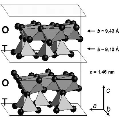
Asbestos chrysotile (Mg3Si2O5OH4) structure consists of wrapped sheets composed of layers of Si-centered tetrahedra in a pseudo-hexagonal network joined to layers of octahedral Mg hydroxides (Fig. 1).
Fig. 1. A prospective view of OT layers in the chrysotile structure. Mg environment is represented by octahedral, Silicon environment bytetrahedral. Different lengths along the b-axis for O-T layer is reported.
[Reproduced by permission of G. Falini, E. Foresti, M. Gazzano, A.F. Gualtieri, M. Leoni, I.G. Lesci, and N. Roveri: Tabularshapedstoichiometric chrysotile nanocristals. Chem.-Eur. J. 10, 3043 (2004).]
Layers are concentrically or spirally wound preferentially around the x-axis (clinochrysotile and orthochrysotile), and seldom around the y-axis (parachrysotile), into a tubular structure of about 22– 27 nm in diameter. The rolls possess a hollow core of about 7 nm, due to the mismatch along the b direction of the smaller layer dimension of the SiO4 sheet with respect to the Mg(OH)2 layer (Figs. 2 and 3).
Fig. 2
These fibers are contaminated with trace metals such as Al, Ni, and Fe, which can be replaced with Mg and Si, usually developing structural defects [122]. This heterogeneity can be invoked to explain the different chrysotile crystal morphologies observed: hollow cylinders, non-hollowed cylinders, tube-in-tube fibers, conically wrapped fibers, cone-in-cone-shaped concentric spiral structures, and multispiral structures (Fig.3) [123].
Fig. 3. Natural chrysotile nanocristals
The high degree of heterogeneity in chrysotile strongly affects the biological–mineral system interactions, which has been widely investigated to single out the causes of health hazards associated with asbestos fibers [124,125]. Actually, several investigations have been reported that asbestos toxicity may be related to the production of reactive oxygen species (ROS) and other free-radical species, even if genetic factors can also be invoked [126-128].
The capability of asbestos fibers to enhance ROS production is believed to depend both on their chemistry and their morphology [129.130]. In addition, their physicochemical properties are also responsible for their solubility, biodurability, and biopersistence [131]. Fiber properties, related to their cytotoxicity [132,133] and mutagenic response [134], are strongly affected by the surface chemical features, which are fully responsible for the interaction with the biological environment. Natural chrysotile, formed by densely packed bundles of multiwalled hollow nanotubes, is a mineral very suitable for nanowires (NW) preparation when their inner nanometer-sized cavity is filled with a proper material. Bundles of chrysotile nanotubes can then behave as host systems, where their large interchannel separation is actually expected to prevent the interaction between individual guest nanoparticles and act as a confining barrier.
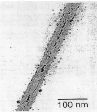
Chrysotile nanotubes have been filled with molten metals such as Hg, Pb (Fig.4), Sn, semimetals, Bi, Te, Se, and with semiconductor materials such as InSb, CdSe, GaAs, and InP using both high-pressure techniques [135] and metal-organic chemical vapor deposition (MOCVD) [136,137]
Fig. 4. TEM image of an isolated mineral chrysotile nanotube partially filled with lead. [Reproduced by permission of C. Metraux, B. Grobety, and P. Ulmer: Filling of chrysotile nanotubes with metals. J. Mater. Res. 17(5), 1129 (2002).]
A variety of semiconducting ultrathin filaments, real quantum wires, inside mineral chrysotile nanotubes have been prepared by means of injection of molten semiconducting materials into the asbestos channels.
CdSe and GaAs have been synthesized inside the channels of mineral chrysotile nanotubes [138,139].
Recently, hydrothermal synthesis of chrysotile has been utilized primarily for the purpose of obtaining pure chrysotile nanocrystals as a reference standard for ongoing investigation of asbestos cytotoxicity and carcinogenicity. A second aim, but not less important, is their use as new synthetic inorganic nanotubes with high potentiality for application in the developing field of nanotechnology.
The optimum hydrothermal conditions to synthesize chrysotile nanocrystals as a single phase are reported to be 300 °C, 82 atm, using SiO2 mesophase MCM41 previously synthesized according to Kresge et al. [140] and MgCl2 as a starting material, Si/Mg molar ratio equal to 0.66, pH 13, reaction time longer than 24 h [141]. The high purity of the synthesized chrysotile nanocrystals can be evinced by microcalorimetric and spectroscopic analysis. DTA and TGA curves do not show any of the additional peaks or bands usually assigned to impurities associated with mineral chrysotile. TEM images of stoichiometric synthetic chrysotile nanocrystals with single-tube morphology is reported in Fig. 5(a), witnessing a high structural and morphological uniformity. Figure 5(b) reports
a selected area electron diffracting (SAED) image of a chrysotile nanocrystal showing the typical split of the diffraction spots due to the cylindrical crystal lattice, resembling what was observed by Yada in 1967 in a mineral sample [142]. The powder diffraction refinement of the nanochrysotile structure was obtained with a model composed of a statistical assembly of six layers, a reasonable approximation of the cylindrical stacking with random disorder along the b-axis. Hydrogen atoms were also included for completeness but not refined; their positions were adapted from those refined for lizardite, the planar analogue of chrysotile [143]. The availability of pure synthetic crystalline crysotile nanocrystals allowed to refine the structural and microstructural parameters using a modified version of DIFFaX software[144]. The new unit cell parameters for the ideal orthogonal layer are: a _ 0.5330(1), b _ 0.9220(1), c_0.73508(9) nm [145]. The structure has been refined also by means of General Structure Analysis System (GSAS) anisotropic broadening parameters along the b-axis, by using the stacking fault model, improving the previous model used for the quantitative phase analysis with the Rietveld method on a natural and obviously impure standard sample [146]. The search for the best space group indicates a Cc space group with c _1.469 nm, which corresponds to an OT layer thickness of 0.734 nm. The present result proves extremely useful in that it fills the gap opened in the literature, since Wicks demonstrated the I inconsistency of chrysotile space group [147]. Transmission electron microscopy analysis was carried out on synthetic chrysotile nanocrystals, showing the cylindrical shape of many microns in length. The central area that runs longitudinally along the crystal indicates that the crystal core is void or containing low electron dense material. The hollow tubular morphology of a typical single crystal is clearly shown in Fig. 5, top, where three tubular subunits are concentrically arranged in a telescopic form.
Fig. 5 (top) TEM image of a single tubular chrysotile nanocrystal;
(bottom) graphical representation of the nanocrystals including dimensions in nm. [Reproduced by permission of G. Falini, E. Foresti, M.
Gazzano, A.F. Gualtieri, M. Leoni, I.G. Lesci, and N. Roveri: Tabularshaped stoichiometric chrysotile nanocristals. Chem.-Eur. J. 10, 3043
A value of 7 ± 1 nm for the central hole diameter as well as for the average wall thickness was determined by TEM. Atomic force microscopy (AFM) imaging has been used to evaluate the mean outer diameter of single, double, and triple tubes concentrically arranged as 21 ± 1, 35 ± 1, and 49 ± 1 nm respectively [148]. A detailed investigation into hydrothermal reaction mechanism leading to chrysotile nanotubes was performed by [149]. They followed the reaction products after using different combinations of reagents, such as MgO, SiO2, MgSiO3, and Mg3[Si4O10](OH)2, at pH >13 and several temperatures between 100 and 450 °C and observed that chrysotile nanotubes formed most quickly in the range of 350–400 °C using SiO2 and MgO as reactants. In addition, compounds with a layered structure are the first step towards the formation of Mg3Si2O5(OH)4 nanotubes under hydrothermal conditions.
The opportunity to have geomimetic sybthetic chrysotyle nanotubes allowed to utilize these inorganic nanotubes to be filled with different metallic nanoparticles.
Biomimetic hydroxyapatite nanocrystals as carriers for Au/Ag alloy anticancer particles
Control at the nano-scale is a fundamental concern in the production of biomimetic materials, particularly in the case of bone substitutes where the chemical and biological properties are very much size dependent. In fact, as the mineral phase of bone is constituted of carbonated
hydroxyapatite (HA) nano-crystals, biomimetic calcium phosphates need to be synthesized with similar nanoscale dimensions, as well as with properties such as low crystallinity,
non-stoichiometric composition, crystalline disorder, and presence of carbonate ions in the crystal lattice. On the other hand, nanoparticles with unique optical properties, facile surface chemistry and appropriate size scale are generating much enthusiasm in nanomedicine.
Noble metal, especially Au nanoparticles has immense potential for both cancer diagnosis and therapy. In the first case in fact, due to the phenomena of the surface plasmon resonance the adsorption and scattering cross-sections of Au nano-particles are significantly superior to the absorbing and fluorescent dyes conventionally used in molecular biology and nanomedicine. In this way conjugation of Au nano-particles to ligands specifically targeted to biomarkers on cancer cells allows molecular specific imaging and detection of cancer. On the other hand, Au nanoparticles efficiently convert the strongly adsorbed light into localized heat, which can be exploited for the selective laser photothermal therapy of cancer.
According to the about, metal nanoparticles-HA nanocrystals composites should have tremendous potential in novel methods for therapy of cancer [150] and represent innovative approach to fine-tune cellular response of implanted nano-biomaterials by environmental stimuli (above all light).
In this work Au-Ag alloy nano-particles have been conjugated to HA nanocrystals trough mercapto-undecanoic acid a linker.
It worth notice that nanomaterials exhibit multi-functional properties if building blocks have been combined through structural and morphological integration at the nanoscale. As a consequence of it, the structural organization of the resulting composite need to be controlled.
Finally, the formation of hydroxyapatite/metal nano-particles assembly opens fascinating perspectives for the biological implication derived from conferring conductive properties to calcium phosphates nanocrystals.
Exsperimental
Reagents and solvents
1-butanethiol (Fluka; 97%), CuCl2 (Aldrich); 4-methylbenzenethiol (Aldrich, 98%) LiBr (Riedel de Haen, pure grade); ascorbic acid (Sigma, 20-200 mesh); lithium chloride (Fluka, 99%); tetrabutylammonium bromide (Fluka, 98%); sodium borohydride (Sigma Aldrich, 98%);
dichloromethane (Sigma-Aldrich, pure grade); methanol (Fluka, 99,8%); diglyme (Aldrich, 99%); Tetrahydrofurane (Riedel de Haen; 99%).
Synthesis of surface protected metallic nanoparticles:
The surface protected metallic nanoparticles were synthesized in a two phase system as reported in literature [151,152]. Reagents and solvents used are listed below: AgNO3(>_99,0%;Acros Organics), AuCl3.HCl(Aldrich; concentred hydrochloric acid 30% solution), KBr(Sigma-Aldrich; 99,5%) dacanethiol (Aldrich; 96%) butanethiol (Aldrich; >_99%), 4-methylbenzenethiol(Aldrich;98%), NaBH4(Aldrich;>_98%); toluene (Sigma-Aldrich; 99,5%), MilliQ-water and tethraoctilammonium bromide (TOABr) (aldrich;98%).
Synthesis of surface protected Ag nanoparticles
Solutions of silver nitrate and potassium bromide were mixed in a 250 ml three naked round bottom
HA
nanocrystals Spacer
Au-Ag alloy nanoparticles [Ca10(PO4)6(OH)2]
respectively. After addition of TOABr (20 ml of a 0,150 molar solution in toluene) under continuous stirring the precipitate in the water phase disappeared in about 30 minutes indicating the complete removal of the metallic salts from the water to the organic phase. 30 ml of thiol solution in toluene (0,0410 molar in the case of 4-methylbenzene thiol and 0,1 molar in that of butanethiol and decanethiol) were then poured in the flask followed by 15 ml of a 0,70 molar solution of NaBH4 in MilliQ-water.
After three hours of continuous stirring, the organic phase was separated from the water phase, repeatedly washed with Milli-Q water and dried with Na2SO4. The toluene was removed under vacuum conditions, the residue redispersed in few ml of dichloromethane and finally dropped in methanol cooled at -10C°. The black brown precipitate was repeatedly washed with cooled methanol and redispersed in 40 ml of dichloromethane (mother suspension).
Synthesis of surface protected Au nanoparticles
Hydrogen tetrachloroaurate (30 ml of a 0,05 molar solution) was mixed with TOABr (20 ml of a 0,150 molar solution in toluene) in a 250 ml three naked round bottom flask under continuous stirring. The orange color in the water phase disappeared in about 30 minutes indicating the complete removal of the auric salt from the water to the organic phase. 30 ml of thiol solution in toluene (0,0410 molar in the case of 4-methylbenzene thiol and 0,1 molar in that of butanethiol and decanethiol) were then poured in the flask followed by 15 ml of a 0,70 molar solution of NaBH4 in MilliQ-water. After three hours of continuous stirring, the organic phase was separated from the water phase, repeatedly washed with Milli-Q water and dried with Na2SO4. The toluene was removed under vacuum conditions, the residue redispersed in few ml of dichloromethane and finally dropped in methanol cooled at -10C°. The black brown precipitate was repeatedly washed with cooled methanol and redispersed in 40 ml of dichloromethane (mother suspension).
Synthesis of surface protected Ag/Au nanoparticles
Solutions of silver nitrate, hydrogen tetrachloroaurate, and potassium bromide were mixed in a 250 ml three naked round bottom flask. The overall metal salt concentration, and the the final volume were 0,05 mmoles/ml, and 30 ml respectively with silver to gold ratios of 1/0, 4/1;1/4; 0/1 in each synthesis. After addition of TOABr (20 ml of a 0,150 molar solution in toluene) under continuous stirring the precipitate in the water phase disappeared in about 30 minutes indicating the complete removal of the metallic salts from the water to the organic phase. 30 ml of thiol solution in toluene
(0,0410 molar in the case of 4-methylbenzene thiol and 0,1 molar in that of butanethiol and decanethiol) were then poured in the flask followed by 15 ml of a 0,70 molar solution of NaBH4 in MilliQ-water.
After three hours of continuous stirring, the organic phase was separated from the water phase, repeatedly washed with Milli-Q water and dried with Na2SO4. The toluene was removed under vacuum conditions, the residue redispersed in few ml of dichloromethane and finally dropped in methanol cooled at -10°C. The black brown precipitate was repeatedly washed with cooled methanol and redispersed in 40 ml of dichloromethane (mother suspension).
Preparation of 11mercaptoundecanoic acid protected Au-Ag nanoparticles
The 1-buthanethiol surface protected Au4/Ag1 nanoparticles isolated as a brown precipitate, were re-dispersed in tetrahydrofuran (30 ml) using TOABr (427,85 mg; 0,8 mille-moles) as solubilizer and the 11-mercaptoundecanoic acid (419,57g; 0,50 mille-moles) to replace the 1 buthanethiol on the nanoparticle surface .
The reaction mixture was kept under continuous stirring in a 100 ml round bottomed flask for 90 minutes. The residue obtained evaporating the solvent, was re-dispersed in 10 ml of methanol, added to a solution of Na2CO3 deca-hydrate (5g; 0,017 moles in 40 ml of milliq water) and stirred for two hours. The obtained dispersion has been washed with 60*3 ml of diethylether. The precipitate formed in the water phase was collected by centrifugation.
Synthesis of CuBr
Ascorbic acid (25 ml of a 0,52 molar water solution) was added dropwise to a water solution of lithium bromide(4, 37g; 0,05 moles) and cupric chloride (4,257g; 0,025 moles) under continuous stirring and nitrogen flux. The white precipitate (sometimes pale yellow) was allowed to stir for half an hour. It was separated by centrifugation at 3000 rpm, washed with water, methanol and finally with diethylether. After dryng under vacuum it was stored in a dessiccator.
Synthesis of butane thiolate protected Cu nanoparticles (I method)
Cuprous bromide (429 mg; 3,0mmoli) was solubilized by means of tetrabuthylammonium bromide TBABr (1,100g; 3,4 mmoli) in 120ml of tethrahydrofuran together with 960 ml (9 mmoles) of 1-butanethiol. The formation of a precipitate was not observed. After ten minutes 45 ml of a LiCl-NaBH4 diglyme suspension* was dropped in the reaction mixture with a dropping time of about 5 minutes under continuous stirring and nitrogen flux. The colour of the solution changed rapidly from pale yellow-green to dark brown and a white precipitate became to form. The reaction mixture
was allowed to stir for 50 minutes and then transferred in a separating funnel filled with 200 ml of dichloromethane. The organic phase was washed with 2 l of Milli-Q water and dried for an hour with Na2SO4., The residue after solvent removal at 30C°, was redissolved in 5ml of dichloromethane and dropped in 50 ml of methanol to remove tetrabutylammonium bromide and the excess of thiol l. The brown precipitate M formed, was collected, washed repeatedly with methanol and redispersed in dichlorometane. The pale brown compound C that was observed to form owing to this operation was washed repeatedly with dichloromethane and desiccated on P2O5. A dark brown residue R was collected after solvent removal from the liquid phase. Both the products were desiccated on P2O5 and stored under inert atmosphere.
The reducing mixture was prepeared by gently adding *NaBH4 (1,14g; 30 mmoles) to 45 ml of a LiCl(1,95g; 46 moles) dispersion in diglyme under nitrogen flux. The synthesis has been also repeated in ambient conditions (without nitrogen flux), but variations in the reaction product physicochemical properties were not detected with respect to those synthesized under nitrogen flux. All the products were desiccated on P2O5 and stored in protected ambient.
Synthesis of butane thiolate protected Cu nanoparticles (II method)
Cuprous bromide (280mg; 1,9 mmoles) and tetrabutylammonium bromide (680mg; 2,1 mmoles) were finely grinded in an agate mortar. The mixture was poured in 80 ml of dichlorometane. After 20 to 30 minutes the solubilization was completed and 20 ml of a 0,25 molar butanehiol solution in the same solvent were added in the flask. The reaction mixture was kept in nitrogen flux under continuous stirring for 30 minutes without precipitate formation. On dropping 20ml of a NaBH4 1 molar water solution in the reaction mixture, the copper-thiol adduct reduction took place. After three hours the organic phase was repeatedly washed with milli-Q water and dried on Na2SO4. The residue after solvent removal at 30°C, was redissolved in 5ml of dichloromethane and dropped in 50 ml of methanol to remove tetrabutylammonium bromide and the excess of thiol. The brown precipitate M formed, was collected, washed repeatedly with methanol and redispersed in dichlorometane. The pale brown compound C that was observed to form owing to this operation was washed repeatedly with dichloromethane and desiccated on P2O5. A dark brown residue (R) was collected after solvent removal from the liquid phase. Both the products were desiccated on P2O5 and stored under inert atmosphere. The synthesis has been also repeated in ambient conditions (without nitrogen flux), but variations in the reaction product physicochemical properties were not detected with respect to those synthesized under nitrogen flux. All the products were desiccated on P2O5 and stored in protected ambient.
Synthesis of butane thiolate protected Cu nanoparticles (III method)
Copper(II) chloride dihydrate (664mg; 4,0 mmoli) tetrabutylammonium bromide (3,10g; 9,6 mmoles) and lithium bromide(684mg; 8,0 mmoles) were finely grinded in an agate mortar. The mixture was gently poured in 160 ml of dichlorometane. On adding 40 ml of a 0,22 molar butanethiolthiol solution in the same solvent, the reaction mixture changed from dark violet to colourless. The reaction mixture was kept in nitrogen flux under continuous stirring for 30 minutes without precipitate formation. On dropping 40ml of a NaBH4 1 molar water solution in the reaction mixture, the copper-thiol adduct reduction took place. After three hours the organic phase was repeatedly washed with milli-Q water and dried on Na2SO4. The residue after solvent removal at 30C°, was redissolved in 5ml of dichloromethane and dropped in 50 ml of methanol to remove tetrabutylammonium bromide and the excess of thiol. The brown precipitate M formed, was collected, washed repeatedly with methanol and redispersed in dichlorometane The pale brown compound C that was observed to form owing to this operation was washed repeatedly with dichloromethane and desiccated on P2O5. A dark brown residue (R) was collected after solvent removal from the liquid phase. Both the products were desiccated on P2O5 and stored under inert atmosphere. The synthesis has been also repeated in ambient conditions (without nitrogen flux), but variations in the reaction product physicochemical properties were not detected with respect to those synthesized under nitrogen flux. All the products were desiccated on P2O5 and stored in protected ambient.
Synthesis of 4-methylbenzenethiol protected Cu nanoparticles (I method)
Cuprous bromide (429 mg; 3,0mmoli) was solubilized by means of tetrabuthylammonium bromide TBABr (1,100g; 3,4 mmoli) in 120ml of tethrahydrofuran together with 960 µl( 9 mmoles )of 4methylbenzenethiol. The formation of a precipitate was not observed. After ten minutes 45 ml of a LiCl-NaBH4 diglyme suspension* was rapidly added to the reaction mixture under continuous stirring and nitrogen flux. The colour of the solution changed rapidly from pale yellow green to yellow orange and a white precipitate became to form. The reaction mixture was allowed to stir for 60 minutes and then transferred in a separating funnel filled with 200 ml of dichloromethane. The organic phase was washed with 2 l of Milli-Q water and dried for an hour with Na2SO4. A pale yellow crystalline solid was observed to form removing the solvent. It was collected, washed repeatedly with methanol and desiccated on P2O5.
*The reducing mixture was prepeared by gently adding NaBH4 (1,14g; 30 mmoles) to 45 ml of a LiCl (1,95g; 46 moles) dispersion in diglyme under nitrogen flux. The synthesis has been also repeated in ambient conditions (without nitrogen flux) but variations in the reaction product
physicochemical properties were not detected with respect to those synthesized under nitrogen flux.
Synthesis of 4-methylbenzenethiol protected Cu nanoparticles (II method)
Cuprous bromide (280md; 1,95 mmoli) and tetrabutylammonium bromide were finely grinded in an agate mortar. The mixture was poured in 80 ml of dichlorometane. After 20 to 30 minutes the solubilization was completed and 20 ml of a 0,25 molar 4methylbenzenethiol solution in the same solvent were added in the flask. The reaction mixture was kept in nitrogen flux under continuous stirring for 30 minutes without precipitate formation. On dropping 20ml of a NaBH4 1 molar water solution in the reaction mixture, the copper-thiol adduct reduction took place. After three hours the organic phase was repeatedly washed with milli-Q water and dried on Na2SO4. The residue after solvent removal at 30°C, was redissolved in 5ml of dichloromethane and dropped in 50 ml of methanol to remove tetrabutylammonium bromide and the excess of thiol. A brown precipitate formed. It was collected, washed repeatedly with methanol and desiccated on P2O5. The synthesis has been also repeated in ambient conditions (without nitrogen flux) but variations in the reaction product physicochemical properties were not detected with respect to those synthesized under nitrogen flux.
Synthesis of 4-methylbenzenethiol protected Cu nanoparticles (III method)
Copper(II) chloride dihydrate (664mg; 4,0 mmoles) tetrabutylammonium bromide (3,10g; 9,6 mmoles) and lithium bromide(684mg; 8,0 mmoles) were finely grinded in an agate mortar. The mixture was gently poured in 160 ml of dichlorometane. On adding 40 ml of a 0,22 molar 4 methylbenzenethiol solution in the same solvent, the reaction mixture changed from dark violet to colourless. The reaction mixture was kept in nitrogen flux under continuous stirring for 30 minutes without precipitate formation. On dropping 40ml of a NaBH4 1 molar water solution in the reaction mixture, the copper-thiol adduct reduction took place. After three hours the organic phase was repeatedly washed with milli-Q water and dried on Na2SO4. The residue after solvent removal at 30°C, was redissolved in 5ml of dichloromethane and dropped in 50 ml of methanol to remove tetrabutylammonium bromide and the excess of thiol. A brown precipitate formed. It was collected, washed repeatedly with methanol and desiccated on P2O5. The synthesis has been also repeated in ambient conditions (without nitrogen flux), but variations in the reaction product physicochemical properties were not detected with respect to those synthesized under nitrogen flux.
A suspension of selenous oxide (0,550g; 5 mmoles) in 25ml of formic acid was kept to boil by means of an oil bath under nitrogen flux.
The initially clear suspension of powdered selenous dioxide in formic acid turned to a dark grey- violet colour while a brown grey precipitate of crystalline selenium was observed to form according to the reaction reported below.
2HC(O)OH + SeO2 Æ 2CO 2 + Se0 + H2O
Heating was arrested after few minutes to limit the formation of dark brown crystalline selenium. The reaction mixture was than allowed to cool at room temperature.
25 ml of a 0,1 molar solution of phenol in formic acid were added to the reaction mixture that was heated again and kept under reflux for an hour. The dark brown precipitate formed A, was separated by centrifugation at 5000 rpm from the liquid phase and repeatedly washed with formic acid, methanol and diethyl ether. The liquid phase was centrifuged at 8000 rpm for 20 minutes and dropped in water kept at 0°C. The dark brown precipitate B formed was repeatedly washed with water, lyophilized and dried on P2O5.
A water bath was also used to heat the SeO2suspension in formic acid before and after the addition
of phenol. A reaction temperature of 96°C was reached without reflux conditions. An increased amount of amorphous selenium was observed to form in this case.
Synthesis of phenol protected selenium nanoparticles (II method)
To increase the phenol protected selenium nanoparticles yield, selenous oxide and phenol in the same molar ratios of the former method were mixed at the same time in formic acid and the reaction mixture was kept to reflux for one hour under nitrogen flux. The dark brown precipitate formed A, was separated by centrifugation at 5000 rpm from the liquid phase and repeatedly washed with formic acid, methanol and diethyl ether. The liquid phase was centrifuged at 8000 rpm for 20 minutes and dropped in water kept at 0°C. The dark brown precipitate B formed was repeatedly washed with water, lyophilized and dried on P2O5.
Synthesis of phloroglucinol protected selenium nanoparticles
A phloroglucinol solution was obtained at room temperature dissolving phloroglucinol dehydrate (FLUKA 98% ) in 50 ml of ethanol obtaining a 0,40 M concentration. Selenous acid solution was prepared dissolving Selenium dioxide (SeO2 Aldrich 98%) previously finely hand-ground in 50 ml of Milli-Q water obtaining a 0,20 M concentration. These two solutions were mixed under stirring
in a 250 ml flask in N2 atmosphere at 84 °C for one hour. The solution turned from colourless to pale yellow, to orange and finally to a persistent brown, indicating that the reaction (1) was complete. The solution was taken to reach room temperature and rotavapored up to ¼ of the initial volume. The orange-red precipitate, probably due to the reaction 2), was removed by centrifugation, repeatedly washed with Milli-Q distilled water, lyophilized, dried on P2O5 and stored for two months. No changes in the colour were noticed in aged samples.
C6H6O3
nH2SeO3 ---Æ Sen (C6H5O3)m
Synthesys of 1 heptane protected Si nanoparticles
Silicon tetrachloride (92 µliters; 0,8 mmoles) along with of Tetrabutilammonium bromide (1,5 g; 2,70 mmoles) were solubilized in 100 ml of anhydrous toluene. 2ml of a 1molar LiAlH4 solution in THF were added to the mixture that was allowed to stirr for three hours. After quenching the residual LiAlH4 with 20ml of methanol, 100µl of a 0,1 M H2PtCl6·6H2O solution along with 2ml of 1-heptene (14,2 mmoles) were added. After three hours of continuous stirring, the solvent was removed under vacuum conditions.
The residue was resolubilized in 20ml of hexane and sonicated for 20 minutes. The solution was filtered and washed with methylformammide (100ml) and milliq water.
Synthesis of Hydroxyapatite nanocrystals
Hydroxyapatite nanocrystals (HA) were prepared from an aqueous suspension of Ca(OH)2 (1.35 M, 1 L) by slow addition of aqueous H3PO4 (1.26 M, 600 ml), at 95 °C. The reaction mixture was kept under stirring at 95 °C, for 24 hours, then stirring was suspended and the mixture left standing for 2 hours to allow deposition of the inorganic phase. This was isolated by filtration of the mother liquor, repeatedly washed with water and freeze-dried at -60 °C under vacuum (3 mbar) for 24 h. In all the above syntheses, the nanocrystals powder fraction having granular dimensions ranging from 100 to 150 µm was selected for the study.
Synthesis of chrysothyle nanotubes
The chrysothyle nanotubes were synthesized according to a method devised in our laboratory [148] using amorphous silica aerosil 380, Magnesium chloride hexahydrate (Sigma Aldrich 99,0-102,02%), NaOH (Riedel de Haen 99%) and Milli-Q water.
The hydrothermal synthesis of chrysothyle nanotubes was performed by means of a 4382 Parr reactor equipped with a 300 ml T316SS steel vessel.
Namely a gel was obtained mixing amorphous silica (aerosil 380: 4,2730g; 0,07 moles) and Magnesium chloride hexahydrate (21,2636 g; 0,105 moles) in 60 ml of Milli-Q water. On adding 240 ml of a 0,70 molar solution of NaOH, the pH was increased up to 9,8 and adjusted at about 13 with 21 ml of a 5,1 molar solution of NaOH. The gel was left to stirr for 80 minutes and 200 ml of it were poured in the reactor vessel. The set out reaction temperature and the pressure reached were of 300 °C and 82 atm respectively. After about 24 hours the heating was stopped and the product allowed to cool at room temperature, was washed with milli-Q water and dried at about 120 °C with an IR lamp.
Chrysotile nanotubes filling method
The nanotubes (30 mg in 2 ml of dichloromethane) were kept under reduced pressure at 0°C and the solvent periodically reloaded. In the following step nanotubes were kept overnight at -20°C. The dichloromethane was then removed with a pipette and replaced with 2 ml of nanoparticle suspention to soak the nanotubes and replaced daily for a week. The solvent was then partially removed under vacuum conditions under reduced pressure and replaced with fresh nanoparticle suspension for two times. Finally the nanotubes were carefully washed with dicloromethane and dried under inert atmosphere. All the nanoparticle suspension amounts used during the filling process were previously centrifugated at 8000 rpm for about 15 minutes.
In another experiment 4-methylbenzenethiol surface protected Silver nanoparticles were successfully used to fill the crysotyle nanotube hollows. In this case nanotubes were added to a dichloromethane filled shlenck tube and sonicated for 2 or 3 seconds repeatedly five or six times to break the molecoular membrane between the outer liquid phase and the empty nanotube cavity so allowing the solvent to occupy the hollows and at the same time to prevent the nanotube disruption due to the ultrasound effect. The nanotube suspension was stirred for one hour and preserved in dichloromethane.
4 ml of nanotube suspension were collected in a screw cap tube. The solvent was gently removed with a pipette after decantation and replaced with a freshly prepeared nanoparticle suspension previously centrifugated at 10000 rpm to remove possible nanoparticle clusters.
The tube content was gently sonicated again and kept to slowly stir for three days.
The liquid phase was carefully removed with a pipette after decantation and the nanotubes washed repeatedly with dichloromethane.
UV-vis absorption spectrophotometry
Optical spectra were recorded with λ6 and λ16 Perkin-Elmer spectrometers in a single beam mode using quartz cuvettes (1cm-path length) filled with metallic nanoparticles suspensions obtained diluting a centrifugated (8000 r. p. m. for 15 minutes) amount of a mother solution (see metallic nanoparticle synthesis section (paragraph)).
Transmission Electron Microscopy (TEM)
The TEM images were obtained with an electron transmission microscope JEM 2010 located at the Modena University C.I.G.S. (Centro Interdisciplinare Grandi Strumenti ). The instrument was equipped with a GATAN 694 SLOW SCAN CCD camera and a LINK INCA 100 microanalysis device.
The sample was prepeared dispersing in Milli-Q water and sonicating for 60 seconds chrysothyle nanotubes previously grinded gently in an agate mortar. Droplets of the dispersion were allowed to evaporate on copper-chrome microgrids coated with a graphite layer. The sample was dried at room temperature or by means of an IR lamp.
An elemental analysis of the sample was carried out using energy dispersive spectroscopy (EDS) technics.
X-ray diffraction investigation
X-ray powder patterns were collected by means of a Philips 1050/81-PW1710 powder diffractometer. The instrument equipped with a graphite monochromator on the diffracted beam and a proportional counter, operated at a voltage and current intensity of 40 kV and 40 mA respectively generating a CuKα radiation with a λ of 1,541873 Å.
The steps were of 0,03° with a time per step of 1 second while the 2Ө range spanned from 10° to 80°. APD, PDF2 programs and Xpert software were used for the diffractogram visualisation and phase identification.
FT-IR Spectroscopy
The infrared spectra were recorded under nitrogen atmosphere from 4000 to 400 cm–1 with 2 cm–1 resolution using a NICOLET 210 FT-IR spectrometer. Other settings include an 8 mm aperture, 32
scans, velocity of 10 kHz, DLATGS detector and a 3-term Blackman-Harris apodization function. KBr pellets were prepared grinding chrisothyle nanotubes (1-2 mg) and FTIR grade KBR (300-350 mg) in an agate mortar. 100 mg of the solid mixture were pressed at 8 tons by means of a Spekak press.
Thermogravimetric and Differential scanning calorimetric analysis
Thermogravimetric (TGA) measurements were carried out using a ThermalAnalysis-SDT Q600 (TA Instruments, New Castle, DE, USA). Heating was performed in nitrogen flow (100 ml min-1) using an alumina sample holder at a rate of 5 °C/min (10 °C/min in the DTA experiment) from room temperature to 1200 °C. The weight of the samples was approximately 10 mg.
Results and discussion
1) Surface protected metallic nanoparticles filling chrysotile nanotubes
The chrysothyle nanotubes were synthesized according to a method devised in our laboratory and described in the exsperimental section.
Morphological characterization
The morphological analysis of inorganic chrysothyle performed by TEM microscopy reveals the nanotubular nature of the reaction product.
Namely the several hundreds nanometers long nanocrystals present inner and external diameters of 7 and 21nm respectively and are caracterized by high structural and morphological uniformity (Fig.6 a, b).
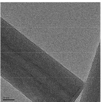
A closer inspection of the synthetic chrysotyle TEM images reveals that the structure of the nanotube walls is made of about ten concentrical layers (Fig. 7).
An EDS (Energy Dispersive Spectroscopy) analysis performed by the same TEM microscope confirms the presence of only Mg, Si and O in the synthetic nanotubes according to the chrysothyle formula Mg3Si2O5(OH)4 (Fig. 8).
Fig. 6. a) TEM micrograph of the synthetic chrysotyle nanotubes. b) single tubular chrysotile nanocrystal electron diffraction pattern
Figure 8. EDS (Energy Dispersive Spectroscopy) analysis confirming the chrysothyle formula Mg3Si2O5(OH)4 (Fig. 7).
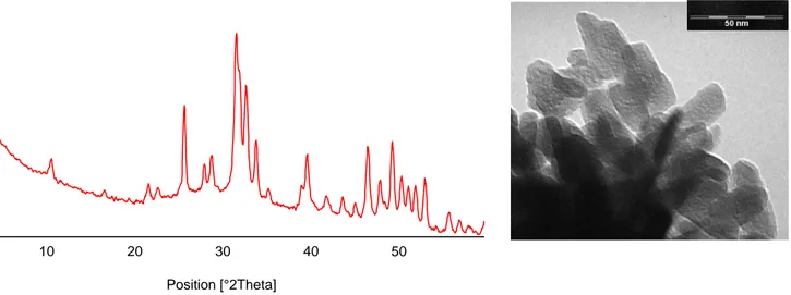
X-ray diffraction investigation
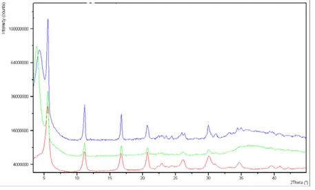
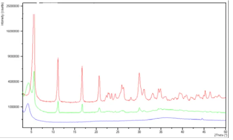
The powder x-ray diffraction pattern of the synthesized chrysotile displays the characteristic reflections of high crystalline chrysotile, as unique crystalline phase (Fig. 9).
Fig. 9x-ray diffraction pattern of the synthesized chrysotile
An attempt was made to determine the cell parameters of the synthetic chrysothyle using the coordinates of an ideal trioctahedral OT unit in the unit cell with c=1.46 nm, space group Cc, as input for a Rietveld structure refinement in GSAS [153].
The refined unit cell parameters were a=0.5340(1) nm, b=0.9241(1) nm, c=1.4689(2) nm, β=93.66(3)°. Although the validity of the structure refinement is limited, the result is very useful because it proves that the refined pattern belongs to a pure chrysotile sample.
Fig.10 TGA-DTA thermogram of the chrysothyle crystals
Thermogravimetric and Differential scanning calorimetric analysis
The TGA-DTA thermogram of the chrysothyle crystals show an endothermic area at 650°C corresponding to a dehydroxylation reaction and a sharp exothermic peak at 810°C which indicates the crystallization of the remaining amorphous anhydrous materials (the so-called meta-chrysotile Mg3Si2O7) into forsterite (Mg2SiO4) (Fig. 10) [154].
Synthetic chrysotile crystals are of high omogeneity and purity as can be recognized from the lack of additional peaks (observed in the mineral chrysotile fibers thermal analyses) together with the sharpness of the transformation to forsterite and silica.
FT-IR analysis
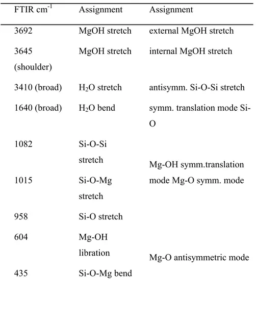
The FTIR spectrum of the synthetic chrysotile nanocrystals is reported in Fig. 11.
(b)
(a)
Figure 11 FTIR absorption spectrum of synthetic chrysotile fibers in the range 4000-400 cm-1. The main
absorption peaks are marked.
The main band frequencies listed in Table 1 agree with the literature data and confirm the absence of other phases.
FTIR cm-1 Assignment Assignment 3692 MgOH stretch external MgOH stretch 3645
(shoulder)
MgOH stretch internal MgOH stretch
3410 (broad) H2O stretch antisymm. Si-O-Si stretch 1640 (broad) H2O bend symm. translation mode
Si-O 1082 Si-O-Si stretch 1015 Si-O-Mg stretch 958 Si-O stretch Mg-OH symm.translation mode Mg-O symm. mode
604 Mg-OH libration
435 Si-O-Mg bend
Mg-O antisymmetric mode
The optical absorption spectra of the synthesized decanethiol capped Au, Ag and Au-Ag alloy nanoparticles dispersed in dichloromethane were recorded in the wavelength range of 250-700 nm and reported in Fig. 12. The spectra show an intense surface plasmon (SP) absorption band whose peak maximum position is directly related to the Au/Ag ratio in the nanoparticle core.
Fig. 12 Optical absorption spectrum, recorded in the 250-700 nm wavelength range, of synthesized decanethiol capped Au, Ag, Au4Ag1, Au1Ag4 nanoparticles.
Peak maxima at about 400 and 540 nanometers appear in the Gold and silver decanethiole surface protected nanoparticles respectively.
The maxima of the alloy clusters fall between and a red shift of the peaks with increased Au content is noted according with the literature data.
The position of the absorption maximum also depends on the nature of the molecules forming the monomolecular layer on the nanoparticle surface as can be seen comparing the absorption maximum positions in the spectra of the silver nanoparticles coated with decilmercaptane (about 400nm) (Fig.12) and buthilmercaptane (450nm) Fig13.
Fig13 Optical spectrum of butilmercaptane capped silver nanoparticles
A well defined absorption maximum is associated to a high degree of monodispersion for Au, Ag and AuAg alloy nanoparticles. This feature is prerequisite for the formation of ordered nanoparticle arrays filling the nanotube hollows and consequently for all their optical and electrical properties. For comparative purpose we report in fig 14 the optical spectrum of
Cisteine coated polidispersed Au nanoparticles synthesized in a one phase method as reported in literature.
Fig.14 Optical spectra of cisteine capped Ag, Au, Au-Ag alloy capped nanoparticles (Ag: yellow; Au1/Ag4: blue; Au4/Ag1: light blue; Au: pink).
0 0, 2 0, 4 0, 6 0, 8 1 1, 2 1, 4 1, 6 1, 8 300 350 400 450 500 550 600 650 700 750 800 Lunghezza d'onda (nm ) A sso rb an z a ( a .u .)
Fig. 15 TEM image of polydispersd cisterne capped Au nanoparticles
The morphology of the thiol surface protected metallic and bimetallic nanoparticles synthesized as reported in experimental section, has been investigated by TEM microscopy. The samples were prepared evaporating small drops of a particle dispersion in booth ethanol or water on a carbon coated copper grid.
In Fig. (16) is reported the TEM micrograph of the decilmercaptane surface coated Au4/Ag1 particles These reveal an approximately spherical shape of about 2 nm in diameter.
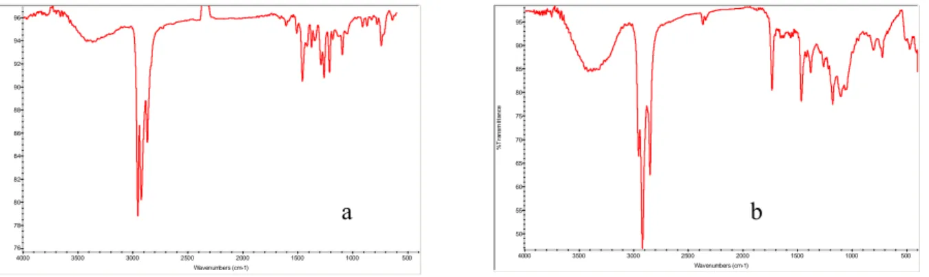
FT-IR Analysis
The FT-IR ATR absorption patterns of decilmercaptane surface protected Au4/Ag1 and buthilmercaptane surface protected Ag nanoparticles are reported in Fig. (17 a,b). These patterns show the typical absorption bands due to the alkilic chains surrounding the nanoparticle surface. Namely sharp and strong absorptions attributed to the symmetrical and asymmetrical stretching modes of the C—H bonds, can be found in the 3000—2800 cm-1 region. Bending and rocking vibrational modes of the C—H, C—C are responsible of the bands below 1500cm-1.
Fig. 17 FT-IR ATR absorption patterns of decilmercaptane surface protected Au4/Ag1 (a) and buthilmercaptane surface protected Ag nanoparticles (b)
Synthetic chrysotyle nanotubes filled with the previously prepared nanoparticles
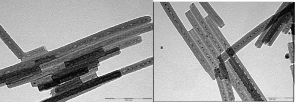
In Fig. (18) is reported the TEM micrograph of the synthetic chrysotyle nanotubes partially filled with the above mentioned nanoparticles.
Fig. 18. TEM micrograph of the synthetic chrysotyle nanotubes partially filled with decanethiol coated Au4/Ag1 nanoparticles.
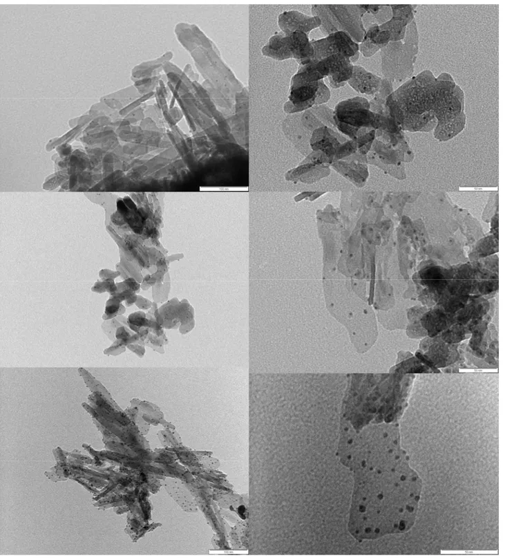
Synthetic crysotyle nanotubes were also filled with buthanethiole Surface coated silver nanoparticles with dimensions ranging between 2-3 nm (Fig. 19). And with 4-methylbenzenethiol surface coated silver nanoparticles (Fig 20)
76 78 80 82 84 86 88 90 92 94 96 %T ra n s m it ta n c e 500 1000 1500 2000 2500 3000 3500 4000 Wavenumbers (cm-1) a b 50 55 60 65 70 75 80 85 90 95 % T ra n s m itta n c e 500 1000 1500 2000 2500 3000 3500 4000 Wavenumbers (cm-1)
Fig. 19 TEM micrograph of butanethiol protected Ag nanoparticles partially filling synthetic chrysothyle hollows
Synthetic chrysotyle nanotubes filled also with 4-methylbenzenethiol protected silver nanoclustersare are reported in Fig. 20.
Fig. 20 TEM micrograph of 4-methylbenzenethiol protected Ag nanoparticles partially filling synthetic chrysothyle hollows.
In many cases, nanoparticles filling the nanotubes appear fused forming clusters separated each other by gaps probably because of gas bubbles due to the decomposition of the molecules on the nanoparticle surface during the morphology characterization by TEM.
The choose of the experimental conditions in the filling process was devoted to the nanotube integrity preservation.
Aprotic solvents were used to soak nanotubes instead of protic ones that in many cases decompose the octahedral magnesium hydroxide layer coating the external nanotube walls.
Hexane diethylether and tethrahydrofurane were discarded because nanoparticles tend to form clusters together greater in size than nanotube hollows in these solvents. Thetrahydrofuran particularly weakens the nanotube walls that are so easily disrupted under the electron beam of the