Alma Mater Studiorum – Università di Bologna
DOTTORATO DI RICERCA IN
SCIENZE ZOOTECNICHE
Ciclo XXIV
Settore Concorsuale di afferenza: AGR 18
TITOLO TESI
IMPORTANZA DELLA DIETA SULL’ATTIVAZIONE E SULLA
MATURAZIONE DEL CONTROLLO ALIMENTARE E DELLA
BARRIERA GASTROINTESTINALE NEL SUINETTO
---
DIET EFFECTS ON ACTIVATION AND MATURATION OF FEED
CONTROL OVER THE GASTROINTESTINAL DEFENCE
BARRIER IN PIGLETS
DAVIDE PRIORI
Presentata da:
___________________________________________
Coordinatore Dottorato
Relatore
Ch.ssimo prof. PAOLO BOSI Ch.ssimo prof. PAOLO BOSI
1
ABSTRACT
Weaning is an important and complex step involving many stresses that interfere deeply with feed intake, gastro-intestinal tract (GIT) development and adaptation to the weaning diet in young pigs. The health of the pig at weaning, its nutrition in the immediate post-weaning period, and the physical, microbiological and psychological environment are all factors that interact to determine food intake and subsequent growth. GIT disorders, infections and diarrhoea increase at the time of weaning, in fact pathogens such as enterotoxigenic Escherichia coli (ETEC) are major causes of mucosal damage in post-weaning disease contributing to diarrhoea in suckling and post-weaned pigs. The European ban in 2006 put on antibiotic growth promoters (AGP) has stimulated research on the mechanisms of GIT disorders and on nutritional approaches for preventing or reducing such disturbances avoiding AGPs. Concerning these aspects here are presented five studies based on the interplay among nutrition, genomic, immunity and physiology with the aim to clarify some of these problematic issues around weaning period in piglets. The first three evaluate the effects of diets threonine or tryptophan enriched on gut defence and health as possible alternatives to AGP in the gut. The fourth is focused on the possible immunological function related with the development of the stomach. The fifth is a pilot study on the gastric sensing and orexygenic signal given by fasting or re-feeding conditions. Although some results are controversial, it appears that both tryptophan and threonine supplementation in weaning diets have a preventive role in E.coli PWD and favorable effects in the gut especially in relation to ETEC susceptible genotype. While the stomach is believed as almost aseptic organ, it shows an immune activity related with the mucosal maturation. Moreover it shows an orexygenic role of both oxyntic mucosa and pyloric mucosa, and its possible relation with nutrient sensing stimuli.
2
Contents
ABSTRACT
1
SUMMARY AND BACKGROUND
4
THE GASTROINTESTINAL TRACT
6
Stomach
Wall structure Oxyntic glands Cardiac glands Pyloric glandsSmall intestine
Wall structure Villi and cryptsAbsorptive enterocytes Globlet cells
Enteroendocrine cells Paneth cells
FAE and M cells
Large intestine
AMINOACIDS
17
Amonoacid balance
Tryptophan
Threonine
WEANING, PDW
and GUT MICROBIOTA
24
Weaning
Post-weaning disease (PWD)
Colibacillosis
F4(K88) fimbiae
AGPS and ALTERNATIVES
29
DEFENCE and IMMUNITY
32
Mucins
Innate immunity
Adaptive immunity
Humoral immunity Cellular immunityToll-like receptors
TLR-4Immunity and Igs in piglets
IgA and pIgR
GALT
GUT SENSING
46
Taste percepition
Gastrointestinal sensing and GPCRs
Sweet and umami receptors Bitter receptors
Calcium-sensing receptor Sinal trasduction
OREXYGENIC SIGNAL
54
Ghrelin
Posttranslational modification system Distribuition
Finctions
Appetite regulation
3
CHAPTER 1
74
“The effect of a threonine-enriched diet on
the growth performance, health, immunity
and gastrointestinal functionality, of
weaning pigs susceptible or not to
Escherichia coli K88ac orally challenged with
this bacterial strain.”
CHAPTER 2
92
“Supplementary tryptophan downregulates
the expression of genes induced by the gut
microbiota in healthy weaned pigs
susceptible to Enterotoxigenic Escherichia
coli F4.”
CHAPTER 3
108
“Effect of susceptibility to enterotoxigenic
Escherichia coli F4 and of dietary
Tryptophan on gut microbiota variability
observed in healthy young pigs.”
CHAPTER 4
122
“Distribution and developmentally
regulated gene expression of polymeric
immunoglobulin receptor in the gastric
mucosa of pre and post weaning pigs.”
CHAPTER 5
134
“Effect of fasting and refeeding on the
expression of the complex of genes involved
in the gastric nutrient sensing and
orexigenic control of pigs.”
GENERAL DISCUSSION
144
4
SUMMARY and BACKGROUND
Weaning is a complex step involving dietary, environmental, social and psychological stresses which interfere deeply with feed intake, GIT development and adaptation to the weaning diet (reviews by Pluske et al, 1997; Lallès et al. 2004).
In the perinatal period the maturational program of the intestinal epithelium is influenced by a complex interplay of local, systemic and luminal factors (Trahair and Sangild, 1997).
The health of the pig at weaning , its nutrition in the immediate post-weaning period, and the physical, microbiological and psychological environment are all factors that interact to determine food intake and subsequent growth.
The influences of dietary change on gastrointestinal mucosa response and development are especially marked at weaning. During the same period, the rapidly changing mucosal surface becomes colonized by successions of gut bacterial groups. The dynamic balance between host physiology, diet and the gastrointestinal microbiota leads to the establishment of a stable microbial ecology characterized by the presence of commensal organisms that exert a positive influence in maintaining and establishing a healthy gut immune system.
However, perturbation of the gut ecosystem can often occur around weaning period and still represents the time of greatest pig morbidity and mortality often associated with reduction of feed intake and post weaning diarrhea (PWD). An important predisposing factor to enteric infections is the prevalence of binding sites for pathogens on the intestinal surfaces of the suckling pig. Pathogens such as enterotoxigenic
Escherichia coli K88 are major causes of PWD and mortality bind to specific intestinal mucins controlled by
pig genome.
The reduction in feed intake, growth rate, PWD and mortality that occurs following weaning is of major economic consequence to the pig industry. In the past a range of antimicrobial growth promoters (AGPs) was utilized in common growth practice but a worldwide concern about development of antimicrobial resistance and about transference of antibiotic resistance genes from animal to human microbiota led to banning the use of antibiotics as growth promoters in the European Union since January 1, 2006.
Thus, there is the need to look for viable alternatives that could enhance the natural defence mechanisms of animals and reduce the massive use of antibiotics.
One way is to use specific feed additives or dietary raw materials to favorably affect animal performance and welfare, particularly through the direct stimulation of animal immune system or indirectly modulation of the gut microbiota which plays a critical role in maintaining host health. A balanced gut microbiota constitutes an efficient barrier against pathogen colonization, produces metabolic substrates (e.g. vitamins and short-chain fatty acids) and stimulates the immune system in a non-inflammatory manner.
Investigations of the interactions between pre- and post-weaning nutrition, gut physiology and immunology and their relevance to body functions and health are fundamental to solving the problems of dietary change and post-weaning performance. The influences of dietary change on intestinal epithelial differentiation and growth are especially marked at birth and weaning.
The most important sensor and effector role in all these responses is the GIT which acts as complex interface between the animal and its environment. It has a remarkable capacity to respond and rapidly adapt to diverse array of endogenous and exogenous stimuli. Vital to this adaptive capacity is a complex mucosa-epithelial surface, which is continually undergoing regeneration and differentiation. The GIT plays a major role in the defence against harmful antigen and pathogen entry into the body thanks to a complex barrier. It includes the secretion of fluid, minerals, mucin and immunoglobulin (IgA). Permeability of the
5
intestinal epithelial cell monolayer is tightly regulated. Intestinal permeability and absorptive and secretory properties of the intestine are largely modulated by cellular and molecular components of immunity, e.g. mast cells and cytokines.
The growth, development and intrinsic differentiation of the digestive tract in neonatal pigs are profoundly influenced through interaction with dietary constituents and the flora.
After an introduction on the main topics considered, here are presented five studies in different chapters with the aim to clarify some of these aspects.
Briefly, the first one proposes to investigate the effects of a threonine enriched diet towards E.coli K88 challenge and the resulting gut effects on host defense parameters like IgA, mucins, villuos changes.
The second study proposes to investigate the effects of a tryptophan enriched diet towards E.coli K88 susceptible genotype and the resulting gut effects on defense related gene expression.
The third is related to the second as experimental design, so it proposes to investigate the effects of a tryptophan enriched diet toward E.coli K88 susceptible genotype but focusing on the microbiota changes. The fourth proposes to investigate a hypothetical immunological function of the stomach in relation to different functional parts of the organ during the piglet development.
The fifth proposes to investigate the effects of fasting and re-feeding treatments towards ghrelin orexygenic signal and specific nutrient receptors probably related in the stomach mucosa.
6
THE GASTROINTESTINAL TRACT
The gastrointestinal system consists of the gastrointestinal tract (GIT)(oral cavity, oesophagus, stomach, small intestine, large intestine and rectum) and the associated glands (salivary glands, pancreas and liver) (Figure 1). The main function of the gastrointestinal system is to assimilate nutrients from the external environment into the animal's internal environment, where they are used for tissue growth and repair and for energy production. This function is carried out through coordinated activities of the entire gastrointestinal system, which include digestion, secretion, motility and absorption. Nutrients in most animal feeds exist as macromolecules, such as proteins and polysaccharides, which are unable to be absorbed across the gastrointestinal wall. These macromolecules need to be broken down into much smaller molecules before being absorbed. The digestion is a breakdown process accomplished through the actions of hydrochloric acid, bile and various digestive enzymes that are secreted by the associated organ glands and glands within the gastrointestinal wall. The motility processes facilitates the digestion and absorption by the contraction of the smooth muscles of the gastrointestinal tract, which mixes the luminal contents with the digestive fluids and moves the resulting digesta along the gastrointestinal tract.
The structure of different parts of the gastrointestinal system is highly adapted to their functions. For example, the stomach has a large lumen for food storage and a strong muscular wall for the mixing of food digesta; the small intestine has a very large luminal surface area facilitated by villus and microvillus projections and is capable of efficient nutrient absorption.
7
Although the gastrointestinal tract is a long tubular structure, different parts of the gastrointestinal tract vary markedly in morphology, the wall of the entire tube (from the oesophagus to the rectum) shares common structural features. When viewed under a microscope, the wall of the gastrointestinal tract can be divided into four layers, namely the mucosa, submucosa, muscularis extema and sierosa.
The mucosa comprises the epithelium, the underlying connective tissue named lamina propria, and the muscularis mucosae. The nature of the epithelium differs clearly along the gastrointestinal tract and is highly adapted to the function and content of the specific part of the tract. New epithelial cells that are derived from the basal region of the epithelium continuously replace the surface cells. The lamina propria consists of connective tissue and is rich in blood capillaries, lymph vessels, diffused leukocites and lymphatic nodules. The diffused leukocytes form an important defence mechanism against harmful luminal microorganisms and molecules that may penetrate through the epithelial lining. The muscularis mucosae consist of smooth muscle cells arranged in a circular and an outer longitudinal layer. Contractions of these smooth muscle cells stir the microenvironment close to the luminal epithelial surface and help local digestion and nutrient absorption.
The submucosa consists of moderately dense, irregular connective tissue. It contains numerous large blood and lymphatic vessels that send branches to the mucosa and muscularis externa The submucosa also contains nerve plexuses and lymphatic nodules. At certain parts of the gastrointestinal tract, the submucosal layer contains numerous exocrine glands with openings into the lumen. The muscularis externa consists of mainly smooth muscle cells. The muscularis externa is divided into two sub layers according to the direction of the smooth muscle cells. In the internal sub layer (close to the lumen), the orientation of the smooth muscle cells is generally circular, and in the external sub layer the orientation is mostly longitudinal.
The myoenteric nerve plexus lies between the two sublayers and controls the motility of the smooth muscles .The serosa is a serous membrane consisting of a single layer of simple squamous epithelial cells and underlying connective tissue. It is continuous with the mesentery, and supports the gastrointestinal tract in the abdominal cavity.
STOMACH
The stomach is a muscular and dilated organ responsible for storage, initiating the breakdown of nutrients, and passing the digesta into the small intestine. Within the stomach, food is mixed with the gastric juices and turned into a pulp-like mass called chyme or digesta. The digesta is then emptied into the small intestine in a controlled manner for further digestion and absorption. Thus, the function of the stomach is mechanical and chemical through the motility of the muscular layer and the secretion of the gastric glands. In addition, the stomach is also an important endocrine organ and secretes various peptide hormones with different functions.
WALL STRUCTURE
The wall of the stomach consists of the usual four layers, i.e., mucosa, submucosa, muscularis externa and serosa. The structure and the appearance of the mucosal layer vary markedly among different parts of the stomach. Accordingly, the stomach can be divided into four distinct zones, the pars oesophagea, the cardia, the fundus and the antrum (Figure 2). The pars oesophagea is a continuation of the oesophagus and its
8
luminal surface is covered with the stratified squamous epithelium. The luminal surface of the other parts of the stomach is covered by simple columnar epithelium. The columnar epithelium invaginates into the lamina propria, forming gastric pits with openings on the luminal surface. Simple columnar epithelial cells that cover the luminal surface and line the gastric pits secrete viscous mucus to protect the gastric lining from the acid and proteolytic enzymes. At the bottom of the gastric pits, there are openings of gastric glands that occupy most of the mucosal layer.
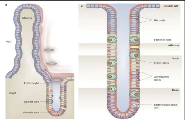
Figura 2. Different mucosal regions in pig stomach.
There are distinct differences in the glandular structures of different stomach zones (Figure 3). The cardiac glands in the cardiac zone are short tubular mucus producing glands and open into shallow, narrow gastric pits. In the fundic zone, the oxyntic glands are long tubular structures opening into wide gastric pits. The pyloric glands in the pyloric region are short tubular glands opening into deep gastric pits. The gastric glandular structures are well developed in piglets at the time of birth but cellular differentiation within the glands continues after birth (Xu et al., 1992b).
9
The surface epithelial cells are continuously desquamated and regeneration takes place at the deep part of the gastric pits and at the neck of the gastric glands by the process of mitosis of undifferentiated stem cells. The newly formed cells are slowly pushed upwards through growth pressure to replace the lost cells. At the base of the mucosal layer there is a well-developed muscularis mucosae in all parts of the stomach (Sloss, 1954). Such well-developed muscularis mucosae may help to empty the gastric glands through contraction. The muscularis externa, unlike that in other parts of the gastrointestinal tract, has three layers of smooth muscle: an outer longitudinal layer, a middle oblique layer, and an inner circular layer (Sloss, 1954). This arrangement of muscle fibres allows the stomach to contract in a variety of ways. At the pylorus, the internal and middle layers are greatly thickened to form the pyloric sphincter. The circular sphincter helps to control the passage of digesta into the small intestine. The serosa covering the stomach is part of the visceral peritoneum.
OXYNTIC GLANDS
Oxyntic glands are long branched tubular glands that are located in the fundic (or oxyntic) mucosa of the stomach. Several of these glands open into a single gastric pit (Figure 4 ). The oxyntic glands contain various types of cells, including mucous neck cells, parietal cells, chief cells, endocrine cells and undifferentiated
stem cells which are found in the neck region of the gland and at the bottom of gastric pits. Stem cells are
short and columnar in shape with oval nuclei near the base and are capable of mitosis. The new cells either move upwards to replace desquamated surface mucous cells or move downwards to differentiate into other types of cells. Mucous neck cells are present in clusters at the neck region of the gland. They are irregular in shape with the nucleus at the base and secretory granules near the apical surface. The secretory granules contain mucinogens and are stained intensely with PAS. In pigs, scattered mucous cells are also found in the lower part of the oxyntic glands.
Figure 4. General cell composition and location in a gastric gland. G and D cells a mainly in pyloric glands, while paretial and chief cells in oxyntic (fundic) mucosa. (Basic and Clinical Pharmacology. Mc Graw-Hill, 2012)
10
Parietal cells produce HCl and are abundant in the oxyntic glands. Parietal cells are more abundant in the
upper half of the gland than in the lower half. A cross-section of a parietal cell appears to be triangular in shape, with its base adjacent to the basal lamina and its apex directed towards the lumen of the gland. The nucleus of the parietal cell is spherical in shape, and some cells contain two nuclei. The striking features that are seen at the ultrastructural level are numerous mitochondria and deep, circular invaginations of apical plasma membrane forming the intracellular canaliculus. Hydrochloric acid is secreted initially into the intracellular canaliculus before being discharged into the glandular lumen. The large number of mitochondria indicates that the production of hydrochloric acid is a process with a high-energy requirement. Chief cells are enzyme-producing cells. For this reason, they are also called zymogenic cells. Chief cells are numerous in the oxyntic glands and are more abundant in the lower region of the tubular glands. Their cytoplasm is basophilic due to the abundance of rough endoplasmic reticulum. The secretory granules that are located at the apical cytoplasm contain inactive proteolytic enzymes. When released into the glandular lumen, the proteolytic enzymes are activated by hydrochloric acid and converted to active enzymes, proteases. Oxyntic glands also contain various types of endocrine cells that secrete regulatory peptides. Endocrine cells are scattered in the basal region of the epithelial layer between basement membrane and other epithelial cells. Some of the endocrine cells send a thin cytoplasmic extension to the lumen of the gland. Their cytoplasm is filled with small secretory granules that can be stained with silver or chromium salts.
CARDIAC GLANDS
Cardiac glands are short tubular glands that are located in the cardiac region of the stomach. The terminal portion of a cardiac gland is often coiled and has a large lumen. The glands consist mainly of
mucus-secreting cells. Undifferentiated stem cells are present in the neck or upper region of the gland. Endocrine
cells are scattered throughout the gland. Parietal cells are frequently seen in the cardiac glands, particularly in newborn pigs (Xu et al., 1992b).
PYLORIC GLANDS
Pyloric glands are branched short-tubular glands opening into deep gastric pits in the pyloric antrum. The glands are primarily made up of mucous cells with scattered parietal and endocrine cells. The dominant type of endocrine cells in the pyloric glands is the G cell, which produces the peptide hormone gastrin.
D cells are another endocrine cell type, which produce somatostatin, presents in all these three glands with
much more prevalence for pyloric gland. Undifferentiated stem cells are also present at the neck region of the pyloric glands.
11
INTESTINE
Small intestine
The small intestine is the longest part of the gastrointestinal tract. It measures about 3.5 meters in length in newborn piglets and up to 20 meters in adult pigs. It receives digesta from the stomach and continues the process of digestion in preparation for absorption of nutrients. Most nutrients are absorbed in the small intestine. The small intestine is divided into three regions: duodenum, jejunum and ileum. The duodenum commences at the pyloric sphincter and comprises about one-twentieth of the small intestine. The ileum is short and is the last part of the small intestine; it terminates at the caecum. The jejunum is the mid- and longest portion of the small intestine. Although the histological structure of the wall differs significantly amongst the duodenum, jejunum and ileum, there is no clear boundary between the regions of the small intestine. In most reports, the divisions of the small intestine into the duodenum, jejunum and ileum are arbitrary.
WALL STRUCTURE
The wall of the small intestine consists of the four layers that are characteristic of the gastrointestinal tract. The mucosal layer of the small intestine has a characteristic morphology with a finger-like or leaf-like projections called villi. The projections increase the luminal surface area and make the small intestine more efficient in the digestion and absorption process. ln the jejunal region of adult pigs, the layers of mucosa and submucosa are folded to form plicae circulares, further increasing the luminal surface area (Sloss, 1954). The newborn piglets, the villi are approximately 0.5-1.0 mm in length, and they are longer in the jejunum than those in the duodenum and the ileum (Xu et al., 1992a).
The intestinal mucosa contains numerous glands, which are also known as intestinal crypts. Intestinal glands are simple tubular glands that open into the intestinal lumen at the base of the villi (Figure 5). The lining of the intestinal mucosa consists of simple columnar epithelium. Underneath the epithelium is the lamina propria. As in other parts of the gastrointestinal tract, the lamina propria contains numerous diffused leukocytes and isolated nodules of lymphatic tissue. In the ileal region the lymphatic nodules aggregate to form Peyer's patches, which often extend into the submucosal layer (Sloss, 1954). Underneath the lamina propria, there are two thin layers of smooth muscle cells, an inner circular and an outer longitudinal layer, forming the muscularis mucosae. Contractile activity of these muscle cells causes movement of the villi and increases their contact with luminal contents.
The submucosa of the small intestine is made up of loose connective tissue and contains numerous blood and lymphatic vessels. In addition, the submucosa of the duodenum contains clusters of ramified, coiled tubular glands called Brunner's glands. These are mucus-producing glands that secrete mucus into the intestinal crypts. The secretory products of the Brunner's glands are alkaline (pH 8.1-9.3), and act to protect the duodenal mucous membrane against the effect of acidic digesta discharged from the stomach, and help to bring the intestinal contents to the optimum pH for pancreatic enzyme action. The muscularis externa consists of inner circular and outer longitudinal smooth muscle cells. The muscle cells are innervated by the autonomic nervous system. The cell bodies if the parasympathetic neurons appear as clumps of light-staining cells between the two layers of muscle cells. The nerve clumps are called the myoenteric plexus which together with the submucosal nerve plexus, comprise an intrinsic innervation of the intestine. Two kinds of muscular contractions take place in the small intestine. One is a local contraction that displaces intestinal contents both proximally and distally. Local contractions serve to mix the digesta with digestive juice and move it into contact with the mucosa for absorption. The second type of contraction, referred to
12
as peristalsis, moves intestinal contents distally. Except for part of the duodenum, the small intestine is completely covered by visceral peritoneum forming the layer of serosa. The small intestinal serosa is a thin layer of simple squamous epithelium supported by connective tissue. It contains blood vessels and lymph vessels that join with larger blood and lymph vessels in the mesentery. The blood vessels eventually drain into the hepatic portal vein.
Figure 5. Intestinal villi and crypt gland cells compared to a stomach gland (Nature Publishing Group, 2009).
VILLI AND CRYPTS
Intestinal villi are finger-like or leaf-like structures that project into the intestinal lumen. The villi are lined by a simple columnar epithelium with a core of connective tissue forming the lamina propria. Embedded in this connective tissue are capillaries and lymphatic vessels. Typically, each villus contains a centrally placed lymphatic that begins in the villus as a blind tube and drains into larger lymphatic vessels in the submucosa. Nutrients absorbed by the surface epithelial cells will pass through the wall of the capillaries and the lacteals, and enter the cardiovascular or lymphatic system.
Multi-potent stem cells residing at the base of each villus, in the so-called crypts of Lieberkühn, give rise to four primary epithelial lineages: absorptive enterocytes, mucin-secreting goblet cells, enteroendocrine cells, and Paneth cells (Fig.6A). Each of these cell types will be discussed in detail below. The epithelium is anchored on a continuous sheet of extracellular matrix, or basement membrane, consisting of a mixture of collagen, laminin, and fibronectin (Beaulieu, 1999). The actual composition of the basement membrane varies along the crypt-villus axis and this is thought to provide signals essential for enterocyte proliferation, survival and differentiation (Beaulieu, 1999). The cellular constituents of the crypts, notably stem cells, goblet cells, enteroendocrine cells, and Paneth cells, collectively are responsible for water and ion secretion, as well as exocrine, paracine and endocrine secretions, whereas the intestinal villi are primarily responsible for fluid and nutrient absorption.
13
Absorptive enterocytes
The epithelium lining the intestinal viIIi consists of primarily columnar enterocytes. Also known as intestinal absorptive cells or absorptive enterocytes. Scattered between the enterocytes are mucus producing goblet cells. Enterocytes are tall columnar cells, with an oval nucleus located at the basal region. The apical surface membrane of the enterocytes is intensively folded, forming the structure that is called the microvilli . When viewed under a light microscope the apical membrane of the enterocytes appears striated. Thus, the apical membrane of enterocytes with microvilli is also called the striated border membrane or the brush border membrane. The brush border membrane of the enterocyte contains various channels and nutrient transporters, and is the active site for nutrient absorption. The projection of microvilli at the apical membrane increases the absorptive surface area of an enterocyte tremendously. Neighboring enterocytes are joined by junctional complexes to stop the diffusion of harmful substances through the intercellular space. The enterocyte not only has an absorptive function, but also digestive functions. Digestive enzymes are anchored to the brush border membrane and their functional groups extend outward to become part of the glycocalyx. Included amongst the enzymes are saccharidases (e.g., lactase, isomallase, maltase and sucrase) and various peptidases. The brush border membrane also contains the enzyme enteropeptidase, also known as enterokinase. This enzyme converts trypsinogen into active trypsin; the latter can then activate additional trypsinogen and other pancreatic zymogens.
Figure 6A. Gut epithelial cell distrubuition where stem cells riding in a proliferative zone of each villus called crypt of Lieberkuhn, give rise to four primary cells: absorptive enterocytes, mucin-secreting globlet cells, enteroendocrine cells and Paneth cells. Figure 6B.The FAE is found exclusively overlying organized lymphoid follicles situated throughout the small and large intestines.
(both: Vajdy M., 2008) (Neutra et al., 2001).
Goblet Cells
Goblet cells, also known as mucin-producing cells, are responsible for the production of the mucus gel that blankets the surface of the intestinal epithelium. Goblet cells, named for their characteristic goblet-like shape, are present in the small and large intestine, and are found along the entire crypt-villus axis. In villi of the small intestine, goblet cells are interspersed among absorptive enterocytes (Fig. 6A)(Karam, 1999). Under normal conditions, individual goblet cells constitutively secrete mucins, high molecular weight glycoproteins that consist of core polypeptides heavily decorated with both N- and O-linked oligosaccharide
14
side chains. This so-called baseline secretion of mucins is necessary for both maintenance and renewal of the mucus layer, which is important in both epithelial function and defense (Deplancke and Gaskins, 2001; Lievin-Le Moal and Servin, 2006). For example, the mucus layer aids in lubrication and protection of the intestinal mucosa, as well as serving as an important defense mechanism against microbial pathogens and toxins. The viscous mucus gel limits diffusion of macromolecues and impedes motility of parasites and bacterial pathogens. Through heterogeneous N- and O-linked oligosaccharide side chains on mucins, the mucus layer also provides “decoy” ligands for lectin like adhesins expressed by microbial pathogens and toxins, thereby competitively inhibiting these agents from gaining access to their receptors on the apical surfaces of enterocytes. In situations where the intestinal epithelium is exposed to microbial pathogens, toxins or other intraluminal irritants, goblet cells can release additional mucins that are normally stored in apically-residing granules.
Enteroendocrine Cells
Enteroendocrine cells are a specialized sub-population of “sensory” epithelial cells that serve as a link between the intestinal lumen and the enteric nervous system (Flemstrom and Sjoblom, 2005). The primary function of this epithelial cell type is to secrete peptide hormones and transmitters. Enterochromaffin cells, for example, are responsible for the production and secretion of serotonin (5-hydroxytryptamine), a hormone which regulates (among other things) intestinal peristaltic and secretory reflexes. Recently, a subpopulation of enteroendocrine cells have been proposed to serve as “taste receptors,” based on the immunohistochemistry and real time PCR analysis demonstrating the presence of taste signaling molecules in the small intestine of mice (Bezencon et al., 2007).
Paneth Cells
Paneth cells migrate to the crypt base where these cells reside for an average of 21–28 days in most mammals (Andreu et al., 2005). Paneth cells possess a well-developed apical secretory apparatus. Their location in the crypt base places them in an ideal position to deliver growth factors to dividing cells in higher compartments of the crypt, and to create gradients of anti-microbial factors which limit, or altogether prevent, microbial colonization of the small intestinal crypts. Key anti-microbial factors produced by Paneth cells include lysozyme, the cryptidins alpha-defensins and other digestive enzymes with known anti-microbial properties (Wehkamp et al., 2006).
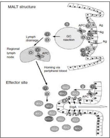
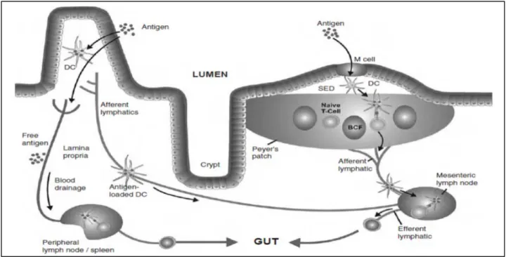
Follicle-Associated Epithelium (FAE) and M Cells
The intestinal epithelium maintains a close collaboration with an underlying local immunological network, collectively referred to as the mucosal immune system (Holmgren and Czerkinsky, 2005; Neutra et al., 2001). Nowhere is this collaboration more apparent than in organized lymphoid follicles, which are present throughout the small intestine and colon. In the small intestine, these macroscopic structures consist of aggregates of between 5 and 10 lymphoid follicles known as Peyer’s patches. These organized lymphoid follicles contain germinal centers which represent the primary sites of mucosal B cell differentiation and somatic cell hypermutation (Brandtzaeg and Johansen, 2005). As mucosa-associated lymphoid tissues lack afferent lymphatics, germinal center activity is driven exclusively in response to antigens present in the intestinal lumen. Uptake and transepithelial transport of macromolecular antigens from the intestinal lumen to the organized lymphoid follicles is achieved the so-called follicle-associated epithelium (FAE). The FAE is distinct from the villus epithelium in both structure and function (fig.6B). Whereas the villus epithelium is specialized for digestion and absorption of nutrients and is dominated by absorptive enterocytes, mucin-secreting goblet cells and enteroendocrine cells, the FAE contains few or no goblet or
15
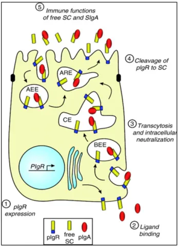
enteroendocrine cells and has reduced levels of certain digestive enzymes. There are also fewer defensin- and lysozyme-producing Paneth cells in follicle-associated crypts. Enterocytes within the FAE, like their counterparts on villi, have well developed microvilli and are coated with a thickfilamentous brush border glycocalyx, but they are not identical to villus cells (Frey et al., 1996). FAE enterocytes express lower levels of the membrane associated hydrolases involved in digestive functions (Savidge and Smith, 1995). It is also apparent that the glycosylation patterns of epithelial cells in the entire FAE differ from those on villi, indicating that glycosyltransferase expression in the FAE is distinct (Mantis et al., 2000). The FAE of Peyer’s patches also express chemokines involved in leukocyte homing, which are not expressed elsewhere in the small intestinal epithelium (Zhao et al., 2003b). Finally, the FAE is devoid of pIgR and therefore it is unable to transport IgA from the interstitium to the lumen. Probably the most distinguishing feature of the FAE is the presence of M cells, a unique epithelial cell type that is specialized in the uptake and transepithelial transport of particulate antigens, including particles and macromolecules, viruses, bacteria, and parasites (Amerongen et al., 1991; Jones et al., 1994; Marcial and Madara, 1986; Pappo and Ermak, 1989). Indeed, M cells have been considered the “gateway” to the gut associated lymphoid tissue (GALT). The apical and basolateral surfaces of M cells have distinct features that enable them to rapidly and efficiently deliver mucosal antigens from the lumen to underlying lymphoid follicles (Fig. 6B). Consequently, M cell apical membranes are more accessible to particles, viruses and bacteria than adjacent enterocytes. In mice and humans, the apical surfaces of M cells have a pattern of glycosylation that is distinct from FAE enterocytes and villus enterocytes (Clark et al., 1993; Giannasca et al., 1999). M cells also selectively express Toll-like receptors and pattern recognition receptors on their apical membrane that may facilitate antigen recognition and contribute to signaling in the local environment (Chabot et al., 2006). The M cell basolateral membrane is deeply invaginated to form a large intraepithelial “pocket” containing specific sub-populations of naive and memory B and T cells (Yamanaka et al., 2001), and occasional dendritic cells (Iwasaki and Kelsall, 2001). The pocket brings the M cell basolateral surface to within a few microns of the apical surface, shortening the distance that transcytotic vesicles must travel to cross the epithelium. Antigens transported by M cells are sampled by adjacent DCs.
Large intestine
The large intestine consists of the caecum, the colon and the rectum, and is about 0.7 meter in length in newborn piglets and up to 4.5 meters in length in adult pigs. The caecum is a cylindrical blind sac that is located at the proximal end of the colon. The proximal part of the colon undergoes four complete turns that spiral towards the center of the coil; it is termed the centripetal colon. A central flexure permits the spirals to reverse and the colon undergoes 3.5 turns from the center of the coil, giving rise to the centrifugal colon. The major functions of the large intestine include the absorption of later, electrolytes and certain nutrients that are produced by bacterial fermentation; so it provides a location for numerous microorganisms. The wall of the large intestine as the four layer structure that is characteristic of the gastrointestinal tract. The structural organizations of each layer are similar to those described for the small intestine, with the exception of the mucosal layer. The mucosa of the large intestine has no villous projections but contains numerous straight tubular glands that extend through the full thickness of the mucosa. The luminal surface and the tubular glands are lined with columnar epithelial cells that generally resemble the enterocytes of the small intestine. However, the brush border at the apical surface of the enterocytes is much thinner than that of the small intestinal enterocytes, numerous mucus-secreting goblet cells and scattered endocrine cells are also found in the tubular glands. At the lower half of the glands,
16
there are numerous stem cells with the capacity for mitotic division. New epithelial cells that arise from the stem cells in the glands migrate upwards and replace the degenerated surface cells.
Although most nutrients are absorbed by the small intestine, the absorption of some nutrients also occurs in the large intestine, especially in its proximal region. In newborn piglets, rudimentary villi are found in the proximal region of the large intestine and these structures gradually disappear during the first few days of postnatal life (Xu et al., 1992a). The enterocytes in the proximal region of the large intestine in a neonatal animal are similar to those cells in the small intestine, having long microvilli at the apical surface, the ability to absorb amino acids and glucose, and digestive enzyme activities at the brush border. It has been suggested that the newborn colon, which is capable of digestion and absorption, may provide an additional capacity to the immature small intestine for nutrient digestion and absorption (Xu, 1996). The lumen of the large intestine is the home for billions of microorganisms. These microorganisms utilize the food residues that are discharged from the small intestine and convert them into useful nutrients such as short-chain fatty acids and vitamins. These nutrients can then be absorbed in the large intestine and be used by the animal. Bacteria that are capable of breaking down cellulose and hemicellulose have been found in the lumen of the large intestine in adult pigs, which indicates that pigs have some capacity to utilize fibrous diets (Varel and Yen, 1997). In addition, bacteria in the large intestine synthesize most of the vitamin K that is needed by the animal.
17
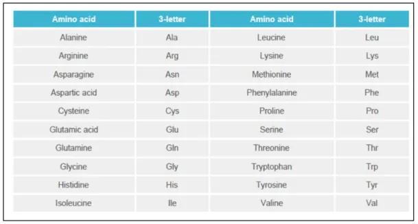
AMINO ACIDS
Twenty amino acids are the building blocks of proteins (Table 1). Some of them can not be synthesised, or not in sufficient quantities, by animals; they must therefore be supplied through feeding and are referred to as essential amino acids. These are listed in Table 2. To maintain good health and growth performance, feeds must provide the indispensable amino acids in sufficient quantities to cover the requirement of animals.
Table 1. The 20 amino acids and their abbreviations. (Ajinomoto Eurolysine S.a.S.)
Table 2. Essential amino acids in pig. (Ajinomoto Eurolysine S.a.S.).
Some amino acids are essential dietary components in certain situations but not in others; they have been referred to as conditional essential amino acids.
18
For example, glutamine is normally regarded as nonessential for the pig. However, under total parenteral nutrition or during the early post-weaning period, supplementation with glutamine prevents intestinal mucosal atrophy (Wu et al., 1996). It is known that glutamine is an important energy source for the enterocytes (Wu et al., 1995). Glutamine also serves as a substrate for the endogenous synthesis of arginine and proline. It has also been reported that supplementing the diet of early-weaned piglets with glutamine preserves normal lymphocyte function following Escherichia coli infection (Yoo et al., 1997). The sulphur-containing amino acid cysteine occurs in two forms, either as itself or as cystine in which two cysteine molecules are joined together by a disulphide bridge. Cystine can be synthesized from methionine; consequently when there is an inadequate supply of cystine in the diet, the methionine requirement will increase. An adequate supply of dietary cystine will have a sparing effect on the methionine requirement. Cystine can satisfy approximately 50% of the sulphur amino acid requirement of growing pigs (Chung and Baker, 1992). In practice, the requirement for sulphur amino acids is usually met by a mixture of methionine and cystine. Similarly, phenylalanine can meet the requirement for the two aromatic amino acids phenylalanine and tyrosine, as phenylalanine can be converted to tyrosine. A mixture of these two amino acids is commonly used to meet the dietary requirements of aromatic amino acids.
AMINO ACID BALANCE
The nutritional value of a protein is primarily dependent on its amino acid composition, especially the content of essential amino acids. A protein that has a perfect balance of amino acids to meet all amino acid requirements has been described as an ideal protein. However, the ideal pattern of amino acid composition for maintenance may differ from that for tissue growth. There are also changes in the amino acid content of body tissues at different stages of development. Therefore, there is no ideal protein that can meet all amino acid requirements for the growing pig. Nevertheless, the porcine milk protein is regarded to be close to an ideal protein for neonatal pigs.
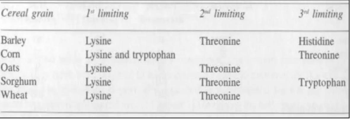
An amino acid that is present in the diet in an inadequate amount relative to its requirement is regarded as a limiting amino acid. Supplementation of the diet with this amino acid will significantly improve its nutritional value and subsequently the performance of the animal that is fed on this diet. Information about which amino acids are most limiting in natural feedstuffs is important in formulating pig diets. The most limiting amino acids of cereal grains commonly used in pig diets are presented in Table 3.
19
As can be seen, lysine is the first limiting amino acid in most cereal grains. In contrast, methionine is the first limiting amino acid in soybean meal (Berry et al., 1966). Soybean meal and canola meal have relatively high lysine and low sulphur amino acid contents, whilst cereal grains arc low in lysine and high in sulphur amino acids. A combination of these two types of proteins can markedly improve the nutritional value of a diet; this phenomenon is known as complementation.
Amino acids that are limiting in common feedstuffs can be produced by chemical synthesis. Currently, four crystalline amino acids (lysine, methionine, tryptophan and threonine) are commercially available as feed additives. Supplementation of the diet with crystalline amino acids requires caution. Crystalline methionine and tryptophan are highly toxic, and improper mixing or using an incorrect dose can cause poisoning. In newly weaned piglets, dietary addition of 4% methionine causes a 52% reduction in body weight gain and a significant decrease in feed intake (Edmonds et al., 1987). Antagonistic effects have been observed between arginine and lysine and amongst leucine, isoleucine and valine, possibly due to competition for carrier proteins during the absorption and metabolism process. When there is an excessive amount of one amino acid in the antagonistic group in the diet the requirement for other competitive amino acids increases.
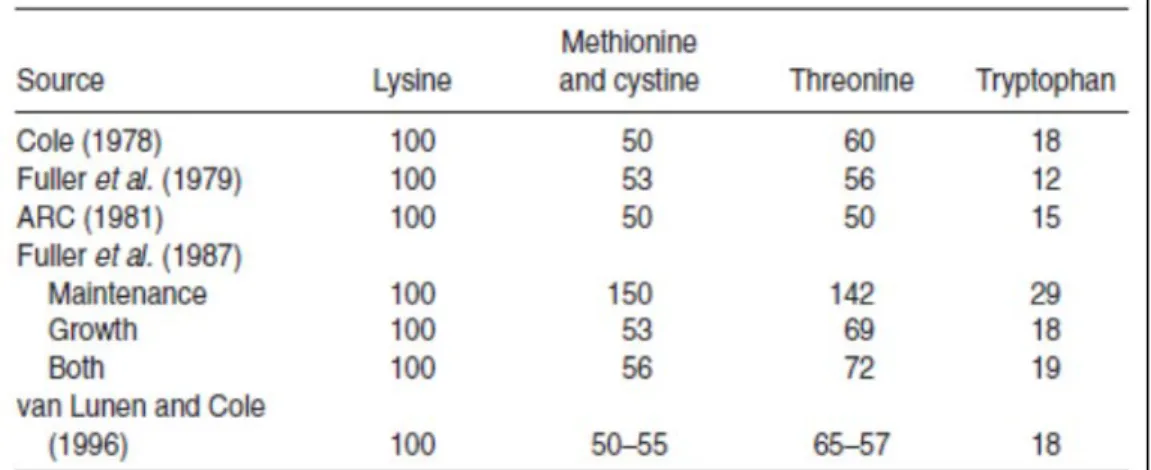
Published balances of amino acids relative to lysine have been well reviewed (Urynek and Buraczewska, 2003) (Table 4). Since ARC (1981), the change that has affected commercial diets the most has been the increase in levels of threonine (usually the second- limiting amino acid in pig feeds) relative to lysine. Fuller
et al. (1987) suggested a level of 72%, having separated out requirements for maintenance and growth and
feeding semi-purified diets. Work by D.J.A. Cole and L. Bong (1989) has found the value to be between 65 and 70%. It is unusual to find commercial diets with tryptophan at a level greater than 17-18% of lysine despite the fact that tryptophan, being a precursor for serotonin, is known to play a role in stimulating feed intake. More recently studies demonstrated that pigs between an average of 8 and 25 kg responded to an increasing level of dietary tryptophan up to 21% of lysine.
Table 4. The ideal amino acid balance of commonly weaner feeds.
Dietary amino acids are major fuels for the small intestinal mucosa, as well as important substrates for syntheses of intestinal proteins, nitric oxide, polyamines, and other products with enormous biological importance. Recent studies support potential therapeutic roles for specific amino acids (including glutamine, glutamate, arginine, glycine, lysine, threonine, tryptophan and sulfurcontaining amino acids) in gut-related diseases. Results of these new lines of work indicate trophic and cytoprotective effects of amino acids on gut integrity, growth, and health in animals and humans.
20
In particular, these amino acids affect the gut because it is an important organ responsible for digestion, absorption and metabolism of dietary nutrients. It contributes to 9–12% of whole-body protein synthesis and is the most important place of entry for foreign antigens, including food proteins, natural toxins, commensal gut flora, and invading pathogens (Li et al., 2007). The intact intestinal tract is lined by a continuous monolayer of intestinal epithelial cells, of which a primary function is to act as a physical barrier, interacting with a complex external environment. The intestinal tract is also one of the largest lymphoid organs in the body, and consists of immune cells in organized gut associated lymphoid tissues (Field et al., 2002). Amino acids are not only important substrates for the synthesis of proteins and other nitrogenous compounds, but also key regulators through major metabolic pathways (Meijer, 2003). Recent studies with animals and humans indicate additional roles for amino acids in maintaining gut health (Wang et al., 2008).
In the contest of the studies presented further here is discussed in particular the importance of threonine and tryptophan.
Threonine
Threonine is the second limiting amino acids in pigs. Among the essential amino acids, threonine is particularly important for mucin synthesis and maintenance of gut barrier integrity (Bertolo et al., 1998) because it is a major component of intestinal mucin and plasma G-globulin in animals. The retention of dietary threonine by the intestine is (up to 60%) high and animal feeding studies indicate that changes in components of the immune system are sensitive to dietary threonine intake (Stoll et al., 1998). It has been shown in piglets that only 38% of dietary threonine appears in portal blood; the remainder is retained by the gut. Nearly 90% of the gut-retained threonine is either secreted as mucosal proteins, threonine-rich mucin, or metabolized. It is known that in intestinal mucosa, a major fate of threonine is incorporation into mucins, which are major glycoproteins protecting the epithelium from injury (Le Floc'h and Seve, 2005; Schaart et al., 2005). A study with rats demonstrated that dietary threonine restriction dramatically and specifically impaired the synthesis of mucins in all segments of the small intestine, reaching the largest reduction of 40% in the duodenum (Faure et al., 2005). In addition, Wang et al. (2007) reported that both a deficiency and an excess of dietary threonine reduced the synthesis of intestinal mucosal protein and mucins in young pigs. The implications of threonine for intestinal health and nutritional requirements have been highlighted in several recent studies. For example, the threonine utilization for synthesis of small intestinal proteins is increased substantially in response to sepsis, representing over two-fold of the threonine intake (Faure et al., 2007). Thus, under inflammatory conditions, threonine availability may become limited for the synthesis of intestinal mucins, which leads to an impairment of gut barrier function. Consequently, an increase in dietary provision of threonine and other amino acids can promote mucin synthesis and rebalance the gut microbiota to favor intestinal protection and mucosal healing (Faure et al., 2006). There are evidences in threonine effects on immune system defence. Parenteral feeding leads not only to atrophy of mucosal epithelial cells, but also to atrophy of the goblet cells, which secrete a large amount of threonine-rich mucin (Bengmark and Jeppsson, 1995). Through protein synthesis and cellular signalling mechanisms, addition of 2mM threonine to the culture medium prevented apoptosis, stimulated cell growth and promoted antibody production in lymphocytes (Duval et al., 1991). Also a dietary supplementation with threonine increased serum levels of IgG in sows (Cuaron et al., 1984). Further, increasing dietary threonine intake increased antibody production, serum IgG levels and jejunal mucosal concentrations of IgG and IgA, while decreasing jejunal mucosal concentrations of IL-6 in young pigs
21
challenged with Escherichia coli. These findings provide support for a role of dietary threonine in modulating immune function in livestock and perhaps humans.
For these reasons it is conceivable that the neonatal small intestine needs high requirement for threonine.
Tryptophan
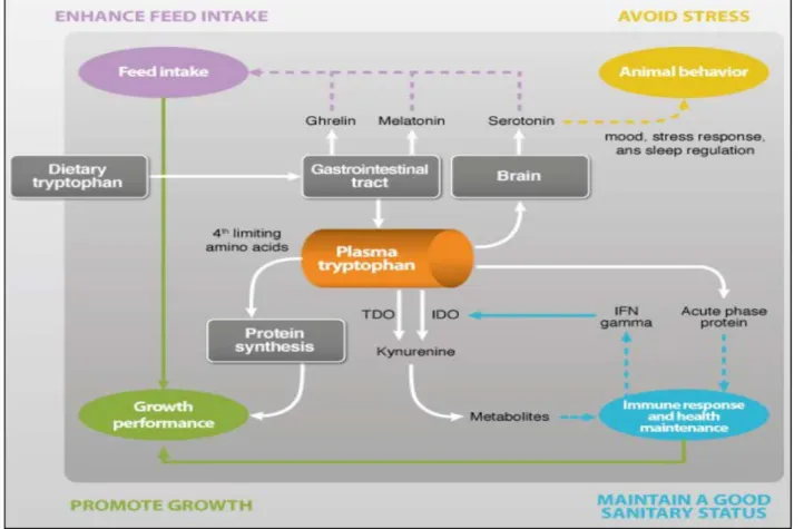
In European diets, tryptophan is the fourth limiting amino acid for growth. This means that when the tryptophan supply is low compared to the other essential amino acids, the protein synthesis and accretion and, finally, the growth rate will be limited.
Tryptophan has the particularity to be involved in several biological functions such as appetite regulation, stress and immune responses. This explains why the young pigs exhibiting a high growth rate potential are especially sensitive to low and inadequate tryptophan dietary supply. Consequently, requirement and practical recommendations can be different according to the biological function to be optimized. In young pigs, the main effect of tryptophan on growth rate is associated to an increase in feed intake when tryptophan is supplied to a level close to recommendations for a maximum growth rate. However, some factors also impact directly on tryptophan metabolism and thus limit its availability for growth and any other functions in which tryptophan is involved.
So, besides being a nutrient that is fundamental to allow maximum growth of the pigs, tryptophan also has other physiological functions of great interest.
Tryptophan has positive effects on voluntary feed intake. This characteristic makes it a very important nutrient in feeds for animals with low appetite and limited feed intake capacity, as it is the case for lactating sows and weaned piglets. This greater feed intake leads to increased growth performance and improved feed conversion ratio in piglets, and to lower body weight losses in sows during lactation. The effect can be very noticeable when extreme levels of tryptophan are used, but even in commercial ranges (22% vs 18% of Trp/Lys) significant improvements can be obtained in piglets. Although recent studies suggest the potential mediation of hormones such as melatonin (pineal gland), insulin (pancreas) or ghrelin (stomach), the effect of tryptophan on feed intake seems to be fundamentally related to its role as a precursor of serotonin, a neurotransmitter synthesized in the brain and in the gastrointestinal tract, which plays a role in regulating feed intake. It has been observed that the amount of dietary tryptophan needed to achieve a certain level of feed intake is greater in diets with higher protein content. High protein diets contain a greater quantity of large neutral amino acids (valine, isoleucine, leucine, phenylalanine and tyrosine), which compete with tryptophan for the same transporter during the intestinal absorption and also to pass the blood-brain-barrier, which reduces the quantity of tryptophan available for the synthesis of serotonin. Tryptophan can modulate aggressive behaviors and improve the stress responses in swine. It has been observed in various species, including humans, for which tryptophan has been used as an anti-depressant in the 1980s. It has also been reported that tryptophan may reduce the negative effects on meat quality caused by stress during transport and before slaughter. The mechanism of coping stress is not well described, but it is most likely due to tryptophan being served as precursor for serotonin synthesis. However, clear effects have been observed only when using levels of tryptophan that are either much lower or much higher than the usual levels found in commercial feeds.
The level of tryptophan in the blood and the quantity of tryptophan available for body protein synthesis are decreased during inflammatory or disease challenge conditions. This is due to the lower level of feed intake during inflammatory states, the increased demand for tryptophan for the synthesis of acute-phase proteins, which are rich in tryptophan, and also due to the greater catabolism of tryptophan via kynurenine
22
pathway caused by the action of the enzyme IDO (indoleamine 2,3 dioxygenase), which is stimulated in the presence of cytokines (inflammatory states) (Fig 7).
Figure 7. Global pattern of the biological roles of tryptophan and their implications in animal growth. (Ajinomoto S.a.S)
An adequate inclusion of tryptophan is especially advisable in low health conditions due to its effect on ingestion capacity and also to counteract the greater demand for tryptophan for non-productive functions. Moreover, in some studies it has been observed that the inclusion of tryptophan in the diet is capable of modifying the inflammatory response. According to these studies, a diet with an adequate level of tryptophan produces a lower inflammatory response than a diet that is deficient in tryptophan, with lower levels of plasma haptoglobin (acute phase protein) and a lower activity of enzyme IDO.
The products of tryptophan catabolism include serotonin, N-acetylserotonin, melatonin and anthranilic acid (Kim et al. 2007). Tryptophan catabolism is increased to generate anthranilic acid through the indoleamine 2,3-dioxygenase (IDO) pathway during inflammation or stimulation by LPS or certain cytokines (Platten et al., 2005). Serotonin, melatonin and N-acetylserotonin can enhance host immunity by inhibiting the production of superoxide, scavenging free radicals and attenuating the production of TNFa (Perianayagam et al., 2005). In addition, N-acetylserotonin is an inhibitor of sepiapterin reductase, an enzyme for the synthesis of tetrahydrobiopterin (Shi et al. 2004). By modulating inducible NO synthesis, this tryptophan metabolite can affect both innate and acquired immunity systems. Excitingly, anthranilic acid was recently found to inhibit the production of proinflammatory Th1 cytokines and prevent autoimmune neuroinflammation (Platten et al. 2005). Because there is a progressive decline in tryptophan concentrations in plasma of animals with inflammation, its catabolism plays a critical role in the functions of both macrophages and lymphocytes (Melchior et al. 2004). Early work indicated that tryptophan
23
starvation resulting from IFNg treatment was associated with the antiproliferative effect of this cytokine on intracellular parasite and tumours (Ozaki et al. 1988). Interestingly, progressively increasing concentrations of IFNg were required for its growth inhibition in the presence of elevated tryptophan concentrations (Pfefferkorn, 1984). Available evidence suggests that tryptophan catabolism plays a role in immune responses by producing a local immunosuppressive environment that is able to control T-cell homeostasis and self-tolerance during inflammation (Platten et al. 2005). A deficiency of dietary tryptophan impaired the immune response in chickens (Konashi et al., 2000). Conversely, oral administration of 300 mg of tryptophan to rats enhanced phagocytosis by macrophages and the innate immune response (Esteban et al., 2004). Dietary supplementation with 0,22% L-tryptophan also increased resistance to bacterial and parasitic infections in rats fed a 20% zein diet (Watson & Petro, 1984). At present, a potential use of crystalline tryptophan for animal health management is not fully developed.
In the literature, recommendations are usually expressed in digestible basis and in relation to lysine, following the concept of ideal protein. In table 1 the values recommended for piglets by different institutions are shown. Traditionally Trp:Lys ratios of 17-18% have been recommended, but there is currently a great deal of evidence that suggests that higher levels (≥ 22%) can be more profitable, especially in low health conditions. In the same way, the levels traditionally recommended for lactating sows oscillate between 18 and 20 % according to the sources, but there are also studies that have shown improvements in feed intake and reduced weight loss with a Trp:Lys ratio of at least 22%.
So, tryptophan is an important amino acid in swine diet formulations due to its positive effect on feed intake, growth performance and health .
24
WEANING, PWD and GUT MICROBIOTA
Weaning
Over the past 50 years, the weaning age of piglets has been decreased from 10–12 weeks to current ages of 3–5 weeks (Nabuurs, 1998), and in our case regards 4 weeks (28 days). Weaning is a stressful time in a pig’s life where it has to adapt rapidly to major changes in environment and nutrition.
The weaning transition is commonly accompanied by adverse changes in intestinal morphology, including reduced villus height, increased villus width, increased crypt depth and reduced absorptive capacity and brush-border enzyme activity (McCracken et al., 1999). It has been even reported that the switch from liquid to solid diets reduces feed intake in neonatal piglets and may subsequently reduce the growth rate. Furthermore, an adequate nutrient intake is essential in providing both the nutrients and hormonal signals required to stimulate gut mucosal growth (Burrin et al., 2000). It has also been shown that weaning of piglets on to solid diets causes anorexia and local intestinal inflammation (McCracken et al., 1999). In contrast, piglets weaned to a liquid diet (e.g., bovine or ovine milk) maintain a normal intestinal structure after weaning (Pluske et al., 1996). These foundings have lead to a sweeping change in the industry practice towards liquid feeding systems for nursery pigs. The length of time it takes piglets to adapt to weaning appears to be quite variable.
The weaning process is known to decrease digestive function in several ways. Although weaning has little effect on gastric lipase activity, the process dramatically decreases exocrine pancreatic lipase activity. Weaning also dramatically reduces both the hydrolytic capacity and the specific activity of major exocrine pancreatic proteases. It is known that brush-border enzyme activities decline along the villus-crypt axis towards to the crypt (Fan et al., 2001). Thus, weaning associated villus atrophy may markedly reduce intestinal digestive and absorptive capacities. These changes are assumed to impair the ability of the small intestine to digest and absorb nutrients and hence to predispose the piglet to malabsorption and diarrhoea. Several biological explanations have been suggested for weaning associated diarrhoea.
Firstly, porcine milk is rich in IgA and glutathione; IgA provides passive mucosal immunity whereas glutathione is an important antioxidant essential for the maintenance of intestinal mucosal integrity (Reeds et al., 1997). Weaning removes these luminal protection agents and increases the susceptibility to enteric infections and diarrhoea.
Secondly, weaning decreases goblet cell on villus-crypt units (Dunsford et al., 1991), thus reducing the protective mucus secretion. It has been proposed that the mucus barrier reduces colonization by pathogens of the intestinal villus membrane. Weaning also disturbs intestinal mucosal active immunity by altering mucosal T-cell subsets, causing intestinal mucosal inflammation and subsequently increases paracellular translocation of pathogenic microbes and their toxins (Spreeuwenberg et al., 2001).
Thirdly, weaning causes villus atrophy and crypt hyperplasia and subsequently alters the ratio between the villus absorptive cells and the crypt secretory cells. Thus, weanling piglets may be physiologically vulnerable to diarrhoea.
The abrupt change from milk to starter solid diets in pigs weaned at about 3-4 weeks is associated with an underfeeding period and a weight lost immediately after weaning. The resulting growth check can have a major impact on the subsequent performance. Feed and alternative strategies limiting growth problems in this sensitive period are important to improve the performance further.An important aim for the pig industry is to formulate economically viable growth-promoting diets to ease the transition from sow’s milk to nursery diets. In addition to satisfying the nutritional requirements of weaned pigs, such diets are
25
increasingly assessed for their ability to modulate microbial succession, stabilize the commensal microbiota, improve immune function and enhance disease resistance in the young animal.
Post-weaning disease - PWD
The gastrointestinal tract performs two major functions: assimilation of nutrients, fluid and electrolytes; and maintenance of a protective barrier to prevent uncontrolled passage of toxins and infectious agents into the systemic circulation. Hence, when gastrointestinal disorders occur, one or both of these functions are compromised, which may lead to severe growth retardation or death.
The gastrointestinal tract of the neonatal pig undergoes two major functional changes that are associated with birth and weaning. At birth, the gut encounters severe challenges when it suddenly takes over the responsibility for nutrient acquisition from the placenta, and it is exposed to a diversity of pathogenic and non-pathogenic microorganisms. Furthermore, during this early period the newborn pig is especially susceptible to gastrointestinal infections because of its immature immune system. The piglet may succumb to infections if it fails to acquire an adequate supply of immunoglobulins through the consumption of colostrum immediately after birth.
The gut encounters a second challenge at the time of weaning. Weaning usually involves a shift from a liquid milk diet to a solid, typically grain-based, diet that has a significantly higher content of carbohydrates and a lower content of protein and lacks many of the growth stimulating and protective substances that are present in milk. In addition, the grain-based diet may not be as palatable as sows milk and may contain substances that have anti nutritional effects. This change in food source, along with other stresses related to weaning such as separation from the sow and littermates, relocation to a new environment and mixing with unfamiliar piglets, often leads to a reduction in feed intake and growth rate and also to gastrointestinal disorders and diarrhoea.
Diarrhoea is a condition characterized by increased frequency and fluidity of bowel movements. The faeces are watery, soft and may contain abnormal amounts of fat, mucus, blood or fibrin, depending on the aetiology of the diarrhoea. The high concentration of water and electrolytes in the faeces during diarrhoea may result from increased gastrointestinal secretion, reduced digestive and absorptive capacity and/or abnormal intestinal motility (O'Loughlin et al., 1991). The common enteric pathogens that cause diarrhoea in young pigs are rotavirus, transmissible gastroenteritis virus, Clostridium perfringens, Escherichia coli,
Isospora suis and Cryptosporidium parvum.
The diarrhoea is initially noninfectious, but at a later stage is often aggravated by opportunistic viruses, bacteria and protozoa. The most common infectious agents of post-weaning diarrhoea are enteropathogenic strains of E. coli. Post-weaning diarrhoea is a complex, multifaceted and poorly understood syndrome that involves the interactions of psychological stress, dietary reactions, enteric pathogens and genetic susceptibility. Changes associated with weaning, e.g., in small intestinal morphology (Mosenthin, 1998; Pluske et al., 1996), enzyme activity (Hampson and Kidder, 1986) and absorption capacity (Miller et al., 1984) are well documented in literature. The changes, which include villous atrophy, crypt hyperplasia, reduced brush border enzyme activity and absorptive capacity, are believed to predispose the weaning pig to post-weaning diarrhoea.
26
Colibacillosis
Escherichia coli is an enterobacteria Gram- that forms part of the normal intestinal microbiota of healthy
animals. Usually, E. coli present in animals are communal antipathogenic strains and they even play a beneficial role, since they compete in several ways with the pathogenic strains in the ecological niche of the lumen.
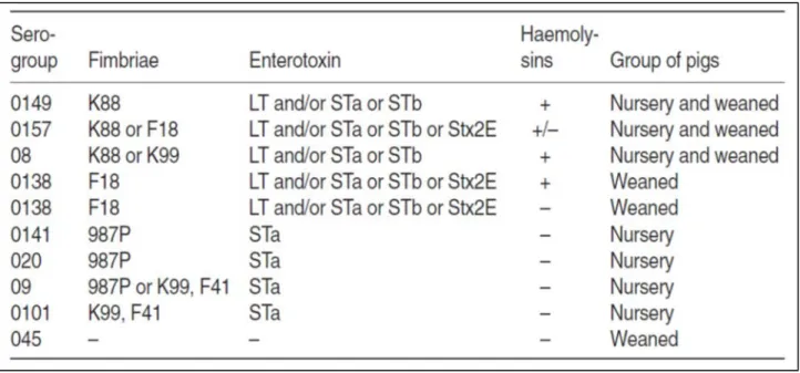
The term E. coli includes around 200 different serotypes that are classified by their antigens of the cell wall (O antigens), of the capsule, (K antigens) of the fimbriae (F antigens) and of the flagella (H antigens).
One of the main characteristics of E. coli is its great capacity to adapt to environmental conditions and experiment change. This is why it acquires a resistance to antibiotics much easier than other bacteria. Colibacillosis is a major cause of illness and death in recently weaned pigs. Usually it is a consequence of enterotoxigenic strains of E. coli (ETEC) producing enterotoxins (heat-labile toxin LT) or VTEC types producing heat-stable toxins (STa/Stb) which act totally in the gut and stimulate hypersecretions of mucus and electrolytes. LT is a heavy molecular toxin and easily induces the formation of specific antibodies, which makes it an important component in vaccines. The toxin acts on the enterocytes, activating the adenylate cyclase which stimulates the production of Cl-, Na+ and HCO3- ions and, consequently, diarrhea
due to hyper secretion. This in turn, provokes dehydration, acidosis, and in serious cases, death. The feces of affected piglets have an alkaline pH. The organism also produces fimbrial adhesions, which mediate the adherence of bacteria to the mucosal surface (Table 5).
Table 5. Serogroups, fimbriae, enterotoxins and haemolysins of nursery and weaned piglets (Varley M.A. and Wiseman J., 2001).
Fimbriae produced include K88 (F4), K99 (F5) and 987P (F6); F41 and F18 (F107 and 2134P) are less common but some may produce a shiga toxin stx 2E. Age and genetic background seem to determine the inherent susceptibility of piglets to E. coli. Pigs are resistant to F18+ E. coli at birth but become susceptible after several weeks (Imberechts et al., 1997), whereas K99 oresistance is substantially complete by 2 weeks (Runnels et al., 1980). Resistance is achieved by failure to produce the receptor on epithelial brush-border membranes to which the fimbriae adhere (Francis et al., 1998).