UNIVERSITÀ DEGLI STUDI DI MESSINA
DIPARTIMENTO DI MEDICINA CLINICA E SPERIMENTALE
CORSO DI DOTTORATO IN
BIOTECNOLOGIE MEDICHE E CHIRURGICHE (XXXII CICLO)
Post-mortem immunohistochemical analysis of
dystrophin and matrix metalloproteinase 9
expression in sudden cardiac death
due to coronary atherosclerotic disease
Tesi di dottorato della:
dott.ssa Cristina Mondello
Tutor:
Chiar.mo Prof. Giovanni Tuccari
1
Contents
1. Introduction
page 2
2. Post-mortem diagnosis of early myocardial ischemia:
summary of the literature evidences
page 7
3. The Study
page 15
- 3.1 Dystrophin and MMP-9 page 16
4. Materials and Methods
page 22
- 4.1 Immunohistochemistry page 23
- 4.2 Statistical analysis page 24
5. Results
page 26
- 5.1 Statistical results page 32
6. Discussion
page 34
- 6.1 Study limitations page 40
7. Conclusions
page 41
2
1.
Introduction
Sudden cardiac death (SCD) represents an unexpected natural death from a cardiac cause that occurs within a short period, generally within the first hour from the onset of symptoms, in a subject without any prior condition that would appear fatal [1]. The annual incidence of SCD in Europe and North America is below 50– 100/100,000 persons [2–5]. In Asia, the reported value is 40/100,000 persons [6].
The most common cause of sudden cardiac death is represented by coronary atherosclerotic disease (CAD) and the related coronary heart disease (CHD), defining CAD as the vascular component alone and CHD as the structural or functional cardiac and vascular consequences of coronary atherogenesis. Moreover, it is followed by cardiomyopathies, myocarditis, valvular disease, and channelopathies.
The total population burden of SCD, resulting from coronary artery disease and coronary heart disease, remains incompletely defined and debated because these data are based largely on retrospective death certificate analyses that could report unprecise etiologic definition of the cardiac cause of death [7,8]. Furthermore, the incidence seems to be underestimated because not all the codes related to heart disease and SCD in death certificates could be taken into consideration for the epidemiological analysis [9,10].
Data obtained from United States community-based studies reported an incidence of CHD-related SCD and its tachyarrhythmic burdens less than 200.000 per year [3]. The accepted proportion of all SCDs resulting from CHD is ≈80 %, and SCD accounts for ≈50 % of all CAD-related deaths [11,12].
In Italy, an ISTAT (national institute of statistics) report of 2017 revealed that the ischemic heart disease is the most common causes of death with an incidence of around 69.000 in 2014 representing 11.6% of total deaths; this data results lower if compared with the incidence of 2003 amounting around 82.000;
3
was also observed that in recent years the mortality rate due to ischemic heart disease in the south of the country is significantly higher than in other regions [13]. The SCDs related to CAD and CHD represent a burden with relevant magnitude and, also, it was highlighted that approximately two-thirds of these SCDs occur either as a first clinical manifestation of the underlying disease or in subjects in whom the disease has been identified but individual risk prediction is low [14].
On the above-exposed, during the past years, many studies explaining the mechanism of SCD due to coronary disease were carried out and the pathophysiology was better defined facilitating the clinicians in the diagnosis and individual management of the patients. On the other hand, these knowledge help the forensic pathologists to explain the vascular and tissue process leading to SCD and to set a diagnostic path aiming at the analysis of specific cellular and/or molecular alterations.
The pathophysiology of CAD-related SCD can be differentiated into vascular pathophysiology, myocardial pathophysiology, and systemic modulations.
The role of dynamic variations of coronaries is well known both as regards the anatomic distribution and severity of atherosclerosis and with regards to the plaques characteristics and their modifications by inflammation, fracturing and fissuring. These acute or subacute changes can determine clinical pictures of myocardial infarction or angina pectoris and, in some cases, a sudden cardiac arrest (SCA) without clinical signs.
Myocardial pathophysiology in CAD contributes independently in, or in conjunction with, the vascular pathophysiology. It could be related to the presence of myocardial scar representing the anatomic basis for arrhythmogenic circuits (static pathophysiology), or resulting from transient ischemic changes in physiology that create opportunistic risk for arrhythmias (dynamic pathophysiology). In particular, some studies highlighted that the arrhythmias can result from transient changes in myocardial tissue or from interaction with
4
abnormal substrates dependent from prior coronary events and, thus, it was suggested as trigger phenomena the superimposition of transient ischemia, mechanical stress, or autonomic influences on healed scars [15,16].
Therefore, in cases of SCDs due to coronary atherosclerotic disease, the sudden cardiac arrest causing death can result from:
- transient myocardial ischemia, occurring in cases with an unstable plaque with platelet aggregation, thrombosis, and/or coronary vasoreactivity states or spasm, or in cases with a very high-grade chronic stable lesion that determines the inability to meet flow requirements in conditions of increase in demand of coronary blood flow. In both cases, potentially fatal arrhythmias can occur when the myocardial perfusion variations determine ischemia and reperfusion events followed by variations in myocardial cell membrane electrophysiology [17];
- fatal arrhythmia, during the acute phase of myocardial infarction, defined as the first 24-48 hours from the onset of ischemic insult, during which ischemia, reperfusion, variations in local conduction velocity, and changes in repolarization interact creating an unstable pattern potentially associated with arrhythmogenic events [18];
- ventricular arrhythmia, occurring during the early convalescent phase after myocardial infarction, the risk of which is associated with factors as infarction size and fraction ejection [19];
- arrhythmia in later convalescent and chronic phase after myocardial infarction, due to the ischemic cardiomyopathy, the ventricular remodeling and the evolution of heart failure.
It follows that sudden cardiac death related to CAD and CHD is based on different pathophysiological substrates to which specific morphological alterations of the myocardial muscle correspond.
This knowledge and the studies carried out to better explain the myocardial response to pathological events associated with the ischemic insult from coronary atherosclerosis are critical for the relevant clinical purposes of estimating patients'
5
risk of mortality and morbidity and for setting the most appropriate diagnostic-therapeutic management. At the same time, they represent the substrates on which research in the field of forensic pathology is based, aimed at promoting an effective postmortem diagnosis in these cases of sudden cardiac death.
Generally, the postmortem diagnosis of myocardial damage due to ischemia in cases of SCD is carried out by the gross examination and the histological analysis of the heart. The routine haematoxylin and eosin staining are very useful because allows evaluating the features of coronary atherosclerosis and, above all, the microscopic cellular/interstitial injuries characterized by the pathognomonic coagulative necrosis. The microscopic analysis can reveal the presence of complicated or less complicated plaques associated with coagulative myocytolysis in different stages, or the presence of complicated plaques with normal or fibrotic myocardium, or an intimal hemorrhage with or without artery thrombosis and normal or fibrotic myocardium (infarct in the initial phase), or an acute or recent infarction with atherosclerosis of any degree. These findings are often associated with signs of ventricular fibrillation (myofiber break-up) that follows to the alterations of myocardial cell electrophysiology due to ischemia [20].
The ischemic myocardial damage becomes histologically detectable after 6-9 hours from the onset of the tissue ischemia with signs as first peripheral leukocyte reaction followed by progressive polymorphonuclear tissue infiltration, gradual general eosinophilia of the myofibers, shrinkage of the heart muscle cells in the infarct area and nuclear dyeability. This evidence becomes more noticeable as the time interval between ischemia and death increases, expressing a clear picture of coagulative necrosis.
However, in some cases, the postmortem diagnosis in this field is a major concern in forensic autopsy cases. This represents a challenge for forensic pathologists when death occurs within minutes to a few hours after an ischemic insult. Moreover, myocardial cell death does not occur instantaneously at the onset of ischemia, and, at least, several hours are required before the myocardial necrosis
6
can be identified with standard macroscopic or microscopic examination. Recognition of early myocardial damage using routine haematoxylin and eosin (H&E) staining is neither specific nor sensitive enough if the death of the patient occurred shortly (<6 h) after the onset of the ischemic injury [21]. This is because the earliest (before this time) histological signs described are mild myofiber eosinophilia, elongation of sarcomeres and nuclei, wavy fibers and interstitial oedema, but these cannot be considered pathognomonic of myocardial infarction [22,23]. Moreover, these cases can show the contraction band necrosis that, although is a form of myocardial necrosis characterized by hyper contraction of cardiomyocytes, markedly thickened Z-lines, extremely short sarcomeres, breakdown of the contractile apparatus, irregular pathological and eosinophilic cross-bands consisting of segments with hypercontracted or coagulated sarcomeres and a granular aspect of the whole cell, does not represent an ischemic change being observed in several conditions as myocardial infarction, drowning, electrocution, cardiopulmonary resuscitation [24].
Many authors have suggested the use of immunohistochemistry to fill the gaps in the histological diagnosis of early myocardial ischemia. Because of this, the current knowledge about the chronology of the myocardial tissue responses to the occurrence of ischemia has an important role [25–27]. Moreover, appears fundamental the role played by the knowledge about the innate immune response, the consequent inflammatory reaction, and the cellular and extracellular alterations. Thus, the immunohistochemical detection of immune-inflammatory and cellular phenomena accompanying the cardiac alterations during the early inflammatory phase of myocardial ischemia could be an excellent diagnostic tool for post-mortem diagnosis.
7
2.
Post-mortem diagnosis of early myocardial
ischemia: summary of the literature evidences
The research of effective and sensible immunohistochemical markers to be used for the diagnosis of early myocardial ischemia has developed since the 1990s. Many molecules were studied with particular attention to cardiac proteins and immune system factors [28,29].
The most studied marker is the membrane attack complex C5b-9, the terminal product of the complement system activation (MAC) that interacts and damages the plasma membrane, causing direct cell lysis [28]. The first studies about the C5b-9 complex demonstrated the immunohistochemical detection of the marker in the myocardial infarction areas, so it was judged to be specific for necrosis [29-31]. Then, this antigen was detected in myocardial ischemic tissues in the cases that do not show histological signs and/or after 30–40 min from the beginning of the ischemic injuries [32,33]. It was also analyzed in cases with body autolysis and putrefaction resulting in reliable and stable.
Based on the experimental results, pathologists agree to consider it the most useful to visualize early ischemic myocardial damage.
Several other molecules were studied as complement components and other inflammatory mediators, cardiac cell proteins and plasma proteins.
In light of the evidence on C5b-9 complex, great attention was paid to other complement system factors, the role of which has been documented in both experimental and clinical studies of myocardial infarction [34–36]. Complement activation can occur through three pathways that begin with the fragmentation of the C3 component, to which follows the activation of C5 and the formation of the membrane attach complex C5b-9.
8
The immunohistochemical studies were conducted for the diagnosis of early myocardial infarction on the complement components concerning C1, C3, C8, and C9. All markers are tested in animal samples demonstrating an early immune positivity already 3 h after the occlusion of the coronary [37]. C9 components were also studied in human samples clearly showing positive immunostaining in half of the myocardial infarction cases within approximately 6 h [38]. Moreover, another study assesses the specificity and sensitivity of C9 expression as 85 % (62.11– 96.79 % confidence interval) in the cases without histological evidence of myocardial infarction and survival times between 3 and 10 h [39].
Knowledge of the chronology of the responses of the myocardial tissue to the ischemic injury, and particularly about the early inflammatory phase, has suggested the possibility to test some inflammatory mediators [40, 41]. Some of these were CD15, IL-1β, IL-6, TNF-α, IL-8, CD18 and tryptase that showed a larger immune reaction gradation over 6 h, and moreover IL-15 and MCP-1 that revealed intense immunostaining earlier than 4 h [42].
Most of the studies on the cardiac cell proteins concerned myoglobin, cardiac troponins I and T (CT-I and CT-T, respectively), heart-type fatty acid-binding protein (H-FABP), dystrophin and cytoskeletal proteins.
About myoglobin and cardiac troponins, the early loss of proteins detectable within 6 h from the onset of the symptoms is documented in all immunohistochemical studies on human samples [43, 44]. The same findings were observed in animal studies in which was also highlighted that the loss of troponin T appeared earlier and is greater than the loss of cardiac troponin I [45,46].
H-FABP is an abundant protein in the cytoplasm of myocardial cells that set in the uptake and oxygenation of long-chain fatty acids. The sarcolemmal damage due to ischemia determines the cellular leaks of H-FABP and its passage in blood vessels. Thus, the immunohistochemical studies about H-FABP expression in myocardial infarcted tissues highlighted the partial depletion of the marker after 15 min and the full loss after 4 h in experimental animal models [47]. The
9
usefulness of the immunohistochemical detection of H-FABP is also demonstrated in human samples where the myocardial infarction was suspected and no or nonspecific histological signs were present [48,49].
The pathophysiological evidence on sarcolemmal damage following myocardial ischemia led the forensic research to analyze the cytoskeletal alterations, highlighting the loss of immunopositivity of desmin, vinculin and α-actinin [32, 49, 50]. The depletion of cellular staining of these markers resulted detectable after 1 h from the onset of the symptoms and, comparing the three proteins, resulted that desmin decrements later than vinculin and α-actinin.
Dystrophin was tested in animal and human samples revealing an early sarcolemmal loss. The immunohistochemistry carried out in the 20- to 30-minutes after induced infarction in animal samples showed well-demarcated foci of complete sarcolemmal loss of dystrophin staining in the region supplied by ligated artery, and areas of partial loss of membranous staining in vicinity to those [51]; the loss of staining was also observed in myocardial sample belonging from subject without histological signs of myocardial ischemia [52].
In the group of myocardial cells, proteins were tested also sorbin and SH3 domain-containing protein 2 (SORBS2), non-phosphorylated connexin 43 (np-Cx43) and S100A1. The adapter protein SORB2 has been proven to be an early marker with different patterns of distribution concerning the histological evidence, showing quantitative patterns of expression ranging from blurred loss to total loss according to findings of contraction bands, wavy fibres or irregular fibres [53]. About np-Cx43, the experimental studies carried out on rat sample showed very early staining (15–30 min), with nuclear distribution [54]. The study conducted on human hearts showed intense staining of the cytoplasm in the cases without specific signs of myocardial infarction, confirming that ischemic injury induces non-phosphorylation of Cx43, normally localized in intercalated discs [55,56]. S100A1 is a small cytoplasmic protein, whose downregulation seems to contribute to the progressive contractile dysfunction of the heart and increases
10
cardiac-related death [57]. The protein was tested for forensic purpose in an experimental sample revealing a very early myocyte protein depletion. This evidence was confirmed in human tissue without microscopic findings of myocardial infarction [58].
Regarding plasma proteins, the most important evidence of the literature concern fibronectin. In experimental studies, the overexpression of the antigen was observed in various areas of the ischemic tissue and, moreover, increased staining in relation to the increase of the interval between the coronary ligation and death of the animals [59]. The positive staining in animal models was highlighted after 4 h from the induced ischemia, and similar results were obtained in human myocardium [49,60].
The research in this field regarded, also, adaptive molecules that are expressed in relation to ischemic damage. Some of these are stress- or hypoxia-induced factors expressed during ischemia as a result of oxygen unavailability. Hypoxia-inducible factor 1α (HIF-1α) was studied both in animal models, resulting rapidly accumulated in the nuclei after exposure to hypoxic conditions [61], and in human tissue of subjects died both before and after 2 h from the onset of the symptoms with evidences of nuclear or mixed (nuclear and cytoplasmic) immunostaining [62]. About the overexpression of HIF-1 α, another study regarded the prolyl hydroxylase PHD2 and PHD3, which are involved in the HIF-alpha chain hydroxylation on which depends the inhibition of HIF function [63]. Comparing with the early increase of HIF, the analysis shows the low expressions of PHD2 and PHD3 [64]. Furthermore, the expression of galectin-1 (GAL-1), a pleiotropic protein previously found in the brain and lung hypoxic conditions, was analyzed. The study showed GAL-1 expression in myocardial ischemic tissue and early positive staining which allows defining the ischemic area within 24 h from the induction of ischemia [65].
Other studies have focused on the protein products of oxidative stress, describing the reactive species produced in ischemic injury that react with the
11
proteins, causing the formation of amino acid radicals [66]. Thus, one of the factors studied is dityrosine, which results from the interaction of the tyrosyl radicals with each other [67]. The most important results highlight the intracellular distribution of the dityrosine and positive immunostaining more documented in heart tissues from subjects with survival times of 4–24 h; also described is the dityrosine non-specificity for myocardial infarction [68].
Some authors investigated the VEGF family consisting of five released growth factors, focusing above all on VEGF-A. This factor initially increases in wavy fibers and, with prolonged ischemia, the positive reaction localizes into hypereosinophilic cardiomyocytes as well as in surviving cardiomyocytes that surround the ischemic lesion [69].
The research regarded also the analysis of expression of pleiotropic molecules as galectin 3 (GAL 3) and Jun B which have a different role in target cells in relation to tissue origin, cellular source, stimulus type, and kinetics of the injury. GAL-3 was associated with myocardial contractile dysfunction and heart failure [70,71], so this protein was tested for the diagnosis of early myocardial infarction, demonstrating positive results as a very early loss (between 30 and 60 min) from cardiomyocytes and endothelial cells in an experimental animal model [72]. JunB is a transcription factor belonging to the activator protein 1 family, which regulates gene expression in response to a variety of stimuli including apoptosis [73]. The evaluation of its expression in myocardial ischemia induced in rat sample showed very early nuclear expression, as soon as 30 min after coronary ligation in the subendocardial region, and a gradient of expression towards the epicardium with increasing duration of ischemia [54]. The same findings were highlighted in human samples even if the expression was observed also in other causes of death with global hypoxia (i.e. hanging) [74].
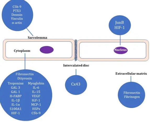
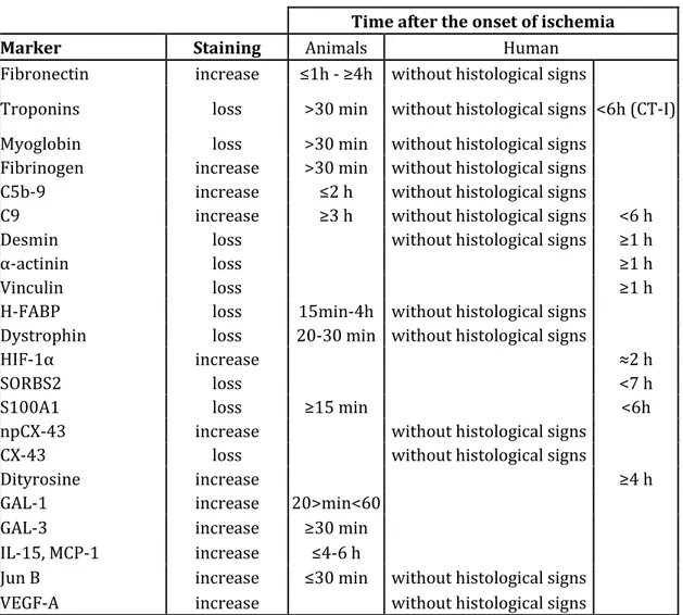
The most important evidence on the studied markers are summarized in Figure 1 and Table 1.
12
Figure 1. Localization of analyzed markers for immunohistochemical detection of early myocardial ischemia.
13
Time after the onset of ischemia
Marker Staining Animals Human
Fibronectin increase ≤1h - ≥4h without histological signs
Troponins loss >30 min without histological signs <6h (CT-I) Myoglobin loss >30 min without histological signs
Fibrinogen increase >30 min without histological signs C5b-9 increase ≤2 h without histological signs
C9 increase ≥3 h without histological signs <6 h
Desmin loss without histological signs ≥1 h
α-actinin loss ≥1 h
Vinculin loss ≥1 h
H-FABP loss 15min-4h without histological signs Dystrophin loss 20-30 min without histological signs
HIF-1α increase ≈2 h
SORBS2 loss <7 h
S100A1 loss ≥15 min <6h
npCX-43 increase without histological signs
CX-43 loss without histological signs
Dityrosine increase ≥4 h
GAL-1 increase 20>min<60
GAL-3 increase ≥30 min
IL-15, MCP-1 increase ≤4-6 h
Jun B increase ≤30 min without histological signs
VEGF-A increase without histological signs
Table 1. Summary of the result for the main evaluate immunohistochemical markers with the description of change induced by ischemia and the temporal timeline of evaluation.
As reported above, several potential EMI markers have been identified and tested during the past decades including those appearing in the very early inflammatory phase and some of these that were analyzed only in animal samples, needs to be validated for human myocardial tissue. The performed studies have focused on molecules with different tissue distributions that could be used in combination to demonstrate cellular and extracellular damage and to study the possible differences in the evolution of damage between these compartments.
14
Although the detection method revealed to be reliable, convenient, affordable, and applicable to formalin-fixed, paraffin-embedded tissues taken from the body for the preliminary routine histological analysis, incorporating a panel of markers into routine diagnostics still requires testing the marker specificity and evaluating as these markers can be affected by factors as global ischemia due to cardiac arrest at death and post-mortem phenomena (i.e. autolysis and putrefaction).
To date, the markers tested for EMI specificity in human are H-FABP, JunB, VEGF-A, HIF-1α, Cx43 and C5b-9 complex. The influence of postmortem phenomena was studied on the C5b-9 complex, which results as a reliable marker even in myocardial tissue with advanced putrefaction and autolysis.
Thus, all studies confirm that the pathophysiological mechanisms involved in the response to myocardial infarction represent very important tools for the forensic investigation of early myocardial ischemia, contributing to identify several markers. Despite this, more researches are needed to deepen the sensitivity, specificity, and stability and to create a validated panel of markers for routine use.
15
3.
The Study
Myocardial infarction is a life-threatening condition and the most frequent cause of sudden cardiac death. Therefore, an accurate diagnosis is crucial for clinicians and pathologists also for its practical implications for counseling among family members.
In the clinical practice, the diagnosis doesn’t represent a challenge if death occurs after the assessment of infarction by electrocardiography, biochemical markers and/or imaging. Nevertheless, if death occurs suddenly and out-of-hospital, mainly in subjects without any prior symptoms, the diagnosis of early myocardial ischemia or myocardial infarction falls under forensic pathologist, raising additional difficulties and practical implications related to assisting public prosecutors in legal cases and requiring accurate autopsy and epidemiological reports.
In light of the current knowledge on post-mortem diagnosis of early myocardial ischemia, an experimental study on myocardial tissue taken from Judiciary autopsy of subjects with sudden cardiac death and coronary atherosclerotic disease was herein carried out. By immunohistochemistry, the aim of the study was to analyze the expression of dystrophin (Dys) and matrix metalloproteinase 9 (MMP-9) in order to evaluate their specific expression, which could be utilized as useful markers for post-mortem diagnosis of early myocardial ischemia. In addition, as second end-point, we will compare the immunohistochemical expression of these proteins to C5b-9 and fibronectin; finally, the temporal timeline of appearance of these four markers has been evaluated.
16
3.1 Dystrophin and MMP-9
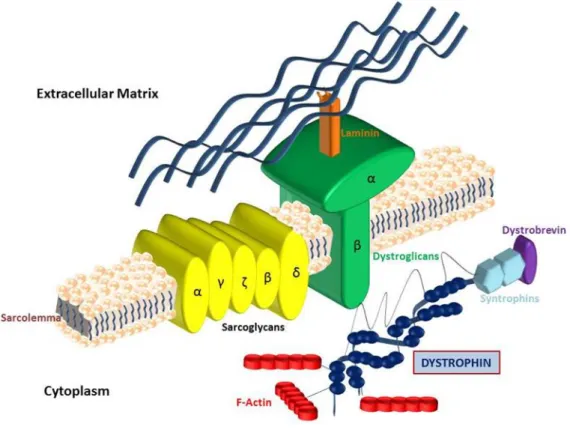
Dystrophin is a cytoskeletal protein belonging to β-spectrin/α-actin protein family, able to create the dystrophin-glycoprotein complex (DGC) [75]. This is a 427-kDa protein with binding affinity to cytoskeletal actin; DGC, in turn, comprises a transmembrane complex linked laterally by β-dystroglycan to α-dystroglycan, a surface membrane receptor for the extracellular matrix component laminin (Fig. 2), suggesting that DGC provides a structural link between the myocyte cytoskeleton and extracellular matrix [75,76]. DGC is involved in contractile force transmission and plasma membrane stabilization.
17
Several studies have been carried out on dystrophin and in general on DGC concerning their involvement in genetic diseases as myopathies characterized by the anatomic and functional alteration of cardiac and skeletal muscle [77,78].
Some of these researches have regarded the localization of dystrophin in cardiac tissue to better understand the protein function and, thus, as it contributes to the myocardial dysfunction. These studies have been performed on both animal and human myocardial tissue by various microscopic techniques, as immunoconfocal fluorescence microscopy and electron microscopy, showing that dystrophin is distributed at the sarcolemma, with concentration at the lateral costameres, suggesting a mechanical role in surface membrane support [79,80]. Moreover, it was observed also in the cardiac T-tubule, hypothesizing a cellular function besides that of mechanical support and transmission of force [81]. Therefore, dystrophin appears to be crucial in maintaining the membrane stability in stress imposed during contraction; moreover, it represents a strong linkage between the sarcolemma and costameric cytoskeleton in order to promote the transmission of contractile force [82,83]. Consequently, the role of dystrophin has been studied with reference to the vulnerability of myocardial mechanical force due to ischemic-reperfusion injuries whose pathogenesis is associated, also, with sarcolemmal membrane fragility [84,85]. The correlation of sarcoglycans and dystrophin decrease with heart failure after an acute myocardial ischemia has been demonstrated in an experimental study [84]. Regarding the myocardial contractile dysfunction due to infarct, some researchers suggested that the dystrophin damage, causally related to disruption of the plasma membrane, could be associated with reperfusion injuries, considering also the observed complete restoration of membrane dystrophin content after the reperfusion in 15 minutes ischemic heart with complete recovery of contractile force [85].
In addition, dystrophin has been analyzed to better understand the pathophysiological mechanism of the transition of ischemic injuries from reversible to irreversible on the basis of accompanying morphological changes in
18
the sarcolemma as formation of subsarcolemmal blebs, detachment of sarcolemmal membranes from the basement membrane and focal discontinuities in the lipid bilayer determining the rupture of plasma membrane detectable in prolonged ischemia or during reperfusion [86,87].
The above-summarized evidence and the forensic studies on the early damage of cytoskeletal proteins after myocardial ischemia, led to carry out the analysis of dystrophin aiming to test if it could be a marker for the post-mortem diagnosis of early myocardial ischemia.
In this regard, there are two studies on animal and human samples respectively [51,52]. Hashmi et al. analyzed the expression of dystrophin in mice killed after induction of myocardial infarction at different time points; the immunohistochemical investigation of the 20- to 30-min post-infarction samples showed well-demarcated foci of complete sarcolemmal loss of dystrophin staining in the region supplied by ligated artery and areas of partial loss of membranous staining in vicinity to those [51]. Successively, in 2018, Mondello et al. tested the use of the same protein in the heart of a subject with CAD-related sudden cardiac death, observing the loss of sarcolemmal dystrophin as different degrees according to more or less significant histological evidence of myocardial ischemia [52].
Matrix metalloproteinase 9 (MMP-9) is a gelatinase being part of MMPs family comprising 23 distinct secreted or membrane-anchored endopeptidases in humans that belong to the metzincin superfamily of metalloproteases [88]. MMP-9, as all the MMPs, is primarily considered an extracellular protease associated with degradation and regulation of the extracellular matrix, but more recently it was also observed the role of MMPs in activation or inactivation of bioactive molecules (i.e. growth factors, cell-surface receptors, cytokines, chemokines) and thus in the regulation of extra and intracellular signaling pathways [89]. The proteomics and degradomics era with the characterization of all proteases, inhibitors, and protease substrates by genomic and proteomic techniques, has
19
supported the above reported functions and exponentially expanded the known substrate repertoire of the matrix metalloproteinases (MMPs), even to include intracellular proteins with newly recognized extracellular functions (Fig. 3) [90,91].
Figure 3. From Morrison et all. [87]: in (a) the percentage of previously identified MMP substrates of which ECM molecules and regulators predominate; in (b) many novel substrates identified by proteomics.
Several researchers had evaluated the MMP-9 in the setting of cardiovascular disease reporting a pivotal role in atherosclerosis, hypertension, myocardial infarction, and heart failure [92,93]. It was observed a low MMP-9 expression at the gene and protein levels under physiological conditions, while a strong elevation was highlighted under pathological conditions including multiple cardiovascular diseases [94,95]. It was described that MMP-9 signaling begins at the extracellular-cell membrane interface, where cytokines, chemokines, growth factors, or ECM proteins engage cell surface receptors to transmit signaling cascades that culminate in MMP-9 release, activation, and proteolysis of substrates [96-99].
The role of MMP-9 in post-infarction left ventricle remodeling was the subject of several studies, which highlighted that, during the post-MI inflammatory response, there was a strong stimulation of MMP-9 in a variety of cell types [93]. Neutrophils and macrophages are the predominant early sources of MMP-9 and, in
20
addition, fibroblasts, cardiomyocytes, and endothelial cells are, also, relevant sources [100,101].
Acute myocardial infarction with irreversible tissue injury is followed by a progressive wound healing process that can evolve to prolonged pathological remodeling of the left ventricle [27].
The interactions between the cardiac cells and the extracellular matrix are fundamental for maintaining the structural and functional integrity of the heart, but after myocardial infarction, the tissue remodeling occurs with the reorganization of the extracellular matrix and the myocytes, determining the loss of heart function [102,103]. For this reason, the research was focused also on MMP-9 to explain the pathobiology associated with MI as well as to provide therapeutic means able to improve outcomes for the post-MI patients.
MMP-9 is involved in many aspects of the inflammatory and proliferative stages of acute myocardial infarction increasing very early post-MI and remains high for the first week in both animal models of MI and human patients with MI [94,104]. In particular, experimental studies on animal samples revealed the expression of MMP-9 within the first 24-48 hours following left coronary ligation, suggesting a role in the initial tissue destruction and the extension of tissue damage [105]. In extracellular matrix degradation, the neutrophil-derived MMP-9 has an early effect, facilitating into the infarctual area also the macrophage cell infiltration, another additional source of MMP-9, which significantly appears at day 3, with a peak at day 5 post-MI [106].
The improvement of the knowledge on MMP-9 substrates in relation to myocardial infarction and left ventricle remodeling is, thus, very important and the matridomics and degradomics approaches can lead to obtaining better evidence on the molecular network for this particular MMP [91]. By the coupling of these approaches, broad substrates were identified and other molecules are currently considered as candidate substrates (Fig. 4) [107].
21
Figure 4. Modified from Patterson et al. [107]: a selection of the MMP-9 known molecular interaction network.
Therefore, considering the analysis of dystrophin expression results to be useful for evaluate the ischemic injury and the related sarcolemmal damage, the analysis of matrix metalloproteinase 9 was chosen to evaluating if the plasma membrane damage is associated with a temporally related extracellular damage.
22
4.
Materials and Methods
A retrospective observational analysis of macroscopic and histopathological data of 564 Judiciary autopsy performed between June 2014 and June 2018 was performed, and 32 cases of sudden cardiac death due to coronary atherosclerosis disease were selected. Cases with cardiopulmonary resuscitation (CPR) were excluded from the selection. All autopsies were performed from 24 to 48 h after death. The post-mortem diagnosis in these cases was based on circumstantial data, negative toxicological investigations, anatomic distribution and severity of coronary atherosclerosis, eventual findings of macroscopic and microscopic myocardial ischemic damage, excluding any other cause of death.
Selected tissue fragments, after neutral buffered formalin fixation for 24-72 hrs, were processed by automatic histokinette engine (Leica) and furtherly embedded in paraffin at 56°C. On corresponding tissue blocks, 4-5 µm thick sections were cut and routinely stained by hematoxylin and eosin. The 32 selected stained cases were divided into two groups: CAD-related SCD with gross and/or histological evidence of myocardial infarction (group 1 or MI group: n = 16 ; 10 men, 6 women, mean age 68,06 yrs.) and CAD-related SCD with no specific histological signs of myocardial ischemia (group 2 or EMI group: n = 16; 13 men, 3 women, mean age 63,12 yrs.).
A third group formed by myocardial tissue of subjects dead by acute mechanical asphyxiation, with absent or minimal signs of coronary atherosclerosis was used as a control group (n = 10; 3 women, 7 men, mean age 61,14 yrs.).
For each case, three routinely stained tissue sections, obtained from the heart and coronary arteries, were histologically re-examined, taking into consideration only samples obtained from the left ventricle. However, it is well known that mainly affects the left coronary arterial tree as well as the left ventricle.
23
4.1 Immunohistochemistry
4-5 µm thick serial sections were mounted on xilane-coated slides for immunohistochemical staining with dystrophin and fibronectin. After deparaffinization and hydration to buffer (water), the sections were subjected to heat-induced epitope retrieval procedure as follows: sections were immersed in a preheated water bath with Tris/EDTA buffer solution in the staining jars and incubated for 20 min at 95 °C. Successively, slides were removed from the water bath and were cooled in the retrieval solution for 20 min at room temperature; finally, the sections were rinsed with Tris-buffered saline solution containing Tween 20 at room temperature for 4 min.
The endogenous peroxidase present in tissue sections was blocked by phosphate buffer containing 3% hydrogen peroxide (EnVision™ Flex Peroxidase-Blocking Reagent, Dako, Italy, code SM801). Subsequently, the sections obtained from group 1 and group 2 were incubated overnight at 4 °C with polyclonal rabbit anti-dystrophin antibody (Novus Biologicals, Abingdon, UK, code NBP1-89953; working dilution 1:2500), polyclonal rabbit anti-MMP-9 antibody (Novus Biologicals, Abingdon, UK, code NB600-1217; working dilution 1:50), monoclonal mouse anti-C5b-9 antibody (Abcam, USA, code ab66768; working dilution 1:100) and polyclonal rabbit anti-fibronectin antibody (Nordic BioSite, Denmark, code BT-BS1644; working dilution 1:150). The sections of the control group were incubated only with polyclonal rabbit dystrophin antibody and polyclonal rabbit anti-MMP-9 antibody to evaluate the protein expression in myocardial tissue without ischemic damage.
The immunoreactions were then visualized using EnVision™ Flex/HRP (Dako, Italy, code SM802), followed by colour development with 3,3’-diaminobenzidine tetrahydrochloride (EnVision™ Flex DAB+ Chromogen, Dako, Italy, code DM827), according to the manufacturer’s instructions.
24
Negative controls were obtained omitting the specific antisera and substituting PBS for the primary antibody.
4.2 Statistical analysis
Immunopositivity for each marker was semiquantitatively scored. In detail: - Dystrophin (Dys) positivity: score 0, negative; score 1, weak and focal sarcolemmal positivity; score 2, scattered and discontinuous sarcolemmal positivity; score 3, strong and diffuse sarcolemmal positivity;
- Matrix metalloproteinase 9 (MMP-9) positivity: score 0, negative; score 1, endovascular leucocytes positivity; score 2, strong endovascular leucocytes positivity and/or weak interstitial positivity; score 3, dense interstitial positivity;
- C5b-9 complex positivity: score 0, negative; score 1, slight positivity in single cells and/or in small cell group; score 2, mild positivity in larger cell groups; score 3, strong and diffuse positivity;
- Fibronectin (FN) positivity: score 0, negative; score 1, weak positivity in the vessels and/or focal interstitial/cytoplasmic positivity; score 2, strong endovascular positivity and/or scattered mild interstitial/cytoplasmic positivity; score 3, dense and strong cytoplasmic positivity.
Data referring to the semiquantitative grading of all markers in group 1 (MI) and 2 (EMI) were expressed as mean, standard deviations, median value, minimum and maximum; the categorical variable (gender) as number and percentage.
Since the examined variables did not present normal distribution, as verified by the Kolmogorov Smirnov test, a non-parametric approach has been utilized.
Mann Whitney test and chi-squared test were applied, respectively, to assess the existence of possible differences in age and gender between-group MI and group EMI. Moreover, Mann Whitney test was used to perform statistical comparisons between MI and EMI groups for each marker. Then, an evaluation by
25
the Wilcoxon test was made for two-by-two comparisons between markers, respectively within group 1 and group 2 (Dys/C5b-9, MMP-9/C5b-9, Dys/FN, MMP-9/FN, Dys/MMP-9).
The nonparametric Spearman correlation test was applied to assess for all subjects the existence of any significant interdependence between examined markers.
A p-value less than 0,05 was considered statistically significant.
26
5.
Results
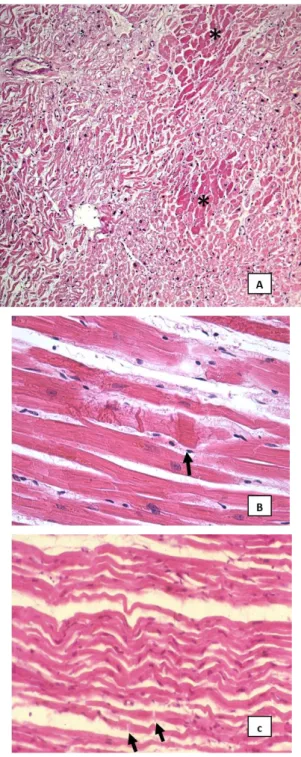
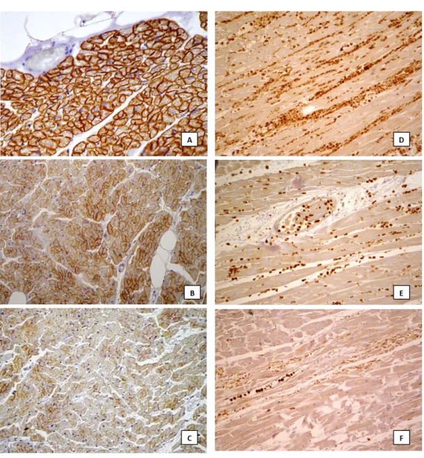
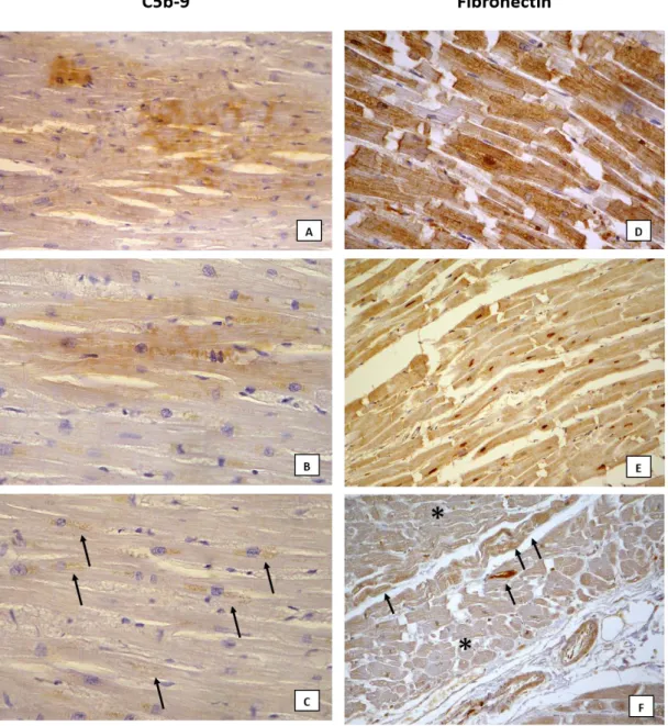
Representative images of the routine histological findings are showed in Figure 5; representative images of the score used for statistical analysis are reported in Figure 6 and 7.
Characteristics of the cases of group 1 and group 2 and their semiquantitative evaluation are reported in the following table.
N. Group Sex Age H&E staining C5b-9 FN Dys MMP-9 1 Group 1 (MI) F 70 Prominent interstitial oedema;
loss of nuclei and cross striation; coagulative necrosis became evident with
neutrophil polymorphs infiltration 3 2 1 2 2 M 57 3 3 1 3 3 F 49 2 2 0 2 4 M 68 3 3 1 2 5 M 81 2 2 0 3 6 M 79 3 3 1 2 7 F 55 3 1 2 2 8 F 76 3 2 0 3 9 M 45 2 2 1 2 10 M 73 2 2 2 3 11 M 61 3 3 1 3 12 F 72 2 3 0 2 13 M 69 3 3 1 2 14 F 74 2 2 1 2 15 M 82 3 1 2 2 16 M 78 3 2 1 1 17 Group 2 (EMI) M 48 Foci of interstitial oedema; wavy cardiac myocytes; contraction band; myofiber break-up 1 0 1 1 18 M 67 2 2 1 1 19 M 65 2 1 2 0 20 M 56 2 2 1 2 21 F 75 1 1 2 0 22 M 61 1 2 2 1 23 M 70 2 1 1 1 24 F 54 1 0 2 1 25 M 68 2 1 1 1 26 M 76 2 2 1 1 27 M 69 1 1 1 0 28 M 66 2 2 1 1 29 M 56 2 0 3 1 30 M 53 2 1 2 1 31 F 74 1 1 2 0 32 M 52 1 2 1 2
27
Statistical analysis showed no significant differences for age (p=0.105) and gender (p= 0.238) between the two groups.
Group 1. The H&E stained sections showed areas of a partial or total loss of
normal myocardial architecture associated with alterations of myocytes and foci of interstitial fibrosis. The ischaemic damage was characterized by the loss of nuclei and cross striation in cells, prominent edema, infiltration of neutrophil polymorphs and in some cases clear coagulative necrosis (Fig. 5A)
Immunohistochemistry revealed well-demarcated areas of complete loss of dystrophin sarcolemmal staining (Fig. 6C). MMP-9 expression was intense in endovascular and interstitial leucocytes, with strong and diffuse immunoreactivity in the interstitial space (Fig. 6D and E). A strong and diffuse expression of C5b-9 was observed in the cytoplasm of cardiomyocytes (Fig. 7A) as well as an intense positivity for fibronectin in blood vessels, interstitial space and myocytes (Fig. 7D).
Group 2. The H&E staining showed partial loss of normal myocardial
architecture with foci of interstitial fibrosis. The most common findings were interstitial edema, wavy cardiomyocytes and break-up of myofibers (Fig. 5B); some cases showed contraction band (Fig. 5C).
The immunohistochemical staining revealed a discontinuous sarcolemmal positivity of dystrophin in groups of cells and/or loss of staining in isolated cells (Fig. 6B). The immunopositivity of MMP-9 was mostly observed in endovascular leucocytes rarely associated with weak interstitial expression (Fig. 6F). The C5b-9 expression was slight and focal in cell groups (Fig. 7B and C). The fibronectin positivity resulted from weak to intense in endovascular space and, in some cases, scattered in the interstitial space and/or weak in the cytoplasm (Fig. 7E and F).
Control group. The H&E stained sections showed mild interstitial edema
and some foci of interstitial fibrosis.
The dystrophin positivity appeared as an intense and thin sarcolemmal staining, encircling the myocardial cells (Fig. 6A). MMP-9 antibodies expression
28
was negative. The C5b-9 staining resulted negative. Fibronectin was negative or scattered.
29
Figure 5. Example of routine histology findings: areas of clear coagulative necrosis (asterisks) associated with zonas of interstitial edema and wavy myocytes, observed in case n. 4, group 1 (A: 10x); myocardial fibers showing contraction band (black arrows) observed in case n. 25, group 2 (B: 40x); wavy myocytes associated with interstitial edema and widening of some intercalated disk (black arrows), observed in case n. 21, group 2 (C: 20x).
30
Figure 6. Example of dystrophin and MMP-9 expression. Dystrophin: score 3 observed in case control group characterized by strong sarcolemmal staining encircling myocytes (A: 40x); score 2 evaluated in case n. 22, group 2, showing discontinuous sarcolemmal staining (B: 20x); score 1 observed in case n. 9, group 1, showing clear areas of complete loss of sarcolemmal staining (C: 20x). MMP-9: score 3 observed in case n. 10, group 1, with dense and strong expression in extracellular matrix (D: 20x); score 2 evaluated in case 6, group 1, with positivity of endovascular and interstitial leucocytes (E: 40x); score 1 evaluated in case n. 30, group 2, with predominant staining of endovascular leucocytes and slight extracellular matrix positivity (F: 20x).
31
Figure 7. Example of C5b-9 and fibronectin expression. C5b-9: score 3 observed in case n. 2, group 1, where the positivity was scattered strong involving a large group of myocytes (A: 40x); score 2 evaluated in case n. 28, of group 2, represented by areas of mild positivity in few myocardial cells (B: 40x); score 1 observed in case n. 22, group 2, in which some of cells showed slight positivity (black arrow) (C: 40x). Fibronectin: score 3 observed in case n. 12, group 1, with strong positivity (H: 40x); score 2 observed in case n. 14, group 1, showing mild positivity in cardiac myocytes (E: 40x); score 1 observed in case n. 19, group 2, with focal but clear cytoplasmic positivity in few cells (black arrows) surrounded by cells with weak expression (asterisks) (F: 20x).
32
5.1 Statistical results
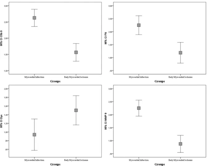
The statistical comparisons between group 1 (MI) and group 2 (EMI) to analyze the expression of all markers showed significant differences, revealing a p<0.05 for Dys and a p<0.001 for MMP-9, while a p<0.001 for both C5b-9 and FN was noted. The expression of MMP-9, C5b-9 and Fibronectin appeared significantly greater in the MI group than in the EMI group, while dystrophin was more expressed in the EMI group (Fig. 3).
The two-by-two statistical comparisons of the expression of the markers within group 1 revealed no significant differences between MMP-9 and C5b-9, and between MMP-9 and Fibronectin; on the other hand, significant differences were observed between dystrophin and each of the other three markers (p<0.005).
The same comparisons within group 2 showed significant differences between dystrophin and fibronectin, dystrophin and MMP-9, C5b-9 and MMP-9 (p<0.05).
The analysis of rank correlation between each pair of markers, in the whole sample (n=32), revealed the existence of a direct correlation between C5b-9 and fibronectin (ρ=0,524; p<0.005), C5b-9 and MMP-9 (ρ=0,593; p<0.001), Fibronectin and MMP-9 (ρ=0,641; p<0.001); an inverse correlation was observed between dystrophin and fibronectin (ρ= 0,572; p<0.005), dystrophin and MMP9 (ρ= -0,449; p<0.05).
33
Figure 8. Error bar graphs show median value, standard deviation, minimum and maximum of each marker, comparing group 1 (MI) to group 2 (EMI).
34
6.
Discussion
The post-mortem diagnosis of CAD-related SCD can be more difficult to perform if it is based only on routine investigations, as cardiac macroscopic findings and haematoxylin-eosin analysis. Consequently, the main interest of our study was to verify whether early myocardial ischemia in cases of sudden cardiac death may determine an early sarcolemmal damage associated with early extracellular matrix injury. This latter phenomenon should be documented by the analysis of dystrophin and MMP-9 expression representing thus additional markers in forensic cases without specific myocardial signs at histological examination.
The correlation between EMI and damage of myocytes sarcolemma has been elsewhere previously demonstrated by an altered immunohistochemical expression of cytoskeletal proteins as desmin, vinculin, and α-actinin [49,50]. However, dystrophin has been firstly analyzed in animal samples [51] and successively in humans [52], exhibiting an important role in DGC, which is in turn involved in myocytes sarcolemma stabilization and contractile force transmission [75,76,79].
The reported previous data on human samples are confirmed in the present study. In detail, considering the control group without any myocardial ischaemia, dystrophin immunostaining results normally localized in the sarcolemma of cells and it appears as thick sarcolemmal staining encircling cardiac myocytes; this pattern should be considered the expression of normal cardiac architecture. On the contrary, the altered expression of sarcolemmal dystrophin is highlighted in both CAD-related SCD with histological evidence of myocardial infarction group as well as CAD-related SCD with no specific histological signs of myocardial ischemia group. Particularly, the loss of sarcolemmal dystrophin is more pronounced in cases with clear histological signs, in which were present well-demarcated areas
35
of complete loss of staining of cardiac myocytes. Loss of staining has been also observed in cases of sudden cardiac death with unspecific histological signs of ischemic damage, although it appears as areas of loss in dystrophin membranous staining of isolated cells or among groups of myocytes.
The observed loss of dystrophin can be associated with structural myocyte alterations correlated to irreversible ischemic injury. This was reported in some researches [108-111] describing the plasma membrane ultrastructural changes in experimental models related to the transition from reversible to irreversible ischemic myocardial injury. One of these morphological changes in experimental animals is the formation of sub-sarcolemmal blebs, which has been ultrastructurally evaluated [108,109]. A study investigating the cause of blebs formation has also shown the correlation between dystrophin loss and sarcolemmal blebs formation as well as the chronological concordance between the onset of blebs formation and dystrophin loss [110]. Moreover, it would seem that dystrophin depletion is associated with the depression of protein transcription and translation [111].
These findings support the results of the present immunohistochemical study highlighting that loss of sarcolemmal dystrophin can be considered a sign of irreversible myocardial ischemic injury in forensic cases of CAD-related sudden cardiac death. Moreover, dystrophin depletion appears to be time-dependent with a different degree of immunostaining (from reduction to absence) in relation to different histological findings (with or without specific signs of myocardial ischemia).
The present study analyzed also the expression of MMP-9, a gelatinase involved in the degradation of the extracellular matrix, which has several substrates as collagenases and laminins [107].
It is well known that the ischemic injury determines dynamic alterations in the cardiac extracellular matrix which are associated with the mechanical properties of the infarcted heart and they are also directly involved in the
36
modulation of the inflammatory and reparative response [112,113]. The MMP-9 represents one of the proteases with rapid activation during the inflammatory phase of tissue healing, being involved in the degradation of the cardiac extracellular matrix with the formation of matrix fragments exerting a potent pro-inflammatory action [114,115].
In our study, the analysis of MMP-9 expression reveals the immunostaining of neutrophils both in CAD-related SCD with histological pieces of evidence of myocardial infarction group and in CAD-related SCD with no specific histological signs of myocardial ischemia group, but with significant differences. In the first group, the expression of MMP-9 is highlighted in endovascular and interstitial neutrophils, associated with an evident immunoreactivity in interstitial spaces. In the second group, the expression was exclusively observed only in endovascular leucocytes. This suggests the dynamics of the gelatinase in myocardial ischemia in which, during the early phase, is mostly released from neutrophils. These leucocytes, from the vessels, pass through the interstitial space to the damaged area, releasing the MMP-9 that is involved in the degradation of extracellular matrix components as collagen.
A study performed by scanning and electron microscope on pig hearts showed the irregular arrangement of collagen network at 40 minutes after the coronary artery ligation [116]; the alterations were more pronounced at two hours resulting as the separation of the collagen fibrils and microfibrils between myocytes from the basement membrane, associated with their breaking and decrease in content [116]. The total content of collagen was better studied in rats, demonstrating that at 1, 2 and 3 hours from the coronary artery ligation, it has been documented a decrease in the infarct zone by 25±8%, 35±7%, and 50±10% respectively [117].
On the basis of these evidences, the collagenolytic activity of MMPs was studied by several techniques as zymography revealing that the increased activity of MMP-9 was detected in the infarct zone after 2 hours from the induced ischemia
37
and it resulted growing at 4 hours and finally very prominent at 24 hours [118]. Moreover, the analysis of MMP-9 activities indexed by densitometer optical reading resulted increased after 2 hours, respect to other MMPs in which the increase was observed at 1 hour, but it was also described that MMP-9 continued to increase over the time, while the others showed fluctuations [118].
On the other hand, the clinical studies of AMI patients offered discrepancies in findings, probably related to differences in patient characteristics, interventions, as well as timing and methods of MMP measurement [119]. Using zymography, the MMP-9 activity was measured in acute STEMI patients within 24 h, revealing that plasma MMP-9 levels at the time of percutaneous coronary intervention were higher than in the control group [120]. Instead, using a sandwich enzyme immunoassay, which could not distinguish between the active and inactive MMP proenzymes, in AMI patients on medical therapy, the MMP-9 showed two different serial changes in fact, in one-half subjects, significant MMP-9 elevations were seen on day 0 if compared to control group, whereas, in the other one-half of patients, MMP-9 levels were similar to those encountered in the control group [121].
The above-described literature reports support the findings observed in the present study showing the immunostaining of endovascular neutrophils in CAD-related SCD with no specific histological signs of myocardial ischemia group. This confirms previous reports on the roles of these leucocytes as both the earliest immune cell type involved in the inflammatory response to ischemia and the first source of MMP-9 [122]. In CAD-related SCD with histological evidence of myocardial infarction group, the immunopositivity was also observed in the interstitial space, together with endovascular and interstitial leucocytes. This may be due to the migration of endovascular activated neutrophils in interstitial space and to the further degranulation to quickly release preformed MMP-9 based on stimulation mediated by molecules as IL-8 and TNF-α, while the inactive pro-form of MMP-9 is stored in neutrophil gelatinase granules and became activated by serine elastase and other proteinases [122-125]. This leads to degradation of
38
collagen, fibronectin, and other ECM components necessary for clearance of necrotic myocardium, and possibly also the degradation of intracellular proteins, including actin, tubulin and annexin 1, representing recently discovered MMP-9 substrates [126].
Thus, as observed for dystrophin, the expression of MMP9 appears to be time-dependent revealing different degrees of immunostaining in relation to localization as well as histologic findings, with or without specific signs of myocardial ischemia.
The analysis of the expression of both dystrophin and MMP-9 showed an early modification in myocardial ischemia as demonstrated by the loss of immunopositivity and the activated neutrophils staining respectively in the EMI group (group 2). Moreover, as resulted from the statistical comparison between group 1 (MI group) and group 2, significant differences were observed in the expression supporting the time-dependent modifications of both molecules.
In the present investigation, the expression of dystrophin and MMP-9 was also compared to C5b-9 and fibronectin. This is because C5b-9 and fibronectin represent two of the most studied markers for the diagnosis of early myocardial ischemia in the forensic setting. The usefulness of both was well documented in experimental studies on animal samples and human samples showing the early overexpression of the corresponding antibody [31,32, 59]. C5b-9, above all, can be considered the closest marker to the ideal one in light of the tested sensibility, specificity, and stability [33,127].
Firstly, the analysis of correlation between each pair of markers, in the whole sample, revealed the existence of a direct correlation between C5b-9 and fibronectin (ρ=0,524; p<0.005), C5b-9 and MMP-9 (ρ=0,593; p<0.001), Fibronectin and MMP-9 (ρ=0,641; p<0.001), while an inverse correlation was observed between dystrophin and fibronectin (ρ= -0,572; p<0.005), dystrophin and MMP-9 (ρ= -0,449; p<0.05). These data are not surprising, taking into consideration the pathophysiological role of the four molecules in myocardial tissue, accordingly to
39
changes observed in the present study as well as the literature reports. Specifically, dystrophin is a cardiac myocyte protein progressively lost after myocardial ischemia and cells damage, while fibronectin is an interstitial protein overexpressed in myocardial infarction; furthermore, MMP-9 and C5b-9 are molecules activated and expressed to respond to the ischemic injury. Consequently, it can be argued that fibronectin, C5b-9 and MMP-9 are increased, while dystrophin is reduced.
Then, the two-by-two comparisons (Dys/C5b-9, MMP-9/C5b-9, Dys/FN, MMP-9/FN, Dys/MMP-9) were performed. In group 1, no significant differences comparing MMP-9 to C5b-9 and fibronectin respectively were observed, suggesting that in cases with clear histological signs of myocardial ischemic damage, a similar semiquantitative expression of the three markers can be appreciated. In these cases, the irreversible damage due to prolonged ischemia is expressed with clear evidence of inflammatory system activation, tissue alteration, and repair phenomena, that influence each other [26,128]. On the contrary, significant differences were observed between dystrophin and each of the other three markers demonstrating the cell death and destruction by the prominent loss of protein expression.
Interesting results were obtained in EMI group in which significant differences of expression between dystrophin and fibronectin, dystrophin and MMP-9, C5b-9 and MMP-9 (p<0.05) were observed, and no significant difference were highlighted between dystrophin and C5b-9, MMP-9 and fibronectin. These data suggest that loss of dystrophin was more evident respect to fibronectin overexpression and MMP-9 positivity. To explain this, it is necessary to remind that the first source of MMP-9 are neutrophils and the overexpression of fibronectin in myocardial ischemic injury depends above all on plasma fibronectin extravasation occurring after the cardiac extracellular matrix network degradation (associated with plasma membrane damage) in which is involved also the MMP-9 [27,93]. In this setting, the fibronectin serves to contribute to the formation of a fibrin-based
40
provisional matrix that works as a scaffold for migration and proliferation of infiltrating inflammatory cells, endothelial cells and fibroblasts [129,130]. This also allows to considerations about the possibility that the expression of these molecules follows a temporal line during myocardial ischemia, supposing that dystrophin damage due to ischemic injury may occur before extracellular matrix degradation mediated by MMP-9 and the fibronectin overexpression related to its plasma extravasation.
6.1 Study limitations
The study presents as bias the limitation related to the small number of cases analyzed. Nevertheless, forensic research, if compared to clinical one, is often performed on small samples in which the subjects/materials enrolled for the analysis belonged from judiciary autopsies above all. Moreover, the sample size can be, also, related to the small number of requests of clinical diagnostic autopsies in the local geographical area, with cases in which the myocardial infarct/ischemia, identified as death cause, represents a not ascertained diagnosis.
Furthermore, the effects of post-mortem changes on molecules expression were not evaluated in this study in which the heart samples belonged from “fresh” bodies. The same limitation must be observed about the analysis of cardiopulmonary resuscitation (CPR) effects. However, some considerations can be highlighted about dystrophin. In fact, its expression could be affected by factors as post-mortem changes or cardiopulmonary resuscitation influencing myocardial integrity and expression of myocyte proteins as has been observed in other studies [43,56]. Thus, when post-mortem changes or CPR occur the loss of dystrophin could be misunderstood.
41
7.
Conclusions
The research on postmortem diagnosis of EMI appears to be in continuous evolution taking inspiration from pathophysiological and clinical studies and, at the same time, offering integrations to the same in light of the possibility of implementing the morphological analysis of human hearts.
The presented study fits in this context analyzing the dystrophin, a cytoskeletal protein of cardiac myocytes, and MMP-9, a protease involved in extracellular matrix degradation due to myocardial ischemia.
The analysis of both markers highlighted the usefulness for the post-mortem diagnosis of CAD-related sudden cardiac death. In fact, the dystrophin expression loss and the MMP-9 immunopositivity were observed in different degrees according to more or less significant histological evidence of myocardial ischemia. These findings suggest that they can be a marker of early myocardial ischemia, describing “direct” cellular and “indirect” extracellular (by activated neutrophils immunopositivity) damage respectively. Moreover, it was reported that the comparison between dystrophin and fibronectin, and dystrophin and MMP-9, respectively, provided some evidence suggesting that dystrophin loss may happen before the fibronectin and MMP-9 appearance.
Obviously, considering also the above-reported study limitations other researches are needed to confirm these findings.
Finally, it must be highlighted that even if immunohistochemistry seems to be a detection method reliable, convenient and applicable to formalin-fixed paraffin-embedded tissue, the post mortem diagnosis of EMI lacks a standardized panel of markers for improving sensitivity and specificity and, thus, for performing an effective evaluation.
However, nowadays the resolution of practical cases is focused on the analysis of clinical records (if present), circumstantial data and supplementing
42
gross evaluation and histological analysis with an immunohistochemical assessment of protein changes. Regards the last one seems to be appropriate to perform the analysis of C5b-9 with, at least, other two markers because only in this way it will be possible to ensure a more accurate Bayesian approach to the diagnosis assessment.
43
References
[1] Zipes DP, Wellens HJ (1998) Sudden cardiac death. Circulation; 98:2334– 2351.
[2] Byrne R, Constant O, Smyth Y, Callagy G, Nash P, Daly K, Crowley J (2008) Multiple source surveillance incidence and aetiology of out-of-hospital sudden cardiac death in a rural population in the West of Ireland. Eur Heart J; 29:1418–1423
[3] Chugh SS, Jui J, Gunson K, Stecker EC, John BT, Thompson B, Ilias N, Vickers C, Dogra V, Daya M, Kron J, Zheng ZJ, Mensah G,McAnulty J (2004) Current burden of sudden cardiac death: multiple source surveillance versus retrospective death certificate-based review in a large U.S. community. J Am Coll Cardiol; 44:1268–1275.
[4] de Vreede-Swagemakers JJ, Gorgels AP, Dubois-Arbouw WI, van Ree JW, Daemen MJ, Houben LG, Wellens HJ (1997) Out-of hospital cardiac arrest in the 1990’s: a population-based study in the Maastricht area on incidence, characteristics and survival. J Am Coll Cardiol; 30:1500–1505
[5] Vaillancourt C, Stiell IG, Canadian Cardiovascular Outcomes Research Team (2004) Cardiac arrest care and emergency medical services in Canada. Can J Cardiol; 20:1081–1090
[6] Murakoshi N, Aonuma K (2013) Epidemiology of arrhythmias and sudden cardiac death in Asia. Circ J; 77:2419–2431.
[7] Gillum RF (1989) Sudden coronary death in the United States: 1980–1985. Circulation; 79:756 –765.
[8] Escobedo LG, Zack MM (1996) Comparison of sudden and nonsudden coronary deaths in the United States. Circulation; 93:2033–2036.
[9] Zheng ZJ, Croft JB, Giles WH, Mensah GA (2001) Sudden cardiac death in the United States, 1989 to 1998. Circulation; 104:2158 –2163.
44
[10] Roger VL, Go AS, Lloyd-Jones DM, Adams RJ, Berry JD, Brown TM, Carnethon MR, Dai S, de Simone G, Ford ES, Fox CS, Fullerton HJ, Gillespie C, Greenlund KJ, Hailpern SM, Heit JA, Ho PM, Howard VJ, Kissela BM, Kittner SJ, Lackland DT, Lichtman JH, Lisabeth LD, Makuc DM, Marcus GM, Marelli A, Matchar DB, McDermott MM, Meigs JB, Moy CS, Mozaffarian D, Mussolino ME, Nichol G, Paynter NP, Rosamond WD, Sorlie PD, Stafford RS, Turan TN, Turner MB, Wong ND, Wylie-Rosett J; American Heart Association Statistics Committee and Stroke Statistics Subcommittee (2011) Heart disease and stroke statistics—2011 update: a report from the American Heart Association. Circulation; 123:e18–e209.
[11] Fox CS, Evans JC, Larson MG, Kannel WB, Levy D (2004) Temporal trends in coronary heart disease mortality and sudden cardiac death from 1950 – 1999: the Framingham Heart Study. Circulation; 110:522–527.
[12] Myerburg RJ (2001) Sudden cardiac death: exploring the limits of our knowledge. J Cardiovasc Electrophysiol; 12:369–381.
[13] https://www.istat.it/it/files/2017/05/Report-cause-di-morte-2003-14.pdf.
[14] Huikuri H, Castellanos A, Myerburg RJ (2001) Sudden death due to cardiac arrhythmias. N Engl J Med; 345:1473–1482.
[15] Furukawa T, Moroe K, Mayrovitz HN, Sampsell R, Furukawa N, Myerburg RJ (1991) Arrhythmogenic effects of graded coronary blood flow reductions superimposed on prior myocardial infarction in dogs. Circulation; 84: 368– 377.
[16] Schmidt A, Azevedo CF, Cheng A, Gupta SN, Bluemke DA, Foo TK, Gerstenblith G, Weiss RG, Marban E, Tomaselli GF, Lima JA, Wu KC (2007) Infarct tissue heterogeneity by magnetic resonance imaging identifies enhanced cardiac arrhythmia susceptibility in patients with left ventricular dysfunction. Circulation; 115:2006 –2014.
![Figure 3. From Morrison et all. [87]: in (a) the percentage of previously identified MMP substrates of which ECM molecules and regulators predominate; in (b) many novel substrates identified by proteomics](https://thumb-eu.123doks.com/thumbv2/123dokorg/4585784.38918/20.892.129.794.327.537/percentage-previously-identified-substrates-predominate-substrates-identified-proteomics.webp)
![Figure 4. Modified from Patterson et al. [107]: a selection of the MMP-9 known molecular interaction network](https://thumb-eu.123doks.com/thumbv2/123dokorg/4585784.38918/22.892.241.667.144.549/figure-modified-patterson-selection-known-molecular-interaction-network.webp)