ALMA MATER STUDIORUM-UNIVERSITA’ DI BOLOGNA
SCUOLA DI FARMACIA, BIOTECNOLOGIE E SCIENZE
MOTORIE
CORSO DI STUDIO IN BIOTECNOLOGIE MOLECOLARI E
INDUSTRIALI
Sea organisms as a source of collagen-derived
polymers for skin tissue engineering
applications
Tesi di laurea in Cellule staminali
Presentata da: Relatori:
Modena Silvia Motta Antonella
Migliaresi Claudio Bonsi Laura Matricola n°
0000708507
2
… to my beloved grandfather and brother, Bruno and Davide
3
Table of contents
ABSTRACT
1. INTRODUCTION
1.1. Skin anatomy and physiology ... 7
1.2. Wound healing ... 9
1.2.1. Adult wound healing ... 11
1.2.2. Fetal wound healing ... 16
1.2.3. Assisted wound healing ... 18
1.3. Tissue Egineering: definition and approaches for skin replacement ... 1.4. Scaffold for skin tissue engineering: strategies for matrix design and fabrication ... 20
1.5. Polymers for skin replacement in Tissue Engineering ... 1.6. Cell sources ... 35
1.7. Open questions with commercially available skin substitutes……..……… 2. RESEARCH STRATEGIES AND OBJECTIVES 2.1. Aims of the thesis ... 37 2.2. Strategies: polymer selection and processing definition...
3. MATERIALS AND METHODS
3.1. Jellyfish collagen extraction ... 3.2. Jellyfish isolated collagen characterization………..
3.2.1. Molecular weight evaluation by SDS-polyacrylamide gel electrophoresis ... 3.2.2. Collagen tertiary conformation analysis by circular dicroism spectroscopy ... 3.2.3. Secondary structure evaluation by Fourier transform infrared spectroscopy .... 3.2.4. Collagen thermal beahviour analysis by differential scanning calorimetry ... 3.2.5. Collagen amino acid composition measurements by high performance liquid
chromatography ... 3.3. Silk fibroin water solution processing ... 3.4. Scaffold fabrication ... 3.4.1. Tissue Culture Plate-well coating ... 3.4.2. Films ... 3.4.3. 3D sponges ... 3.5. Scaffold characterization ... 3.5.1. Morphological analysis by scanning electron microscopy ... 3.5.2. Secondary structure evaluation by Fourier transform infrared spectroscopy .... 3.5.3. Thermal behaviour by differential scanning calorimetry ... 3.6. Preliminary biocompatibility evaluation: in vitro testing ...
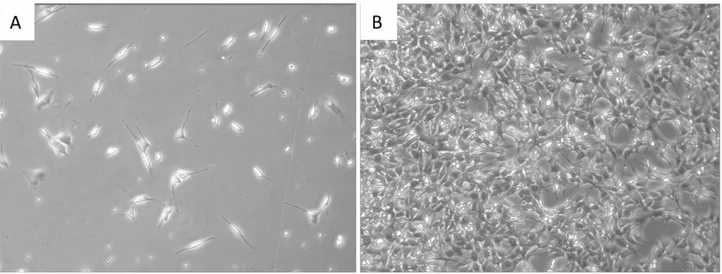
3.6.1. Cell lines: MRC-5, NIH 3T3 ...
3.6.2 Design of cell culture ... 3.6.3 Cytotoxicity: LDH-based assay ...
4
3.6.4 Confocal laser scanning microscopy (image acquisition) ...
3.6.5. Cell viability: Live/Dead ... 3.6.6 Metabolic activity: Alamar blue® assay ...
3.6.7 Cell proliferation: PicoGreen assay ... 3.6.8 Collagen synthesis: Immunocytochemistry ... 3.6.9 Specific markers expression: Reverse transcription quantitative PCR in real time ... 3.6.10 Statistical analysis ...
4. RESULTS AND DISCUSSION
4.1. Optimization of jellyfish collagen extraction ... 4.2. Material evaluation on MRC-5 cell culture ... 4.2. Material evaluation on NIH 3T3 cell culture ... 4.3. Collagen expression and assembly ...
5. CONCLUSIONS AND FUTURE PERSPECTIVES
BIBLIOGRAPHY ... ACKNOWLEDGMENTS ...
5
Abstract
Advanced therapies combating acute and chronic skin wounds are likely to be brought about using our knowledge of regenerative medicine coupled with appropriately tissue engineered skin substitutes. At the present time, there are no models of an artificial skin that completely replicate normal uninjured skin and they are usually accompanied by fibrotic reactions that result in the production of a scar. Natural biopolymers such as collagen have been a lot investigated as potential source of biomaterial for skin replacement in Tissue Engineering. Collagens are the most abundant high molecular weight proteins in both invertebrate and vertebrate organisms, including mammals, and possess mainly a structural role in connective tissues. From this, they have been elected as one of the key biological materials in tissue regeneration approaches, as skin tissue engineering. In addition, industry is constantly searching for new natural sources of collagen and upgraded methodologies for their production. The most common sources are skin and bone from bovine and porcine origin. However, these last carry high risk of bovine spongiform encephalopathy or transmissible spongiform encephalopathy and immunogenic responses. On the other hand, the increase of jellyfish has led us to consider this marine organism as potential collagen source for tissue engineering applications.
In the present study, novel form of acid and pepsin soluble collagen were extracted from dried
Rhopilema hispidum jellyfish species in an effort to obtain an alternative and safer collagen.
We studied different methods of collagen purification (tissues and experimental procedures). The best collagen yield was obtained using pepsin extraction method (34.16 mg collagen/g of tissue). The isolated collagen was characterized by SDS-polyacrylamide gel electrophoresis and circular dichroism spectroscopy. The results revealed the existence of triple helical arrangements of collagen and its fair closeness in molecular weight with mammalian collagen. The structural properties were analyzed by using Fourier transform infra-red spectrum, the stability of collagen was given the single transition peak in differential scanning calorimetry and the amino acid analysis emphasized a possible presence of like-type I collagen. Tissue Culture Plate-well coating by using jellyfish collagen was compared with standard Tissue Culture Plate and commercially available bovine type I collagen in terms of cell viability (Live/Dead analysis) and proliferation (AlamarBlue® assay). The in vitro results show that
jellyfish collagen exhibited higher cell viability than the other samples. Film-like scaffolds and porous three-dimensional scaffolds composed of jellyfish collagen and a water-stable natural
6
polymer, silk fibroin, presented smooth surface with micro roughness and highly porous and interconnected pore structure, respectively. The latter appeared to be useful for an high-density cell seeding and an efficient nutrient supply. Adhesion, spreading and proliferation of MRC-5 and NIH 3T3 fibroblasts on silk fibroin, silk fibroin/collagen blend and jellyfish coated fibroin scaffolds were also observed to investigate the biocompatibility and a potential bioactive role of collagen. The biological evaluations suggested that the adding of collagen, as bioabsorbable coating containing RGD sequence, could facilitates fibroblasts attachment and proliferation in vitro. Additionally, a preliminary study was made in order to determine the potential of collagen-based scaffold in promoting wound healing biomarkers by fibroblasts. The secretion of type I and III collagens in collagen coated fibroin scaffolds was assessed by immunocytochemistry and reverse transcription quantitative PCR in real time. Our data confirmed the broad biocompatibility of jellyfish collagen, its biological effects on mammalian cells and its important role in promoting cell adhesion and proliferation. Given the bioavailability of jellyfish collagen and its biological properties, this marine material is thus a good candidate for replacing bovine or human collagens in selected biomedical applications.
7
1 1. Introduction
1.1 Skin anatomy and physiology
The skin is the largest organ of the body in vertebrates, accounting for approximately 16% of the total body weight of an adult and covering its entire surface. It performs many vital functions, including protection against external physical, chemical, and biologic assailants like ultra-violet radiation and pathogens, as well as prevention of excess water loss from the body, regulation of electrolyte balance, thermos- and immunoregulation. It plays a central role as a sensory organ due to a widespread system of sweat glands and cells sensitive to touch, pain, pressure, itching, and temperature changes [1]. When this barrier is disrupted due to any cause ̶ surgery, ulcers, burns, neoplasms or traumas, the functions of the skin are no longer adequately performed. It is therefore vital to restore the integrity of the skin as soon as possible [2]. In order to understand skin functioning, it is necessary to focus on this integumentary system organization, formed by the skin and its derivative structures.
The skin is composed of three layers, the epidermis and dermis with a complex nerve and blood supply. A third subcutaneous layer, the hypodermis, is composed mainly of fat and a layer of loose connective tissue.
The outermost level of the cutis, the epidermis, is thin and highly cellular, but has sufficient thickness to provide vital barrier function. The epidermis is a stratified, squamous epithelium layer and consists of a specific constellation of cells known as keratinocytes, which function to synthesize keratin, a long, threadlike protein with a protective role. The keratinocytes, which represent the majority of the cells, differ from the “clear” dendritic cells by possessing intercellular bridges and ample amounts of stainable cytoplasm. This layer harbours a number of other cell populations, such as melanocytes, responsible for producing the pigment melanin, Langerhans cells and Merkel cells, involved in the immune response and in the sensation of touch, respectively. The epidermis commonly is divided into four layers according to keratinocyte morphology, position and maturation as they differentiate into horny cells, including the basal cell layer (stratum basale or germinativum, the deepest layer adjacent to the basal lamina), the squamous cell layer (stratum spinosum), the granular cell layer (stratum granulosum), and the cornified or horny cell layer (stratum corneum) (Figure 1.1). The epidermis renews itself continually and gives rise to derivative structures, such as pilosebaceous apparatuses, nails, and sweat glands. The basal layer, in fact, is the primary location of mitotically active cells in the epidermis, the basal cells, which undergo
8
proliferation cycles that provide for the renewal of the outer epidermis. However, not all basal cells have the potential to divide. Epidermal stem cells in the basal layer are clonogenic cells with a long lifespan that progress through the cell cycle very slowly under normal conditions. Moreover, in healthy skin, a balance between the formation of new keratinocytes in the stratum basale and the shedding of dead keratinocytes from the stratum corneum is maintained. Hyperplasiogenic conditions, such as wounding, can instead increase the number of cycling cells in the epidermis by stimulating division of stem cells. The epidermis is a dynamic tissue in which cells are constantly in unsynchronized motion, as differing individual cell populations pass not only one another but also melanocytes and Langerhans cells as they move toward the surface of the skin [1]. This level is avascular (without blood vessels) and is dependent on blood vessels of the underlying dermis for oxygenation, metabolite provision and removal of metabolic waste products [3].
The dermis, lied below the epidermis, is an integrated system of fibrous, filamentous, and amorphous connective tissue that accommodates stimulus-induced entry by nerve and vascular networks, epidermally derived appendages, fibroblasts, macrophages, and mast cells. Other blood-borne cells, including lymphocytes, plasma cells, and other leukocytes, enter the dermis in response to various stimuli as well. The dermis comprises the bulk of the skin and is composed of two layers: the reticular and papillary layers. The papillary layer contains the nerves and capillaries that nourish the epidermis, whereas the reticular layer is made up of strong connective woven network of fibres containing collagen with some elastic fibres and glycosaminoglycans (GAGs), which provide physical support, pliability, elasticity and tensile strength to the epidermis. The dermis, in fact, interacts with the epidermis in maintaining the properties of both tissues. These matrix components also vary in a depth-dependent manner and undergo turnover and remodelling in normal skin, in pathologic processes, and in response to external stimuli [1]. The major cell type present in the dermis is fibroblast (mesenchymal cell), which is responsible for collagen and elastin synthesis and is capable of producing remodelling enzymes such as proteases and collagenases, which play an important role in the wound healing process [4]. The dermal vasculature is made up of endothelial cells forming two intercommunicating plexuses: the subpapillary or superficial plexus and lower
plexus at the dermal-subcutaneous interface. This thick connective tissue is separated from
the dermis by a multi-layered membrane, the basement membrane, it is an extremely complex and dynamic structure, which is responsible of the tight junction of the upper epidermis and underlying connective tissue.
9
The dermis lies on the subcutaneous tissue, the hypodermis, which contains small lobes of adipose tissue (fat cells known as lipocytes) that is well vascularized and contributes to both the mechanical and the thermoregulatory properties of the skin [4]. Considered an endocrine organ, the subcutaneous tissue provides the body with buoyancy, hormone synthesis and functions as a storehouse of energy.
The thickness of these layers varies considerably, depending on the geographic location on the anatomy of the body; however, they form an effective barrier to the external environment, allow the transmission of sensory information, and serve a significant role in maintaining homeostasis [1]. Intact, healthy skin is essential for patient wellbeing. In fact, because the skin serves as a protective barrier against the outside world, any break in it must be rapidly and efficiently mended.
Figure 1.1: A schematic of the structure of skin. A) epidermis; B) dermis; C) hypodermis. (https://answersingenesis.org/human-body/skin/)
1.2 Wound healing
A wound is a disruption in the epithelial integrity of the skin and may be accompanied by breakdown of the structure and function of underlying normal tissue. Wounds can also occur as a part of a disease process (ex. diabetic ulcers) or have an accidental or intentional etiology
10
[5]. There are multiple reasons for skin damage, including genetic disorders (bullous conditions), acute trauma, chronic wounds or even surgical interventions. One of the most common reasons for major skin loss is thermal trauma, where substantial areas of skin can be damaged, often without the possibility of skin regeneration.
Wounds can be divided into epidermal, superficial thickness, and deep partial-thickness and full-partial-thickness with increasing depth of the injury. Treatment approaches differ accordingly.
Epidermal injuries, typical of sunburns, light scalds or grazing, do not require specific surgical treatment as only the epidermis is affected and this regenerates rapidly without scarring, as no extracellular matrix (ECM) deposition occurs to contribute towards the scar tissue.
Superficial partial-thickness wounds affect the epidermis and superficial parts of the dermis and heal by epithelialization from the margins of the wound, where basal keratinocytes change into a proliferating migratory cell type and cover the damaged area. Cells migrate either from the wound edge, hair follicle or from sweat gland remnants that lie in the deeper dermis, which has been preserved in this depth of injury. Each hair follicle and sweat gland is lined with epithelial cells capable of contributing to epithelial regeneration across the wounded surface and contains a reserve of self-renewal stem cells, located in the bulge region of the follicle.
Deep partial-thickness injuries involve greater dermal damage that results in fewer skin appendages. Remaining and pronounced scarring as fibroplasia is more intensive when compared with superficial partial-thickness wounds.
Full-thickness injuries are characterized by the complete destruction of epithelial-regenerative elements. This type of injury heals by contraction, with epithelialization from only the edge of the wound, leading to cosmetic and functional defects and extensive scarring [6].
The continuity of the skin must be restored expeditiously because it plays a crucial role in maintaining homeostasis. Tissue repair is normally a rapid process that has been devised through evolution to allow animals to escape danger and rapidly recover tissue integrity using scarring to join the wound edges or to fill tissue voids. Wound healing has often been described as a sequential natural restorative response to tissue injury and it is more specifically an event-driven process, whereby signals from one cell typeset off cascades in other cell types, which propel the wound through the phases of healing.
11
In order to fully understand the differences between regeneration and the normal outcome of tissue repair, namely fibrosis and scarring, it is useful at this point to briefly review the processes of normal wound repair or adult wound healing, the scarring process and to consider early fetal tissues, which undergo scar-free healing. In designing a smart skin replacement for grafting, it is obvious that the mechanisms of both wound healing and regeneration must be at the forefront of the tissue engineers’ mind [4].
1.2.1 Adult wound healing
Adult wound healing is essentially a repair process, which normally exhibits scarring [4]. Regardless of the aetiology of the wound, the repair processes are similar. A wound results in tissue damage, which stimulates a coordinated physiological response or cellular events to provide homeostasis and restoration of the tensile strength of injured skin. Acute wounds, including surgical incisions, usually pass through the wound healing phases relatively quickly. Wounds that demonstrate delayed healing 12 weeks after the initial insult are termed chronic wounds, often because of prolonged pathological inflammation. Surgical incisions are usually clean and cause minimal tissue loss and disruption. These wounds can be closed immediately with sutures and tend to heal rapidly. As a result, epithelial regeneration predominates over fibrosis. This is termed closure by primary intention or healing. When the tissue loss, in particular soft tissue, has been more extensive, the edges cannot be approximated, or the wound must be left open due to sepsis, the reparative process is prolonged, as the defect must fill with extensive granulation tissue. This process is termed as closure by secondary intention and involves wound contraction and epithelialization [6, 7].
At the time of insult, healing occurs as a carefully regulated, systemic cascade of overlapping processes that require the coordinated completion of a variety of cellular and extracellular activities, with the aim of restoring tissue integrity. These activities occur in a cascade that correlates with the appearance of different cell types in the wound during various stages of the healing process. These processes (triggered by tissue injury) involve four overlapping but well-defined phases of hemostasis, inflammation, proliferation, and tissue remodeling. The regulation of these events is multifactorial (Figure 1.2, 1.3) [5].
12
Figure 1.2: Major cells and their effects on wound during various stages of the normal healing process.
The vascular response and hemostasis
Bleeding occurs immediately after tissue injuries as a result of the disruption of blood vessels on a macro- or micro vascular scale. The immediate response of the body is to prevent exsanguination and promote hemostasis. Damaged arterial vessels rapidly constrict through the contraction of smooth muscle in the circular layer of the vessel wall, mediated by increasing cytoplasmic calcium levels. Within a few minutes, the reduced blood flow mediated by arteriole constriction leads to tissue hypoxia and acidosis. This promotes the production of vasoactive metabolites to cause a reflex vasodilatation and to increase vascular permeability, facilitating the entry of platelets and inflammatory cells into the extra-cellular space around the wound [7]. Upon activation, platelets, which represent the first cells to appear after injury, undergo adhesion as well as aggregation and at the same time release many mediators (e.g. serotonin) and adhesive proteins (e.g. fibrinogen, fibronectin). With the conversion by thrombin of fibrinogen to fibrins during platelet aggregation, a fibrin clot is formed to stop the bleeding [8]. The clot consists of platelets embedded in a mesh of cross-linked fibrin fibers, provides the provisional matrix for cellular migration and serves as a reservoir of cytokines and growth factors [9]. The second component of hemostasis is coagulation achieved via intrinsic and extrinsic coagulation pathways.
The cellular response and inflammation
The cellular response of the inflammatory phase takes place 24-72 hours after wounding and is characterized by the influx of leukocytes into the area of injury, whose activity is to
13
minimize bacterial contamination of the wound, thus preventing infection. Neutrophils and monocytes are recruited to the wound by chemotactic factors released during hemostasis by platelets, such as transforming growth factor-β (TGF- β) and platelet-derived growth factor (PDGF), which leads to them passing down a chemical gradient towards the wound.
In the early inflammatory state, neutrophils (polymorphonuclear leukocytes), once in the wound environment, can perform their function of killing and phagocytizing bacteria and damaged matrix proteins within the wound bed by releasing degrading enzymes and free radicals derived from oxygen. Within 24–48 hours, epithelial cells from both edges begin to migrate and proliferate along the dermis, depositing components of basement membrane as they progress [5]. Later in inflammation, the number of neutrophils declines and macrophages (tissue-derived monocytes) predominate. They appear to act as the key regulatory cells for repair. They function as phagocytic cells as well as being the primary producer of growth factors, such as epidermal growth factor (EGF) and vascular endothelial growth factors (VEGF), which are important in regulating the inflammatory response, stimulating angiogenesis and enhancing matrix production. During this late phase, collagen fibers are evident at the incision margins of the wound and epithelial cell proliferation continues, yielding a thickened epidermal-covering layer [7, 8].
Proliferative phase
The initial inflammatory responses to injury provide the necessary framework to the subsequent production of a new functional barrier. In this phase of healing, cellular activity predominates. The major events during this phase are reinforcement of the injured dermal tissue (i.e. fibroplasia), the establishment of appropriate blood supply (i.e. angiogenesis), the creation of a permeability barrier (i.e. reepithelialization), and wound contraction, which occur simultaneously [7].
Fibroplasia describes a process in which fibroblasts are stimulated to proliferate by growth factors, released from the hemostatic clot, and then migrate into the provisional matrix of wound fibrin clot, where they lay down a collagen-rich matrix (proteoglycans and elastin) and subsequently produce collagen and fibronectin. The resulting vascular, fibrous tissue, which replaces the clot at the wound site, is termed as granulation tissue. It represents a scaffold for contact guidance, a reservoir for growth factors and is composed of a different range of collagens (a higher proportion of type III collagen). Once sufficient matrix has been laid down, fibroblasts change to a myofibroblast phenotype and participate in wound contraction, by
14
connecting to the surrounding proteins like collagen. Collagens synthesized by fibroblasts are the key component in providing strength to tissue [7, 8]. Crucial interaction exists between the fibroblasts and the extracellular matrix, which helps to regulate further synthesis of the ECM and subsequent remodeling [5].
Angiogenesis refers to new blood vessel growth by the sprouting of preexisting vessels adjacent to the wound (neovascularization). As in most normal adult tissues, dermal blood vasculatures remain quiescent. In response to the injury, microvascular endothelial cells initiate an angiogenic process consisting of activation of endothelial cells, local degradation of their basement membrane, sprouting into the wound clot, cell proliferation, tubule structure formation, reconstruction of the basement membrane, and, eventually, regression and involution of the newly formed vasculature as tissue remodeling [8]. Those processes are triggered and guided by molecules that act as angiogenic factors, including fibroblast growth factors (ex. FGF), VEGF, which are secreted by fibroblasts and platelets at the healing site [10]. Angiogenic capillary sprouts invade the fibronectin-rich wound clot and organize into a microvascular network throughout the granulation tissue, providing nutrition and oxygen to growing tissues. During angiogenesis, endothelial cells also produce and secrete biologically active substances or cytokines, such as vascular endothelial growth factor and angiopoietins. Development of new capillary vessels is dependent on not only the cells and cytokines present but also the production and organization of extracellular matrix components, such as laminin [8].
Re-epithelialization is the process of restoring an intact epidermis after cutaneous injury. It generally involves several processes. The epidermal keratinocytes initially respond to an epidermal defect by migrating from the edges of the wound within 24 hours, thanks to several elements, including the extracellular matrix, integrin receptors and matrix metalloproteinases (MMPs). Reepithelialization also involves increased proliferation of keratinocytes located near the cells of the migrating front tongue. This proliferating source of keratinocytes ensures an adequate supply of cells to form a delicate covering over the exposed raw area. Further growth and differentiation of these epithelial cells restore an intact basement membrane that connects the epidermis and the underlying dermis, re-establishing the stratified epidermis [8].
Contraction of the wound begins about 7 days after injury, mediated mainly by myofibroblasts. Interactions between actin and myosin pull the cell bodies closer together decreasing the area of tissue needing to heal [7].
15
Remodeling and scar formation
The final stage of adult wound healing is the remodelling of the extracellular matrix and its subsequent changes over time, which results in the development of normal epithelium and, in the adult, maturation of the scar tissue. This phase involves a balance between synthesis and degradation of collagen and other ECM components, through mechanisms of proteolysis (metalloproteinases) and new matrix secretion (tissue inhibitors of metalloproteinases) [7]. It occurs throughout the entire wound repair process as fibrin clot formed in the early inflammatory phase is replaced by the granulation tissue that is rich, similar to the case in the fetal dermis, in immature type III collagen and blood vessels. During the proliferative phase, it is subsequently substituted by a collagenous scar predominantly of type I collagen with much less mature blood vessels [8]. As the scar, the end point of the normal mammalian tissue repair, matures, collagen bundles increase in diameter, corresponding with increasing tensile strength of the wound. However, these collagen fibers never regain the original strength of unwounded skin, and a maximum of 80% strength of unwounded skin can be achieved [5].
16
Figure 1.3: Principal events that occurs after a cutaneous injury. (http://www.pilonidal.org/_assets/pdf/phase_healing.pdf)
Certain mammalian tissues, in human in specific stage of development, have a capacity for complete regeneration without scarring. Good example include embryonic or fetal skin, which might be useful to further clarify the regenerative process, identify the factors expressed during regeneration with the aim to incorporate them to produce novel replacement skin [11].
1.2.2 Fetal regeneration
Fetal wound repair is essentially a regenerative process, characterized by an absence of scarring and fibrosis. These differences in the healing processes have sparked great interest
17
and have lead to the development of several animal models in which fetal scarless healing has been described.
Wound on early fetal mammalian tissues heal via different mechanisms with no sign of scarring and complete restitution of the normal skin architecture or result in much less scarring than an equivalent wound in adult tissues. Such healing is believed only to occur through a gestational age equivalent to the first third of human development; after that time, normal scarring as evident in the adult occurs.
The transition from scar-free embryonic wound healing to scar-forming adult wound healing is a gradual one. It is characterized by the abnormal organization of the neo-dermis, predominantly the abnormal deposition of small parallel bundles of extracellular matrix (consisting largely of collagen types I and III and fibronectin) to form the scar in the adult. This process is opposed to minimal inflammation and complete restoration of normal skin structure, with the correct deposition of large bundles of extracellular matrix and regularly distributed hair follicles, capillaries and glands in the neo-dermis of an embryonic wound. Scarring is therefore a morphogenetic problem, i.e. a failure of the regeneration of the normal skin structure as opposed to a biochemical problem, e.g. abnormal composition of the scar tissue [12].
There are a large number of differences between the healing of embryonic and adult wounds. Many of these differences are epiphenomenon, i.e. not causative of the scar-free healing phenotype because, of course, embryos are still developing and do not have the same stable phenotype as the adult. Consequently, many obvious differences between embryonic and adult wounds have been shown to be irrelevant to scar formation or the lack of it. Originally, it was thought that the sterile aqueous environment provided by the amniotic fluid was important in fetal scar-free healing, whereas adult wounds are exposed to air and numerous potential contaminating agents, e.g. bacteria, viruses, foreign bodies, etc. A particularly elegant demonstration of the irrelevance of the sterile, fluid, embryonic environment to scar-free healing was an ontological investigation of wound healing and scarring in the pouch young of the marsupial Monodelphis domestica (Armstrong & Ferguson 1995, 1997), whose skin wounds heal perfectly with no scars. Moreover, it point out that repair and regeneration of the skin appear to be correlated with the degree of skin differentiation and the inflammatory response to wounding [13].
Many other differences between embryonic scar-free healing wounds and adult scar-forming wounds have been shown, but only a few of the myriad of cellular and molecular differences
18
between embryonic and adult healing remain as potential mechanisms and therapeutic targets involved in skin scarring, including:
̶ Lack of fibrin clots and platelet degranulation,
̶ Markedly reduced inflammatory response, consisting of small numbers of poorly differentiated inflammatory cells,
̶ Markedly elevated levels of molecules involved in skin growth, remodeling and morphogenesis.
As a consequence of these principal variablesthe growth factor profile at a healing embryonic wound is very different qualitatively (i.e. the types of growth factor present), quantitatively (i.e. the amounts of such growth factors present) and temporally (i.e. the length of time the growth factors are present) compared with an adult wound. Thus, for example, there are major differences in the TGFβ isoforms present in embryonic and adult wounds. Embryonic wounds express very high levels of TGFβ3, a skin morphogenetic factor predominantly synthesized by keratinocytes and fibroblasts and very low levels of TGFβ1, TGFβ2 and PDGF, which is virtually absent in embryonic wounds (owing to the lack of platelet degranulation). By contrast, adult wounds contain predominantly TGFβ1, TGFβ2 and large quantities of PDGF. Experimental manipulation of the growth factor profile of adult wounds by exogenous addition of TGFβ3 or neutralization of TGFβ1, TGFβ2, PDGF, etc., results in markedly reduced or absent scarring [12]. Such implications may be very important in artificial skin substitutes or grafts to enhance host take and minimize scarring at the perimeter and base of the graft.
1.2.3 Strategies for guided wound healing: state of the art
Serious injury to the skin, such as burns, trauma or chronic ulcers, requires immediate coverage to facilitate repair and restore skin function and so to fulfill the wound healing process.
In the field of skin repair, the clinical ‘gold standard’ for skin replacement is the autologous skin graft in which an area of suitable skin is separated from the tissue bed and transplanted to the recipient area on the same individual from which it must receive a new blood supply. Grafts may be full thickness in which a complete section of epidermis and dermis is transplanted, or split thickness that includes only part of the dermis. After application of a graft to a full-thickness wound, its capillaries merge with the capillary network in the excise
19
wound. This “graft take” is essential for a proper supply of nutrients and ensures graft survival. The split skin donor site heals within one week and can be used for skin graft harvesting up to 4 times; however, repeated harvesting is associated with scarring at the donor sites as well as lengthy hospital stays. Moreover, in the case of a more extensive injury, donor sites are extremely limited and might leave the patient with too little undamaged skin to harvest enough autologous split skin grafts (SSGs). To address the problem of limited SSG harvesting sites, a meshing technique is used that stretches the graft and therefore can cover a larger wounded area at the expense of cosmetic and functional outcome [14].
Another possibility is the use of allografts or allogeneic transplantation (from a non-genetically identical individual of the same species), which include syngeneic grafts, performed between genetically identical individuals such as monozygotic twins, xenogeneic skin grafting that involves the transfer of tissue between species. Allografts of cadaver skin are also used as a temporary cover for full thickness burns but all these methodologies are subject to rejection, because antigens present in the donor tissue may elicit an immune reaction in the recipient [4]. These allografts can be obtained for example from non-profit European skin banks; however, there is not enough tissue available to meet the current demand and only a few of these tissue banks exist worldwide. Allografts incorporate into deep wounds and provide pain relief; however, ethical as well as safety issues remain, as the rigorous screening for viral diseases and standardized sterilization techniques
cannot completely eliminate the possibility of infective agent transmission. In comparison to autologous SSGs, a major disadvantage of allografts is that they leave the patients for weeks with wounds prone to complications. Eventually, allografts undergo immunogenic rejection and the site of injury needs to be covered with an autologous SSG. Delayed rejection can occur in patients with extensive burns due to their pathologically suppressed immune response, but eventually can be triggered by the highly immunogenic epithelial cells of the allograft during its vascularization [14]. Accordingly, there is a great need for an alternative that can provide a more permanent solution, like tissue-engineered skin substitutes, which aim for skin regeneration.
20
1.3 Tissue engineering: definition and approaches for
skin replacement
The definition of Tissue Engineering (TE) was first established in 1988, during a conference organized by the National Science Foundation in Lake Tahoe (California). It was defined as the “application of principles and methods of engineering and life sciences toward fundamental understanding of structure–function relationship in normal and pathological mammalian tissues and the development of biological substitutes to restore, maintain, or improve functions” [15]. TE refers to an interdisciplinary field of therapeutic or diagnostic products and processes, which are based upon the combination of three different components that is living cells, engineering biomaterials and suitable biochemical factors. The aim of Tissue Engineering is therefore to replace or regenerate structures and tissues in the organism, which have been compromised by traumas or pathologies implanting in the damaged site new artificially produced tissue that allows cell growth and thus restoring the original tissue. The term regenerative medicine is often used synonymously with tissue engineering, although it is a broad field that includes tissue engineering but also incorporates research on self-healing, where the body uses its own systems, sometimes with help foreign biological material to recreate cells and rebuild tissues and organs [16].
The paradigm of TE is schematically shown in Figure 1.4. Functional tissue can be fabricated starting from a scaffold that is a tridimensional structure able to guide tissue development, eventually combined with a cells and/or growth factors able to regenerate tissues in a critical size defect. Several cell sources can be employed, from cells extracted from the patient in the damaged tissue to stem cells of different origin, cultured so that they express the suitable phenotype.
From the previous considerations, it becomes evident how the approach of Tissue Engineering implies the transfer of the medical treatment to a cellular level: the resources needed for tissue regeneration are introduced in the pathologic environment in a suitable configuration to optimize their activity and help the natural processes of self-repair. The main challenge of this strategy is to control the 3D new tissue development in a functional way. The achievement of this goal is highly dependent on many variables: the use of a cell-free or a pre-seeded scaffold, the choice of the most appropriate cell source, the material of the scaffold, the
21
design of the morphological, chemical and physical properties of the scaffold, the parameters of the preliminary in vitro culture, the concentration of growth factors, and so on [17].
Fig. 1.4: The paradigm of tissue engineering [15].
The strategy generally followed consists in combining these components, in order to mimic characteristics and properties of the biological system to be regenerated. This principle is well known as biomimetic. It implies that the cellular behavior has to be known in detail, both during normal tissue morphogenesis and during the repair and regeneration phases when damage or pathology is present. In fact, only in this case we will be able to evaluate how cells can be guided towards tissue regeneration, that is, which signals have to be provided, both in
vitro and in vivo [18].
Many challenges have to be handled to reach its final aim, i.e. to gradually substitute organ transplantation. However, efforts have been done towards this objective and several tissue engineered applications are today commercially available, for instance for skin regeneration to heal burns or diabetic ulcers.
Alternative life-saving approaches in the treatment of extensive full-thickness wounds, where donor sites for SSG harvesting are not available, in fact, include the use of cultured autologous fibroblasts and/or keratinocytes and bioengineered skin substitutes. ‘Off-the-shelf’ availability or the possibility of producing, in a relatively short period of time, sufficient quantities of epithelium capable of permanent wound closure sometimes make these approaches the treatments available in extensive deep injuries.
22
Because of the great importance and demand for skin-replacement products, there is a long history of material development, and many research groups worldwide have focused on creating biomaterials for skin substitution [6].
On the other hand, these skin substitutes can be utilized as complex human-based organ-like test systems for basic or pharmaceutical research. In skin TE, various biological and synthetic materials are combined with in vitro-cultured cells to generate functional tissues (Figure 1.5). A critical issue is the ex vivo expansion that is required to obtain sufficient numbers of the needed cells, while preserving the cells' normal phenotype and functionality. Only then, these cells can be used for either the generation of skin substitutes that are suitable for transplantation or as in vitro test systems [14].
Figure 1.4: Scheme of principles of skin tissue engineering. Primary keratinocytes and fibroblasts are isolated from human donor tissues, which are then in vitro expanded prior seeding onto suitable scaffold materials/matrices. For a full-thickness skin equivalent, the fibroblasts and the matrix are initially used to establish the dermal part. The keratinocytes are seeded afterwards on the top of the dermis to ultimately form the epidermal part of the skin substitute. The in vitro-engineered skin can serve as skin graft or can be used as human-cell based in vitro test system [14].
Promising results have been obtained in these fields thanks to the development of bioreactors, i.e. systems able to provide suitable cell culture conditions in terms of nutrient flow and mechanical stimulation.
23
Skin formation in vitro has been improved, but many difficulties has still to be ridden out: stimuli have to be designed specifically according to the application and the mechanisms underlying the integration of a graft after implantation in vivo need to be understood [19]. Finally, a crucial remark has to be done about the economical, social and ethical impact that a clinical application of skin tissue engineering will have. Many issues need to be solved, such as the debate about the use of embryonic stem cells or the necessary requirements to start a clinical trial on humans. However, results remain promising and more and more benefits will soon be available for patients.
1.4 Scaffold for skin tissue engineering: strategies for
matrix design and fabrication
Nowadays the importance of the extracellular matrix structure is well known, especially for connective tissues like skin. For this reason, the main approach of Tissue Engineering consists in producing a functional tissue starting from a scaffold. Scaffolds are three dimensional structures or matrices made of natural and/or artificial materials and populated by cells, before or after implantation. The chemical, physical and biological properties of the scaffold have to be designed carefully, to guide the tissue regeneration in a functional way. To improve tissue restoration, specific chemical (i.e. growth factors) or mechanical (i.e. hydrostatic pressure or compressive stimulation) signals can be conjugated to the material in order to help, in the damaged site, tissue morphogenesis, ECM functional distribution throughout the scaffold and cell differentiation [15].
Scaffolds have mainly a structural role, supporting cell growth and allowing a certain integrity during tissue regeneration [21]. On these bases, the ideal properties of a scaffold can be outlined: a high tridimensional and interconnected porosity to allow cell migration, waste removal and nutrient diffusion; morphology and elasticity have to be designed according to the physiological properties of the damaged tissue; its surface has to be chemically suitable to induce cell adhesion, proliferation and differentiation. The biodegradability of the material has to be carefully evaluated: the scaffold has to degrade in a controlled fashion, so that its 3D structure can be gradually substituted with new tissue and the loss of mechanical stability is compensated by the formation of fresh ECM. Finally, a scaffold needs to be implanted easily;
24
its production should be reproducible also on a large scale for a clinical application and has to be sterilized to avoid any contaminations which may cause the failure of an implant [20, 21]. To fabricate a scaffold, several so-called biomaterials have been proposed and used: polymers, metals, ceramics and composites. The action of a biomaterial, which is treating, augmenting or replacing any tissue or function in the body, is governed by the interaction between material and organism, that is, by the reciprocal effect of the biological environment on the material and vice versa. The property, which describes this interaction, is called biocompatibility and it is a fundamental requirement of a scaffold for TE. This term was defined exhaustively by Williams as: “the ability of a biomaterial to perform its desired function with respect to a medical therapy, without eliciting any undesirable local or systemic effects in the recipient or beneficiary of that therapy, but generating the most appropriate beneficial cellular or tissue response in that specific situation, and optimizing the clinically relevant performance of that therapy” [22].
With the aim of skin regeneration, the current focus of research is towards developing a tissue-engineered skin equivalent that combines living cells with natural or synthetic cellular components. An engineered skin substitute bioconstruct would need to comply with major requirements and specific criteria. They must be safe for the patient, be clinically effective and be convenient in handling and application [4]. Properties of the ‘ideal’ skin substitute for in
vivo use have been described elsewhere and recently reviewed by MacNeil (2007). In general,
such biomaterials must not be toxic, immunogenic or cause excessive inflammation, and should also have no or low level of transmissible disease risk. The biomaterial for skin reconstruction should be biodegradable, repairable and able to support the reconstruction of normal tissue, with similar physical and mechanical properties to the skin it replaces. It should provide pain relief, prevent fluid and heat loss from the wound surface and protect the wound from infection. It is also of great advantage if the skin substitute bioconstruct is cost-effective, readily available, user-friendly and possesses a long shelf life.
Shakespeare (2005) outlines four groups of functions which bioengineered skin-replacement products can offer: protection—by establishing a mechanical barrier to micro-organisms and vapour loss; procrastination—following early wound debridement some wound cover is needed until permanent wound closure can be achieved with serial skin grafts or cultured autologous cell applications; promotion—delivery to the wound bed of dermal matrix components, cytokines and growth factors, which can promote and enhance natural host wound-healing responses; provision—of new structures, such as dermal collagen or cultured
25
cells, which are incorporated into the wound and persist during wound healing and/or thereafter. There are many different classifications of currently available skin-substitute products and they can be summarized as follows:
(i) Anatomical structure:
— dermo-epidermal (composite), — epidermal,
— dermal.
(ii) Duration of the cover: — permanent, — semi-permanent, — temporary.
(iii) Type of the biomaterial:
— biological: autologous, allogeneic, xenogeneic, — synthetic: biodegradable, non-biodegradable.
(iv) Skin substitute composition regarding cellular component: — cellular,
— acellular.
(v) Primary biomaterial loading with cellular component occurs:
— in vitro, — in vivo.
However, the anatomical structure classification is the most commonly used, also if many more are still in the process of investigation and are not reported in this context (Table 1.1) [6].
Dermo-epidermal or composite skin substitutes aim to mimic the histological structure of normal skin where both epidermal and dermal layers are present. This similarity also provides some functional resemblance to the normal skin. Most of these products are based on allogeneic skin cells (keratinocytes and fibroblasts), incorporated into a scaffold. Although mimicking the functional architecture of normal skin, the epidermal/dermal skin substitutes should be considered as temporary biologically active wound dressings, providing growth factors, cytokines and ECM for host cells while initiating and regulating wound healing. Major pitfalls are the high manufacturing costs and their failure to close the wound permanently due to tissue rejection.
Epidermal substitutes. A key step in the designing and production of epidermal substitutes is the isolation of keratinocytes from a donor and the subsequent in vitro culture of these cells, to obtain the necessary number of keratinocytes for therapeutic needs. To initiate a culture of autologous cells, a skin biopsy of 2–5 cm2 is usually taken along with initial
26
dermis and single keratinocytes are enzymatically released and cultured on mitotically inactivated mouse fibroblasts or also in xenogeneic-free conditions [14]. Allogeneic products have the advantage of reduced manufacturing costs compared to autologous products. Nevertheless, a shortcoming of both products is that they show poor attachment rates that can lead to the formation of blisters [23].
Dermal substitutes. For the treatment of full-thickness burns, both the epidermal and the dermal layers of the skin need to be replaced, as the treatment with cultured epidermal (keratinocyte) sheets alone would result in an inferior outcome [14]. In contrast to cultured epidermal sheets, engineered dermal constructs can prevent wound contraction and they provide a greater mechanical stability. The dermal and epidermal equivalents must be applied consecutively, as good dermal vascularization by the debrided wound bed needs to be achieved prior to application of the epidermal layer [24].
Despite all efforts, an off-the-shelf, full-thickness skin replacement is not yet available. A future prospective is to incorporate cellular growth-enhancing substances or additional cell types, besides keratinocytes and fibroblasts, in the bioengineered skin substitutes to obtain constructs with improved function and higher resemblance to native skin. Since poor vascularization of skin grafts is still an unsolved problem, many attempts have been made to improve the angiogenesis in transplanted grafts. A common approach is to stimulate the formation of capillary network by applying growth factors such as vascular endothelial growth factor (VEGF) [25].
27
28
Table 1.1: Examples of currently commercially available or marketed dermo-epidermal (a), epidermal (b) and dermal (c) skin constructs, with their specific characteristics. [6]
1.5 Polymers for skin replacement in Tissue Engineering
The individual cells of the skin are orchestrated to behave in such a way that skin integrity is re-established in an evolutionarily proven, most robust way. It is highly challenging to design experiments capturing how this orchestration actually takes place. Although 2D monolayer experiments are ideal for analyzing individual cellular functions such as migration mechanistically on the single cell level, wound healing cannot be reduced merely to cell migration [27]. Thus, for understanding wound healing, the analysis of the orchestration of the individual processes taking part in wound healing has to be performed. This can only be undertaken in 3D wound-healing models, which include the use of a proper scaffold, which have to be systematically and quantitatively characterized.
In this context, materials used to fabricate specific constructs play a critical role for example in providing for epidermal cover, dermal and epidermal/dermal replacement, through different interactions that can establish with cells.
Synthetic polymers, such as polycaprolactione (PCL), poly(lactic acid) (PLA), polyglycolic acid (PGA), or blends are generally less biocompatible than natural polymers and it is necessary to evaluate the possible toxicity of the released monomers. On the other hand they have high and tunable properties and are easily processed and adapted to the final purpose.
A number of natural derived polymers have excellent properties that have led to their use in skin tissue engineering because of similar cellular properties to human skin tissue, including those pertaining to adhesion and infiltration. The most common used materials in this field are represented by collagen, silk fibroin, gelatin et al. [28, 29, 42], which are obtained by extraction from living organisms. They are closer in composition and structure with the ECM, possess usually a good interaction cell-material, low toxicity, and an ideal environment for cell growth and activity but exhibit poor engineering properties, and are characterized by a certain variability.
Collagen is the most abundant family of proteins constituting the extracellular matrix in animals and it represents one third of the total protein in humans [30, 31, 32].
29
Collagens are the major structural and biologically active components of all connective tissues including skin where provide proper stability and structural integrity of tissues and organs [33].
These molecules are ubiquitously found in living beings, in quite conserved forms, both in terms of gene and amino acid sequences, especially in the triple helix structure. They have a complex structural and hierarchical organization, with 28 genetically distinct collagen types reported up to now in vertebrates [29, 35, 36]. The different collagen types are characterized by considerable complexity and diversity in their structure, their splice variants, the presence of additional, non-helical domains, their assembly, distribution and their function. Based on their structure and supramolecular organization, they can be grouped in different families. Collagen Type I and Type III represent essential components of skin during human development and the Type I is the most abundant and best known among the wide types [64, 65, 66].
Generally, collagens are formed by polypeptide chains constituted by repeating triplets Gly-X-Y of Glycine and two other amino acids, where proline and hydroxyproline (Hyp) are the most common, of about 1000 total amino acids.
Collagen, as other secreted protein, follows a standard biosynthesis pathway. Collagen chains are synthesized as longer precursors called procollagens, delivered and packaged within the Golgi apparatus into secretory vesicles and released into the extracellular space, where they are finally assembled and cross-linked. The resultant protein structurally consists almost completely of three α-helices forming its secondary structure, a triple-stranded helix, which is 1.5 nm in diameter and 300 nm long [29, 34]. This is called tropocollagen, a protein consisting of three polypeptide units. Mammal collagen I typically results of the association of two identical α1(I) chains and one α2(I) chain, slightly different in chemical composition. In other collagen types the three α-helices are distinct or can be all the same such as in collagen III. The polypeptide chains wrap around one another forming a characteristic triple helix-tertiary structure. As in all protein complexes, the organization of several protein molecules – quaternary structure – is determinant of protein function and in collagen, in proper condition of temperature and pH, the triple helices self-assemble and cross link in staggered formation to form collagen fibrils, ranging from 10 to 300 nm in diameter. Those fibrils are also packed together to form collagen fibers between 0.5 to 3.0 µm diameter (Figure 1.5) [44].
30
Figure 1.5: Representation of collagen synthesis, secretion and assembly [22].
Characteristic fibrillar and micro-fibrillar networks, formed by collagen types I, II, III, V and XI that represent about 90% of the total collagen, contribute to the basement membrane structure as well as other structures of the extracellular matrix.
The importance of collagen is attributed to many of its essential characteristics, including thermal stability, mechanical strength and the ability to interact with other biomolecules and cells. These properties are derived from the triple-helix structure with its interstrand hydrogen bonds, hydroxylation of Proline residues in the Y position and covalent crosslinking within the triple helices. The hierarchical nature of collagen organization permits the assessment of its mechanical properties of tissues like skin, at different levels of structural complexity, including the tropocollagen monomer, individual collagen fibrils, and collagen fibres [32].
In addition to these characteristics, its biodegradability and its poor immunogenicity explain that collagen has a wide range of applications in the health-related sectors, namely in cosmetics, pharmaceutical industries and tissue engineering field.
31
Figure 1.6: Representation of collagen assembly.
The high potential of collagen use has been the rational for intense research on collagen applications over the years. On Figure 1.7, it is possible to observe the impressive and continuous increase in the number of papers on collagen published in this 21st century (data collected from search in ISI Web of Knowledge™ using the terms “collagen” and “marine collagen”, the total number of papers, taking 2001 as reference), from about 15 thousand in 2001 to more than 26 thousand in 2013, experiencing the same increment rate of the total number of papers [37].
32
Figure 1.7: Increment on the number of papers on collagen, marine collagen and in total, published in the last 13 years (XXI century), taking 2001 as reference.
Regarding its industrial exploitation, collagen has mainly bovine and porcine origins, which have been a matter of concern in the last years. In fact, due to religious constraints related with avoidance of porcine and bovine products and to the recent episode of the wide scale bovine spongiform encephalopathy (BSE) outbreak in bovines, other collagen sources are being debated. In this regard, besides the use of recombinant technology (more expensive and not always effective), the use of collagen with marine origin is being considered highly attractive by the industry as an important alternative source. There is also significant research on marine origin collagens, as can be seen by the number of papers published in the last years on this subject depicted in Figure 1.7, indicating the growing attraction of such material in the last years [37].
Marine collagens can be obtained from different sources, such as fishes, or invertebrate marine animals, such as jellyfish. The latter is often considered as gelatinous animals (mostly water and a developed collagen-rich mesoglea), and their increasingly frequent outbreaks generate ecological and economic consequences from the formation of ocean jellyfish to beach closures. In this way, this alternative source takes into account the management of natural wastes and/or ecological problems and its by-products could acquire an important increase of the economic value [37]. Additionally, from genomic programs, molecular cloning,
33
biochemical and/or ultrastructural studies, it has been demonstrated that invertebrate fibrillary collagens would share the same characteristics than their human counterparts [38]. Under these conditions, jellyfish might be a model of great interest.
Silk is a typical fibrous protein produced by epithelial cells of a variety of insects including
Bombyx mori silkworm. The raw silk thread presents two inner fibroin filaments (75-80 wt %)
embedded into a sericin matrix (20-25 wt %), which acts as a glue (Figure 1.8) [39].
Figure 1.8: The structure of a silk filament.
(www.dermasilk.com.au/content.php?view=HEALTHCARE_PROFESSIONALS)
Silk fibroin is a natural fibrous polymer that forms the filaments of Bombyx mori cocoons and has been used in clinic as biomedical sutures for decades. Because of its impressive biological compatibility, non-immunogenic and mechanical properties, and good elasticity, silk fibroin have also been explored for many other biomedical applications including fibroblast and osteoblast cell support matrixes [41]. It is a protein mainly comprising of glycine, alanine, serine and tyrosine amino acids that form a crystalline β-sheets silk fibres, leading to its unique mechanical properties and hydrophobic domain structure [42]. When properly degummed – sericin removal – and sterilized, silk fibroin products have good biocompatibility, comparable with other biomaterials like collagen, good oxygen and water vapor permeability, biodegradability and minimal inflammatory reaction [43]. Host immune system plays a key role in degradation of silk fibroin mediated by macrophages, proving that silk is not only biodegradable but also bio-resorbable [44]. In addition, this protein is able to support endothelial cells attachment and growth to improve the formation of microcapillary
34
structures in vitro [45]. All these properties make silk fibroin suitable for the production of scaffolds especially for skin tissue engineering purpose.
Not only the material choice has to be considered, but also the different types of structures that material(s) can acquire, after fabrication, may influence significantly cells adhesion, proliferation and differentiation.
In this study, an initial simplified 2D model has been favoured to move later into a more complex 3D environment. Firstly, water-insoluble silk films, jellyfish collagen/fibroin blend and jellyfish collagen coated fibroin films have been prepared with a low β-sheet content, by using an all aqueous process. This type of constructs allows to evaluate some properties not directly linked with scaffold geometry. In fact, while the bulk structure and properties of these cast fibroin and collagen/fibroin blend films are of significant importance, the surface characteristics appear to be equally important, especially in connection with the growing interest in biomedical applications of the films. The surface properties of a material are the key factors controlling the interactions that occur when it is exposed to biological environments. Understanding of the surface features of these films could therefore provide new opportunities for these materials in biomedical fields.
Additionally, scaffold 3D geometry should define the space and the outer shape of the defect or lack to finally be properly adapted, also matching the healthy tissue stiffness and strength while maintaining an interconnected pore network for cell migration and nutrient transportation. It must also provide an environment in which cells can maintain their phenotype and synthesize proteins and molecules. The structure of the tissue scaffolds should be designed to have high macro- and micro-porosity, high surface area, fully interconnected geometry, structural strength and a specific three-dimensional shape [15]. For these reasons, three-dimensional (3D) jellyfish collagen/fibroin scaffolds and jellyfish collagen coated bioabsorbable fibroin scaffold using a freeze drying technique without methanol treatment were reported in this study. Especially, collagen in the form of sponge is useful in the treatment of different wounds, such as pressure sores, donor sites, leg ulcers and decubitus ulcers, as it adheres well to wet wounds, absorbs large quantities of tissue exudates, preserve a moist environment, and encourages the formation of new granulation tissue and epithelium on the wound.
35
1.6 Cell sources
One of the postulates tissue engineering is to combine scaffold with cells to produce new-engineered tissue. For this purpose, it is essential to understand cell behaviour in two situations: the normal morphogenesis and repair/regeneration phase in case of tissue damage. In both cases, cells create and/or recreate functional structures using signal molecules and the information encoded in the genome, synthesizing proteins according to a precise pattern. If we acquire knowledge of these processes, both in the case of healthy tissue and in the case of pathological tissue, it is possible to use them to design a functional instructive scaffold for new tissue formation.
In skin tissue engineering, scaffolds can be populated by cells before or after implantation. In the first approach, cells are an integral part of the graft and it involves a time of pre-culture in
vitro. In the latter case, cells available in the implantation site are supposed to migrate inside
the 3D structure and synthetize new extracellular matrix.
In this context, pre-seeded scaffolds were used in combination with fibroblast cells, which represent a primary cell component in the inflammatory phase of wound healing process and the cell type able to produce certain type of collagen, such as collagen I and III essential for skin regeneration.
Additionally, there are many reports of host immunogenic tolerance to allogeneic fibroblasts (Coulomb et al 1998) and their survival in the host up to more than three weeks. Long-term preservation of allogeneic fibroblasts and their proliferation up to two months in the host without signs of immune rejection have also been reported. On the contrary, allogeneic keratinocytes provide effective pain relief and accelerate wound healing, but they do not survive longer than a few weeks when applied to the wound because they are rejected by the host.
Therefore, in order to produce permanent dermo/epidermal skin substitutes, it appears that either allogeneic or autologous fibroblasts can be used. In particular, two different fibroblast cells of different origin were chosen: NIH 3T3 and MRC-5 cell lines (Paragraph 3.6.1).
36
1.7 Open questions with commercially available skin
substitutes
In this context, we highlighted the most important principles that submit to tissue engineering of skin and reported some skin substitutes available in the treatment of skin injuries in humans along with the advantages and disadvantages associated with their use. At present, no manufactured skin substitute has provided an outcome consistently comparable to an autograft because no currently skin replacement biomaterials possess all the above-mentioned properties nor can they fully replace the functional and anatomical properties of the native skin [6]. One of the most important problems of skin constructs concern with reduced vascularization when a skin graft is placed on a recipient wound bed. Presently available skin substitutes that integrate well often suffer from scarring problems at the graft margins and absence of differentiated structures, with regard to lack of temperature control provided by sweat and sebaceous glands, as well as hair follicles. Additionally, in skin substitutes an adequate vascular supply from adipose tissue and nerve supply do not exist; critically these constructs have no resident Langerhans cells, which play an important function in immune regulation in the skin. At the last, understanding the mechanisms by which foetal wounds heal together with new skin substitute biomaterials could result in a real breakthrough in adult wound healing with the possibility of real skin regeneration rather than defective and inferior scar-like skin repair.
The goal is hereby to combine jellyfish collagen and silk fibroin with fibroblast cells to produce a novel prospective skin equivalent, which is both functional and durable, and allows the integration and manipulation of the cell biology of host cells and the multitude of signals that control their behavior. Conventionally in fact, tissue-engineered skin exists as cells grown in vitro and subsequently seeded onto a scaffold or some porous material, which is then placed in vivo at the site of injury.

![Fig. 1.4: The paradigm of tissue engineering [15].](https://thumb-eu.123doks.com/thumbv2/123dokorg/7436195.99995/21.892.246.655.198.521/fig-paradigm-tissue-engineering.webp)