Arch Orthop Trauma Surg ( 1985) 104:164-174
Archives of Orthopaedic
and Traumatic Surgery
© Springer-Verlag 1985Involvement of Metal Particles
in Loosening of Metal-Plastic Total Hip Prostheses
U E Pazzaglial, L Cecilianil, M J Wilkinson 3, and C Dell'Orbo 21 Institute of Orthopedics and 2Institute of Anatomy of the University of Pavia, Via Taramelli, I-27100 Pavia, Italy 3 Institute of Orthopaedics (University of London), Brockley Hill, Stanmore, Middlesex HA 7 4 LP, United Kingdom
Summary Four loosened metal-on-plastic total hip prostheses and associated tissues were examined. Each implant showed an uncommonly high forma-tion of metal particles produced by wear or corrosion of the femoral stem The granulation tissue between bone and cement was characterized by macrophages containing metal particles Histological, histochemi-cal, and ultrastructural investigations have been per-formed to assess cellular reactions to ingested metal particles Pathogenesis of loosening in these cases is discussed in relation to the role of macrophages in bone resorption.
Zusammenfassung Von 4 gelockerten Hiftgelenk-totalendoprothesen wurden die aus Metall und Poly-mer/Kunststoff bestehenden Implantate und das um-gebende Gewebe untersucht Bei allen Implantaten fanden sich ungewbhnlich reichlich Metallpartikeln, die durch Abrieb oder Korrosion des Femurstiels entstanden waren Das Granulationsgewebe zwischen Knochen und Zement war charakterisiert durch Makrophagen, die Metallpartikeln enthielten Es wurden histologische, histochemische und ultrastruk-turelle Untersuchungen durchgefiihrt, um die zellu-laren Reaktionen auf die gespeicherten Metallparti-keln zu erfassen Die Pathogenese der Lockerung in diesen Fillen wird in Zusammenhang mit der Rolle von Makrophagen bei der Knochenresorption disku-tiert.
Wear particles released from the articulating surface of total joint prostheses initially accumulate in the adjacent neosynovial tissue The reaction of this tis-sue to wear debris has been widely studied, mainly by
Reprint requests to: Ugo E Pazzaglia (see above address)
light microscopy of sections prepared from specimens obtained at the time of implant removal or revision. These observations have implicated adverse tissue responses to particulate material as a factor in prosthetic loosening l 2, 4, 5, 8, 13, 16, 18, 30 l.
An abundance of metallic debris is especially associated with wear of the cup and head of metal-on-metal hip prostheses Such prostheses suffer a high incidence of failure through loosening, and a number of factors may be involved, including metal sensitivity l 3, 14, 17, 26 l, and phagocytosis of metal particles l 17 l.
The quantity and effects of wear debris liberated by metal-on-plastic hip prostheses with a wear-resis-tant cup of high-density polyethylene are usually much less dramatic Nevertheless, both polymeric and metallic debris have been observed in inflamma-tory tissues surrounding this type of prosthesis l 1, 8, 20, 28, 30 l.
The tissue reaction to a mixed population of plas-tic and metallic wear parplas-ticles is determined by fac-tors such as particle size, morphology, and chemical composition l 5, 24, 26 l Large polymeric particles on the order of 1 glm are usually surrounded by foreign-body giant cells or encapsulated within fibrous tissue. Smaller polymeric particles and most metallic debris are phagocytosed by macrophages l 12, 20, 21, 31 l.
Wear debris may be removed from the site of release by lymphatic drainage or stored within a granulomatous tissue l 19 l Extensive evidence has been obtained to support the view that the balance between particle production and particle transport and storage may be upset by excessive wear, usually of the polymeric cup This leads to an extension of foreign-body granulation tissue along the fibrous cap-sule at the bone-cement interface, with consequent bone resorption and loosening of the prosthesis l 5, 22, 30 l.
U E Pazzaglia et al : Metal Particles and Loosening
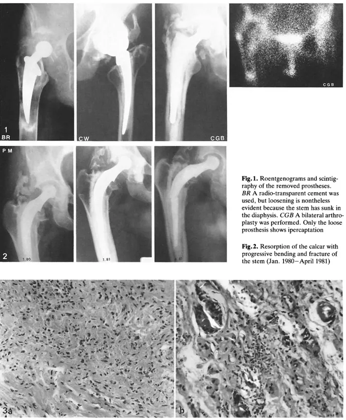
Fig 1 Roentgenograms and
scintig-raphy of the removed prostheses. BR A radio-transparent cement was used, but loosening is nontheless evident because the stem has sunk in the diaphysis CGB A bilateral arthro-plasty was performed Only the loose prosthesis shows ipercaptation
Fig 2 Resorption of the calcar with
progressive bending and fracture of the stem (Jan 1980-April 1981)
Fig 3 a, b Tissue adjacent to the cup and the head; H&E a Mononuclear histiocytes with many black particles; x 83 b Large,
strongly birefringent polyethylene fibers engulfed by multinucleated giant cells; x 210
In contrast, this paper considers cases of loosen-ing of metal-plastic hip prostheses in relation to cellu-lar reactions due to metal particles released from the femoral stem as a result of abrasion and corrosion.
Case Reports
1 CGB, male, aged 70 In October 1970 a bilateral Charnley stainless-steel hip arthroplasty for osteoarthritis was perform-165
U E Pazzaglia et al : Metal Particles and Loosening
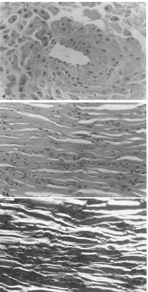
Fig 4 Mononuclear histiocytes with many black particles sur-rounding a vessel H&E; x 300
Fig 5 a, b Tissue between bone
and cement; H&E a Mono-nuclear histiocytes with foamy cytoplasm and black particles between collagen fibers; x 187.
b The same field in polarized
light; black particles are faintly birefringent
U E Pazzaglia et al : Metal Particles and Loosening
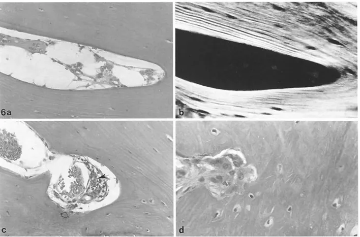
Fig 6 a-d Relationships between mononuclear histiocytes and bone; H&E a Mononuclear histiocytes with black inclusions inside
vessels of bone; x 175 b The same field in polarized light; some particles are faintly birefringent; x 175 c A mononuclear histiocyte outside the vessel wall and in contact with bone (white arrow), another is observed inside the vessel (black arrow); x 175 d Direct bone resorption by macrophages; x 220
Fig 7 Mononuclear histiocytes show a strongly
positive reaction for acid phosphatase; x 74
ed The result was good, pain was reduced significantly, and functional recovery was almost complete for the requirements of the patient After 5 years he started to complain of pain in the right hip and was readmitted in October 1975 X-ray ex-amination and scintigraphy showed loosening of the right pros-thesis (Fig 1) In November 1975 both the cup and the stem were removed and replaced.
2 CW, male, aged 30 In 1975 a Judet total-hip arthroplasty was performed for post-traumatic necrosis of the left hip Be-cause of pain the prosthesis was removed 1 year later and replaced with a Miiller prosthesis After 2 years the pain recur-red The patient was admitted for the first time to the Orthopedic Clinic of the University of Pavia in December 1978 X-ray examination and scintigraphy showed loosening of 167
U E Pazzaglia et al : Metal Particles and Loosening
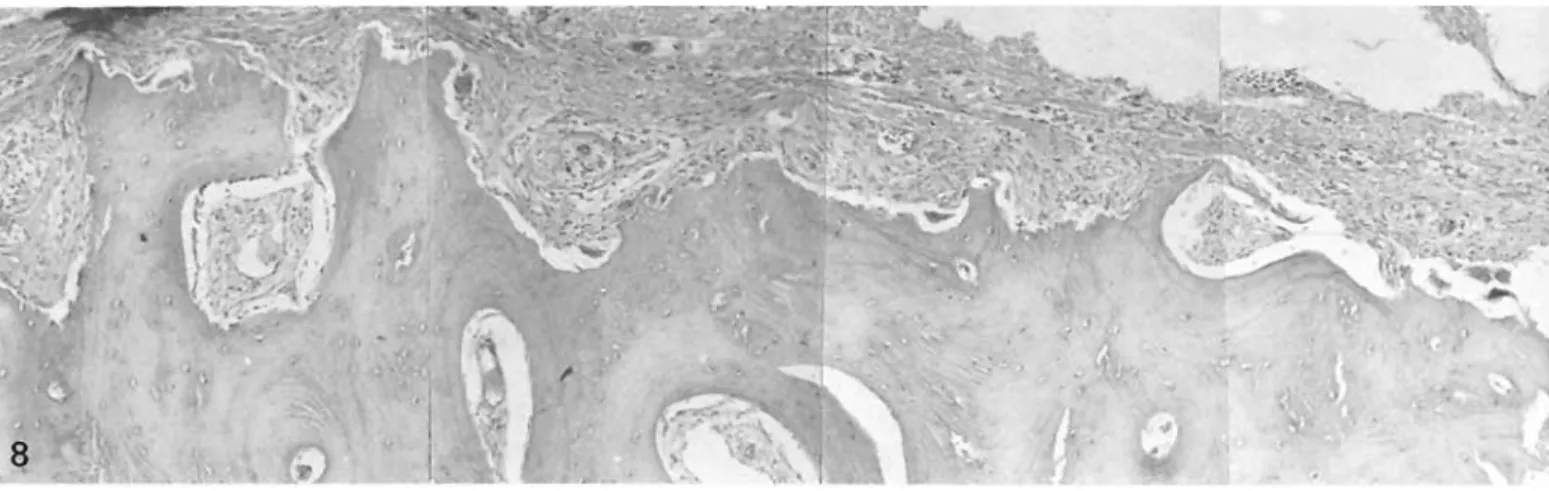
Fig 8 Tissue between diaphyseal bone and cement (but not from the calcar) of prosthesis PM Many osteoclasts are present on the
surrounding bone surface; H&E; x 73
the stem (Fig 1) The prosthesis was removed and not replaced.
3 BR, female, aged 73 IN 1977 a Co-Cr prosthesis (Rizzoli type) was inserted for osteoarthritis of the right hip After 2 years the patient started to complain of pain Resorption of bone around both the cup and the stem was observed She was admitted for the first time to the Orthopedic Clinic of the Uni-versity of Pavia in March 1980 X-ray examination and scintig-raphy showed a complete loosening of both the cup and the stem (Fig 1) They were removed and replaced.
4 PM, female, aged 61 In June 1971, a Charnley stainless-steel hip prosthesis was inserted for osteoarthritis of the right hip The result was good, with a full functional recovery After 9 years the patient started to complain of pain, and X-ray examination showed bone resorption of the calcar and loosen-ing between metal and cement In 1980 there was a progressive increase in pain and the patient was compelled to use a cane to walk Progressive loosening and bending of the stem occurred, until it finally fractured (Fig 2) In April 1981 the stem was removed and replaced.
Materials and Methods
Specimens of neosynovia and of the tissue between bone and cement were taken when prostheses were removed Bacterio-logical cultures for aerobic organisms were performed on the neosynovial tissue Formalin fixation, paraffin embedding (after 5 % nitric acid decalcification, if required), and hemato-xylin-eosin staining were used in preparing histology sections. Fresh-frozen sections were prepared for detection of acid phosphatase l 6 l The sections were observed with a light mi-croscope (Leitz Orthoplan) Coverslips were removed with xylene and a gold layer of approximately 4 nm was applied by sputter coating The same sections that had been examined optically were then examined with a scanning electron micro-scope (JEOL JSM 35 C), using the secondary and backscatter-ed electron modes X-ray microanalysis was performbackscatter-ed using an energy-dispersive spectrometer Samples for transmission electron microscopy were fixed in 4 % glutaraldehyde, buf-fered at p H 7 4, and postfixed in 1 % O 5004 Stained thin sec-tions were observed with a Philips 300 transmission electron microscope equipped for energy-dispersive X-ray analysis.
Observations
At operation, two prostheses exhibited loosening of both cup and stem The cement was removed easily because contact between bone and cement had been lost In two cases (CGB and BR) a soft gray tissue was present adjacent to the ball and socket This tis-sue extended between bone and cement behind the socket and down the diaphysis Its thickness was not uniform, but was never less than 1 mm The adjacent bone had lost its cancellous architecture and the bone surface was smooth and sclerotic, with some recesses into which the tissue penetrated.
In one case (CW), only the stem was loose, with the same gray tissue extending down the diaphysis In the fourth case, the broken prosthesis (PM), only the proximal part of the stem was loose The distal por-tion was firmly anchored to the bone A soft gray tis-sue was interposed between the ball and the socket, extending toward the calcar Otherwise the tissue between bone and cement at the level of the proximal stem in this particular case was unpigmented, thin, and firmer in consistency compared with the others. Tissue from comparable sites was macroscopically similar in the first three prostheses However, there were differences between the tissue from different sites, but only at the microscopical level, namely, those adjacent to the socket and the head and those from the bone-cement interface All bacteriological cultures were negative.
The following microscopical observations were made in the first three cases: tissue adjacent to the socket and the head was mostly very cellular with little or no intercellular substance Some cell-free material with amorphous, eosinophilic, fibrinous character was present (Figs 3 and 4) Two cell types predominated:
U E Pazzaglia et al : Metal Particles and Loosening
Fig 8
1 Mononuclear histiocytes with a diameter of 20 to 40 pm; their cytoplasm was foamy with many faintly birefringent black particles of various shapes and sizes, but as a rule no larger than 1 m Some elon-gated, strongly birefringent polyethylene particles were visible.
2 Multinucleate giant cells, diameter 100-400 m, which incorporated larger, strongly birefringent poly-ethylene fibers; similar cells also surrounded voids which had originally contained globules of bone cement Neither granulocytes nor lymphocytes nor plasma cells were apparent.
The tissue between bone and cement was moder-ately fibrotic, with collagen fibers following a pre-dominant orientation, mainly parallel to the bone surface There were few fibroblasts and many mononuclear histiocytes with a foamy cytoplasm con-taining black particles under 1 m in size (Fig 5). Small polyethylene particles were rare, and no giant cells or large polyethylene fibers were found here. The adjacent bone surface was smooth and the bone was dense and viable The mononuclear histiocytes formed recesses in the bone (Fig 6) These cells had the same cytoplasmatic inclusions as those described above The irregular surface which they presented to the bone suggests active resorption by them, and they proved strongly positive for acid phosphatase (Fig 7).
The neosynovium and the neocapsule of the fourth prosthesis contained mononuclear histiocytes with black cytoplasmic inclusions extending exclu-sively in the medial part of the diaphyseal cortical bone, corresponding to the calcar Elsewhere the tis-sue between bone and cement was poorly cellular and densely collagenous with fibers running parallel to the cement surface No macrophages with
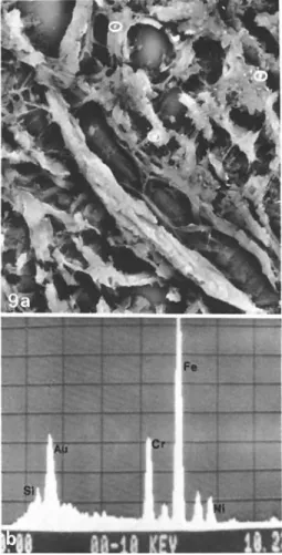
inclu-Fig 9 a, b a Scanning electron micrograph (backscattered
elec-tron image) of a gold-coated histological section mounted on a glass slide (coverslip removed) Intracellular metal particles are revealed as bright spot (circles) against the darker background of the organic material; x 900 b Typical X-ray analysis spectrum of the particles encircled in a showing the Fe, Cr, and Ni composition of stainless steel The gold (Au) peak results from the coating and the silicon (Si) peak from the glass substrate
U E Pazzaglia et al : Metal Particles and Loosening
Fig 10 a-c Transmission electron microscopy a Mononuclear histiocytes with
many lysosomal cisternae which contain highly electron-dense material There are no electron-dense particles in the intercellular substance; x 12800.
b Secondary lysosomes with electron-dense particles at higher magnification;
x 34000 c X-ray analysis of electron-dense material indicates Co-Cr alloy
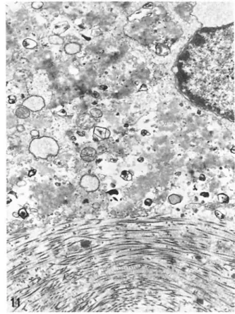
Fig 11 Transmission electron microscopy A mononuclear histiocyte in very
poor condition with many large lysosomes, electron-dense particles, wide-spread myelinic figures, and a discontinuous plasma membrane; x 6400
sions were identified here Many osteoclasts were present on the surrounding bone surface, suggesting active bone resorption (Fig 8).
Scanning electron microscopy combined with X-ray microanalysis was used to identify the particles. The same macrophages observed by light microscopy appeared in the SEM (backscattered electron mode) to contain inclusions of high atomic number X-ray analysis confirmed the Co-Cr-Mo composition of particles found within specimens of tissue close to Co-Cr alloy prostheses and the Fe-Cr-Ni spectrum for particles related to stainless-steel prostheses (Fig 9).
The transmission electron microscope observa-tions in all four cases showed the detailed character
of the mononuclear cells They exhibited many lyso-somal cisternae containing highly opaque electron-dense material Lanceolate spaces in lysosomes sometimes contained highly electron-opaque parti-cles which varied in size up to 100 nm Their appear-ance was not altered by staining with lead citrate or uranyl acetate X-ray analysis confirmed the metallic nature of these particles Many of the macrophages were in poor condition, with numerous large lyso-somes, signs of cell necrosis, a discontinuous plasma membrane, and widespread myelinic figures (Figs 10 and 11).
The surfaces of the removed prosthesis were ex-amined Both Co-Cr prostheses (CW and BR) show-ed marks of wear on the stem, characterizshow-ed by a
U E Pazzaglia et al : Metal Particles and Loosening
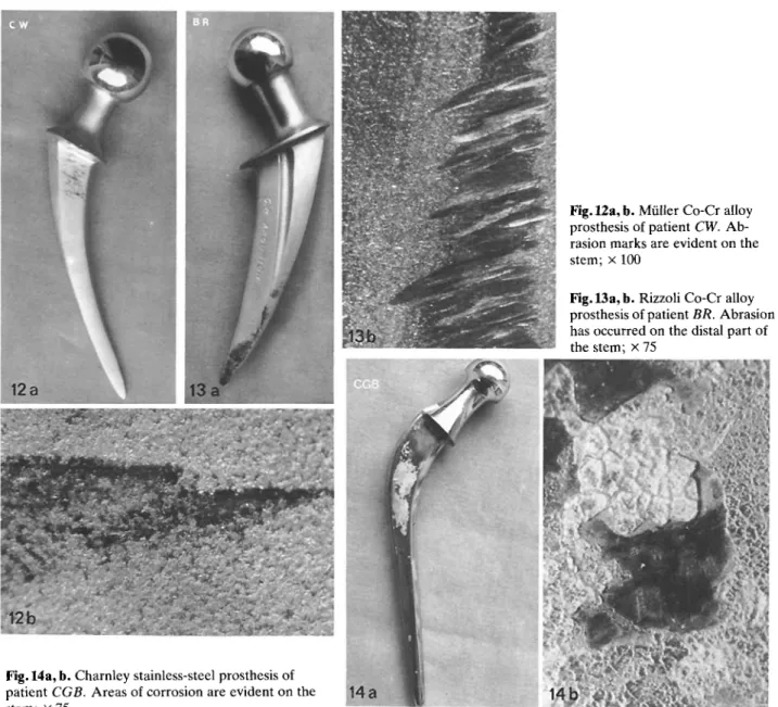
Fig 12 a, b Miller Co-Cr alloy
prosthesis of patient CW Ab-rasion marks are evident on the stem; x 100
Fig 13 a, b Rizzoli Co-Cr alloy
prosthesis of patient BR Abrasion has occurred on the distal part of the stem; x 75
Fig 14 a, b Charnley stainless-steel prosthesis of
patient CGB Areas of corrosion are evident on the stem; x 75
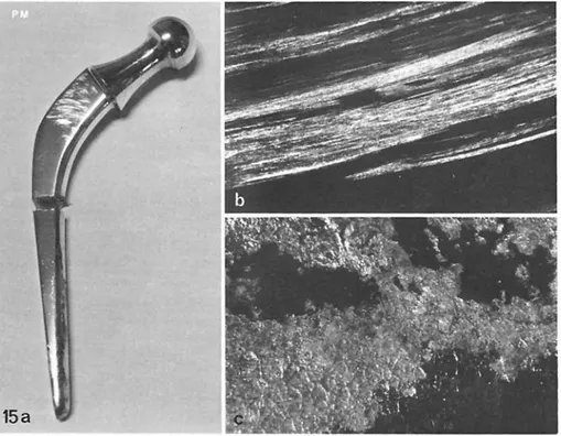
ular parallel pattern of scratches (Figs 12 and 13). One stainless-steel prosthesis (CGB) exhibited a wide area of pitting corrosion across the stem surface in contact with the cement (Fig 14) The other (PM) showed an area of pitting corrosion on the distal part of the stem (the part still firmly fixed), while the loose proximal part showed wear marks but no cor-rosion (Fig 15).
Discussion
Histological studies of necropsy material led Charn-ley l 9, 10, 11 l to conclude that a successful hip pros-thesis, even after 10 or more years, exhibits direct contact between bone and cement in the femoral diaphysis A thin, ca 1-mm layer of connective tissue
divides the cement from the marrow Such charac-teristics may be taken as proof of prosthetic stability. In contrast, loose prostheses are characterized by a thickened layer of connective tissue at the bone-cement interface This thickened layer probably develops as a result of micro-movements of the cement and associated small lacerations of adjacent bone and soft tissue with consequent exudation and hemorrhage l 27, 29 l.
A critical factor in prosthetic loosening is damage to the bone-cement interface Such damage may arise from microfracture of the bone This could be related to, for example, inadequate packing of cement with insufficient contact area, resulting in high stress at a few sites.
Bone may also be damaged by avascular necrosis following infiltration of marrow spaces by
U E Pazzaglia et al : Metal Particles and Loosening
Fig 15 a-c a Charnley
stainless-steel prosthesis of patient PM. b Wear is evident on the proximal portion of the fractured stem; x 100 c An area of corrosion on the distal part of the stem; x 75
tion tissue containing excessive quantities of wear debris l 22 l We did not observe this reaction in these four cases, all of which exhibited viable osteocytes.
Damage to the bone-cement interface could also result from bone resorption This process is normally associated with osteoclasts Our results strongly suggest, however, that in the first three cases pre-sented bone resorption by macrophages of inflam-matory tissue has occurred to a significant extent We are led to this conclusion by the absence of osteo-clasts from bone surfaces in contact with inflamma-tory tissue, by the extensive lysosomal development within macrophages suggesting their potential to gen-erate large quantities of hydrolytic enzymes, and by the presence of these acid-phosphatase-rich, mono-nuclear, phagocytic cells within bone recesses.
These observations also support the suggestion made by Salthouse l 23 l that metal-particle uptake may induce increased lysosomal enzyme activity among macrophages associated with orthopedic im-plants.
Freeman et al l 15 l recently hypothesized that progressive osteoclasis results when macrophages at the interface of bone and polymethylmethacrylate cement are provoked by foreign particulate matter, by bacteria, or by the products of cell death Of the foreign debris observed in the present cases (poly-ethylene, acrylic cement, and metal), only metallic debris (stainless-steel, and Co-Cr-Mo particles) ex-cited macrophages Furthermore, only metallic
debris was present almost exclusively in the critical areas around the stem and behind the cup This debris was intracellular, associated with lysosomal vesicles, and characterized by dead and dying cells. As yet, we have no information concerning the effects of other types of metallic debris such as that liberated by wear of titanium prostheses on macro-phage excitation in vivo The particles of polyethy-lene and polymethylmethacrylate observed seemed to elicit comparatively little cellular response and were not associated with cell necrosis.
The four cases showed no evidence of infection, and it seems unlikely that bacteria were responsible for the macrophage response On the other hand, cell necrosis certainly occurred, and the products of cell death presumably reinforced the effects of metal par-ticles on macrophages by encouraging phagocytosis and lysosomal development.
It should not be assumed that the release of hyd-rolytic enzymes from macrophages depends entirely on cell death and disintegration These cells may leak enzymes as well as secrete the contents of their lysosomal vesicles l 7 l Macrophages at the bone-cement interface could therefore cause bone resorp-tion by liberating hydrolytic enzymes in several ways, including passive leakage, active secretion, and ulti-mately cellular disintegration A self-perpetuating cycle might become established where phagocytosed particles of stainless steel or Co-Cr-Mo alloy at first provoke benign macrophages to overproduce
U E Pazzaglia et al : Metal Particles and Loosening
somal vesicles and hydrolytic enzymes This would result in cell damage and eventual deaths, together with associated bone resorption Cell remains would be engulfed by yet more macrophages of the inflam-matory tissue, along with re-released metal particles. These particles would retain their toxicity to provoke a further cycle, and so on.
Although the pattern of bone resorption seen in the first three cases indicates a distinct macrophage involvement, it cannot be concluded that resorption by osteoclasts has been bypassed entirely Osteo-clasts are highly motile Their absence from sections of histology specimens fixed at one point in time does not exclude the possibility that they were once pre-sent and functional within the tissue.
Revell et al l 22 l noted that resorption of both living and dead bone adjacent to macrophage-con-taining fibrous tissue appeared to be mediated by a mixture of mononuclear cells and osteoclasts Unfor-tunately, when examining clinical specimens obtain-able only at a late stage of loosening, it is not possible to establish fully the relative contributions that may have been made by each of these two cell types at dif-ferent times during the overall process of bone resorption.
Metal-on-plastic total hip prostheses liberate metal particles by corrosion and/or wear of the femoral stem When corrosion occurs particle pro-duction may precede loosening Wear debris, how-ever, results mainly from abrasive micromovements between stem and cement A minimal degree of movement between stem and cement must therefore precede wear This relative movement between two surfaces presumably aids the passage of metal parti-cles from the metal interface to the cement-tissue interface in the absence of direct contact via fissures in the cement Metallic wear debris may also be shed from the femoral head articulating in the cup if the latter is contaminated with an abrasive, such as bone cement, although this was not noticed in these four cases.
Whatever the mechanism of their production and release, particles of stainless steel and Co-Cr-Mo alloy at the bone-cement interface encourage macro-phage-related bone resorption This, we suggest, rep-resents a contributing, and in some cases not incon-siderable, factor in loosening of metal-plastic total hip prostheses.
Acknowledgements The authors are grateful to Miss Giovanna Bodini and Mr Giovanni Pesci for technical assistance This work was supported by grant No 82 02034 04 of the National Research Council of Italy.
References
1 Amstutz HC, Lurie L, Bullough P ( 1972) Skeletal fixation with self-curing polymethylmethacrylate Clin Orthop 84:
163-178
2 Beneke G, Kuprosch R, Mohr W, Paulini K, Mohing W ( 1973) Die Reaktion der Gelenkkapsel nach Totalarthro-plastik des Hiftgelenkes Arch Orthop Unfall-Chir 75: 289-301
3 Benson MKD, Goodwin PG, Brostoff J ( 1975) Metal sen-sitivity in patients with joint replacement arthroplasties Br Med J 4:374-375
4 Brinkmann KE, Heilmann K ( 1974) Klinische, rntgeno-logische und feingewebliche Untersuchungen an ausgelok-kerten Hiiftgelenksprothesen Arch Orthop Unfall-Chir 80:333 342
5 Buchhorn GH, Willert HG ( 1982) Effects of plastic wear particles on tissue In: Williams DF (ed) Biocompatibility of orthopedic implants CRC Press, Boca Raton, pp 249-267
6 Burstone MS ( 1962) Enzyme histochemistry Academic Press, New York
7 Chambers TJ ( 1982) The response of the macrophage to foreign material In: Williams DF (ed) Fundamental aspects of biocompatibility, vol 1, pp 145-158
8 Charosky CB, Bullough PG, Wilson PD ( 1973) Total hip replacement failures (a histological evaluation) J Bone Joint Surg lAml 55:49-58
9 Charnley J ( 1970) The reaction of bone to self-curing acryl-ic cement (a long-term histologacryl-ical study in man) J Bone Joint Surg lBrl 52: 340-353
10 Charnley J ( 1970) Acrylic cement in orthopaedic surgery. Livingstone, Edinburgh London
11 Charnley J ( 1979) Low-friction arthroplasty of the hip. Theory and practice Springer, Berlin Heidelberg New York
12 Cohen J ( 1959) Assay of foreign-body reaction J Bone Joint Surg lAml 41:152
13 Cotta H, Schulitz KP ( 1970) Komplikationen der Hiiftallo-arthroplastik durch periartikulare Gewebereaktionen. Arch Orthop Unfall-Chir 69:39-59
14 Evans EM, Freeman MAR, Miller AJ, Vernon-Roberts B ( 1974) Metal sensitivity as a cause of bone necrosis and loosening of the prosthesis in total hip replacement J Bone Joint Surg lBrl 56:626-642
15 Freeman MAR, Bradley GW, Revell PA ( 1982) Obser-vations upon the interface between bone and poly-methylmethacrylate cement J Bone Joint Surg lBrl 64: 489-493
16 Gui L, Pizzoferrato A, Ranieri L, Gualtieri 1 ( 1978) Rea-zione tissutale articolare e para-articolare negli impianti protesici Giorn Ital Ortop Traum lSuppll 4:215-227 17 Langlais F, Postel M, Berry JP, Le Charpentier Y, Weill
BJ ( 1980) L'intol 6 rance aux debris d'usure des protheses. Bilan immunologique et anatomopathologique de 30 cas.
Int Orthop 4:145-153
18 Masshoff W, Neuhaus-Vogel A ( 1974) Die Gelenkkapsel nach Alloplastik Arch Orthop Unfall-Chir 78:175-198 19 Mendes DG, Walker PS, Figarola F, Bullough PH ( 1974)
Total surface hip replacement in the dog A preliminary study of local tissue reaction Clin Orthop 100:256-264 20 Mirra JM, Amstutz HC, Matos M, Gold R ( 1976) The
pathology of the joint tissue and its clinical relevance in prosthesis failure Clin Orthop 117:221-240
U E Pazzaglia et al : Metal Particles and Loosening
21 Rae T ( 1975) A study on the effects of particulate metals of orthopedic interest on murine macrophage in vitro J Bone Joint Surg lBrl 57:444-450
22 Revell PA, Weightman B, Freeman MAR, Roberts BV ( 1978) The production and biology of polyethylene wear debris Arch Orthop Trauma Surg 91:167-181
23 Salthouse TN ( 1976) Cellular enzyme activity at the poly-mer-tissue interface: a review J Biomed Mater Res 10:197-229
24 Stinson NE ( 1964) Tissue reaction induced in guinea-pigs by particulate polymethylmethacrylate, polythene and nylon of the same size range Br J Exp Pathol 46: 135-146 25 Stinson NE ( 1963) The tissue reaction induced in rats and guinea-pigs by polymethylmethacrylate (acrylic) and stain-less steel Br J Exp Pathol 45: 21-29
26 Uchida S, Yoshino S, Doi M, Kudo H ( 1980) Side-effects of prosthetic materials on the human body Int Orthop 3:285 291
27 Willert HG, Puls P ( 1972) Die Reaktion des Knochens auf Knochenzement bei der Allo-Arthroplastik der Huifte. Arch Orthop Unfall-Chir 72:33-71
28 Willert HG ( 1973) Tissue reactions around joint implants and bone cement In: Chapchal G (ed) Arthroplasty of the hip Thieme, Stuttgart
29 Willert HG, Ludwig T, Semlitsch M ( 1974) Reaction of bone to methacrylate after hip arthroplasty (a long-term gross, light-microscopic and scanning electron-microscopic study) J Bone Joint Surg lAml 56: 1368-1382
30 Willert HG, Semlitsch M ( 1970) Tissue reactions to plastic and metallic wear products of joint endoprostheses In: Gschwend N, Debrunner HV (eds) Total hip prosthesis. Huber, Bern
31 Winter GD ( 1974) Tissue reactions to metallic wear and corrosion products in human patients J Biomed Mater Res 8:11-26
Received January 17, 1984 174