DOTTORATO DI RICERCA IN
SCIENZE BIOTECNOLOGICHE E FARMACEUTICHE
XXXII Ciclo
Settore Concorsuale: 03/D2Settore Scientifico Disciplinare: CHIM 09
DEVELOPMENT OF INNOVATIVE DELIVERY SYSTEMS
TO COUNTERACT VAGINAL INFECTIONS
Presentata da:
Dott.ssa Barbara Giordani
Coordinatore Dottorato
Supervisore
Chiar.ma Prof.ssa Maria Laura Bolognesi
Prof.ssa Barbara Luppi
The present thesis was focused on the development of innovative delivery systems to counteract vaginal infections and dysbiosis, particularly bacterial vaginosis and vulvovaginal candidiasis. These conditions widely compromise the quality of life of women worldwide and also expose to complications, including higher susceptibility to viral and sexually transmitted infections and increased risks of preterm birth and late termination of pregnancy.
In particular, the project aimed to overcome some drawbacks associated with the currently available therapies, such as the high incidence of recurrent vaginal infections and the growing emergence of drug resistance, and with the use of conventional dosage forms, like the poor retention at the vaginal site that reduces efficacy and patient compliance. In this contest, both formulations based on well-established drugs (chlorhexidine and econazole nitrate) delivered in innovative systems or formulations containing alternative active agents (probiotics cells and polyphenols) able to counteract vaginal infections have been taken into account. Moreover, in order to improve the vaginal health status, local as well as oral administration routes have been investigated.
In the first proposed work (paper 1), the strain Bifidobacterium breve BC204 was isolated from the vagina of a healthy woman and indagated for its probiotic potential. It showed good ability to adhere to Caco-2 cells, moderate ability to resist to gastrointestinal stress and strong antimicrobial activities towards both urogenital and enteric pathogens. Given these promising features, B. breve BC204 was subsequently protect by spray-drying encapsulation and formulated inside orally time-dependent erodible lipid tablets, with the aim to exert beneficial effects both at intestinal and vaginal level. The final dosage forms provided high loading and survival of B. breve BC204, as well as a delayed release and mucoadhesive abilities, which are required to guarantee the presence of an adequate amount of beneficial probiotic cells at the treatment site.
In the second work (paper 2), the freeze-drying technique was applied to produce lyophilized polymeric matrices for the local delivery of chlorhexidine, with the aim to prolong the vaginal release of this disinfectant, thus reducing the daily dose frequency. To achieve this objective, chlorhexidine was first complexed by means of ionic interactions with different polyanion polymers, then freeze-dried matrices containing either the isolated complex or the complex along with free drug and polymers were prepared. The selection of suitable polymers and the use of the adequate preparative
and the best profile of drug release, as well as excellent antimicrobial properties.
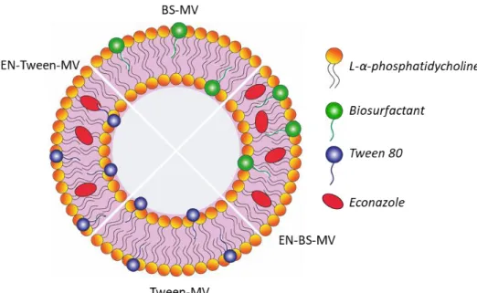
The last two papers focused on the development of nanosystems based on lipid vesicles for treatment of candidiasis. In this regard, two different strategies were followed. In a first approach (paper 3), econazole nitrate was vaginally delivered in phosphatidylcholine vesicles containing a biosurfactant, which was selected as “green” alternative excipient over chemical surfactants (i.e. Tween 80). The biosurfactant was isolated from a vaginal probiotic strain and characterized. It revealed a peptide-like structure and good surfactant and emulsifying properties. Mixed vesicles were further obtained by film rehydration and extrusion method and presented optimal diameter range for vaginal administration. Compared to vesicles containing Tween 80, those prepared with the biosurfactant showed higher encapsulation efficiency and mucoadhesion ability, as well as a sustained drug release. Notably, they also significantly improved the ability of econazole to eradicate Candida biofilm.
The second approach (paper 4) aimed to develop new liposomes for the simultaneous vaginal delivery of two polyphenols that could act in combination to eradicate the infection and alleviate candidiasis symptoms, like burning and irritation. Thus, quercetin and gallic acid were proposed as alternative to conventional antifungals to deal with the problem of azole resistance. Film rehydration and sonication method was used to obtain liposomes carrying both polyphenols with the desired size of 200 nm. They showed good entrapment efficiencies and better release profiles with respect to formulations containing only one polyphenol. Moreover, liposomes displayed strong oxidant and anti-inflammatory activities and resulted not cytotoxic to cells. Finally, they were able to counteract the growth of Candida owing to the antifungal effect exerted by gallic acid.
Lo scopo del presente lavoro di tesi è lo sviluppo di sistemi di veicolazione innovativi efficaci nel trattamento di infezioni e disbiosi vaginali, con particolare attenzione alla vaginosi batterica e alla candidosi vulvovaginale. Queste condizioni, oltre ad impattare pesantemente sulla qualità di vita di molte donne in tutto il mondo, espongono anche a complicazioni legate a un’aumentata suscettibilità alle infezioni virali e a trasmissione sessuale e a una maggiore probabilità di parto pretermine e aborto tardivo.
In particolare, il progetto di tesi si propone di superare alcune delle criticità associate alle terapie attualmente disponibili, come l’elevata incidenza di infezioni vaginali ricorrenti e il problema crescente della farmaco resistenza, e all’utilizzo delle forme di dosaggio convenzionali, come la scarsa capacità di ritenzione a livello vaginale che riduce l’efficacia del trattamento e la compliance della paziente.
A tal fine, sono stati proposti sia sistemi innovativi per la veicolazione di farmaci impiegati comunemente (clorexidina ed econazolo nitrato), che formulazioni contenenti agenti attivi alternativi (probiotici e polifenoli) in grado di contrastare le infezioni vaginali. Inoltre, sia la via di somministrazione locale che quella orale sono state prese in considerazione per ottenere un miglioramento delle condizioni di salute a livello vaginale. Nel primo lavoro presentato (paper 1), il ceppo vaginale Bifidobacterium breve BC204 è stato isolato da donna sana e caratterizzato per il suo potenziale probiotico. Il ceppo ha mostrato una buona capacità di adesione alle cellule Caco-2, una moderata resistenza all’ambiente gastrointestinale e una pronunciata attività antimicrobica nei confronti sia di patogeni urogenitali che enterici. Date queste promettenti proprietà, B. breve BC204 è stato successivamente microincapsulazione mediante spray-drying e formulato in compresse orali a base lipidica in grado di cedere il probiotico secondo un fenomeno di erosione tempo-dipendente. Le forme di dosaggio finali presentavano un profilo tecnologico ottimale per garantire la presenza di una quantità adeguata di cellule vitali al sito di trattamento, ovvero un elevato caricamento e sopravvivenza di B. breve BC204, un rilascio ritardato del probiotico e capacità mucoadesive.
Nel secondo lavoro (paper 2), è stata applicata la tecnica di freeze-drying per produrre matrici polimeriche liofilizzate contenenti clorexidina, allo scopo di prolungare il rilascio vaginale di questo disinfettante e ridurre quindi la frequenza di somministrazioni giornaliere. La clorexidina è stata dapprima complessata con diversi polimeri polianionici tramite interazione ionica e successivamente sono stati preparati due tipi di matrici, uno
matrici tramite la scelta dei polimeri adeguati e del metodo preparativo più idoneo. Nello specifico, le matrici a base di acido ialuronico hanno mostrato una struttura flessibile, le migliori proprietà di idratazione e mucoadesione e il miglior profilo di rilascio del farmaco, oltre che eccellenti proprietà antimicrobiche.
Gli ultimi due lavori sono stati invece incentrati sullo sviluppo di nanosistemi basati su vescicole lipidiche per il trattamento della candidosi e, al riguardo, sono state adottate due strategie.
In un primo approccio (paper 3), l’econazolo è stato veicolato per via vaginale in vescicole a base di fosfatidilcolina contenenti un biosurfattante, selezionato come eccipiente “green” alternativo ai surfattanti chimici (ad esempio Tween 80). Il biosurfattante è stato isolato da un ceppo probiotico di origine vaginale e caratterizzato. La molecola ha mostrato una struttura a base peptidica e buone proprietà tensioattive ed emulsificanti. Le vescicole miste, ottenute col metodo dell’idratazione del film sottile seguito da estrusione, presentavano un diametro ottimale per la somministrazione vaginale. Rispetto alle vescicole contenenti Tween 80, quelle preparate con il biosurfattante mostravano una più alta efficienza di incapsulazione, migliori proprietà mucoadesive e un rilascio sostenuto del farmaco. Inoltre, erano in grado di potenziare significativamente la capacità dell’econazolo di eradicare i biofilm di Candida.
Il secondo approccio (paper 4) ha consistito nello sviluppo di nuovi liposomi per la contemporanea veicolazione di due polifenoli che potessero agire in combinazione per eradicare l’infezione e alleviare allo stesso tempo i sintomi della candidosi, come bruciore e irritazione. La quercetina e l’acido gallico sono stati quindi proposti come alternativa ai convenzionali antifungini per affrontare il problema della resistenza agli azoli. I liposomi formulati con entrambi i polifenoli, ottenuti col metodo del film sottile seguito da sonicazione, avevano le dimensioni volute di 200 nm, buone efficienze di incapsulazione e migliori profili di rilascio rispetto alle formulazioni contenenti un solo polifenolo. I liposomi esplicavano inoltre ottime attività anti-ossidanti e anti-infiammatorie e sono risultati non citotossici per le cellule. Infine, si sono rivelati in grado di contrastare la crescita di Candida, grazie all’azione antifungina attribuibile all’acido gallico.
i
Table of contents
Theoretical part
I. Introduction ... 1
I.1 Vaginal environment ... 1
I.1.1 Overview of the vaginal cavity ... 1
I.1.1.1 Anatomy of the vagina ... 1
I.1.1.2 Physiology of the vagina ... 3
I.1.2 Vaginal microbiota of healthy women ... 4
I.1.2.1 Vaginal microbiota of reproductive-age women ... 4
I.1.2.2 Vaginal microbiota during different stages of woman’s life ... 7
I.1.2.3 Protective factors of healthy vaginal microbiota ... 9
I.1.3 Cervical-vaginal infections and dysbiosis ... 11
I.1.3.1 Bacterial vaginosis ... 12
I.1.3.2 Aerobic vaginitis ... 13
I.1.3.3 Vulvovaginal candidiasis ... 14
I.1.3.4 Sexually transmitted infections ... 16
I.1.4 The vagina as a route for drug delivery ... 18
I.1.4.1 Advantages of vaginal delivery ... 18
I.1.4.2 Factors affecting the vaginal delivery of drugs ... 20
I.1.4.3 Vaginal drug delivery systems ... 20
I.1.4.4 Limitations of conventional vaginal formulations ... 22
I.2 Active agents selected for the project ... 23
I.2.1 Probiotics ... 23
I.2.1.1 Probiotic bacteria ... 23
I.2.1.2 The requirements for probiotics ... 25
I.2.1.3 Microencapsulation of probiotics ... 26
I.2.1.4 Prebiotics ... 28
I.2.1.5 Probiotics for treatment of urogenital infections ... 28
I.2.2 Chlorhexidine ... 30
I.2.2.1 General characteristics of chlorhexidine ... 30
I.2.2.2 Antimicrobial activity of chlorhexidine ... 31
I.2.2.3 Vaginal application of chlorhexidine ... 31
I.2.3 Econazole nitrate ... 32
I.2.3.1 Antifungal activity of econazole ... 33
I.2.3.2 Vaginal application of econazole ... 33
I.2.4 Polyphenols ... 34
I.2.4.1 Polyphenol classification ... 35
I.2.4.2 Quercetin ... 36
I.2.4.3 Gallic acid ... 38
I.2.4.4 Anti-Candida activity of phenolic acids ... 39
I.3 Dosage forms and carries selected for the project ... 41
I.3.1 Solid lipid dosage forms ... 41
I.3.1.1 Beeswax ... 42
ii
I.3.2.1 Mucoadhesion process ... 44
I.3.2.2 Mucoadhesive polymers ... 46
I.3.2.3 Sodium alginate ... 47
I.3.2.4 Sodium carboxymethylcellulose ... 48
I.3.2.5 Xanthan gum ... 49
I.3.2.6 Hyaluronic acid ... 50
I.3.2.7 Freeze-drying technique ... 51
I.3.3 Liposomes ... 53
I.3.3.1 Conventional liposomes ... 54
I.3.3.2 Liposome preparation ... 55
I.3.3.3 Liposomes as drug carrier ... 57
I.3.3.4 Surfactant lipid vesicles ... 58
I.3.3.5 Synthetic surfactants ... 59
I.3.3.6 Biosurfactants ... 60
I.4 References ... 62
Experimental part II. Overview of the project ... 83
II.1 Aim of the thesis ... 83
II.2 List of the papers ... 84
II.3 Summary of the papers ... 85
II.4 References ... 91
III. Vaginal Bifidobacterium breve for preventing urogenital infections: development of delayed release mucoadhesive oral tablets (paper 1) ... 93
III.1 Abstract ... 94
III.2 Introduction ... 95
III.3 Materials and methods ... 97
III.3.1 Materials ... 97
III.3.2 Isolation, cultivation and taxonomic characterization of B. breve BC204 ... 97
III.3.3 Adhesion of B. breve BC204 to HeLa and Caco-2 cells ... 98
III.3.4 Tolerance of B. breve BC204 to gastric acids and bile salts ... 98
III.3.5 Antimicrobial activity of B. breve BC204 ... 99
III.3.5.1 Anti-Candida activity ... 99
III.3.5.2 Anti-Chlamydia activity ... 99
III.3.5.3 Antibacterial activity against extracellular urogenital and gastrointestinal pathogens ... 100
III.3.6 Microencapsulation of B. breve BC204 ... 101
III.3.6.1 Preparation of microparticles by spray-drying ... 101
III.3.6.2 Preparation of microparticles by freeze-drying ... 101
III.3.6.3 Size and morphology of microparticles ... 101
III.3.6.4 Survival of B. breve BC204 after microencapsulation and during storage .... 101
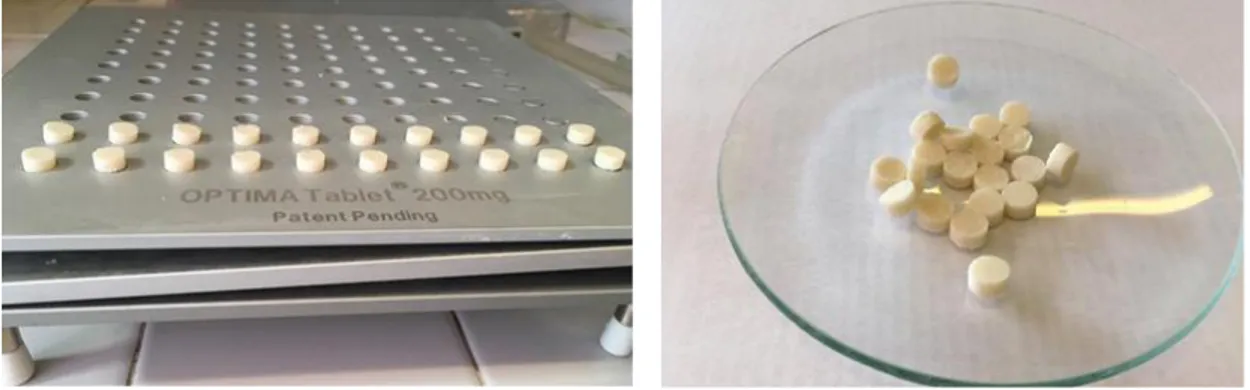
III.3.7 Formulation of microencapsulated B. breve BC204 in oral tablets ... 102
iii
III.3.7.2 Size, weight and friability of the tablet ... 102
III.3.7.3 Survival of B. breve BC204 after tablet production and during storage ... 103
III.3.7.4 Water-uptake ability ... 103
III.3.7.5 In vitro mucoadhesion ... 103
III.3.7.6 In vitro B. breve BC204 release ... 103
III.3.8 Statistical Analysis ... 104
III.4 Results and discussion ... 105
III.4.1 Adhesion of B. breve BC204 to HeLa and Caco-2 cells ... 105
III.4.2 Tolerance of B. breve BC204 to gastric acids and bile salts ... 105
III.4.3 Antimicrobial activity of B. breve BC204 ... 106
III.4.4 Microencapsulation of B. breve BC204 ... 107
III.4.4.1 Size and morphology of microparticles ... 107
III.4.4.2 Viability of B. breve BC204 after microencapsulation ... 108
III.4.5 Formulation of B. breve BC204 in oral tablets... 110
III.4.5.1 Preparation of oral tablets by spreading and cooling ... 110
III.4.5.2 Water-uptake ability ... 111
III.4.5.3 In vitro mucoadhesion ... 111
III.4.5.4 In vitro B. breve BC204 release ... 112
III.4.5.5 Survival of B. breve BC204 during tablet storage ... 113
III.5 Conclusions ... 114
III.6 References ... 115
IV. Freeze-dried matrices based on polyanion polymers for chlorhexidine local release in the buccal and vaginal cavities (paper 2) ... 118
IV.1 Abstract ... 119
IV.2 Introduction ... 120
IV.3 Materials and methods ... 122
IV.3.1 Materials ... 122
IV.3.2 Freeze-dried matrix preparation ... 122
IV.3.3 Fourier transform infrared spectroscopy ... 123
IV.3.4 Matrix dimensions, weight and drug content ... 123
IV.3.5 Scanning electron microscopy ... 124
IV.3.6 Differential scanning calorimetry ... 124
IV.3.7 Moisture content ... 124
IV.3.8 Mechanical characterization ... 125
IV.3.9 Water-uptake ability ... 125
IV.3.10 Mucoadhesion ability ... 125
IV.3.11 In vitro release studies ... 126
IV.3.12 Antimicrobial studies ... 127
IV.3.13 Statistical analysis ... 127
IV.4 Results and discussion ... 128
IV.4.1 Solid complex weight measurement ... 128
IV.4.2 Fourier transform infrared spectroscopy ... 128
IV.4.3 Matrix dimensions, weight and drug content ... 131
IV.4.4 Scanning electron microscopy ... 132
IV.4.5 Differential scanning calorimetry ... 133
iv
IV.4.7 Mechanical characterization ... 134
IV.4.8 Water-uptake ability ... 135
IV.4.9 Mucoadhesion ability ... 136
IV.4.10 In vitro release studies ... 138
IV.4.11 Antimicrobial studies ... 139
IV.5 Conclusions ... 141
IV.6 References ... 142
V. Novel mixed vesicles containing lactobacilli biosurfactant for vaginal delivery of anti-Candida agent (paper 3) ... 145
V.1 Abstract ... 146
V.2 Introduction ... 147
V.3 Materials and methods ... 149
V.3.1 Materials ... 149
V.3.2 Microorganisms and culture conditions ... 149
V.3.3 Biosurfactant production and isolation ... 149
V.3.4 Chemical characterization of biosurfactant ... 150
V.3.4.1 Fourier transformed infrared spectroscopy ... 150
V.3.4.2 Mass Spectrometry (ESI-MS) ... 150
V.3.5 Surface-activity determination and critical micelle concentration ... 150
V.3.6 Emulsification properties ... 151
V.3.7 Mixed vesicles preparation ... 151
V.3.8 Determination of encapsulation efficiency ... 151
V.3.9 Mixed vesicle size distribution and zeta potential ... 152
V.3.10 Stability studies ... 152
V.3.11 Mucoadhesion studies ... 152
V.3.12 In vitro drug release studies ... 153
V.3.13 Anti-Candida activity ... 153
V.3.13.1 Preliminary evaluation of activity against Candida spp. ... 153
V.3.13.2 Inhibitory activity against C. albicans planktonic culture ... 154
V.3.13.3 Eradication of C. albicans biofilm ... 154
V.3.14 Statistical analysis ... 155
V.4 Results and discussion ... 156
V.4.1 Chemical characterization of biosurfactant ... 156
V.4.1.1 Fourier transformed infrared spectroscopy ... 156
V.4.1.2 ESI-MS analysis ... 156
V.4.2 Surface-activity determination and critical micelle concentration ... 157
V.4.3 Emulsification properties ... 158
V.4.4 Determination of encapsulation efficiency ... 158
V.4.5 Mixed vesicle size distribution and zeta-potential measurements ... 158
V.4.6 Stability studies ... 159
V.4.7 Mucoadhesion studies ... 161
V.4.8 In vitro drug release studies ... 162
V.4.9 Anti-Candida activities ... 162
V.4.9.1 Activity against Candida spp. ... 162
V.4.9.2 Inhibition of planktonic cultures ... 163
v
V.5 Conclusions ... 166
V.6 References ... 167
VI. Utilizing liposomal quercetin and gallic acid in localized treatment of vaginal Candida infections (paper 4) ... 170
VI.1 Abstract ... 171
VI.2 Introduction ... 172
VI.3 Materials and methods ... 175
VI.3.1 Materials ... 175
VI.3.2 Preparation of liposomes ... 175
VI.3.3 Determination of vesicle size distribution ... 176
VI.3.4 Liposomes zeta potential ... 176
VI.3.5 Determination of polyphenol entrapment efficiency ... 176
VI.3.6 Evaluation of liposomes storage stability ... 177
VI.3.7 Mucoadhesion studies ... 177
VI.3.8 In vitro polyphenol release studies ... 177
VI.3.9 Anti-oxidative assays ... 178
VI.3.9.1 ABTS •+ radical scavenging ... 178
VI.3.9.2 DPPH radical scavenging ... 179
VI.3.10 Cell culture ... 179
VI.3.11 In vitro cell viability study ... 179
VI.3.12 Anti-inflammatory activity determination ... 180
VI.3.13 Anti-Candida activity testing ... 181
VI.3.14 Statistical analysis ... 181
VI.4 Results and discussion ... 182
VI.4.1 Technological characterization of liposomes ... 182
VI.4.1.1 Liposomal size and zeta potential ... 182
VI.4.1.2 Polyphenols entrapment efficiency ... 184
VI.4.1.3 Stability of liposomes during storage ... 186
VI.4.1.4 Mucoadhesion studies ... 188
VI.4.1.5 In vitro release of polyphenols from liposomes ... 189
VI.4.2 Biological characterization of liposomes ... 190
VI.4.2.1 Anti-oxidant activity ... 191
VI.4.2.2 Anti-inflammatory activity of free and liposomal polyphenols ... 193
VI.4.2.3 Effect of free and liposomal polyphenols on cell viability ... 194
VI.4.2.4 Antifungal potential ... 195
VI.5 Conclusions ... 198
VI.6 References ... 199
1
I. Introduction
I.1 Vaginal environment
I.1.1 Overview of the vaginal cavity
I.1.1.1 Anatomy of the vagina
Figure I.1 – Mid-sagittal section of the female pelvis [Konar 2014].
The vagina is one of the most important organs of female genital apparatus and plays a focal role in reproduction. Anatomically, the vagina can be described as a slightly S-shape, fibromuscular and strong tubular organ that extends from the cervix of the uterus to the vestibule of the external genitalia. It is situated between the rectum, bladder and urethra with dimensions ranging from 8-11 cm in length and 2-5 cm in diameter (figure I.1) [Baloglu 2009; de Araújo Pereira 2012].
Normally, the vagina appeared as a curved organ, in which two distinct portions can be distinguished: a lower convex portion and a broader upper part that lies in an almost horizontal plane when the woman is in upright position. The two axes form an angle of 130 degrees. The vagina is a collapsed organ; indeed, when viewed in transverse cross-section, the anterior and posterior walls are in contact with each other [Choudhury 2011]. Once the vagina enters to the pelvic region it passes two diaphragms. The bulbocavernosus muscle and the pubococcygeus, from the urogenital and pelvic diaphragms respectively, behave as sphincters to the vaginal introitus [Alexander 2004]. The vaginal wall (figure I.2) is composed of four layers and it’s estimated that a turnover of about 10-15 layers happens every 7 days [Valenta 2005; Konar 2014]. (i) The most superficial layer consists of non-cornified stratified squamous epithelium, which has no secretory glands. Its thickness varies by approximately 200-300 μm and basically depends
2
on hormonal fluctuations that occur during different life stages (new born, child, adult and menopause) and periodically during menstrual cycle. In particular, with hormonal activity the vaginal epithelium increases in thickness and is highest in proliferative stage. In women of reproductive age, the vagina is characterized by numerous folds, called “rugae”, that offer a high distensibility, support and allow to increase surface area of vaginal wall. (ii) The second layer is the lamina propria, or tunica, made of collagen and elastin, which is highly vascularized and rich in lymphatic channels. (iii) The muscle layer is the third, with smooth muscle fibers running in both circular and longitudinal directions, that confer excellent elasticity to the vagina. (iv) The last layer is formed by areolar connective tissue and a large plexus of blood vessels.
Figure I.2 – Structure of the vaginal wall [Konar 2014].
The vagina’s nerve supply arises from two sources [Artico 2012]. The lower quarter of the vaginal is innervate by peripheral nerves, that make it a very sensible area, while autonomic fibers innervate the upper three quarters and respond to stretch but are not very sensitive to pain. For this reason, women rarely feel great discomfort or localized sensations during the use of vaginal products (i.e. suppositories, ovules, vaginal rings and tampons) [Alexander 2005].
The vascular supply involves an extensive network of arteries that surround the vagina from multiple origins, including the uterine artery, the pudendal artery, and the middle and inferior hemorrhoidal arteries. Drugs adsorbed from the vagina avoid the first-pass metabolism because blood leaving the vagina enters the peripheral circulation via a rich venous plexus, which empties primarily into the internal iliac veins. Since vascular circulations of the vagina and uterus are strictly connected, a “first uterine pass effect” has been hypothesized after vaginally administration of hormones [Hussain 2005].
3 I.1.1.2 Physiology of the vagina
The physiology of the vagina is influenced by age, hormone status, pregnancy and pH changes induced by several factors including semen, menstruation, estrogenic concentration, and bacterial colonization [Alexander 2004].
In healthy women of reproductive age, vaginal pH is generally maintained between 3.8 and 4.2. Lactobacilli that inhabit vaginal niche contribute to the preservation of an acidic environment through the conversion of glycogen from exfoliated epithelium cells into lactic acid and production of hydrogen peroxide [Valenta 2005]. The pH varies according to age, stages of menstrual cycle, pathological states and frequency of coitus. In particular, menstrual, cervical and uterine secretions, and semen cause an increase in pH by acting as alkaline agents.
Despite the absence of glands, a large amount of fluid is secreted in the vaginal cavity. Cervical secretion and transudation from the blood vessels, together with desquamated vaginal cells and leukocytes, mainly form the vaginal fluid. Secretions from the endometrium, fallopian tube and Bartholin’s glands, microorganisms and their metabolic products also contribute to formation of vaginal fluid [Hussain 2005]. Thus, the human vaginal fluid is a mixture of a large variety of substances comprising enzymes, enzymatic inhibitors, proteins and amino acids, carbohydrates, alcohols, hydroxylketones and aromatic compounds. This aspect is important because the high enzymatic activity of vaginal epithelium could possibly affect short- and long-term stability of intravaginal dosage forms and devices [Choudhury 2011].
Some factors, like sexual arousal and hormonal cyclical fluctuations, may induce variations in volume and composition of vaginal fluid, thus affecting drug release behaviour from vaginal delivery systems [Choudhury 2011]. In absence of sexual stimulation, the vaginal fluid is principally made up of plasma transudate from the vaginal wall and secretions from the cervical and vestibular glands [Valore 2002]. On arousal, the vaginal lubrification increases as consequence of locally release of vasoactive peptides, which cause arteriolar dilatation and vaginal distention and suppress venous return [Levin 1991]. The rate of discharge produced by reproductive-age women is of 3-4 g every four hours, while the amount of fluid decreases by 50% in postmenopausal women.
For vaginally administered formulations, should be pointed out that the physiologically presence of fluids may cause an overall dilution of active compounds and reduction of drug efficacy, increase leakage and decrease drug residence time at the site of application
4 [Andrews 2009; Rohan 2009].
Furthermore, vaginal fluid is characterized by the presence of cervical mucus, which in turn can either facilitate or hamper the efficacy of a dosage form [Valenta 2005; de Araújo Pereira 2012].
Mucus is a secretion synthesized by specialized goblet cells and forms an incessantly renewed, semi-permeable, adherent and viscoelastic barrier that lines all female reproductive tract. Within the vaginal cavity, the mucus appeared as a permeable gel layer that assures a proper lubrification and hydration and allows for the exchange of gases and nutrients to and from epithelium. Moreover, it acts as a barrier to protect the underlying layers against pathogens and harmful substances [Carvalho 2010; Lai 2010]. The cervical mucus is composed of mucins and other glycoproteins, inorganic and organic salts, proteins, cholesterol, lipids, carbohydrates, urea, enzymes, amino acids and 95% water by mass, making it a highly hydrated system. Mucin is the most secreted glycoprotein of mucus and is responsible for its structure [de Araújo Pereira 2012].
At physiological pH the mucus network is negatively charged for the presence of sialic acid and sulfate residues, which play an important role in bioadhesion and mucoadhesion processes.
I.1.2 Vaginal microbiota of healthy women
I.1.2.1 Vaginal microbiota of reproductive-age women
The term microbiota (or microbiome) refers to the whole assortment of microorganisms in a particular body niche, including the human gut, oral cavity and vaginal tract [Leyva-Gómez 2019].
The vaginal microbiota of healthy women consists of a plethora of anaerobic and aerobic microorganisms that establish a mutualistic relationship with the host and constitute the first line of defence against the colonization by opportunistic pathogens responsible for urogenital diseases, such as bacterial vaginosis, aerobic vaginitis, fungal infections and sexually transmitted infections [Oakley 2008; Smith and Ravel 2016].
The composition of the vaginal microbiota is not static but dynamic and undergoes modifications in response to hormonal fluctuations throughout the women’s reproductive life (i.e. from puberty to menopause and during pregnancy, see section I.1.2.2). Other external factors, such as pathological states, drug intake, sexual intercourses and hygienic behaviours, may also affect the composition of microbiome [Borges 2014].
5
Despite the proximity of the vagina to the anus, the variety of microorganisms present in the vaginal tract is much lower than in the gut; indeed, about 50 microbial species have been identified in the vaginal microbiome whereas the gut microbiome harbours more than 800 species. The reason for the limited microbial diversity of the vaginal niche is still not completely understood but may be ascribed to reduced competition with indigenous organisms and differences in nutrient availability and immune activity [Cribby 2008].
The vaginal microflora was first described in 1982 by the German gynaecologist Albert Döderlein, who observed the presence of Gram-positive, non-spore-forming rods (referred to as Döderlein’s bacilli), sometimes quite long and slim, with square or very tapering ends, occurring single or in chain, producing lactic acid that could inhibit the growth of pathogens [Petrova 2013]. Until 1980 it was believed that Lactobacillus acidophilus was the dominant constituent of the vaginal microbiota, as described by culture-dependent and microscope techniques. Knowledge on the composition of the vaginal microbiota has significantly expanded after the development and diffusion of cultivation-independent methods that rely on the analysis of 16-S ribosomal gene sequences [Ma 2012; Borges 2014]. In fact, the culture-dependent methods fail to identify several Lactobacillus species, such as L. iners that is not able to growth on de Man, Rogosa and Sharpe medium commonly employed for lactobacilli cultivation.
The vaginal ecosystem of healthy adult women is mostly dominated by Lactobacillus spp., that generally constitute more than 80% of total microbiome and are present at a concentration of 107-108 CFU per g of vaginal fluid [Farage 2010; Borges 2014].
Lactobacilli belong to the group of LAB (Lactic Acid Bacteria) and the genus is composed of more than 170 species of facultative, anaerobic or microaerophile, catalase-negative, Gram-positive, non-spore-forming rods. The most recurrent species in vaginal microflora are L. crispatus, L. gasseri, L. jensenii and L. iners [Pavlova 2002] but other microbial genera and species may also be present at lower concentrations. These include Bifidobacterium, Staphylococcus, Ureaplasma, Corynebacterium, Streptococcus, Peptostreptococcus, Gardnerella, Bacteroides, Mycoplasma, Enterococcus, Escherichia, Veillonella and Candida [Hyman 2005].
Several studies revealed that the Lactobacillus spp. composition in vaginal tract varies among women on the basis of geographic locations, races, ethnicities and individual susceptibility. In a study by Ravel et al. the vaginal bacterial communities of 396 asymptomatic North American women representative of four ethnic groups (white, black,
6
Hispanic and Asian) were investigated through pyrosequencing of barcoded 16S rRNA genes (figure I.3) [Ravel 2011]. According to these authors, microbial communities categorized in group I (26.2%), II (6.3%), III (34.1%), and V (5.3%) are dominated by L. crispatus, L. gasseri, L. iners and L. jensenii, respectively, and are isolated mostly from white and Asian women. The remaining group IV (27%), mainly formed by black and Hispanic women, is a polymicrobial mixture of strict and facultative anaerobes, including species of the genera Prevotella, Dialister, Atopobium, Gardnerella, Megasphaera and Peptoniphilus.
Lactobacillus-dominated vaginal microbial communities have been associated with healthy conditions and are characterized by the production of large amounts of lactic acid, that maintains the vaginal pH below 4.5. This acidic environment is thought to be highly protective against infections or colonization of the vaginal mucosa by opportunistic (i.e. Escherichia coli) or non-indigenous pathogens (i.e. Neisseria gonorroheae and Chlamydia trachomatis). However, additional studies also reported that some women’s vaginal ecosystems can be healthy without a Lactobacillus-dominant vaginal microbiota. In these cases, other species belonging to the LAB (i.e. Atopobium vaginae, Megasphera and/or Leptotrichia spp.) can be identified as dominant vaginal phylotype [Zhou 2004; Zhou 2007; Srinivasan 2012; Petrova 2013]. The same species were also detected by Ravel et al. in group IV, suggesting that some key functions of vaginal community, such as the production of lactic acid and hydrogen peroxide, must be preserved in order to guarantee an adult healthy vaginal ecosystem [Witkin 2007; Ravel 2011].
Figure I.3 – Composition of vaginal microbiota in healthy adult women [Petrova 2013]. The figure summarizes the studies conducted by Ravel et al. [Ravel 2011] and Gajer et al. [Gajer 2012].
7
A similar study conducted by Gajer et al. confirmed the previous observations of Ravel and colleagues. The authors identified five community groups since they split the group IV into two subgroups, IV-A and IV-B (figure I.3). Both of them are heterogenous in composition but group IV-A is characterized by a modest proportion of either L. crispatus or L. iners together with a low number of strict anaerobic bacteria, while IV-B has a higher proportion of members of the genus Atopobium, Prevotella, Parvimonas, Sneathia, Gardnerella and Mobiluncus.
The authors highlight that some of the vaginal bacterial communities noticeably change over time, switching from one to another group, whereas others are relatively stable. For instance, the vaginal communities dominated by L. crispatus often transform to group III dominated by L. iners, or to IV-A. Group III in turn shifts more often to group IV-B, but in rare cases to IV-A. On the contrary, the community group dominated by L. gasseri is quite stable and rarely transits to other types [Gajer 2012].
Furthermore, a strong association between group IV-B (Nugent score > 6) and increased risk to turn into a dysbiosis (i.e. bacterial vaginosis) and to get sexually transmitted infections (i.e. HIV) has been established through epidemiological studies [Smith and Ravel 2016]. Hence, while group IV may be normal (asymptomatic) in some women, in other conditions may lead to adverse sequelae.
I.1.2.2 Vaginal microbiota during different stages of woman’s life
The composition of the vaginal microbiota undergoes several changes during the maturation of women as consequence of variations in oestrogen levels (figure I.4) [Cribby 2008; Amabebe and Anumba 2018].
Figure I.4 – Changes in the vaginal mucosae and in microbiome during different stages of the life of a woman [Petrova 2013].
8
Immediately after birth, the vaginal epithelium is colonized by a large number of microorganisms, that mainly originate from gastrointestinal tract or from the surrounding skin epithelium.
In particular, it seems that vaginally delivered infants acquire bacterial communities that are similar to that of their own mother’s vaginal microbiota, usually dominated by lactobacilli. On the contrary, after caesarean delivery infants result more frequently colonized by bacteria present on skin surface, especially Staphylococcus, Corynebacterium and Propionibacterium [Petrova 2013].
During the early stage of infancy, maternal oestrogens induce thickening of the vaginal epithelium and promote the deposition of glycogen in the epithelial cells, which is used by glucose-fermenting microorganisms as carbohydrate source [Boskey 1999].
As soon as the maternal oestrogens are metabolized, a thinning of the mucosa and reduction of glycogen occur. The consequent reduction in glucose-fermenting microorganisms, including lactobacilli, facilitates an increase in the vaginal pH, encouraging the proliferation of a wide range of aerobes and facultative anaerobes. Indeed, during childhood the vaginal microbiome mostly harbours Gram-positive (i.e. Actinomyces, Bifidobacteria, Peptococcus, Propionibacterium) and Gram-negative (i.e. Veillonella, Bacteroides, Fusobacteria) anaerobe bacteria, as well as some aerobic bacteria (i.e. Staphulococcus, Enterococcus, Corynebacterium and Diphteroides) [Randelovic 2012].
With the beginning of puberty, the vaginal epithelium, under estrogenic control, once again thickens and the glycogen-rich environment selects for glucose-fermenting microorganisms. The microbiota presents in this stage of life is predominantly colonized by Lactobacillus spp. (see section I.1.2.1).
During pregnancy the vaginal microbiota is predominated by lactobacilli and is more stable than in non-pregnant state. This is due to the high levels of oestrogens during pregnancy, resulting in increased vaginal glycogen deposition [Amabebe and Anumba 2018].
In postmenopausal period the level of oestrogens decreases, with consequent reduction or completely absence of glycogen and atrophy of vaginal mucosa. In women who don’t take hormone replacement therapy, the postmenopausal microbiota is dominated by L. iners and G. vaginalis while the presence of lactobacilli is strongly reduced, and therefore growth of potential pathogenic bacteria is increased [Petrova 2013].
9
I.1.2.3 Protective factors of healthy vaginal microbiota
The vaginal ecosystem plays a pivotal role in maintaining a healthy equilibrium, that acts to supply a barrier to new colonization by pathogenic microorganisms and to overgrowth of species that are otherwise commensal.
The switch from homeostasis to a perturbed state can occur through several means, including physical injuries to the mucosa, impairment of the indigenous microbiota by the use of antimicrobials, expression of specific virulence factors and access to the site by excessive quantity of pathogens [Reid 2011].
Restoration of a healthy microbiota is driven by multiple species, especially Lactobacillus spp., and requires considerable and prompt reorganization after insult.
The mechanisms adopted by lactobacilli, as well as by exogenous probiotic strains, to stabilize and rapidly re-establish the normal vaginal microbiota are diverse and are listed below (figure I.5) [Reid 2011; Borges 2014; Zhang 2018].
Figure I.5 – Protective mechanisms exerted by vaginal microbiota [Reid 2011].
Co-aggregation – The phenomenon of co-aggregation consists in the assembly of
microbial communities into distinct and interlinked structures. Lactobacilli can bind pathogenic microorganisms and form co-aggregates. This mechanism allows to restore the homeostasis of the vaginal tract because it creates a microenvironment biochemically hostile for the growth of the harmful strain and prevents its access to the target epithelial tissues, thus hindering its proliferation in the vaginal niche.
10
gasseri and L. jensenii, possess the ability to co-aggregate with E. coli, vaginal staphylococci and C. albicans [Younes 2012].
Biosurfactant production – Biosurfactants are surface-active compounds synthetized as
secondary metabolites by a variety of microorganisms. Biosurfactants produced by vaginal lactobacilli are composed of a mixture of proteins, lipids and carbohydrates and show an activity in displacing dense polymicrobial cultures of uropathogenic E. coli, Enterococcus faecalis and Gardnerella vaginalis [Saunders 2007; Reid 2011]. These effects occur even with only few lactobacilli, suggesting that the secreted biosurfactants spread out over the surface of the vaginal mucosa and the resulting modification in surface tension repels hydrophobic pathogens [Banat 2010; Reid 2011].
Production of antimicrobial compounds – Lactobacilli synthetize several metabolites
(like lactic acid, hydrogen peroxide and phenyllactic acid) able to exert direct antimicrobial activity against bacteria, viruses and fungi [Zhang 2018].
Lactobacilli metabolize glycogen, deposited in the vaginal epithelium by hormonal activation of oestrogens, with the production of organic acids, especially lactic acid. Lactobacilli are acid tolerant while most vaginal pathogens are sensitive to low pH values. Lactic acid acts as antimicrobial agent by inhibiting the growth and production of virulence factors of Gram-negative bacteria. Furthermore, the acidic environment induced by accumulation of lactic acid can permeabilize the outer membrane of Gram-negative bacteria and can inhibit urease activity [Alakomi 2000; Zhang 2018].
Hydrogen peroxide is produced approximately by 80% of vaginal strains, in particular by L. crispatus and L. jensenii, and may also prevent invasions of pathogens [Aroutcheva 2001; Borges 2014]. Indeed, H2O2 and its metabolites (hydroxyl radical and superoxide
anion) are powerful oxidizing agents, toxic to catalase-negative bacteria like most anaerobe microorganisms. On the contrary, lactobacilli seem to protect themselves from the killing action of free radicals thorough the production of Fe3+-activated extracellular peroxidase [Reid 2011].
Bacteriocins are protein molecules secreted by lactobacilli that have a narrow killing spectrum, being able to control closely related bacteria residing in the same ecological niche via mechanisms that include cytoplasmatic membrane pore formation and permeabilization, interference with cellular enzymatic reaction (i.e. cell wall synthesis) and nuclease activity [Mokoena 2017; Zhang 2018].
Signaling effects – Signaling between bacteria can lead to downregulation of toxin
11
some strains of lactobacilli that are known to be present in the vagina and gut, as well as in some probiotics. For example, L. reuteri RC-14 produces a signaling molecule that inhibits the expression of toxic shock toxin 1 in several strains of S. aureus and interferes with the P2 and P3 promoters of the staphylococcal global regulatory system agr [Laughton 2006].
Competitive exclusion – Lactobacilli can inhibit undesired microbial colonization by
adhering to the vaginal epithelium and thus occupying or masking (by steric hindrance) the potential binding sites of pathogens in the mucosa.
The blockage of vaginal pathogens adherence by lactobacilli may occur through three different mechanisms, namely exclusion, competition for receptor sites and displacement of adherent pathogens [Borges 2013].
Immunomodulation – The regulation of immune response by the microbiota can lead to:
the production of host factors such as antimicrobial peptides (like defensins), lactoferrin and lysozyme, which can kill pathogens; the production of alkaline phosphatases, which bind to lipopolysaccharide and abolish its toxicity; the deregulation of nuclear factor-κB (NF-κB) signalling in host epithelia [ Reid 2011].
Enhancement of epithelial barrier function – The integrity of the vaginal epithelial is
crucial to maintaining health. Lactobacilli play important roles in preserving epithelial barrier integrity by acting through diverse mechanisms, such as modulation of the cytoskeleton, induction of mucus production and phosphorylation of tight junction protein, which results in the improvement of tight junction function and the immune response [Kozakova 2016; Zhang 2018].
When this lining is destroyed or breached, the microorganisms on the outer surface gain access to the tissue and induce infection.
For example, HIV entry into the host is facilitate by the capability of the virus to decrease trans-epithelial resistance through disruption of tight junction proteins, the transmembrane protein occludin and the scaffold protein zonula occludens 1 (ZO1). Lactobacilli could potentially counteract this effect, as they upregulate ZO1 and occludin [Reid 2011].
I.1.3 Cervical-vaginal infections and dysbiosis
As mentioned above, several factors (summarized in figure I.6) may lead to transition from a health condition (also referred to as eubiosis) to an imbalance in vaginal homeostasis. This can promote colonization by potential pathogenic species, usually
12
causing a state of “abnormal vaginal microflora”, called dysbiosis.
The most prevalent dysbiosis include bacterial vaginosis (BV), aerobic vaginitis (AV) and vulvovaginal candidiasis (VVC). An alteration of vaginal ecosystem can also expose to cervical-vaginal infections, mainly sexually transmitted infections (STIs).
Figure I.6 – Factors associated with dysbiosis or eubiosis of the vaginal microbiota in reproductive age women [Kroon 2018].
I.1.3.1 Bacterial vaginosis
Bacterial vaginosis (BV) is the most prevalent vaginal disease in women of reproductive ages, with a prevalence rate of 12-40% [Kenyon 2013]. Although the exact aetiology of BV is still unclear, the unset of this dysbiosis has been associated with the depletion of lactobacilli, especially of those producing H2O2, that in turn favours the proliferation of
anaerobes commonly present in the vagina, such as Gardnerella, Atopobium, Mobilincus, Prevotella, Streptococcus, Mycoplasma, Ureoplasma, Dialister and Bacteroidetes [Kaambo 2018]. Thus, BV can be considered a polymicrobial state that leads to a more heterogeneous vaginal environment, with low lactic acid levels and pH above 4.5. These anaerobic bacteria engage in synergistic interactions and may form a mixed biofilm, which is considered one of the factors responsible for the chronicity and recurrence of the disease [Leyva-Gómez 2019].
BV is asymptomatic in 50% of BV-positive women and no inflammatory processes are detected at the vaginal epithelium. However, symptoms could appear in the form of non-itchy but irritating, white-grey, creamy vaginal discharges, containing exfoliated epithelial cells and, attached to their membranes, Gram-variable polymorphic bacteria. They are also characterized by a fishy odour due to the production and accumulation of
13
amines (such as putrescine, cadaverine and triethylamine) subsequent to the overgrowth of anaerobic bacteria [Amabebe and Anumba 2018].
Clinically, BV is diagnosed by using Amsel criteria, that include the evaluation of vaginal acidity, the presence of discharges, the appearance of clue cells (desquamated vaginal epithelial cells studded with anaerobes) and a positive “whiff test” (a fishy odour perceived when KOH 10% is added to vaginal discharges).
Interestingly, it seems that BV can increase the risk of acquisition of STIs (i.e. N. gonorrhoea, C. trachomatis, T. vaginalis HSV, HPV and HIV) and other infections (i.e. pelvic inflammatory disease and endometritis). Moreover, women who develop BV during pregnancy are more prone to spontaneous abortions, post abortion sepsis and preterm labour [Donders 2009; Combs 2014].
I.1.3.2 Aerobic vaginitis
Vaginal dysbiosis also manifest as aerobic vaginitis (AV), first described in 2002 by Donders and colleagues as a need to distinguish it from BV [Donders 2002; Leyva-Gómez 2019]. In AV, Lactobacillus microflora is dramatically destroyed. In particular, L. crispatus and L. jensenii in healthy conditions are able to prevent the growth of urogenital pathogens through the action of the surface proteins, and thus their depletion can predispose to the onset of a pathological state [Donders 2017]. The alteration of vaginal microflora triggers an increase in the pH, that is always above 6 in AV, and the proliferation of enteric aerobic microorganisms. In particular, the most isolated bacteria in AV-positive women are Escherichia coli, Staphylococcus aureus, coagulase-negative staphylococci (such as S. epidermidis), group B Streptococcus (such as S. agalactiae) and Enterococcus faecalis [Donders 2002; Donders 2007].
The lack of oestrogens, which induces the redness and atrophy of vaginal mucosa, is also believed to play a role in AV origin. In addition, the numbers of intermediate and parabasal cells increase due to the enhanced turnover and desquamation of superficial epithelial-cell layers, that elicit vaginal inflammation. Indeed, unlike BV, AV is characterized by the presence of a noticeable inflammation response, accompanied with a strong recruitment of leukocytes and neutrophils as well as pro-inflammatory cytokines, specifically IL-6 and IL-1β [Smith and Ravel 2016]. As consequence, AV clinically manifests through redness, itching, burning, pruritus, dyspareunia and purulent yellowish sticky discharges devoid of fishy odour.
14
(AV and BV) can be found, representing either a transient form or prolonged co-infection. In fact, the vaginal milieu should not be considered as a static system, but rather as a complex dynamic one. AV is still not widely known and remains underdiagnosed by many clinicians, or may even be mistaken as BV [Vieira-Baptista 2016].
AV affects 2-25% of women and has been associated with severe gynecological and obstetric outcomes (i.e. ascending genital infection, preterm premature rupture of membranes, preterm labour and preterm birth) and with STIs (i.e. N. gonorrhoea, C. trachomatis, T. vaginalis) [Amabebe and Anumba 2018].
I.1.3.3 Vulvovaginal candidiasis
After BV, vulvovaginal candidiasis (VVC) is the second most common cause of vaginitis and the most prevalent vaginal fungal infection. In fact, it’s estimated that VVC affects approximately 70-75% of women, especially those of reproductive ages, at least once during their life. Moreover, 40-50% of patients will experience a recurrence, while 5-8% of adult women manifests recurrent VVC, defined as four or more episodes per year [Sobel 2007]. In the United States alone, 13 million of cases of VVC are observed annually, which further results in 10 million of gynaecologic visits and estimated cost of 1 billion dollars.
The causative agents of VVC are Candida species and, among them, C. albicans accounts for 85-95% of total vaginal fungal infections. C. albicans is a dimorphic fungus, being able to grow both in the yeast and in the filamentous form, and is a resident of the normal vaginal microbiota, colonizing about 20% of women without eliciting any explicit symptoms. VVC may also be related to non-albicans species. In particular, C. glabrata is the one most commonly associated with candidiasis and can reach a prevalence of 10-20% in many parts of the world [Corsello 2003]. Other non-albicans spp., such as C. parapsilosis, C. tropicalis, C. krusei, C. dubliniensis and C. guillermondii, are less frequently isolated from VVC-positive women [Nyirjesy 2005]. Vaginitis induced by such species are clinically indistinguishable from that caused by C. albicans but are often more resistant to treatment, for example to fluconazole [Holland 2003].
Furthermore, non-albicans spp., in particular C. glabrata, are frequently responsible for recurrent VVC [Sobel 2007].
The pathogenesis of VVC is still not completely clear. However, it seems that when the balance of vaginal ecosystem gets disturbed, the overgrowth of Candida spp. and a simultaneous depletion of lactobacilli occur. Some factors, such as individual
15
susceptibility, pregnancy, prolonged treatment with wide-spectrum antibiotics, use of contraceptives and spermicide, frequent sexual intercourse, diabetes and immunosuppression, can increase the risk for development of VVC. This happens after initial vaginal epithelial colonization by Candida and switch between yeast asymptomatic form and symptomatic pathogenic hyphal form [Johal 2016].
Although VVC is not a life-threatening disease, it can seriously impair the quality of life of patients, leading to physical, psychological and even sexual complications.
Clinical manifestations of candidiasis include vaginal itchiness, dyspareunia (pain during sexual intercourse), external dysuria (pain during urination), vulvar burning, irritation and vaginal soreness. The most well-known symptom, cottage cheese-like discharge, is often minimal and sometimes absent.
Examination reveals erythema and swelling of the labia and vulva, often accompanied with fissures and pustulopapular peripheral lesions. Indeed, one of the hallmarks of vaginal candidiasis is the robust recruitment of neutrophils to the site on infection that, along with cytokine response, actually exacerbates disease symptoms yet fails to properly contain and clear the fungus [Fidel 2004].
The vaginal pH is normal (4-4.5) in VVC, and pH above 4.7 usually indicates the presence of bacterial vaginosis, trichomoniasis or a mixed infection [Sobel 2007].
Nowadays, the major factors that hinder the effective treatment of candidiasis are the difficulty of quick and accurate diagnosis, since VVC is often mistaken for bacterial infection, and the limited number of therapeutic options [Ksiezopolska 2018].
VVC therapy is mainly based on azole drugs (i.e. fluconazole, econazole, ketoconazole, itraconazole, clotrimazole, miconazole, terconazole) that can be administered orally (in the form of tablets or capsules) or topically (in the form of creams, tablets or suppositories) [Johal 2016]. Azoles act as fungistatic agents, blocking ergosterol synthesis, targeting the enzyme lanosterol 14α-demethylase and leading to an accumulation of toxic sterol pathway intermediates.
Unfortunately, the emergence of resistance to multiple antifungal drugs has been increasingly reported in the last years, making the treatment of VVC very challenging. On one hand, the up-regulation of gene coding for efflux pump is likely the major cause for the acquired planktonic cell resistance. On the other hand, the capability of Candida spp. to rapidly form biofilm on host mucosal tissues is an important virulence factor, associated with both resistance and recurrent outcomes (figure I.7) [Tsui 2016; Silva 2017].
16
Generally, a biofilm consists in a community of microorganisms that are irreversibly attached to a surface, behaving very differently from planktonic cells [Cavalheiro 2018]. Biofilm formation of C. albicans is a multifaced process that starts with adhesion of yeast cells to a surface, followed by the formation of a discrete colony. Subsequently, cells become organize and begin to produce and secrete extracellular polymeric matrix, that allows the maturation of a three-dimensional structure. Once the formation of mature biofilm, daughter cells can be released from the biofilm and migrate to other niches, thus propagating the fungus infections and colonization.
Figure I.7 – Summary of mechanisms involved on Candida spp. biofilm resistance [Silva 2017].
Biofilms of C. albicans exhibit five- to eightfold higher resistance to all azole drugs compared to planktonic cells due to several factors, such as high concentration of Candida cells inside biofilm communities and the presence of extracellular matrix, that provides protection from host immunity and limits the diffusion of antifungal agents. In particular, the major carbohydrate polymer of the matrix, β-1,3 glucans, is responsible for sequestering azoles, acting as a sponge and conferring resistance to C. albicans biofilm. The presence of persister cells – a subset of dormant, non-dividing cells located in the deeper layers of biofilm – is also related to a high tolerance to multiple antifungal classes. Moreover, cells from mature biofilms rely less on ergosterol for maintaining their membrane fluidity, and this aspect limits the efficacy of drugs targeting the sterol, including all azoles as stated above [Silva 2017; Cavalheiro 2018].
I.1.3.4 Sexually transmitted infections
Various catastrophic microorganisms can produce severe sequelae when they infect the cervical-vaginal tract. In particular, Chlamydia trachomatis, Neisseria gonorrhoeae,
17
Trichomonas vaginalis, Treponema pallidum, Mycobacterium tuberculosis, human papillomavirus (HPV), human immunodeficiency virus (HIV) and herpes virus (HSV-2) should be mentioned as potential sources of STIs. Among them, C. trachomatis, N. gonorrhoeae, and less frequently T. vaginalis, are responsible for the onset of cervicitis, defined as an inflammation of the cervix that can manifest in acute or chronic form.
Chlamydia trachomatis – C. trachomatis (in particular serovars D-K) is one of the most
common STIs worldwide, infecting more than 130 million of people every year with an incidence rate of 38 per 1000 females [Chi Wai Wong 2019]. C. trachomatis is a Gram-negative obligate intracellular bacterium whose only natural host is humans.
Chlamydia has a unique biphasic developmental cycle of 30-72 h, alternating two distinct bacterial forms. The elementary bodies (EBs) are spore-like, infectious but non-dividing. After the binding with sensitive cells, EBs are internalized through endocytosis to form a phagosome termed inclusion. Within the inclusion EBs differentiate into the reticulate bodies (RBs), the metabolically active and replicative form of the pathogen. Midway through the infectious cycle RBs begin to differentiate back into EBs, which are released to initiate new rounds of infection.
Most women with urogenital Chlamydia experience a subclinical and often asymptomatic infection and, therefore, do not pursue treatment. Unfortunately, protracted exposure of the fallopian tube epithelium to C. trachomatis, or to antigens released by this bacterium, may compromise tubal integrity, thus leading to infertility and, if conception occurs, increased susceptibility to ectopic pregnancy and pre-term birth.
Other consequences of an upper genital tract infection in women include pelvic inflammatory disease, endometritis, and perihepatitis [Witkin 2017].
Neisseria gonorrhoeae – The host-adapted human pathogen N. gonorrhoeae is a
Gram-negative coccoid bacterium, accountable for gonorrhoea. In women, urogenital gonococcal infections are often asymptomatic, thus remaining ignored and untreated, and providing an important reservoir for further transmission. Since N. gonorrhoeae does not express potent exotoxins, the pathogenesis mainly results from genital mucosa damage caused by the activation of innate immune responses. Symptoms of gonorrhoeal infection in women are usually nonspecific and the vaginal discharges originated from neutrophil influx may be mistaken for BV or yeast infections [Quillin 2018].
If left untreated, gonorrhoea can result in severe complications like pelvic inflammatory disease, infertility, ectopic pregnancy, first trimester abortion, and less frequently, disseminated infections [Foschi 2017].
18
Trichomonas vaginalis – Trichomoniasis is caused by a single-cell parasite also known
as trichomonad. This parasite affects vagina, urinary bladder and urethra in females and penis in males. The infected women present stinking foamy yellow-grey-green vaginal discharges, together with other symptoms as painful urination and sexual intercourse, vaginal soreness, burning, redness and irritation [Gupta 2019].
I.1.4 The vagina as a route for drug delivery
Until 1920s it was believed that the vagina was incapable of absorbing drugs systematically. However, the mucus permeability, the dense network of blood vessels and the large surface area make the vagina a valid route to achieve both local and systemic effects. In 1918, Macht observed for the first time that some drugs, specifically morphine, atropine and potassium iodide, were actually absorbed after vaginal administration [Macht 1918].
Traditionally, the vaginal cavity has been used for the delivery of locally acting drugs, such as antibacterial, antifungal, antiprotozoal, antiviral, anti-inflammatory and spermicidal substances, prostaglandins and steroids [Valenta 2005]. Even if most of the drugs approved for vaginal administration are intended to treat local conditions, a part of them reaches blood circulation at sufficiently high levels to elicit systemic effects. Moreover, the vaginal route can be potentially used for the uterine targeting of active agents like progesterone and danazol [Cicinelli 2000; Einer-Jensen 2002]. Indeed, after vaginal administration, the plasma concentration of progesterone results higher in the uterine artery than in the radial one, suggesting a preferential distribution of this drug to the uterus. Such evidence underlines the existence of direct local transport from the vagina to the uterus, called “first uterine pass effect” [De Ziegler 1997].
I.1.4.1 Advantages of vaginal delivery
Despite the fact that vaginal delivery is only exploitable for females, it offers severe advantages. One of the major benefits of vaginal administration over oral intake is the avoidance of gastrointestinal absorption and hepatic first-passage [de Araújo Pereira 2012]. Absorption from the gastrointestinal tract may be unpredictable and influenced by several issues, such as vomiting, drug-drug interference, presence of food or limited intestinal absorption capacity due to physical-chemical features of drugs (i.e. water solubility) [Valenta 2005].
19
liver. The possibility of by-passing hepatic first-passage is particularly desirable for drugs that undergo a high degree of hepatic metabolism, as in the case for oestrogens that are 95% metabolized by the liver when are taken orally [Alexander 2004]. Another example is propranolol, whose bioavailability is greater after vaginal delivery compared with oral administration [Patel 1984]. The vaginal drug delivery could therefore permit to lower the dose and systemic exposure, thus reducing incidence of side effects while achieving the same pharmacodynamic effect.
The decrease in plasma fluctuations, characteristic of the daily oral intake, may also contribute to limit side effects. Considering that the onset of side effects is the most important aspect associated with discontinuation of oral contraception, their reduction will increase the acceptability of a dosage form and consequently patient compliance [Rosenberg 1995].
Some compounds have been shown to be more effective when administered vaginally with respect to other routes. For example, indomethacin employed in case of preterm labour appears to have greater effects when used intravaginally as compared with an intrarectal plus oral regimen [Abramov 2000]. Vaginal administration allows also to reduce gastrointestinal mucosal irritation – as observed during the vaginal delivery of bromocriptine [Vermesh 1988] – and hepatic side effects of steroids used in hormone replacement therapy or contraception [Dezarnaulds 2003]. In addition, the vaginal application overcomes the inconveniences caused by parenteral routes such as pain, tissue damage and possible infections [Valenta 2005]. Ease of self-insertion and removal of the final formulations, as well as the possibility of maintaining them for extended periods of time (i.e. daytime and night time) thereby lowering dosing frequencies, are other advantages of this route [Wang 2018]. Indeed, a prolonged contact of a delivery system with the vaginal mucosa can be obtained more easily than at other absorption sites, like rectum and gut mucosa. Several studies also report that a wide range of compounds, including peptides and proteins, show a good permeability trough vaginal mucosa. It’s also worth to note that vaginal drug delivery permits a selective local exposure where needed, producing little or no chance in exposure throughout the rest of the body [Alexander 2004]. This is particularly important for steroids employed for vaginal treatment of urogenital atrophic diseases.
However, some drawbacks, including cultural sensitivity, personal hygiene, gender specificity, local irritation and influence of sexual intercourse should be considered during the design of a vaginal formulation [Hussain 2005].
20
I.1.4.2 Factors affecting the vaginal delivery of drugs
Several factors and physicochemical properties of delivered substances may influence the release, and eventually the absorption, of drugs in the vaginal cavity.
As mentioned in section I.1.1.2, cyclic changes in thickness of vaginal epithelium, fluid volume and composition, pH and sexual arousal could potentially affect drug release at the administration site and drug bioavailability. For instance, the thickness of the vaginal epithelium influences the absorption of steroids like oestrogen, which is higher absorbed in postmenopausal women compared to premenopausal ones [Pschera 1989].
Moreover, cervical mucus of the vagina, made of a glycoprotein gel, could be exploited for bioadhesive drug delivery but, on the other side, can represent a permeability barrier for several drug candidates.
Even volume, viscosity and pH of vaginal fluid can positively or negatively influence drug release and absorption. In general, poorly water-soluble drugs are better absorbed when the fluid volume is higher; however, huge volumes of fluid may wash the drug out of the vaginal cavity and subsequently decrease absorption. Variations in vaginal pH can also alter degree of ionization of weak electrolytic molecules and hence impact on the release profile of pH sensitive drugs [Choudhury 2011].
Vaginal absorption can also be influenced by physicochemical properties of drugs, such as molecular weight, lipophilicity, ionization, surface charge and chemical nature. Generally, low molecular weight lipophilic substances have a better chance to be absorbed with respect to large molecular weight lipophilic and hydrophilic drugs [Hussain 2005]. However, considering that vaginal fluid contains a large percentage of water, any compound intended for vaginal delivery requires a certain degree of solubility in water.
I.1.4.3 Vaginal drug delivery systems
Dosage forms traditionally employed for vaginal delivery comprise solutions, suppositories, gels foams and tablets. More recently, vaginal rings have been developed for hormone replacement and contraceptive therapy.
Vaginal drug delivery systems intended for local effect (i.e. spermicidal or antibacterial effect) should be able to distribute uniformly throughout the vaginal cavity and, for this purpose, semi-solid or fast dissolving solid systems are preferred.
Contrariwise, systems able to promote sustained release of drugs, such as vaginal rings, are more useful to obtain a systemic effect (i.e. contraceptives).
![Figure I.3 – Composition of vaginal microbiota in healthy adult women [Petrova 2013]](https://thumb-eu.123doks.com/thumbv2/123dokorg/8079949.124388/18.892.194.690.793.1081/figure-composition-vaginal-microbiota-healthy-adult-women-petrova.webp)
![Figure I.4 – Changes in the vaginal mucosae and in microbiome during different stages of the life of a woman [Petrova 2013]](https://thumb-eu.123doks.com/thumbv2/123dokorg/8079949.124388/19.892.137.752.853.1116/figure-changes-vaginal-mucosae-microbiome-different-stages-petrova.webp)
![Figure I.7 – Summary of mechanisms involved on Candida spp. biofilm resistance [Silva 2017]](https://thumb-eu.123doks.com/thumbv2/123dokorg/8079949.124388/28.892.252.645.391.626/figure-summary-mechanisms-involved-candida-biofilm-resistance-silva.webp)
![Figure I.14 – Entanglement between polymeric chains of the dosage form and glycoproteins of the mucus [Carvalho 2010]](https://thumb-eu.123doks.com/thumbv2/123dokorg/8079949.124388/57.892.212.678.281.449/figure-entanglement-polymeric-chains-dosage-glycoproteins-mucus-carvalho.webp)