INTRODUCTION
Thymic enlargement in childhood may be ascribed to various causes including thymoma, thymolipoma, lym-phoid follicular hyperplasia and benign thymic hyper-plasia. This latter disorder is considered rare with few cases described in the world literature [1]. True thymic hyperplasia corresponds to a thymic enlargement due to a generalized increase in number of thymic cells without alteration of normal architecture and histology [2]. Al-though rare, true thymic hyperplasia should be consid-ered in the differential diagnosis with others anterior mediastinal masses in children and young adolescents. These lesions infrequently can cause acute respiratory symptoms that are related to compression of adjacent structures. In these cases, a surgical management has been advocated as a definitive therapeutic option. Con-ventionally, a trans-sternal approach is used for thymec-tomy since the advent of the minimally invasive surgical technique, a video-assisted thoracoscopic approach has been reported in order to obtain a less invasive access and excellent cosmetic results [3-4]. We report a case of a patient with a thymic mass, diagnosed occasionally for presence of respiratory symptoms, who was treated by video assisted thoracoscopic surgery.
CASe RePORT
A 10 years-old boy presented to our hospital for sev-eral episodes of dyspnea after physical practice and ret-rosternal pain with restriction of daily activities from four months. Physical examination revealed an healthy boy weighting 40 kg with normal vital signs. Diagnos-tic imaging was performed including a chest x-ray and magnetic resonance imaging (MRI), revealing a large homogeneous left-sided mediastinal mass contiguous to the anterior mediastinum (Figure 1 A and B). The additional laboratory analysis, considered essential for differential diagnosis with myasthenia gravis and lym-phoma, resulted negative. In view of these findings, we submitted our patient to a video assisted thora-coscopy with left-sided approach to facilitate the dis-section of both thymus and perithymic fatty tissue. Surgical technique
The technique is performed, under general anesthesia, from the left side with the patient in the 30° right lateral decubits position under single right lung ventilation. Three trocars of 5 mm are placed at the fifth intercostal space of the left anterior axillary line for introduce a 30° telescope, at the third and seventh intercostal space of
54
JOURNAL OF THeSIeNAACADeMY OFSCIeNCeS, PUbLISHeD SINCe1761 - VOL. 4 - 2012
Correspondence to:
Mario Messina
Division of Pediatric Surgery, Dept of Pediatrics, Obstetrics and Reproductive Medicine. University of Siena. Policlinico “Le Scotte”, Viale Bracci 53100, Siena, Italy.
Telefono: +39 577 586501, Fax :+39 577 586174 E-mail: [email protected]
H
YPERPLASIA OF THYMIC GLAND:
LEFT VIDEO-
ASSISTED THORACOSCOPIC APPROACHCerchia E., Bulotta A.L., Angotti R., Molinaro F., Di Maggio G., Messina M.
Section of Pediatric Surgery, Department of Pediatrics, Obstetrics and Reproductive Medicine,University of Siena, Siena , Italy
Abstract. Hyperplasia of thymic gland is a rare benign entity that should be considered in the differential diagnosis of anterior
mediastinal masses in children and young adolescents. We report a case of a patient with a thymic mass, diagnosed occasion-ally for respiratory symptoms and treated by video-assisted thoracoscopic surgery. A previously healthy 10 years-old boy presented to our hospital for retrosternal pain and dyspnea with restriction to daily activities from four months. Diagnostic imaging was performed, including a chest x-ray and a magnetic resonance imaging, showing a large homogeneous antero-superior mediastinal mass, more extended on the left side. The additional laboratory analysis, considered essential for dif-ferential diagnosis with myasthenia gravis and lymphoma, resulted negative. In view of these findings, our patient underwent to video assisted thoracoscopy with left-sided approach for a total resection of thymus and perithymic fat. The patient made an excellent recovery without postoperative complications and was discharged from the hospital four days later. Histopatho-logical examination showed a normal thymic architecture like a true thymic hyperplasia. At follow up, chest x-ray was normal in absence of pleural and parenchimal alterations. Thoracoscopic thymectomy is a safe technique that allows to achieve the goal of early thymectomy with the advantages of less invasive procedure.
55
C
LINICALC
ASeSthe midaxillary for instruments. After general explo-ration, the dissection was begun caudally along the cap-sule of the thymus gland with progressive exsposure of the lower pole by using harmonic scalpel. Lateral dis-section included pericardial tissue preserving the phrenic nerve and proceeding towards the innominate vein. We continued to dissect the thymic mass, from the right parietal pleura and the left perithymic fat, cranially until the mammarian artery and vein (Figure 3). At the end resected gland was brought into an endo-bag. After control of hemostasis, a drainage tube was placed in the seventh intercostal space.
ReSULTS
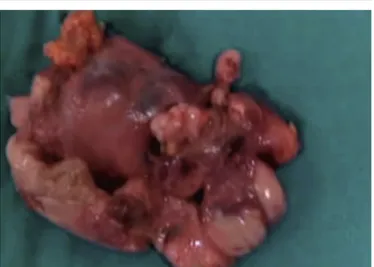
The procedure was well tolerated with a mean oper-ation time of 90 min and without complicoper-ation, except for a minimal controlled bleeding. The oral feeding was restored on the first postoperative day and the drainage was removed in the second postoperative day. The patient made an excellent recovery and was dis-charged from the hospital four days later, after a nega-tive radiological control (Figure 4). Histopathological examination showed thymic tissue of 60 g with a preservation of normal architecture compatible with a true thymic hyperplasia (Figure 5). At follow up the patient was totally asymptomatic during daily activi-ties with a good cosmetic result.
DISCUSSION
The thymus is embriologically derived from the third and fourth pairs of pharyngeal pouches. At birth, it weights from 10 to 35 g and continues to grow in size until puberty, when it achieves a maximum weight of 10 to 50 g, then it undergoes to progressive atrophy [5].
The thymus gland increases in size without a known etiology is defined benign thymic hyperplasia. Histo-logically, two types of thymic hyperplasia are recog-nized: the first, “true hyperplasia”, is defined as an increase in both size and weight of the gland with re-tention of a normal architecture as in our case; the sec-ond type, “lymphoid hyperplasia”, is characterized by the presence of lymphoid follicles with germinal cen-ters, usually not accompanied by a marked increase in thymic size [6]. Clinically, thymic hyperplasia can mimic others important diseases, including thymoma, lypoma and germ cell tumors. Thymic enlargement is generally asymptomatic, but when respiratory symp-toms are present, they may influence the management [7]. In these cases, most of authors support surgical management as the best therapeutic option [8-9]. Sev-eral surgical approaches to thymectomy exist, the tho-racoscopic one was first reported by Sugarbaker from Boston and also the Belgium group in 1993 [10-11]. After, it has evolved with several variants including video-assisted thoracic surgery (VATS) unilateral thymectomy through three ports, the bilateral thoraco-scopic approach combined with a cervical incision for extended thymectomy and more recently endoscopic robot-assisted thymecthomy. Minimally invasive tech-niques have become popular due to their low proce-dural mortality and morbility associated to minimal surgical trauma with the best cosmetic results. Yim et al. first proposed a right-sided approach for VATS thymectomy justifying this for the greater maneuver-ability of instruments in the wider right pleural cavity and easier identification of the left innominate vein using superior vena cava as a landmark [12]. Mineo et al. advocated a left-sided approach because the dissec-tion maneuvers are safer for the posidissec-tion of the superior vena cava out of the surgical field with less risk of ac-cidental injury [13]. In addition, the resection of perithymic fatty tissue around the left
pericardio-A B
56
JOURNAL OF THeSIeNAACADeMY OFSCIeNCeS, PUbLISHeD SINCe1761 - VOL. 4 - 2012
phrenic angle and aortopulmonary window can be per-formed more readly from the left side. Even if the thy-mus can be completely removed using a left or right approach, in our case we chose the left access because the thymic gland was larger in the left side and to allow an extended resection of potential ectopic thymic tissue including the aortopulmonary window. Video-assisted thoracoscopic thymectomy is a successful tech-nique in experienced hands and represents an increasingly popular alternative approach to conven-tional open techniques because it allows to achieve the goal of early thymectomy with the advantages of less invasive procedure.
ReFeReNCeS
1. Ocal T., Turken A., Ciftci A., et al. Thymic enlargement in child-hood. Turk J Pediatric 2000; 42:298-303.
2. Ricci C, Pescarmona E., Rendina EA, et al. True thymic hyperplasia: a clinicalpathological study. Ann Thorac Surg 1989; 47:741-5. 3. Linegar AG., Odell JA., Fennel WMP, et al. Massive thymic
hyper-plasia. Ann Thorac Surg 1991;57:21-3.
4. Yim AP., Izzat MB. VATS approach to the thymus. Minimal Access Cardiothoracic Surgery 2000;209-20.
5. Rice HE., Flake AW., Hori T., et al. Massive thymic hyperplasia: carachterization of a rare mediastinal mass. J Ped Surg 1994;29:1561-4.
6. Zheng T., Li-Yang Y., et al. True thymic hyperplasia in an infant. J Ped Surg 2010; 45:1711-1713.
7. Szarf G., De Andrade M, De Oliveira R., et al. Massive thymic hy-perplasia presenting with respiratory insufficiency in a 2-year-old child. Thorax 2010; 65:555-556.
8. Cheuk W., Tsang W.Y., Chan J.K. Microthymoma: definition of the entity and distinction from nodular hyperplasia of the thymic ep-ithelium (so-called microscopic thymoma). Am Surg Pathol 2005; 29:415-419.
9. Landrenau RJ., Dowling RD., Castillo WM. Thoracoscopic resection of an anterior mediastinal tumor. Ann Thorac Surg 1992; 54:142-4. 10. Sugarbaker DJ. Thoracoscopy in the management of anterior
me-diastinal masses. Ann Thorac Surg 1993;56:653-6.
11. Coosemans W., Lerut TE, Van Raemdonck DE. Thoracoscopic sur-gery: the Belgian experience. Ann Thorac Surg 1993;56:721-30. 12. Ng CS., Wan IY., Yim AP. Video-assisted thoracic surgery
thimec-tomy: the better approach. Ann Thorac Surg 2010; 6:2135-2141. 13. Mineo TC, Pompeo E., Lerut TE., Bernardi G, Coosemans W.,
Nofroni I. Thoracoscopic thymectomy in autoimmune myasthenia gravis: results of left sided approach. Ann Thorac Surg 2000;69:1537-41.
Figure 3. Thoracoscopic isolation of upper thymic pole.
Figure 4. Chest X-ray at post operative control.