AUTHOR COPY
IOS Press
Research Report
Myasthenia Gravis: Unusual Presentations
and Diagnostic Pitfalls
Carmelo Rodolico
a,∗, Daniela Parisi
a, Simona Portaro
b, Fiammetta Biasini
a, Stefano Sinicropi
a,
Annamaria Ciranni
a, Antonio Toscano
a, Sonia Messina
a, Olimpia Musumeci
a, Giuseppe Vita
aand Paolo Girlanda
aaDepartment of Clinical and Experimental Medicine - University of Messina, Messina, Italy bIRCCS Centro Neurolesi Bonino Pulejo, Messina, Italy
Abstract.
Background: Myasthenia gravis (MG) is an autoimmune disorder presenting with fluctuating, fatigable muscle weakness.
Initial symptoms classically involve ocular and proximal limb muscles. Rarely, MG may onset with unusual features, so it can be misdiagnosed with other neuromuscular diseases.
Objective: To describe unusual and atypical presentations of MG in a large cohort of patients, considering and discussing
diagnostic difficulties and pitfalls.
Methods: We report on 21 out of 508 MG patients, coming to our department in the last 27 years and presenting with atypical
or unusual features. The diagnosis was achieved performing a careful clinical examination, a proper neurophysiological assessment, the neostigmine test, the AChR and MuSK antibodies assay and chest CT-scan.
Results: Patients with atypical/unusual MG onset were the 4.4% of all MG patients population. We describe seven different
clinical categories: asymmetric distal upper limbs weakness, foot drop, isolated triceps brachii weakness and foot drop, post exertional axial weakness with dropped head, acute facial dyplegia, limb-girdle MG and MG with sudden lower limbs weakness and recurrent falls.
Conclusions: Atypical and unusual presentations may increase the risk to misdiagnose or delay MG diagnosis. Isolated
limb-girdle presentation is the most frequent atypical form in our series.
Keywords: Myasthenia gravis, limb girdle myasthenia, distal myasthenia, foot drop
INTRODUCTION
Myasthenia gravis (MG) is an autoimmune dis-order, characterized by a wide spectrum of clinical presentations, ranging from purely ocular symp-toms to severe generalized forms. It may be related to different antibodies profiles against sev-eral neuromuscular junction components (NMJ). The most frequent form of MG is related to antibod-ies against the acetylcholine receptors (AChR-abs), whereas MG associated to muscle-specific kinase
∗Correspondence to: Prof. Carmelo Rodolico, Department of Neuroscience, via C. Valeria, Messina, Italy.; Tel.: +39 090/2213501; Fax: +39 090/2212789; E-mail: crodolico@ unime.it.
receptors antibodies (MuSK-abs) is generally char-acterized by a major involvement of bulbar muscles, atrophic tongue, worsening with acetylcholinesterase inhibitors (AChEI) and absent thymic pathology [1–4]. Most recently, several patients with MG associated to lipoprotein related protein 4 (LRP4) antibodies have been reported [5–7]. The clinical hallmark of MG is the presence of fatigable mus-cle weakness. Classically, onset symptoms involve the ocular muscles, including eyelid ptosis, fluctu-ant diplopia, or both. Approximately 20% of patients with MG present, at onset, bulbar symptoms, includ-ing dysarthria, dysphagia and dysphonia [1–3, 8]. Rarely, patients with MG may present at onset with unusual features, such as head drop syndrome, acute
AUTHOR COPY
bilateral facial weakness or isolated bulbar weak-ness, sudden lower limbs weakness and falls, or with atypical presentations including foot-drop, isolated distal weakness without atrophy, limb-girdle muscle weakness, in absence of the classical fluctuation of the disturbances and without extra-ocular muscles involvement. For these cases, the diagnosis of MG represents a diagnostic challenge [10–17]. Herein, we report the atypical and unusual presentations of MG in a large cohort of patients, examined at our Neuro-logical department over the past 27 years, considering and discussing diagnostic difficulties and pitfalls.
PATIENTS AND METHODS
In the last 27 years (1987–2014) we followed-up 508 patients affected by MG. Age at onset of this cohort (263 males and 245 female) ranged between 11 and 90 years. Serum antibodies assay revealed the presence of AChR-abs in 413 patients, whereas MuSK-abs were in 23 present. The remain-ing 72 patients were negative. The diagnosis was achieved performing a careful clinical examination, a proper neurophysiological assessment, including single fiber electromyography (SFEMG) and repet-itive nerve stimulation (RNS), the neostigmine test and a chest CT-scan. All patients were tested for AChR- and MuSK-abs; LRP4-abs were not tested in negative patients. The frequency of follow-up visits and hospital admissions was variable, depending on the severity of symptoms and on treatment response. Herein, we report on 21 out of 508 patients, 10 males (M) and 11 females (F), aged from 14 to 70 years, presenting with atypical or unusual clinical phe-notypes. In the following paragraphs we describe different categories of unusual MG presentation, clas-sified by symptoms at onset.
ATYPICAL MG PRESENTATIONS: CLINICAL CATEGORIES
Asymmetric distal upper limbs weakness without atrophy (cases 1-2-3)
Three patients (M 30-y, F 20-y, F 40-y) complained of subacute muscle weakness and distal limb mus-cles fatigability. In particular, in case 1, intrinsic hand muscles and wrist extensor muscles involvement had been preceded by a long history of ocular myasthe-nia, whereas, in cases 2 and 3, no other muscles have been previously involved. AChR-abs were increased
in all patients, thymoma was found in two out of three patients. They all responded to a combined AChEI and steroid treatment; thymectomy was performed in patients with tymoma. Because of the diagnostic challenge, we reported, in detail, an illustrative case
(case 1).
Case 1
A 22-year-old man came to our Clinic because of bilateral eyelid ptosis and diplopia, started after a fever. These symptoms fluctuated during the day, so that he was investigated for MG. Serum AChR-abs were negative. SFEMG, performed on the orbic-ularis oculi muscles, disclosed an abnormal NMJ function, whereas SFEMG of the extensor indicis muscle was normal. Chest CT-scan did not show thymus abnormalities. Patient received a diagnoses of ocular myasthenia and AChEI and prednisone were administered without benefit. After 6 months, he improved and prednisone was gradually tapered. Eleven years later, he was forced to use crutches for 1 month because of a traumatic right tibia and fibula fracture; after two weeks, he was unable to extend his right wrist and, one month later, also the left wrist. Neurophysiological examination, per-formed in another hospital, ruled out a lesion, due to compression, of the radial nerves or of the pos-terior interosseous branches, hereditary neuropathy with liability to pressure palsy (HNPP) or multi-focal motor neuropathy (MMN). For this reason, the patient come back to our Clinic. Neurological examination disclosed a severe weakness of the right extensor, left wrist and of the third and fourth hand fingers muscles (video 1 - supplementary materials). No sensory symptoms were referred. A generalized form of MG was then suspected. RNS of the right ulnar nerve, recording from the abductor digiti min-imi muscle, disclosed a decremental pattern, whereas the SFEMG, performed on the right extensor indicis muscle, evidenced the presence of 80% of record-ings with increased jitter, 30% of recordrecord-ings with conduction blocks and a mean MCD of 95s. The neostigmine test showed a considerable improvement of symptoms (video 2 - supplementary materials). Serum AChR-abs were positive (1.90 nmol/L with normal values <0,4 nmol/L), whereas chest CT-scan did not reveal thymic abnormalities. The patient was diagnosed of generalized myasthenia gravis with dis-tal involvement; he was treated with AChEI and prednisone 50 mg/die, with a progressive clinical improvement.
AUTHOR COPY
MG with isolated foot drop, without atrophy:(Case 4)
A 16-year-old man came to our attention because of subacute and fluctuant onset of a bilateral foot drop. Neurological examination showed isolated bilat-eral foot and ankle dorsi-flexors muscles weakness. These symptoms worsened while walking. No sen-sory symptoms were referred. Details about patient’s immunological profile, neurophysiological study and treatment are reported in Table 1.
Isolated triceps brachii weakness and foot drop, without atrophy: (Case 5)
A 40-year-old man complained a 5-years history of progressive difficulty and fatigability to keep his arms raised, worsened by repeated exercise. Neu-rological examination showed mild facial weakness (orbicular oris and oculi) and bilateral triceps brachii muscle weakness; he was able to keep the upper limbs raised only for few seconds (video 3 - supplementary materials). There was a mild right ankle dorsi-flexors muscles weakness with a slight “steppage”. Serum AChR-abs were positive (0.65 nmol/L). SFEMG of triceps brachii muscle showed 40% of recordings with increased jitter, 20% of recordings with blocks and a mean MCD of 112s. RNS of the peroneal nerve, recording from the anterior tibial muscle, con-firmed a neuromuscular transmission disorder. Chest CT-scan was normal. He had a good response to steroids, which is still ongoing.
Limb-girdle myasthenia gravis (LGMG): (Cases 6-7-8-9+10-11-12-13-14)
Four patients, cases 6–9, (M 24-y, M 43-y, M 32-y, F 30-y) were admitted to our Clinic during a 15-years period and their data have been already published [15]. In the last 12 years, we have evaluated other 5 additional cases, presenting with a LGMG phenotype
(cases 10–14) (F 38-y, F 44-y, M 41-y, F 56-y, M
30-y). Details are reported in Table 1.
UNUSUAL MG PRESENTATIONS: CLINICAL CATEGORIES
Post exertional axial weakness with dropped head: (Cases 15-16-17)
We report three patients (M 70-y, F 65-y, F 43-y) who complained of a progressive neck extensor
muscles weakness, worsened by walking, even for short distances (video 4 – supplementary materi-als). A RNS of the right ulnar nerve, recording from the abductor digiti minimi muscle, was nor-mal in all patients. SFEMG of the extensor digitorum communis muscle (EDC) disclosed, in case 15, an increased jitter with conduction blocks, but, in
cases 16 and 17 it was normal at the same site
and abnormal on the trapezius muscle, revealing an increased jitter and conduction blocks. Serum AChR-abs were absent, whereas MuSK-abs were found elevated in all patients (ranged from 1.6 to 5.30 nmol/l [n.v. <0.05]). A diagnosis of MuSK pos-itive MG with predominant axial involvement was made. Case 15 was treated with prednisone and aza-thioprine, case 16 received prednisone, azathioprine and plasma exchange (PE), whereas prednisone, cyclosporin, rituximab and intravenous immunoglob-ulin (IVIG) were administered in case 17. All three patients improved with residual symptoms.
Acute facial dyplegia and bulbar weakness: (Cases 18-19)
Two patients (M 47-y and F 26-y) presented with severe and isolated bulbar and respiratory muscles weakness. Both patients presented an acute onset of breathing and swallowing difficulties. They denied weakness and fatigability. Neurological examina-tion disclosed severe facial dyplegia, dysphonia with paralysis of the soft palate, but no ptosis neither ophthalmoplegia were evident. Data about immuno-logical, neurophysiological assessment and therapy are summarized in Table 1.
Myasthenia with sudden lower limbs weakness and falls: (Cases 20-21)
Two patients (F 14-y and F 15-y) complained of episodic, severe lower limb muscles weakness, lead-ing to recurrent falls, often in occasion of fever. Detailed data are summarized in Table 1.
RESULTS
Patients with an unusual/atypical presentation were the 4.4% of all MG patients referred to our department. As reported in Fig. 1, 19% of these (n = 4) showed an isolated distal weakness at four limbs: the most commonly involved muscles were: intrin-sic hand muscles and forearm extensors at the upper limbs (n = 3), and foot and ankle extensor muscles
AUTHOR COPY
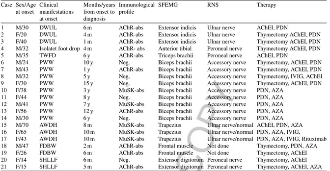
Table 1Summary of clinical course, immunological profile, instrumental data and treatment of patients Case Sex/Age Clinical Months/years Immunological SFEMG RNS Therapy
at onset manifestations from onset to profile at onset diagnosis
1 M/30 DWUL 6 m AChR-abs Extensor indicis Ulnar nerve AChEI, PDN
2 F/20 DWUL 4 m AChR-abs Extensor indicis Ulnar nerve Thymectomy AChEI, PDN
3 F/40 DWUL 1 m AChR-abs Extensor indicis Ulnar nerve Thymectomy AChEI, PDN
4 M/32 Isolatet foot drop 4 m AChR- abs Anterior tibial Peroneal nerve Thymectomy AChEI, PDN
5 M/35 TWFD 6 y AChR-abs Triceps brachii Peroneal nerve AChEI, PDN
6 M/24 PWW 10 y Neg. Biceps brachii Accessory nerve Thymectomy, AChEI, PDN
7 M/43 PWW 1 y AChR-abs Biceps brachii Accessory nerve Thymectomy, AChEI, PDN
8 M/32 PWW 5 y Neg. Biceps brachii Accessory nerve Thymectomy, IVIG, AChEI
9 F/30 PWW 15 y Neg. Biceps brachii Accessory nerve Thymectomy, AChEI, PDN
10 F/38 PWW 3 y MuSK-abs Biceps brachii Accessory nerve PDN, AZA
11 F/44 PWW 8 y Neg. Biceps brachii Accessory nerve PDN, AZA
12 M/41 PWW 7 y MuSK-abs Biceps brachii Accessory nerve PDN, AZA
13 F/56 PWW 12 y AChR-abs Biceps brachii Accessory nerve PDN, AZA
14 M/30 PWW 6 y Neg. Biceps brachii Accessory nerve PDN, AZA
15 M/70 AWDH 8 m MuSK-abs Trapezius Ulnar nerve/normal AChEI, PDN, AZA
16 F/65 AWDH 10 m MuSK-abs Trapezius Ulnar nerve/normal PDN, AZA, IVIG,
17 F/43 AWDH 10 m MuSK-abs Trapezius Ulnar nerve/normal PDN, AZA, IVIG, Rituximab
18 M/47 FDBW 2 m AChR-abs Frontal muscle Not done Thymectomy, PDN, AZA
19 F/26 FDBW 6 m AChR-abs Frontal muscle Not done Thymectomy, AChEI
20 F/14 SHLLF 6 m Neg. Extensor digitorum Peroneal nerve Thymectomy, AChEI 21 F/15 SHLLF 5 m AChR-abs Extensor digitorum Peroneal nerve Thymectomy, AChEI, AZA Legend - DWUL: Distal weakness at upper limbs; TWFD: Triceps brachii weakness and foot drop; PWW: Proximal wasting and weakness; AWDH: Axial weakness with dropped head; FDBW: Facial dyplegia and bulbar weakness; SHLLF: Sudden hyposthenia at lower limbs and falls.; SFEMG was abnormal in the reported muscles; RNS was decremental stimulating the mentioned nerves, if RNS was normal or not done, it is reported.
Categories of unusual and atypical MG presentaƟon
Limb-girdle MG Isolated distal weakness at upper limbs
Post exerƟonal axial weakness with dropped head
Acute facial dyplegia and bulbar weakness
Sudden hyposthenia at lower limbs and recurrent falls Foot droop
Isolated triceps brachii weakness and foot droop 42% 14% 14% 10% 10% 5% 5%
Fig. 1. Diagram of atypical and unusual clinical phenotypes percentages.
at the lower limbs (n = 1); 4.7% (n = 1) presented with isolated triceps brachii weakness and foot droop; 14% (n = 3) had post exertional axial weakness with dropped head; 9.5% (n = 2) manifested acute facial dyplegia and bulbar weakness. LGMG was the most frequent atypical phenotype (43%) (n = 9). Further-more, 9.5% (n = 2) presented with sudden weakness at lower limbs with recurrent falls. Clinical course,
immunological profile, instrumental data and treat-ment are summarized in Table 1.
DISCUSSION
Herein, we have reported the unusual and atypical presentations of MG in a large cohort of patients, mimicking other neuromuscular conditions, such
AUTHOR COPY
as polymiositis, limb-girdle muscular dystrophies, facio-scapulo-humeral muscular dystrophy, periph-eral neurophaties. RNS, which is the most easily and commonly performed test, is not sensitive and it is often normal, especially in ocular and mild general-ized MG. On the other hand, SFEMG is usually more sensitive, but it can be abnormal in muscles which appear clinically unaffected or in other neuromuscu-lar conditions, as well [18]. In our study, we confirm the importance of SFEMG to reach a proper diagnosis of MG, even though it is much more time consuming and it needs more expertise. Furthermore, SFEMG is crucial for diagnosis when performed on affected muscles, which are not routinely tested (cases 1, 5,
15, 16, 17). In conclusion, we suppose that
atypi-cal and unusual presentations of MG might increase the risk to delay the diagnosis of a treatable disease; to recognize these phenotypes is mandatory. Experi-ence from various centers all over the world, in the last 2 decades, has contributed to define atypical MG phenotypes [10–25]. Prior studies reported on asym-metric distal upper limbs weakness: Nations et al, in 1999 described a retrospective study on 236 patients, 9 of whom presenting with upper limbs weakness (hand muscles and finger extensors) [21]. Later on, in 2003, Werner et al. reported on 2 out of 84 patients with distal limb weakness at onset [11]. In our cases, 4 patients out of 508 had this similar clinical pre-sentation at onset. As described, i.e. for case 1, a misdiagnosis with a neuropathy could be a frequent pitfall. Helpful features to make a MG diagnosis are: fluctuations of symptoms, no sensitive disturbances, no involvement of muscle bulk and tone and normal electrophysiological screening for peripheral nerve involvement. Clinical surveillance and follow-up is mandatory, since MG can become generalized, even after 10 years from onset, as described in case 1. Fur-thermore, comparing the time from symptoms onset to diagnosis, it lasts from 1–6 months in distal MG to 1–15 years in LGMG, confirming that the diag-nosis for the latter forms is more difficult. These data are relevant since the majority of patients pre-senting with unusual forms belong to the LGMG group (9/508). Previous studies performed by Oh and Kuruoglu reported on 12 cases diagnosed after 20 years from onset [23]. This persistent and iso-lated limb-girdle involvement, sparing cranial nerves, without fluctuations, is peculiar of these forms. For these cases, RNS is really helpful to reach the proper diagnosis and to rule out the diagnosis of Lamber Eaton myasthenic syndrome, that may mimic LGMG [20]. We speculated that the high frequency of LGMG
in our series, (1.7% of MG patients in 27 years of observation) could be related to a possible latitude influence. Another possible explanation is that our department is a reference center for muscular dis-eases and LGMG patients could have been referred to us with a suspicion of a myopathy. However, mis-diagnosis with a myopathy is not only related to these forms in fact, recently, Devon et al. reported on a patient with triceps brachii and tibialis anterior muscles weakness, mimicking a primarily myopathic disorder [22], as in case 5. Moreover, isolated mus-cle weakness may involve the extensor neck musmus-cles, configuring the “dropped head syndrome” (DHS), that imply a larger differential diagnosis [24–26]. It is known that DHS is not a rare finding in MG: fluctu-ations, worsening during the day with improvement after rest could be considered the “red flags” to reach a clinical diagnosis [24, 25]. In our series of MG patients with atypical and unusual manifestations, cases with other atypical aspects such as sphincter incontinence, pseudohemiplegic form, atrophic MG or MG mimicking blephatospams, already reported in the literature [27–32] were not evident. In con-clusion, we confirm that the combined use of a proper neurophysiological assessment (SFEMG and RNS), antibody dosage, neostigmine test and treat-ment response, are essential to support the clinical suspicion. We are aware that a limit of this study is represented by the scarce number of cases in some categories, but we would test like to focus the atten-tion on these atypical presentaatten-tions, as a warning for clinicians to consider that MG may mimic other, sometimes untreatable, neuromuscular disorders.
FOOTNOTES
Carmelo Rodolico and Daniela Parisi contributed equally to this study as first authors.
CONFLICT OF INTEREST
The authors have no conflict of interest to report.
SUPPLEMENTARY MATERIAL
Supplementary videos are available in the online version of this article: http://dx.doi.org/10.3233/ JND-160148
REFERENCES
[1] Berrih-Aknin S, Frenkian-Cuvelier M, Eymard B. Diag-nostic and clinical classification of autoimmune myasthenia gravis. J Autoimmun. 2014;48-49:143-8.
AUTHOR COPY
[2] Grob D. Course and management of myasthenia gravis. JAm Med Assoc. 1953;153(6):529-32.
[3] Sieb JP. Myasthenia gravis: An update for the clinician. Clin Exp Immunol. 2014;175(3):408-18.
[4] Berrih-Aknin S, Le Panse R. Myasthenia gravis: A com-prehensive review of immune dysregulation and etiological mechanisms. J Autoimmun. 2014;52:90-100.
[5] Tsivgoulis G, Dervenoulas G, Kokotis P, Zompola C, Tzartos JS, Tzartos SJ, Voumvourakis KI. Double seroneg-ative myasthenia gravis with low density lipoprotein-4 (LRP4) antibodies presenting with isolated ocular symp-toms. J Neurol Sci. 2014;346(1-2):328-30.
[6] Verschuuren JJ, Huijbers MG, Plomp JJ, Niks EH, Molenaar PC, Martinez-Martinez P, Gomez AM, De Baets MH, Losen M. Pathophysiology of myasthenia gravis with antibodies to the acetylcholine receptor, muscle-specific kinase and low-density lipoprotein receptor-related protein 4. Autoimmun Rev. 2013;12(9):918-23.
[7] Zisimopoulou P, Brenner T, Trakas N, Tzartos SJ. Sero-logical diagnostics in myasthenia gravis based on novel assays and recently identified antigens. Autoimmun Rev. 2013;12(9):924-30.
[8] Statland JM, Ciafaloni E. Myasthenia gravis: Five new things. Neurol Clin Pract. 2013;3(2):126-133.
[9] Le Panse R, Bismuth J, Cizeron-Clairac G, Weiss JM, Cufi P, Dartevelle P, De Rosbo NK, Berrih-Aknin S. Thymic remodeling associated with hyperplasia in myas-thenia gravis. Autoimmunity. 2010;43(5-6):401-12. [10] Khan KA, Bennett JD. Undiagnosed myasthenia gravis
owing to a very unusual primary presentation. Oral Surg Oral Med Oral Pathol Oral Radiol. 2014;118(4):e101-4. [11] Werner P, Kiechl S, L¨oscher W, Poewe W, Willeit J. Distal
myasthenia gravis frequency and clinical course in a large prospective series. Acta Neurol Scand. 2003;108(3):209-11. [12] Rodolico C, Toscano A, Autunno M, Messina S, Nicolosi C, Aguennouz M, Laur`a M, Girlanda P, Messina C, Vita G. Limb-girdle myasthenia: Clinical, electrophysiological and morphological features in familial and autoimmune cases. Neuromuscul Disord. 2002;12(10):964-9.
[13] Scaioli V, Andreetta F, Mantegazza R. Unusual neurophys-iological and immunological findings in myasthenia gravis: A case report. J Peripher Nerv Syst. 2004;9(2):92-7. [14] Spengos K, Vassilopoulou S, Papadimas G, Tsivgoulis
G, Karandreas N, Zambelis T, Manta P. Dropped head syndrome as prominent clinical feature in MuSK-positive Myasthenia Gravis with thymus hyperplasia. Neuromuscul Disord. 2008;18(2):175-7
[15] Kini PG. Juvenile myasthenia gravis with predominant facial weakness in a 7-year-old boy. Int J Pediatr Otorhi-nolaryngol. 1995;32(2):167-9.
[16] Maher J, Grand’Maison F, Nicolle MW, Strong MJ, Bolton CF. Diagnostic difficulties in myasthenia gravis. Muscle Nerve. 1998;21(5):577-83.
[17] Mongiovi PC, Elsheikh B, Lawson VH, Kissel JT, Arnold WD. Neuromuscular junction disorders mimicking myopa-thy. Muscle Nerve. 2014;50(5):854-6.
[18] Witoonpanich R, Dejthevaporn C, Sriphrapradang A, Pulkes T. Electrophysiological and immunological study in myas-thenia gravis: Diagnostic sensitivity and correlation. Clin Neurophysiol. 2011;122(9):1873-7.
[19] Pallaver F, Riviera AP, Piffer S, Ricciardi R, Roni R, Orrico D, Bonifati DM. Change in myasthenia gravis epidemiol-ogy in Trento, Italy, after twenty years. Neuroepidemiolepidemiol-ogy. 2011;36(4):282-7.
[20] Oh SJ, Head T, Fesenmeier J, Claussen G. Peroneal nerve repetitive nerve stimulation test: Its value in diagnosis of myasthenia gravis and Lambert-Eaton myasthenic syn-drome. Muscle Nerve. 1995;18(8):867-73.
[21] Nations SP, Wolfe GI, Amato AA, Jackson CE, Bryan WW, Barohn RJ. Distal myasthenia gravis. Neurology. 1999;52(3):632-4.
[22] Rubin DI, Litchy WJ. Severe, focal tibialis anterior and tri-ceps brachii weakness in myasthenia gravis: A case report. J Clin Neuromuscul Dis. 2011;12(4):219-22.
[23] Oh SJ, Kuruoglu R. Chronic limb-girdle myasthenia gravis. Neurology. 1992;42(6):1153-6.
[24] Sawa N, Kataoka H, Eura N, Ueno S. Dropped head with positive intravenous edrophonium, progressing to myasthe-nia gravis. BMJ Case Rep. 2013;31:2013.
[25] Rodolico C, Messina S, Toscano A, Vita G, Gaeta M. Axial myopathy in myasthenia: A misleading cause of dropped head. Muscle Nerve. 2004;29(2):329-30.
[26] Goh KJ, Wong KT, Tan CT. Myopathic dropped head syn-drome: A syndrome of mixed aetiology. J Clin Neurosci. 2000;7(4):334-6.
[27] Husillos-Alonso A, Sim´on-Rodr´ıguez C, Bolufer-Moragues E, L´opez-Mart´ın L, Carbonero-Garc´ıa M, Gonz´alez-Enguita C. Greenlight-XPS laser vaporization, the new standard of treatment in men with myasthenia gravis and benign prostatic obstruction? Arch Esp Urol. 2015;68(4):441-3.
[28] Khan Z, Bhola A. Urinary incontinence after transurethral resection of prostate in myasthenia gravis patients. Urology. 1989;34(3):168-9.
[29] Ong BK, Chong PN. Acute hemiparesis with hemichorea and crossed hemiparesis; unusual presentation for myasthe-nia gravis. Singapore Med J. 1993;34(1):60-1.
[30] Roberts ME, Steiger MJ, Hart IK. Presentation of myasthenia gravis mimicking blepharospasm. Neurology. 2002;58(1):150-1.
[31] Nikoli´c AV, Baˇci´c GG, Dakovi´c M ˇZ, Lavrni´c S-D, Rakoˇcevi´c Stojanovi´c VM, Basta IZ, Lavrni´c DV. Myopa-thy, muscle atrophy and tongue lipid composition in MuSK myasthenia gravis. Acta Neurol Belg. 2015;115(3):361-5. [32] Zouvelou V, Rentzos M, Toulas P, Evdokimidis I.
AChR-positive myasthenia gravis with MRI evidence of early muscle atrophy. J Clin Neurosci. 2012;19(6):918-9.