Università degli Studi di Ferrara
DOTTORATO DI RICERCA IN
"FARMACOLOGIA ED ONCOLOGIA MOLECOLARE"
CICLO XXVI
COORDINATORE Prof. Antonio Cuneo
Dental implants osseointegration:
in vitro, preclinical and clinical research results.
Settore Scientifico Disciplinare MED/04
Dottorando Tutore
Dott. Sivolella Stefano Dott. Rimessi Alessandro
_______________________________ _______________________
TABLE OF CONTENTS
ABSTRACT ………... 5
1. INTRODUCTION ……….. 7
1.1 Dental Implant Osseointegration ………. 7
1.2 Haemostasis ……….. 10
1.3 Inflammatory phase ………... 12
1.4 Proliferative phase ………... 14
1.5 Remodelling phase ………... 18
1.6 Introduction to the in vitro, preclinical and clinical studies on osseointegration ………... 22
2. AIM OF THE THESIS ………... 29
3. MATERIALS AND METHODS ………... 31
3.1 IN VITRO STUDIES ……… 31
3.1.1 From Brun et al, 2013. Mechanisms underlying the attachment and spreading of human osteoblasts: From transient interactions to focal adhesions on vitronectin-grafted bioactive surface ………. 31
3.1.2 From A novel in vitro technique to evaluate dental implant osseointegration ……… 37
3.2 IN VIVO STUDIES ………. 41
3.2.1 PRECLINICAL STUDIES ……….. 41
3.2.1.1 From Sivolella et al., 2012. Osteogenesis at implants without primary bone contact – An experimental study in dogs ………. 41
3.2.1.2 From Sivolella et al., 2013. Deproteinized bovine bone mineral particles and osseointegration of implants without primary bone contact: an experimental study in dogs ………. 45
3.2.1.3 From Bressan et al., 2013. Healing of buccal dehiscence defects at implants installed immediately into extraction sockets – an experimental study in dogs ………. 49
3.2.1.4 From Bressan et al., 2012. Short implants (6 mm) installed immediately into extraction sockets: An experimental study in dogs ………... 52
3.2.2 HUMAN STUDY ………. 55
3.2.2.1 From Sivolella et al. 2012. Splinted and Unsplinted Short Implants in Mandibles: A Retrospective Evaluation With 5 to 16 Years of Follow-up ... 57
4. RESULTS ……….. 60 4.1 IN VITRO STUDIES ……….. 60 4.1.1 From Brun et al. 2013 Mechanisms underlying the attachment and
spreading of human osteoblasts: From transient interactions to focal
adhesions on vitronectin-grafted bioactive surfaces …... 60
4.1.2 From A novel in vitro technique to evaluate dental
implantosseointegration ……….. 65
4.2 RESULTS FROM IN VIVO STUDIES ……… 67 4.2.1 PRECLINICAL STUDIES ………. 67 4.2.1.1 From Sivolella et al., 2012. Osteogenesis at implants without
primary bone contact – An experimental study in dogs ………. 67
4.2.1.2 From Sivolella et al., 2013. Deproteinized bovine bone mineral particles and osseointegration of implants without primary bone contact: an
experimental study in dogs ………. 68
4.2.1.3 From Bressan et al., 2013. Healing of buccal dehiscence defects at implants installed immediately into extraction sockets – an experimental
study in dogs ………. 70
4.2.1.4 From Bressan et al., 2012. Short implants (6 mm) installed
immediately into extraction sockets: An experimental study in dogs ………... 71
4.2.2 HUMAN STUDIES ………. 73 4.2.2.1 From Sivolella et al. 2012 Splinted and Unsplinted Short Implants in
Mandibles: A Retrospective Evaluation With 5 to 16 Years of Follow-up ... 75
5. DISCUSSION ……… 76
6. CONCLUSIONS ………... 94
REFERENCES ………. 97
APPENDIX 1– Tables and figures
APPENDIX 2 – Publications related to the thesis APPENDIX 3 – Dichiarazione di conformità
ABSTRACT
Osseointegration is described as the close contact between bone and an implant surface, and the interest on surface engineering has to be understood as an important and natural trend. The biological fixation between the dental implant surfaces and jaw bones should be considered a prerequisite for the long-term success of implant-supported prostheses. In this context, the implant surface modifications gained an important and decisive place in implant research over the last years. The bone response, which means rate, quantity and quality, are related to implant surface properties. For example, the composition and charges are critical for protein adsorption and cell attachment. Hydrophilic surfaces seem to favor the interactions with biological fluids and cells when compared to the hydrophobic ones, and hydrophilicity is affected by the surface chemical composition. Various techniques of surface treatments have been studied and applied to improve biological surface properties, which favors the mechanism of osseointegration. This strategy aims at promoting the mechanism of osseointegration with faster and stronger bone formation, to confer better stability during the healing process, thus allowing earlier loading of the implant. Some of the objectives for the development of implant surface modifications are to improve the clinical performance in anatomical sites characterized by poor quantity or quality of bone, to accelerate the bone healing and thereby allowing immediate or early loading protocols and also stimulating bone growth in order to permit implant placement in sites that lack sufficient residual alveolar ridge, thus providing them a jumping gap ability, for example. Implant morphology influences bone metabolism: rougher surfaces stimulate differentiation, growth and attachment of bone cells, and increase mineralization; furthermore, the degree of roughness is important. Implants may have "smooth" (machined) or rough surfaces. The main methods that are reported in the literature to create implant roughness are acid etching, sandblasting, titanium plasma spraying and hydroxyapatite (HA) coating. A current tendency is the manufacturing of implants with micro and submicro (nano) topography. Furthermore, the bio-functionalization of implants surfaces, by adding different substances to improve its biological characteristics, has also been recently investigated.
surface modifications in order to improve the clinical performance in areas with poor quantity or quality of bone.
In detail this aim has been reached trough 3 steps: 1. in vitro studies:
a) in vitro studies of osteoproperties of glass and titanium surfaces grafted with fibronectin sequence motif for integrin binding (Arg-Gly-Asp, RGD)/ human vitronectin protein (HVP)
b) development of an in vitro model to evaluate the degree of implants osteointegration by passing in vivo test.
2. in vivo studies, in big animal model, dogs, of osteointegrative properties of implants with different degree of surface modification.
3. clinical research on short implants, to underline the effect of surface modifications on dental implant survival in the long term in the clinic.
As final results of these studies, we can conclude that there are a huge number of types of implant surfaces in the market, from different implant manufacturers, all of them claiming to have better clinical results. It is important that the clinician choose the surface that have shown the best results in the scientific literature.
The majority of currently available in vitro and in vivo studies seem to indicate that implant surfaces with micro and submicro (nano) topography bring forward benefits to the process of interaction between bone cells and implant surfaces, accelerating and increasing the quality of bone-to-implant-contact (BIC).
Finally, based on the state of the art of implant development, it is possible to predict that, within some time, implant surfaces coated with substances with biomimetic capacity will be available for clinical use. This process of implant bio-functionalization aims at modulating new bone formation around implants, and it represents the next step in implant development.
1. INTRODUCTION
1.1 Dental Implant Osseointegration
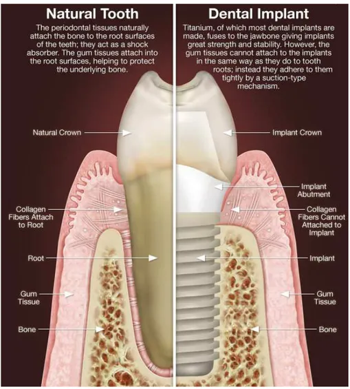
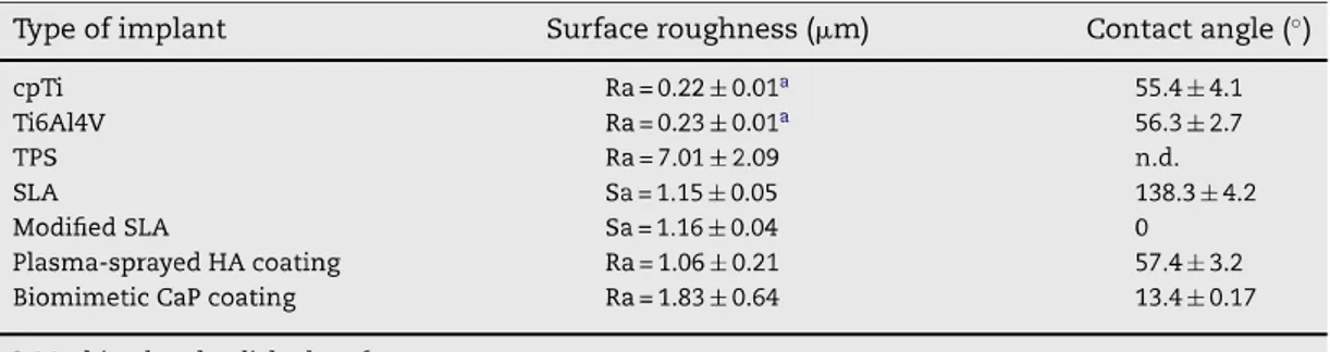
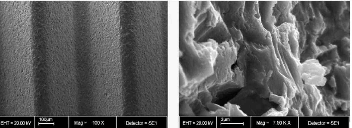
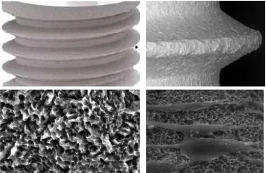
A dental implant (also known as an endosseous implant or fixture) is a surgical device that interfaces with the alveolar bone of the jaws (or skull) to support a dental prosthesis such as a crown, bridge, denture, facial prosthesis, or to act as an orthodontic anchor (Fig. 1). The basis for modern dental implants is a biologic process called osseointegration where materials, such as titanium, form an intimate bond to bone. Osseointegration derives from the Greek osteon, bone, and the Latin integrare, to make whole. The term refers to the direct structural and functional connection between living bone and the surface of a load-bearing artificial implant. Osseointegration is also defined as: "the formation of a direct interface between an implant and bone, without intervening soft tissue" (1, 2). The osseointegration rate of titanium dental implants is related to their composition and surface roughness (fig. 2 and 3). Rough-surfaced implants favor both bone anchoring and biomechanical stability. Osteoconductive calcium phosphate coatings promote bone healing and apposition, leading to the rapid biological fixation of implants. Different methods have been used for increasing surface roughness or applying osteoconductive coatings to titanium dental implants. Surface treatments, such as titanium plasma-spraying, grit-blasting, acid-etching, anodization or calcium phosphate coatings, and their corresponding surface morphologies and properties have been studied. Most of these surfaces are commercially available and have proven clinical efficacy (>95% over 5 years) (3). A huge number of the experimental investigations have demonstrated that the bone response was influenced by the implant surface topography (evaluated using
Sa value, that is the arithmetic average of the 3D roughness); smooth (Sa<0.5 µm)
and minimally rough (Sa 0.5–1 µm) surfaces showed less strong bone responses
than rougher surfaces. Moderately rough (Sa>1–2 µm) surfaces showed stronger
bone responses than rough (Sa>2 µm) in some studies (4) (figs. 5-12).
Finally, osseointegration is the growth action of bone tissue, as it assimilates surgically implanted devices or prostheses to be used as either replacement parts (e.g., hip) or as anchors (e.g., endosseous dental implants).The direct contact of bone and implant surface can be verified microscopically. Osseointegration has
enhanced the science of medical bone and joint replacement techniques as well as dental implants and improving prosthetics for amputees.
As recently described by Terheyden H et al (2012) (5) the healing of an osseous wound around a dental implant is a coordinated and sequentially organized repair mechanism of the organism (6). The main players in this process are cells. Cells communicate with each other via exchange of molecules which are read by specific receptors on the cell surface. The different cell types appear in a chronological sequence with a certain overlap. This sequence is known as the four phases of wound healing, a concept that originates from the observation of soft tissue healing (7). However, this concept can be transferred to bone healing and, in particular, to intraoral bone healing of an implant wound – haemostasis, the inflammatory phase, the proliferative phase and finally the remodelling phase. In a physiological soft tissue wound, the haemostasis takes minutes to hours, the inflammatory phase hours to days, the proliferative phase days to weeks and the remodelling phase begins at approximately 3 weeks and lasts for years (7).
The temporal sequence of bone healing around dental implants has been investigated histologically in animals (dogs) (8, 9) and in humans (10, 11). In the animal study, the first biopsy showing erythrocytes and inflammatory cells was taken after 2 h at the transition between haemostasis and inflammatory phase. The second biopsy was taken after 4 days and showed new vessels as well as fibroblasts and osteoclasts on the old bone (early proliferative phase). After 1 week, woven bone had appeared (late proliferative phase). After 2 weeks, a load oriented remodelling of the woven bone by osteoclasts was noted in the areas of the tips of the threads (early remodelling phase). After 4 weeks, the remodelling at the tips of the threads was most intense. After 6 weeks, woven bone formation continued and remodelling also took place in the grooves of the implant threads. After 8 and 12 weeks, most woven bone was replaced by lamellar bone. In the human volunteer study, four time points after 1, 2, 4 and 6 weeks were examined. After 1 week in humans, new bone was observed occasionally on the implant surface in humans – comparable to what had been seen in the dog study. After 2 weeks, woven bone formation had increased, but only in the grooves. In contrast to the dog study, no marked osteoclastic activity was observed in humans (proliferative phase). After 4 weeks, bridging between the parent bone and the implant took place in humans. After 6 weeks, first signs of transition to the
remodelling phase were noted, 2 weeks later than in the dog. The direct comparison of the bone-implant contact rates revealed a delay of at least 2 weeks for humans compared with dogs (9). A microarray analysis of the transcriptome of the material of the human volunteer study showed genes associated with inflammation upregulated at day 4, for angiogenesis at day 7 and for skeletogenesis at day 14 (12, 13). Thus, the duration of the phases of bone healing around dental implants in humans approximates the duration of the same phases in physiological soft tissue healing as a biological constant.
For Terheyden H et al (2012) (5) the key players in this process are the different cell types. We observe coordinated action of several cell types and numerous individual cells in the defect. The action of cells is controlled by sequential activation of typical genes, which in turn are activated by soluble cytokines (soluble protein factors), small molecules (e.g. histamine, prostaglandins etc.) or molecules from the extracellular matrix (14). These messenger molecules interact with specific receptors on the surface of the cells. Usually, this causes a change of the conformation of transmembrane receptor proteins which become enzymatically active and start an intracellular second messenger system that amplifies or modifies the information and transports it through the nuclear membrane to the DNA. The cellular response is then initiated by activation of genes and expression of certain proteins, either secretory products or intracellular regulatory proteins. Adjacent cells can communicate with each other through direct membrane channels. However, over distances, the cells communicate through chemical messenger molecules. The most important classes of messenger molecules are cytokines and hormones. Cytokines are proteins (interleukins, growth and differentiation factors). Hormones are subdivided into peptide hormones (e.g. bradykinin), lipid hormones (e.g. prostaglandins or steroid hormones) and amine hormones (e.g. histamine). Although there is an overlap between the definitions of cytokines and hormones, hormones are usually active in nanomolar concentrations and longer ranges, whereas cytokines can be active in femtomolar concentrations through very specific protein receptors within a more restricted area. In addition, cells receive information through interaction with the extracellular matrix, to which they attach with specific receptors (15). On a very local level, small molecules like nitric oxide or even ions like calcium play a role in signalling.
1.2 Haemostasis
Haemostasis (exudative phase) begins with the surgical trauma exerted by the dental implant drill followed by the insertion of the implant (5). The duration of this phase is minutes to hours. With the bone trauma matrix proteins, growth and differentiation factors which are stored in the bone matrix become soluble and active. Usually stored deep in the bone matrix, the factors are unmasked by the bone trauma and liberated from their heparin binding domains by heparin hydrolases from blood platelets (16). Mechanical crushing of the bone matrix in form of bone debris created by the implant drill may facilitate the liberation of such molecules from the matrix (10). Bleeding from injured blood vessels lays the foundation of the polymerization of fibrinogen to create a first extracellular matrix in the defect. The polymerization of fibrinogen is performed by thrombin and initiated by platelets (extrinsic system) and the intrinsic clotting cascade (Hageman Factor). Immediately after implantation, the implant surface interacts with water molecules and ions. This can change the charge pattern of the surface, and bivalent ions like calcium can potentially link equally negatively charged partners (a reason for the requirement of calcium ions in blood clotting) (17). Ions are followed by plasma proteins like albumin, globulins or fibrin. The process of protein adsorption is very effective, increasing the concentration of proteins on the surface rapidly by a factor of 1000 compared with the surrounding aqueous solution (18). The first proteins to bind are those that are present at high concentrations in blood such as albumin. These will slowly be replaced by proteins with a lower concentration, but a higher affinity for the surface such as vitronectin or fibronectin. In this process, size and thus mobility of the proteins also play a role (also referred to as Vroman effect) (19). The adsorption of proteins is determined by various factors such as properties of proteins and the solid substrate surface as well as environmental conditions. With respect to the protein properties, the charge, size, stability of the structure, amino acid composition and steric conformation may play a role. Proteins with low internal stability (soft) adsorb mainly based on a gain in conformational entropy as they change their shape. On hydrophobic surfaces, these changes can occur to a great extent and can lead to protein denaturation and loss of protein function, as hydrophobic residues usually hidden in the protein interior are exposed. To a much smaller extent this also applies to very stable (stiff) proteins, but these will only adsorb if there is electrostatic attraction even on
hydrophobic surfaces (20). Overall shape also plays a role, as rod-like proteins with a higher surface to volume ratio will have more interaction sites and thus bind more strongly than globular ones. Thus, hydrophilic titanium surfaces may better preserve the protein conformation and function. Clinically, a faster osseointegration was observed for ultrahydrophilic surfaces compared with standard titanium surfaces (11). On the metal side also, topography and surface energy are important factors. Little is known about spatial distribution of these properties on a nanometre scale. Patterns may bind and select effective proteins more specific than uniform surfaces.
Through protein absorption, cells are able to attach to the titanium surface. The subsequent cell attachment is influenced extensively by this initial coating of the titanium with blood proteins (21). Fibronectin, for example, contains cell binding sites (RGD sequence) that can interact with cellular adhesion proteins (integrins). At the sites of vascular injury, platelets aggregate and form a white thrombus closing the vascular leak. Bioactive molecules such as thrombin, ADP, collagen, fibrinogen and thrombospondin are generated. Vitronectin bound to the metallic surface can bind platelets. These stimuli activate platelets, converting the major
platelet integrin ανβ3 from a resting state to an active conformation. Integrin
activation refers to the change required to enhance ligand-binding activity. The
activated ανβ3 interacts with the fibrinogen and links platelets together in an
aggregate to form a platelet plug. ανβ3 bound to fibrin generates more intracellular
signals (outside-in signalling), causing further platelet activation and platelet plug retraction. Platelets also bind to collagen with collagenspecific glycoprotein Ia/IIa receptors. This adhesion is further strengthened by von Willebrand factor, which forms additional links between the platelets glycoprotein Ib/ IX/V and the collagen fibrils. Surface bound fibrin on the metal surface of the implant can bind thrombocytes over the glycoprotein IIb/IIIa receptor to the titanium implant surface. This binding results in activation and degranulation of the thrombocytes. Haemostasis is supported by vasoactive substances from the platelets like serotonin, which results in vasoconstriction. Also, thromboxane from platelets plays a role in the initial vasoconstriction. The release of cytokines from degranulating platelets is the beginning of the inflammatory phase.
1.3 Inflammatory phase
Terheyden H et al (2012) (5) report that the inflammatory phase begins approximately after 10 min and lasts for the first days after surgery. The phase begins with the degranulation of the platelets. The platelets release growth factors like transforming growth factor beta (TGF-b), platelet-derived growth factor (PDGF), Basic fibroblast growth factor (bFGF). Bradykinin from degranulated platelets increases the vascular permeability for fluids, serum proteins and white blood cells. Vasodilative histamine derived from the platelets increases blood flow, decreases the blood stream velocity and induces hyperaemia. The initial vasoconstriction in the haemostatic phase turns into vasodilatation, clinically detectable as swelling and warming of the skin overlying the wound.
In the very early stages of the inflammatory phase, the innate host defence systems are activated (22). The innate immune system is activated by unspecific molecules of bacterial origin, and is not adaptable. It consists of molecular (e.g. complement system) and cellular elements: polymorphonuclear leucocytes (PMN, also called neutrophil granulocytes) and macrophages. The complement system is a group of glycoproteins which form membrane perforating channels (perforins) that damage bacterial cells. Complement C3b binds to bacterial glycoproteins and labels (opsonization) bacteria and other foreign bodies for phagocytosis by the immune cells. The PMN invade the blood clot by amoeboid migration, squeezing through little gaps in the walls of the blood vessels. This process is known as the diapedesis. Diapedesis is initiated by loose adhesion of lectins in the inner lining of blood vessels. These first bindings are reversible. The leucocytes move to the periphery of the blood stream, attach and detach and roll along the inner lining of the blood vessel mediated by adhesion of L-selectin on the leucocyte with E-selectin on the endothelial side. Later, stronger chemical adhesions occur until the cells finally attach. Intercellular adhesion molecule-1 (ICAM-1), ICAM-2 (similar to immune globulins) and the vascular cell adhesion molecule-1 (VCAM-1) catch the granulocyte out of the blood stream, binding to integrins on the leucocyte (22). After adhesion, endothelial cells open a small gap, and the granulocyte migrates in amoeboid fashion through the gap. PMN produce elastase and collagenase which helps them in digesting the basal lamina of the blood vessel and pass beyond the
basal lamina. After the cell has left the blood vessel, its further amoeboid migration is directed by chemotaxis (22).
Chemotactic substances for PMN include: fibrinopeptides from fibrin activation through thrombin, products from fibrinolysis, complement 5a, leucotriene B4 from present PMN, bacterial proteins (N-formyl methionyl peptides), platelet activating factor (PAF), tumour necrosis factor alpha (TNF-alpha), Platelet factor 4 (PF4), PDGF and interleukin8 (IL-8). Some of these factors are produced from PMN or macrophages already present which had antigen contact. If the granulocytes encounter large numbers of bacteria, they recruit more PMN by releasing proinflammatory cytokines (TNF-alpha, IL-8).
Thus, abundance of bacteria prolongs and amplifies the cellular immune response. PMN kill bacteria through reactive radicals (oxygen species and hydroxygroups, chlorine radicals and hypochlorites) which are also toxic for the host cells and the healthy tissue surrounding the wound. Thus, a fulminant neutrophil granulocyte response can induce loss of healthy surrounding tissues (22).
Furthermore, PMN secrete digestive enzymes like collagenase and elastase. These factors can further enhance the tissue damage in the neighbouring tissues. If granulocyte action is prolonged through a high concentration and prolonged presence of bacteria in the wound, a toxic wound environment can develop. In a toxic wound environment, concentrations of proinflammatory cytokines and toxic radicals are high. An elevated activity of the urokinase-type plasminogen activator uPA results in plasmin activity, fibrinolysis and degradation of the extracellular matrix (23). The fibrin network can dissolve. Under these conditions, the concentrations of protective extracellular matrix glycoproteins and proteoglycans such as fibronectin and decorin are low. These proteins normally can bind and protect growth factors from the digesting proteolytic enzymes. A high concentration of these digesting enzymes therefore is typical for a toxic wound environment. If the number or virulence of bacteria further increases, the tissues can be liquified and pus is formed. The early inflammatory phase within the first 3 h is rather decisive for the further fate of the wound. High numbers of bacteria enhance inflammation. Contaminated foreign bodies in the wound, which unlike living tissue have no own defence mechanisms against bacterial colonisation, can increase bacterial counts in the wound. To limit the inflammatory phase, the
cleanest possible surgical work with low bacterial inoculation is likewise important as antibacterial measures including antibiosis and local disinfection. Clean conditions help the organism to move as quickly as possible through the inflammatory phase into the proliferative phase.
PMN are relatively short-lived in acute wounds and are replaced by lymphocytes and macrophages. The roll of lymphocytes is not well defined in the repair process, but they appear to assist by secreting cytokines that are mitogens and chemoattractants for fibroblasts, while simultaneously clearing the wound of old neutrophils (7). If bacteria have to be eliminated, the number of macrophages increases. In presence of bacteria, they secrete proinflammatory cytokines, but they can act as a switch to end the inflammatory phase. After having removed tissue debris, macrophages secrete angiogenic and fibrogenic growth factors. The level of the radical nitric oxide (NO) in the wound formed by inducible nitric oxide synthase (iNOS) by macrophages correlates positively with cyclooxygenase activity and prostaglandin production, which is necessary for subsequent fibroblast activation (24, 25). Under the conditions of a healing wound which was successfully cleaned from bacterial contamination these cells secrete TIMPs (tissue inhibitors of metalloproteinases). These molecules antagonize the digesting enzymes of PMN and therefore protect the extracellular matrix proteins like proteoglycans. These in turn can protect growth factors which are stored in the extracellular matrix (26). The concentration of growth factors is further increased by secretion of growth factors like bFGF and PDGF from macrophages. A high concentration of fibronectin allows attachment of fibroblasts via integrin binding sites. These cells can hereupon crawl into the wound. This is the beginning of the proliferative phase.
1.4 Proliferative phase
The transition into the proliferative phase is characterized by the formation of new extracellular matrix and by angiogenesis (5). This newly formed tissue is called granulation tissue. The duration of this phase ranges from a few days to a few weeks.
Stimulated by FGF from macrophages, fibroblasts from the surrounding healthy tissue migrate by amoeboid movement into the blood clot. These cells drill tunnels through the provisional extracellular matrix of the fibrin clot by secreting matrix
metalloproteinases. The metalloproteinases degrade the blood derived fibrin in the clot und uncover integrin binding sites in the fragments. The fibroblasts attach via integrins to the RGD peptides of fibronectin and crawl from attachment to attachment deeper into the wound (27). To replace the degraded provisional clot matrix, they produce insoluble cellular fibronectin and other insoluble proteins of the extracellular matrix like collagens, vitronectin, decorin and other proteoglycans. The movement of the fibroblasts is directed by the concentration gradient of growth factors produced by the macrophages (PDGF, TGF-b, basic FGF, connective tissue growth factor (CTGF)).
In parallel, angiogenesis is stimulated by hypoxia. Hypoxia attracts macrophages (28), which are able to survive under hypoxic conditions by adjusting their metabolism to an oxygen independent generation of ATP. In macrophages, vascular endothelial growth factor (VEGF) expression is stimulated by an intracellular transcription factor called hypoxia inducible factor (HIF-1). The macrophage is able to function under low oxygen tension (29) and releases VEGF, which stimulates the production of endothelial cell precursors and is chemotactic for these cells. Furthermore, end products of lipid oxidation, ω-(2-carboxyethyl) pyrrole (CEP) and other related pyrroles stimulate endothelial cells over the toll like receptor 2 (30). Also, other growth factors like PDGF from platelets and FGF from macrophages act angiogenic.
In response to VEGF, pericytes detach from the outer walls of the vessel. These cells use matrix metalloproteinases to digest the basal lamina around the vessels (31). The pericytes give rise to the new endothelial progenitor cells, which migrate to the place of low oxygen tension where they are chemotactically attracted by the chemokine stromal cell derived factor (SDF-1) which is produced by cells in the wound (32). This process is called homing of endothelial cells. The cells proliferate to form condensed groups and they arrange themselves to form tubes. The room needed for that is created by matrix metallo-proteinases. Finally, these newly formed tubes are connected to an existing blood vessel. A new vascular loop is created and blood can flow through.
Angiogenesis is the prerequisite for osteogenesis. New bone forms only in close connection to blood vessels. The mature bone cell does not survive more than 200 µm away from a blood vessel. First, the blood vessel develops and then the bone
follows, a process called angiogenetic osteogenesis. The formation of new bone needs a mechanically stable environment.
An osteoprogenitor cell attaches to the surface of an implant via integrins. Integrins attach to extracellular matrix proteins such as fibronectin via the RGD motif. An osteoblast does not directly attach to metal, but to the protein layer on top of the implant. The bone precursor cell itself produces insoluble cellular fibronectin needed for cellular attachment to titanium (33). After firm attachment to the surface, the osteoprogenitor cell that becomes secretory active is called osteoblast. As a molecular marker, the osteoblast starts to express osteocalcin and alkaline phosphatase.
Osteoblasts derive from mesenchymal stem cells and there is growing evidence that these stem cells are pericytes in the walls of smallest blood vessels (34). The precursors of pericytes originate from bone marrow cells (35). Bone morphogenetic proteins bind to receptors on the cell surface of the bone precursor cells (36). Binding to preformed complexes of receptors I and II will lead to activation of the Smad pathway, where the activated SMAD protein ultimately binds to DNA and in turn activates SMAD responsive genes like Runx. Bone morphogenic proteins (BMP) may also bind to single receptors, which induces their oligomerization, caveolae-dependent internalization and the activation of non-SMAD pathways such as ERK (extracellular signal regulated kinases) and MAPK (mitogen activated protein kinase). These will activate ATF2, c-jun or c-fos, which regulate BMP target genes like osteopontin, alkaline phosphatase or collagen typeI (37).
It is unclear from where the first BMPs in the wound originate. BMPs are stored in the bone matrix, bound in an inactive form to the glycosaminoglycane heparan sulphate. This allows the organism to store large quantities of active growth readily available and independent of new protein synthesis (16). With bone trauma matrix proteins, growth and differentiation factors which are stored in the bone matrix become soluble and active. Usually stored deep in the bone matrix, the factors are unmasked by the bone trauma and liberated from their heparin binding domains by heparin hydrolases from blood platelets (16). Heparan sulphate binding growth factors (e.g. BMPs, FGF, PDGF, VEGF) can also be released from the matrix by soluble heparin degrading enzymes (heparin hydrolases), which can be released
by platelets, lymphocytes or mast cells. The factors can also be released by competition with heparin, with proteins that bind to the growth factor or with other heparin binding proteins. A number of factors can be released by special proteolytic enzymes, e.g. PDGF-B (thrombin) or VEGF (plasmin) or TGF-b (multiple serin proteases) (16). The growth factors may be also unmasked or synthesized by osteoclasts (38, 39). They are produced by myofibroblasts and osteoblasts. BMPs appear in the bone wound after 3 days. Therefore, new bone forms with a latency.
With implant insertion, a dental implant gains primary stability. The implant is passively stabilized in the bone wound through friction with the primary bone contacts. The denser the host bone is, the more primary bone contacts are available and the higher primary stability of the implant will be. Primary stability implies that the friction holding the implant is higher than the highest dynamic load forces applied. Micromovement caused by load peaks higher than friction hold is critical. Micromovement of the implant can grind and slowly smoothen the bone surface, reducing the interlock between bone and titanium and ultimately resulting in a loss of primary stability. Therefore, it is critical to overload the implant occlusally in the early phase. Primary stability is important during the first days after implant installation. Under normal conditions the first weeks are a vulnerable phase because primary stability can decrease to critical levels before secondary stability has developed.
As early as 1 week after implant, placement new bone formation starts and the primary bone contacts are supplemented by newly formed secondary bone contacts (8). The first bone that forms after an injury is woven bone. Histologically, this bone is characterized by the fact that its collagen fibres are not parallel, but randomly oriented. Woven bone usually grows along the existing bone surfaces and along the dental implant surface towards the groves of the threads. Bone debris created by the implant drill was demonstrated to be important for early bone formation, and is incorporated into the immature trabelculae of woven bone (10). In the beginning, these bone contacts are not load oriented and randomly distributed. In a human volunteer study, new bone apposition amounted to a bone-implant contact of 62% of the intraosseous bone-implant surface after 6 weeks, irrespective whether a SLA (sandblasted large grit and acid etched) or a modified ultrahydrophyllic SLA (SLActive) was used. However, the modified ultrahydrophilic
surface yielded more bone early contacts after 2 and 4 weeks compared with the standard SLA surface (11) (Fig. 8).
New bone formation begins with the secretion of a collagen matrix by osteoblasts. Depending on the process of ossification (endochondral or intramembraneous), this can be collagen type II or type III, which is ultimately replaced by collagen type I. Bone formation within the alveolar process is a process of intramembranous ossification, starting by the secretion of collagen type III. This matrix is subsequently mineralized by hydroxyapatite. The exact mechanism of this process is still widely debated, but in all probability is based on the concept of heterogenous nucleation, where organic or inorganic precursor seeds direct the formation of apatite from soluble inorganic ions (40). Opinions diverge on the nucleation site and the molecular nature of the nucleator: One theory proposes matrix vesicles (small vesicles derived from mineral forming cells such as chondroblasts or osteoblasts) as the site of an initial mineralization prerequisite for the following secondary mineralization of collagen (41). An alternative view proposes direct nucleation of apatite by matrix molecules such as collagen and noncollagenous proteins.
The mineralization process during primary bone formation is rapid, but relatively unorganized and not in close association to collagen (extrafibrillar). During the following remodelling phase, woven bone is removed by osteoclasts and replaced by lamellar bone. Next, in this process nanometre-sized, uniaxially oriented hydroxyapatite crystal plates are formed within the collagen fibres (interfibrillar) (42). This nanostructural architecture gives rise to the unique mechanical and biological properties of bone, making it rigid enough to resist pressure and traction forces while maintaining elasticity.
Removal of the woven bone by osteoclasts is the beginning of remodelling and thus the fourth and last phase.
1.5 Remodelling phase
One of the cellular key players of the remodelling phase is the osteoclast (5). Osteoclasts appear in the wound after a few days. They start to create space for new bone formation and remove primary boneimplant contacts. The remodelling phase can last several years until most woven bone and old bone from the primary
bone contacts is replaced by newly formed and load oriented bone.
Bone being formed after remodelling is called lamellar bone, named after the parallel orientation of its collagen fibres under polarized light. In contrast to woven bone which is oriented parallel to the titanium surface in the grooves of the threads, lamellar bone attaches rather to the tips of the macrothreads. These trabeculae usually attach at the tip of a thread of the implant in a little extended foot plate. The trabeculae distribute the occlusal loads to the surrounding bone and, if present, neighbouring tooth sockets. The new trabecular network is oriented similar to the supporting arches of a gothic church. According to Wolfs Law in bone, such a structure is built as light as possible. Therefore, between the insertion areas of the trabeculae, non-covered titanium surface areas appear on the implant surface. The so-called bone-implant contact can decrease during the remodelling phase and usually balances at approximately two-thirds of the surface after some time (43, 44).
Osteoclasts and osteoblasts act interdependently (45, 46). The so-called bone balance is necessary, because otherwise, the skeleton would become more porous (osteopenic) or denser (osteopetrotic). Both situations can be pathologic. At the beginning, osteoclast action depends on osteoblasts which control osteoclastsogenesis by the balance between RANKL and its counterpart osteoprotegerin, both produced by the osteoblast (47). Osteoblasts secrete RANKL, the ligand of the RANK (receptor activator of nuclear factor kappa beta) receptor which activates osteoclastogenesis together with M-CSF (macrophage colony stimulating factor). RANKL is membrane bound and can be masked by soluble osteoprotegerin which is also synthesized by osteoblasts and is a decoy receptor for RANKL (48). Thus, osteoprotegerin preserves bone by inhibition of osteoclastogenesis. The ratio of RANKL/ osteoprotegerin can be modulated, and the osteoblast is the target for various bone enhancing and inhibiting messenger molecules including IL-11, sclerostin, prostaglandin E2, parathyroid hormone (PTH) related protein, vitamin D and estradiol (39). PTH inhibits osteoprotegerin secretion from the osteoblast and thus increases osteoclast activation and bone degradation (48). In addition, soluble RANKL and the related messenger molecule TNF produced by lymphocytes can upregulate osteoclastogenesis under inflammatory situations (49).
The origin of osteoclasts is blood borne monocytes. They attach to the walls of the blood vessels by SDF-1/CXCR-4 interaction, and SDF-1 is bound to the endothelial cells surface (50). By diapedesis, these cells leave the blood stream. For the transmigration through the collagen of the basal lamina, they secrete matrix metalloproteinase MMP-9 (50). By chemotaxis, the cells are directed towards the bone. Soluble SDF-1 was identified as chemo-attracting molecule for osteoclast precursors (50), but being originally immune cells also, other
immunoregulating molecules like IL-8 (cytokine-induced neutrophil
chemoattractant; CINC-1) and monocyte chemotactic protein (MCP-1/CCL2) were demonstrated to be chemoattractive for the osteoclast precursor cells. Precursor cells fuse to form multinuclear giant cells. Osteoclast formation requires the presence of RANKLigand and M-CSF (51).
These membrane bound proteins are produced by neighbouring osteoblasts, thus requiring direct contact between these cells and osteoclast precursors. Proinflammatory cytokines like IL-6 or TNF-alpha can intensify this activation (52). There is some evidence that osteoclast precursors, like many other immune cells, need a costimulation via the ITAM receptor (immunoreceptor tyrosine-based activation motifs) (53).
The life span of an osteoclast was calculated to average 12 days in humans (54). The bone lining cells (terminally differentiated osteoblasts) digest remnants of osteoid by collagenases and thereby liberate RGD peptide endings from non-collagenous bone matrix proteins like osteopontin. The lining cells then detach from the bone surface. The so-prepared surface attracts migrating osteoclast precursors. The osteoclasts form a structure comparable to a suction cup on the bone surface, sealing the margin with a ring of integrin attachments. These integrins attach to bone matrix proteins like osteopontin (55). Between the osteoclasts and bone, a secluded space is created – the resorption lacuna – to protect neighbouring cells from acid and aggressive enzymes and to limit the extent of bone resorption. Under the suction cup, the osteoclasts increase the surface of their cell membrane by forming microscopic folds, the so-called ruffled border, a sign of the actively resorbing osteoclast (39). The cell membrane in the folds contains ion pumps that are comparable to gastric ion pumps. Producing hydrochloric acid, the acid demineralizes bone matrix and liberates bone collagen. Special enzymes, one of which is cathepsin K, digest bone collagen.
The coupling of osteoclasts and osteoblast and the molecular mechanism of how osteoclasts control and activate osteoblasts to fill up the bone void after resorption is still unclear in detail (56). As discussed earlier, growthand differentiation factors like BMP, IGF, TGF beta are stored in the bone matrix bound in an inactive form to heparan sulphate. They can be liberated from the bone matrix and activated by cleavage from the glycosaminoglycan by proteolytic enzymes. These enzymes are located on the surface of many cell types including osteoblasts (16). The role of the osteoclast in unmasking these factors by their proteases in the resorption lacuna is unclear. It is unlikely that these growth factors are transferred in intact form through endocytosis and through the osteoclasts cytoplasm. However, it is known that osteoclasts express and secrete BMP-6 and may thereby amplify the BMP signal which they have received from the degraded matrix (38). BMP-6 and the chemokine sphingosine 1 phosphate (S1P) are released on the tissue side by the osteoclast (56). BMP-6 is a coupling factor of bone resorption and refill involved in the osteoblast recruitment (57). BMP-6 differentiates mesenchymal stem cells to osteoblasts to build new bone (58). With other types of messenger molecules like ephrin and cardiotropin, osteoclasts may control the osteoblasts (45, 39). It has been shown that a bidirectional signalling exists between osteoclasts and osteoblasts in direct neighbourhood by the exchange of membrane bound ephrinB2 and EphB4 ligands. According to this theory, the osteoclasts retract themselves and directly differentiate osteoblasts in direct cell contact to fill the void with new bone (59). Osteoblastic precursor cells can sense the surface topography in the resorption lacuna by creating pseudopodia and thus attain information about how much bone is needed to fill the void (39). At this point, there is a scientific parallel to the different osteoconductivity of microand nanostructured titanium implant surfaces, which can also be sensed by the osteoblasts (60).
The formation of new osteons and remodelling of cortical bone is organized in form of so-called cutting cones. This is mainly a vessel loop with multiple osteoclasts on its tip. These groups of osteoclasts dig a tunnel into the old bone. The tube behind the tip of the tunnel is conclusively lined by concentric layers of newly formed lamellar bone. In the final state, the newly formed unit, containing a central blood vessel is called osteone or Haversian system.
have to be translated into a cytokine signal to control the action of the osteoblast. This so-called mechanotransduction is thought to be a task of the osteocyte. The osteocyte is buried in bone and has tiny cytoplasmatic processes in nanoscale bone channels. According to the fluid shift theory, loading of bone causes interstitial pericellular fluid shifts within these channels (61) which stimulate primary cilia organs in the cell membrane that in turn induce an intracellular signal (62). These signals propagate through cellular junctions to neighbouring osteocytes, a network that is called the osteocyte syncytium (63). This communication process involves ion streams through gap junctions, small messenger molecules like nitric oxide and prostaglandin signalling (64, 65). This signalling precedes a protein signalling. Osteocytes can inhibit osteoblasts through the messenger sclerostin (66), a soluble inhibitor of canonical Wnt signalling which is closely connected to the PTH signal transduction system (66).
1.6 Introduction to the in vitro, preclinical and clinical studies on osseointegration
The influence of implant surface variation, in terms of surface roughness and application of bioactive molecules, is one of the most important field of research in the context of dental implants osseointegration
The features of implant devices and the reactions of bone-derived cells to foreign surfaces determine implant success during osseointegration. In an attempt to better understand the mechanisms underlying osteoblasts attachment and spreading, the first in vitro study was about adhesive peptides containing the fibronectin sequence motif for integrin binding (Arg-Gly-Asp, RGD) or mapping the human vitronectin protein (HVP). They were grafted on glass and titanium surfaces with or without chemically induced controlled immobilization. Several experimental studies have made reference to the role of heparin-binding motives in the selective binding of osteoblasts [67, 68]. With respect to the various extracellular matrix glycoproteins, the FRHRNRKGY peptide mapped on human vitronectin promotes osteoblast-like but not human umbilical vein endothelial cell (HUVEC) adhesion [69]. Heparin-binding sites of vitronectin and fibronectin are adjacent to the tripeptide sequence motif for integrin binding (Arg-Gly-Asp, RGD) and show a pattern of charged groups making contact with integral cell membrane proteins, namely heparan sulfate proteoglycans. Syndecans and glypicans, members of the proteoglycan family, consist of a core protein covalently bound to
long side-chain sulfated (heparan sulfate) glycosaminoglycan (GAG) or non-sulfated (hyaluronic acid) carbohydrates [70]. These membrane structures make contact with the extracellular environment through electrostatic and polar bindings, largely favoured by the hydrophilic layer described on device surfaces during the post-implantation phases. The energy involved in each of these interactions is relatively small. Large numbers of proteoglycan molecules are, however, expressed on the cellular membrane and the simultaneous cooperation of these contacts leads to a strong, even if transient, interaction [71]. Moreover, these dynamic interactions occurring in the time scale of milliseconds are required to elicit the receptor-mediated intracellular signalling involved in the modulation of subsequent cellular adhesion and in the osteoblasts differentiation [72]. Indeed, formation of new mineralized bone is a multistep, temporally and spatially coordinated process requiring membrane adhesive receptors, such as members of the integrin family [72]. The integrin-mediated signalling requires time frames of the order of minutes, a time-lag depending on the low densities of both the receptors and the ligands and on the lack of their appropriate spatial orientation [73]. An ideal implant surface should, therefore, exhibit electrostatic interactions to promote early osteoblasts attachment, preventing contact with other cell populations while ensuring the specific orientation of cellular receptors leading to platform for subsequent long-term cellular binding.
Tissue engineering procedures can be applied for the study of osseointegration in vitro. In vitro experiments can nowadays be based on tissue engineering methods. The use of 3D scaffolds loaded with human adipose-derived stem cells (ADSCs) has been investigated in the field of bone tissue engineering (74, 75). In this context, blocks of hydroxyapatite used as scaffolds provided an excellent porous architecture for ADSC spreading, adhesion, growth, and proliferation. In vitro ADSC osteo-endothelial commitment, which is a prerogative to mimic native bone, was also described (76) The insertion of a dental implant into a natural bone block that can be secondarily seeded with stem cells was considered one of the in vitro strategies to study osseointegration.
As described, the events related to the bone-to implant interface are within the complex phenomena of healing and bone remodeling, leading to new bone formation on the implant surface and to the intimate contact between the two opposing surfaces. The bone formation in vivo can be affected by various factors,
among them, the distance between the bone wall and the implant, in particular towards the buccal, lingual and apical walls.
Bone bridging in hard tissue defects is dependent on the size of the gap (77). This applies to implant dentistry as well and implies that osseointegration may be compromised at implants with marginal gaps >1mm in width between the implant surface and host bone (for review see: 78, 79). In fact, reports from experiments in dogs confirmed this hypothesis (e.g. 80, 81). These studies have demonstrated that the coronal level of bone-to-implant contact was dependent on the size of the gap between the titanium surface and the hard tissue walls of the recipient site. Conversely, other experimental studies showed that also marginal defects >1mm, even >2mm around implants may heal similarly to control sites (78, 82). It was further observed that new bone formed from the parental bone independently of the dimensions of the defects or irrespective of the use of GBR procedures (83, 84, 85, 86). In the studies cited, primary contact between bone and implants was always achieved in order to guarantee primary (mechanical) stability of the implant.
It had previously been demonstrated that defects up to 1mm gave rise to new bone formation and bridging the gap with one “single jump” while, in larger defects, multiple “jumps” were necessary (77). It has been shown in dogs experiments (83, 78; 87) that, in the presence of a marginal defect around implants, bone formation starts from the lateral and apical bony walls, towards the implant surface. However, the front of the newly formed bone does not reach the implant surface in the early phase of healing, leaving a space of about 0.4mm occupied by connective tissue. After 3-4 weeks of healing, osseointegration processes will start from the bottom of the defect, towards the margin of the implant.
An important role in osteogenesis around implants, and consequently osseointegration, is played by primary stability and by the surface characteristics of the implant (88, 89, 90, 4).
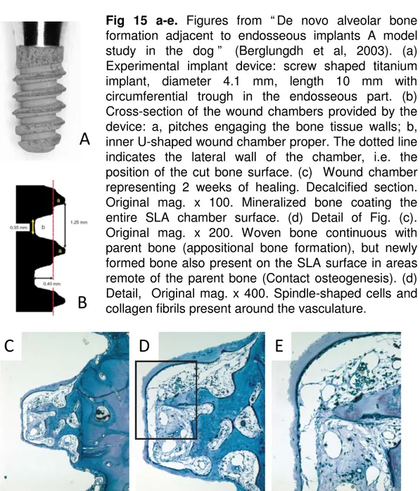
Two different patterns of bone formation at implant sites have been described (89, 91) (figs 12-15). Contact osteogenesis implies bone formation in direct contact with the implant surface, and distance osteogenesis implies new bone formation on the surfaces of the parent bone. This may subsequently become attached to the implant surface as well. When a gap results between the bony wall of the implant bed and the implant surface, a series of events occurs (8, 9, 87). A clot will
initially fill the space and will be replaced by a provisional connective matrix that will act as scaffold for woven bone formation. Contact and distance osteogenesis have been described as biological processes that lead to dental implant osseointegration (89, 91, 92). The existence of these processes has been studied in some animal experiments (93, 8, 9, 83, 84, 87). Contact osteogenesis has been described in an experimental study in dogs (9) in which secluded chambers were used. Newly formed woven bone was found attached to the implant surfaces and occasionally not connected with the parent bone. In another experimental study in dogs, marginal defects around implants were used (83). The healing indicated that both processes, contact and distance osteogenesis, may have participated in the closure of the marginal defects.
It should be emphasized that in the above mentioned experimental studies initial bone contact was provided to the implants by the threads of the implant bodies. However, in a rabbit model, implant contact with the parent bone was avoided (93). Without such initial contact, osseointegration was not achieved. It has to be further realized that a turned implant surface was used in that experiment. This fact may have influenced the healing outcomes as well (90, 4). In any case, the Carlsson et al experiment showed that either contact or distance osteogenesis were not completely impossible, even though with very reduced predictability. In the study mentioned (93), a device was applied to assure primary stability of the implant, since implant instability has been shown to induce a fibrous encapsulation of the implant rather than osseointegration.
The effect of demineralized bovine bone matrix (DBBM) as a bone substitute and filler of marginal defects around implants has been evaluated in several experimental (e.g. 95, 96, 97, 98) and clinical studies (e.g. 99, 100). This biomaterial appeared to be completely integrated in newly formed bone over time, appearing to offer optimal osteoconductivity (96). Nevertheless, the advantages of DBBM in improving osseointegration when applied at marginal defects around implants is still under debate (97, 101, 102, 103, 104, 105).
The influence on osseointegration of the quality and condition of the alveolar bone in relation to the timing of extraction can affect the pattern of bone healing and osseointegration.
A systematic review (106) has documented that implants installed into alveolar sockets immediately after tooth extraction yielded a similar survival rate as implants placed in healed alveolar bony ridges. The results obtained were
maintained for at least 5 year (107). The use of this surgical approach has been scrutinized in several clinical (e.g. 108; 109) and experimental studies (e.g. 102, 103, 110-118). These studies, however, were not able to confirm the maintenance of the vertical and horizontal dimensions of the peri-implant hard tissue, due to the alveolar bone resorption taking place after tooth extraction (119, 110). The buccal plate of the alveolar extraction socket may be compromised as a consequence of the disease process before tooth extraction. As result, a buccal dehiscence may occur after implant installation. Several different procedures have been proposed to improve the outcomes after healing of such compromised sites (e.g. 120, 121, 122, 123). In a controlled clinical study (123), implants were installed into molar alveolar sockets immediately after tooth extraction, and deproteinized bovine bone mineral covered with a collagen membrane was used to fill the residual dehiscence buccal defects (test sites). As control sites, implants installed in healed molar sites were used. The survival rates were similar in both groups (100%), while better results were obtained at the control sites in relation to probing depths and clinical attachment levels. In animal experiments, buccal dehiscences were produced at extraction sockets and different regenerative methods applied (e.g. 124, 125, 126, 127, 128, 129, 130, 112). In a experiment in monkeys (130), dehiscence-type defects, about 4 mm large and 6 mm deep, were filled with rhBMP-2, while the control sites were left untreated. A vertical bone gain of approximately 4 mm was observed in both groups, the difference between test and control group not being statistically significant. Also the positioning of the implants within the extraction sockets has been shown to affect the final outcome. The closer the implant surface was to the outer contour of the bony crest, the higher was the supracrestal exposure of the buccal portion of the implant (117, 118, 112, 131, 132, 133)
The development of implant surfaces, together with a better understanding of the mechanisms the peri-implant bone healing, leads to variation of some clinical paradigms. Among these, the need to use implants with a length not less than 10 mm, especially in case of immediate post extractive implants
Implants installed into alveolar sockets immediately after tooth extraction have been shown to yield predictable outcomes (e.g. 134, 135). Furthermore, a systematic review (106) has established the fact that such implants present with similar survival rates (estimated annual failure rate: 0.82%) as conventionally
placed implants. The use of this procedure reduces the number of surgical sessions and may also reduce the time between surgery and prosthetic delivery (136). This technique, however, cannot prevent the physiological bone resorption that occurs after tooth extraction (108, 137, 106.)
For this placement modality (type 1; Hämmerle et al. 2004) (136), the need for implants that are longer than the remaining extraction sockets has been propagated under the assumption that implant stability may be guaranteed in the area beyond the apex of the extraction socket (138, 139, 140). Because of the presence of anatomical structures such as the maxillary sinus or the inferior alveolar nerve, however, bone may not be available beyond the apex of the socket. Moreover, it was shown in experimental animals that immediate implants became osseointegrated irrespective of their length in relation to the extraction socket (118). In these experimental studies, however, the length of the implants was at least of 10 mm.
Moreover, the use of short implants (6 mm) has been reported with good outcomes (141, 142, 143) and promising results have been published on the installation of short implants in comparison with alveolar bone augmentation techniques (144, 145).
However, the use of short implants (6 mm) in immediate installation has not been studied as yet.
In vitro and preclinical animals studies demonstrate that the development of implant surfaces can influence the clinical utilization in human. In particular, the overall increase of the implant surface that contacts the alveolar bone – bone to implant contact , related to the increase of surface roughness secondary to treatments at micro and nanoscale, may allow the treatment of edentulism associated with severe alveolar bone atrophy with implants of reduced dimensions (eg length of the intraosseous component <10mm).
Osseointegrated implants have become a viable option for replacing missing teeth in totally and partially edentulous patients, as established by systematic reviews, especially in the case of single-tooth gaps (146-149). However, in many clinical situations, placing long implants could prove difficult as a result of limitations such as the location of the mandibular canal, pneumatization of the maxillary sinus, and alveolar ridge deficiencies. In patients with severe alveolar resorption, there are
different surgical procedures available to facilitate future implant placement. More complex implant techniques include the use of bone grafts harvested extraorally or intraorally and placed as inlays or onlays, distraction osteogenesis, zygomatic implants, transposition of the inferior dental nerve, guided bone regeneration (GBR), and maxillary sinus elevation (150-153).
However, these surgical procedures are case sensitive, technically demanding, and time consuming and may increase postoperative morbidity and the total cost and duration of the therapy. Minimally invasive surgical techniques are currently advocated to reduce patients’ postoperative discomfort and contain the risk of complications. The use of short implants was introduced as an alternative treatment in patients with limited amounts of bone available. There is no consensus in the dental literature on the definition of a short implant: in various reviews, it has come in lengths of 7, 8, or <10 mm (141, 154). An implant can also be inserted at different levels, so a short implant has also been defined as an implant designed to have an intrabony length of 8 mm (141).
Recent reviews have compared short implants with conventional implants. Kotsovilis et al (155) concluded in their systematic review that short implants (8 or 10 mm) with rough surfaces are no less effective than implants of conventional length (10 mm) with rough surfaces. Romeo et al (156) wrote that the recent literature has demonstrated a similar survival rate (SSR) for shortand standard-length implants. In a systematic review of horizontal and vertical bone augmentation techniques for the purposes of dental implant treatment, Esposito et al (157) concluded that short implants appear to be a better alternative to vertical bone grafting of resorbed jaws. Conversely, when the clinical outcome of short implants was discussed, non-homogeneous SSRs were reported (158-161). probably because the surface treatment of the implants was not always taken into account.
2. AIM OF THE THESIS
Aim of the present thesis has been to focus on dental implant surface modifications in order to improve the clinical performance in areas with poor quantity or quality of bone. To achieve this objective, the thesis has been developed by identifying specific aims for the in vitro, preclinical and clinical studies.
1) In vitro studies:
a) to better understand the mechanisms underlying osteoblasts
attachment and spreading, in this study adhesive peptides containing the fibronectin sequence motif for integrin binding (Arg-Gly-Asp, RGD) or mapping the human vitronectin protein (HVP) were grafted on glass and titanium surfaces with or without chemically induced controlled immobilization.
b) to reproduce the osseointegration process in vitro in order to study the
dynamic of bone-implant interactions. To this end, a tissue engineering approach was used by positioning dental implants into stem cells seeded 3D bone-derived scaffolds. The final goal was to overcome the limitations concerning in vitro methods, and to complement and eventually replace animal studies in this field.
2) in vivo studies (in big animal model, dogs, of osteointegrative
properties of implants with different degree of surface modification).
a) to evaluate the healing at implants with a moderately rough surface
placed and stabilized in recipient sites of dimensions deeper and larger than that of the implants to avoid any contact between parent bone and the implant surface.
b) to evaluate the influence on osseointegration of DBBM particles used to
fill defects of at least 1mm around implants having no primary contact with bone.
c) to evaluate the influence of bucco-lingual implant positioning into
extraction sockets on bone formation at buccal alveolar dehiscence defects.
d) to compare the bone-to-implant contact of 6 mm osseointegrated
tooth extraction.
3) clinical research (on short implants, to underline the effect of surface
modifications on dental implant survival in the long term in the clinic).
a) to assess the mediumto long-term prognosis of short implants (7 or 8.5 mm in length), with machined (M) and rough (R) surfaces, placed in partially or totally edentulous arches, in a retrospective clinical trial.
3. MATERIALS AND METHODS 3.1 IN VITRO STUDIES
3.1.1 From Brun et al, 2012. Mechanisms underlying the attachment and spreading of human osteoblasts: From transient interactions to focal adhesions on vitronectin-grafted bioactive surfaces
Peptide synthesis and surfaces preparation
The vitronectin peptide is mapped on the human vitronectin protein (HVP, sequence 351-359: FRHRNRKGY). The RGD peptide is a linear sequence of human fibronectin presenting four GRGDSP motifs per chain (RGD, sequence: GRGDSPGRGDSPGRGDSPGRGDSPK). The RAD peptide is the control peptide with respect to RGD (RAD, sequence: GRADSPGRADSPGRADSPGRADSPK). In its sequence the Arg-Gly-Asp motif is substituted with a similar but not adhesive motif, Arg-Ala-Asp. Both peptides were synthesized as C-terminal amides using the Fmoc Chemistry on Applied Biosystems 431A Instrument, a traditional solid-phase technique (162).
Side-chain protected peptides (t-Butyl (tBu), Tyr; 2,2,5,7,8-Pentamethylchromane-6-sulfonyl (Pmc), Arg; Trityl (Trt), His and Asn; t-butoxycarbonyl (Boc), Lys; tBu, Ser) were synthesized using a Sieber Amide resin. The cleavage from the resin was obtained by incubation for 15 min in (1) 1% 2,2,2-trifluoroacetic acid (TFA)/dichloromethane (DCM) for the full side-chain protected sequences and in (2) 10% TFA/DCM for 20 min for the partially deprotected sequences. The deprotection method (2) produced the loss of Boc and Trt for HVP and of Boc for RGD. The products were ascertained by electrospray ionization/time of flight mass spectrometry. To prepare the glass coverslips (1.76 cm2), glasses were washed several times in an ultrasonic bath (acetone, 30% ethanol in MilliQ water) and treated with 1 N NaOH for 1 h. Glasses were washed in MilliQ water, dried at 100 °C for 10 min, washed with acetone and dried under vacuum. The glass coverslips were then immersed in 2% (3-aminopropyl) triethoxysilane (APTES) in acetone solution at 40 "C overnight, washed three times with dichloromethane (DCM), acetone and finally with MilliQ water. After drying for 10 min at 100 °C the