International Orthopaedics (SICOT) (1991) 15 : 143-144
International
O hopaedics
© ~Springer Verlag 1991
Fracture with loss of the proximal femur in a child
A case report
U. E. Pazzaglia, E. Finardi, L. Pedrotti, and G. Zatti
Clinica Ortopedica, 2 A Facolta di Medicina e Chirurgia dell'Universitfi di Pavia, Ospedale F. del Ponte, 1-21100 Varese, Italy
Summary. An 8 year old child was involved in a
road accident and sustained a large wound in the
left groin; radiographs showed a fracture with loss
o f the proximal femur. After skeletal traction f o r 80
days, there was bony regeneration o f the proximal
femur. At 8 months she was able to walk without
support and her left leg was 2 cm only shorter than
the right.
R6sum+.
Une petite fille de 8 ans a ktb victime d'un
accident de voiture. Elle prbsentait une vaste plaie
de la rbgion inguinale. Les radiographies mon-
traient une fracture avec avulsion de l'extrkmitb su-
pbrieure du fbmur. Apr~s 80 jours de traction on a
constatb la reconstruction de la partie proximale de
la diaphyse fbmorale. Au 8~me mois la blesske pou-
vait marcher sans canne et son membre infbrieur
n'btait raccourci que de 2 cm par rapport au c6tk
oppos&
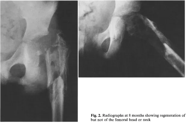
She regained consciousness after 3 days and traction was maintained for 80 days until consolidation of the tibial frac- tures had occurred. At this time bone had been laid down in the proximal femur; by 4 months a piece of diaphysis about 10 cm long had formed. There had been no regeneration of the femoral head and neck after 8 months (Fig. 2).
Weight-bearing in a plaster cast was allowed at 4 months and with 2 sticks at 5 months.
At follow up at the 8th month the child could walk with- out support or pain, although she limped with a Trendelen- burg gait. The left leg was 2 cm shorter than the right and was externally rotated 15 ° .
Discussion
T h e e n e r g y c r e a t e d b y t h e t r a u m a m u s t h a v e b e e n e x c e p t i o n a l l y h i g h b e c a u s e it p r o d u c e d a f r a c t u r e o f t h e s h a f t , d i s l o c a t i o n o f t h e f e m o r a l h e a d , a c o m p l e t e t e a r o f t h e c a p s u l e a n d t h e l i g a m e n t u mCase report
A girl, aged 8 years, was involved in an accident when riding with her mother on a motor cycle. She was unconscious on ad- mission and had sustained fractures of both legs and her right wrist. Apart from several superficial abrasions, she had a large wound in her left groin through which the acetabulum could be palpated. The femoral vessels were intact. No trace could be found of the femoral head.
Radiographs showed a fracture with loss of the proximal femur and only a small shell from the greater trochanter re- maining (Fig. 1). The avulsed fragment of the femur had not been found.
The wound was sutured and skeletal traction was applied through both heels and the distal left femur by 3 Kirschner wires.
Offprint requests to:
U. E. PazzagliaFig. 1. Radiograph after the accident showing absence of the proximal femur with only a thin shell of the greater trochanter remaining
144 U.E. Pazzaglia et al.: Fracture with loss of the proximal femur
/
teres, and detachment of the periosteum, as well
as laceration of the soft tissues and skin (Fig. 3).
The proximal shaft regenerated, which suggests
that the germinal layer of the periosteum was left
intact; in this respect the fracture reproduced the
pattern of a benign subperiosteal fracture of
childhood where there is mechanical failure of the
bone, but not the periosteum.
Reposition and osteosynthesis of the detached
piece would have been a more straightforward
method of treatment. However, skeletal traction
allowed regeneration of the lost segment w i t h
only slight loss of length and a fair functional re-
covery. Further management will have to deal
with the possibility of further leg length discrep-
ancy.
Fig. 2. Radiographs at 8 months showing regeneration of the proximal shaft, but not of the femoral head or neck
Fig. 3. Diagram showing the extent of the injury and the struc- tures damaged