PROCEDURE FOR HIGH-YIELD SEPARATION
AND RADIOACTIVE LABELLING
BY
51CR AND
111IN OF HUMAN PLATELETS
A.F. SEDDA Fusion and Technology for Nuclear Safety
and Security Department Casaccia Research Centre, Rome
R. LUCIANINI Nuclear Medicine Division ASST Settelaghi Hospital,
ASST Settelaghi Hospital, Varese, Italy
S. BOEMI
Nuclear Medicine Division S. Eugenio Hospital, Rome
RT/2017/43/ENEA
ITALIAN NATIONAL AGENCY FOR NEW TECHNOLOGIES, ENERGY AND SUSTAINABLE ECONOMIC DEVELOPMENT
A.F. SEDDA
Fusion and Technology for Nuclear Safety and Security Department Casaccia Research Centre, Rome
R. LUCIANINI
Nuclear Medicine Division ASST Settelaghi Hospital, Varese, Italy
PROCEDURE FOR HIGH-YIELD SEPARATION
AND RADIOACTIVE LABELLING
BY
51
CR AND
111
IN OF HUMAN PLATELETS
S. BOEMI
Nuclear Medicine Division S. Eugenio Hospital, Rome
RT/2017/43/ENEA
ITALIAN NATIONAL AGENCY FOR NEW TECHNOLOGIES, ENERGY AND SUSTAINABLE ECONOMIC DEVELOPMENT
I rapporti tecnici sono scaricabili in formato pdf dal sito web ENEA alla pagina www.enea.it I contenuti tecnico-scientifici dei rapporti tecnici dell’ENEA rispecchiano
l’opinione degli autori e non necessariamente quella dell’Agenzia
The technical and scientific contents of these reports express the opinion of the authors but not necessarily the opinion of ENEA.
PROCEDURE FOR HIGH-YIELD SEPARATION AND RADIOACTIVE LABELLING BY 51CR AND 111IN OF HUMAN PLATELETS
A.F. Sedda , S. Boemi, R. Lucianini
Abstract
Platelets are non-nucleated corpuscles of the circulating blood, produced in bone marrow and derived from fragmentation of bone marrow precursors called megakaryocytes. Recently the platelets have found wide use in clinical therapy for their ability to release growth factors; the platelet concentrate, PRP (Platelet Rich Plasma) has been shown to possess anti-inflammatory, antibacterial, regenerative and hemostatic properties. The radiolabelling and re-injection of autologous platelets is also a nuclear medicine technique useful in the scintigraphic detection of many clinical diseases, from splenic disor-ders to thrombi visualization. In the present paper the procedure of platelets separation from whole blood of human subjects was optimized by using a disposable kit aimed to PRP preparation, that al-lowed a platelet separation with high yield, in aseptic conditions and in short times. The obtained platelets were efficiently labelled by using radioactive 51Cr in the form of sodium chromate and 111In
in the form of 111In-oxine.
Key words: platelets, PRP, radioactive isotopes, 51Cr, 111In, radioactive labelling
Riassunto
Le piastrine sono corpuscoli non nucleati del sangue circolante, prodotti nel midollo osseo e derivati dalla frammentazione dei precursori del midollo osseo chiamati megacariociti. Recentemente le pia-strine hanno trovato largo impiego nella terapia clinica per la loro capacità di rilasciare fattori di cre-scita; il concentrato piastrinico, PRP (plasma ricco di piastrine) ha mostrato di possedere proprietà antinfiammatorie, antibatteriche, rigenerative ed emostatiche. La radiomarcatura e reiniezione di pia-strine autologhe è anche una tecnica di medicina nucleare utile nella rilevazione scintigrafica di molte patologie cliniche , dai disturbi della milza alla visualizzazione dei trombi. Nel presente lavoro é stata ottimizzata la procedura di separazione di piastrine dal sangue intero di soggetti umani, utilizzando un kit monouso finalizzato alla preparazione del PRP, che ha consentito una separazione delle piastrine con alta resa, in condizioni asettiche e in tempi brevi. Le piastrine ottenute sono state efficacemente marcate con 51Cr radioattivo sotto forma di sodio cromato e 111In sotto forma di 111In-ossina.
Introduction
Material and methods
Platelets separation
51Cr preparation and platelet labelling 111In platelet labelling
Radioactivity and scintigraphy measurement Quality control on the separation procedure Quality control on platelets activation Results and discussion
Conclusions Annex 1 References 7 9 9 11 11 12 12 13 14 25 26 28
INDEX
7
Introduction
Platelets are non-nucleated corpuscles of the circulating blood, 1.0 to 1.5 micrometers in diameter. They are produced in bone marrow and derive from fragmentation of bone marrow precursors called megakaryocytes, which originate from stem cells that undergo repeated division of the cell nucleus. The cytoplasm begins the production of immature platelets, which are then transferred in the bloodstream. Platelets are present in the circulating blood in number 150 000-400 000 for blood mm³ and have an average life of 5-9 days. They are not cells, so have no nucleus, but possess granules, many cytoplasmic organelles and RNA; they have round or oval shape of about 2-4 microns. Circulating platelets play a primary role in thrombosis, under physiological and pathological conditions. Characterization of in vivo platelet kinetics and uptake is useful in diagnosing primary platelet pathologies (thrombocytopenia, thrombocytosis), hypercoagulability disorders, atherosclerosis. With the use of labeled platelets, sites of active thrombus formation can be easily localized in vivo.
The in vitro labeling with radioactive tracers of platelets starts from the withdrawing of a blood sample from which platelets to be marked are isolated. For the marking of platelets is commercially available the radiopharmaceutical 111In-oxine (8-hydroxyquinoline), supplied as a radiopharmaceutical ready-to-use. The minimum number of platelets for a successful labelling is ≥15000 / microliter. The lipophilic 99mTc-HMPAO complex can also be used for labelling platelets. Platelets are isolated according to standard isolation procedures. The labelling efficiency % depends on incubation temperature (22° C: 40%; 37° C: 50%), incubation time (3 min: 20%, 25 min: 55%) and the incubation medium (plasma: 40%; saline 50%). The 60 min 99mTc elution found out of the platelets ranged around 8%. The platelet recovery, used as a quality control parameter, is around 25%±4% and is stable for at least 240 min. The high elution rate out of the platelets leads to renal excretion of the label and hence to significant kidney and bladder activity. Intestinal excretion of the label can also be frequently demonstrated. Fresh thrombotic lesions can normally be detected 4 h after reinjection of the labelled platelets, and in some patients as early as 1 h after reinjection.
99m
Tc-HMPAO seems to be a promising platelet label for imaging thrombotic lesions but not for platelet survival studies, because of the short physical half life of 99mTc 1.
111
In platelet imaging can identify sites of active intravascular platelet deposition, and may help define patients at risk of embolization. Both platelet imaging and echocardiography detect ventricular thrombi, but platelet imaging may detect only the most haematologically active thrombi. Both techniques may help define patients at risk of embolization and may be useful for in vivo assessment of antithrombotic drugs 2.
8
A group of 23 patients was studied to determine whether platelet deposition could be detected in patients with vascular aneurysms or in patients in whom Dacron prosthetic grafts had been placed, and it was found that platelet imaging is highly valuable for studying platelet physiology in vivo and for assessing platelet-active drugs and the thrombogenicity of prosthetic graft materials in human beings 3.
The method of autologous platelet labelling with 111In-oxine, and labelled platelets lifespan, was used for production and sequestration site determination in 555 patients with clinical diagnosis of primary immune thrombocytopenia (ITP), as well as in 6 control healthy subjects. On the basis of labelled platelets sequestration site determination, it was possible to predict the efficacy of splenectomy in ITP patients. Unstable atherosclerotic plaques and acute thrombosis were also well visualized in ITP patients 4.
51
Cr can also be used for platelets labelling, in the form of sodium chromate. The in vivo kinetics of simultaneously injected 51Cr- and 111In-labelled platelets was investigated in 20 healthy male volunteers. The studies were carried out using both fresh platelets and platelets stored for 5 d at 22 °C; the disappearance of platelet-bound radioactivity was measured on whole blood samples as well as platelet pellets. 51Cr yielded a slightly higher platelet recovery and longer T1/2 than
111
In, when whole blood samples were used for calculations; no differences were seen when using platelet pellets. When stored platelets were studied, 51Cr gave significantly longer T1/2 and mean life-span (MLS) than did
111
In. It is concluded that both 51Cr and 111In are acceptable as radiolabels for both fresh and stored platelets. However, it appears that the viability of stored platelets may be influenced by the choice of label, and caution must be taken when these isotopes are used together in dual tracer experiments. A meticulously standardized processing of blood samples and experimental data is required to enable inter-laboratory comparisons of the results 5.
Recently the platelets have also been used in clinical therapy for their ability to release growth factors. It is well known that platelets contain a large number of growth factors in their granules, which are released following platelet activation, and are of particular importance for tissue healing (Transforming Growth Factor-β (TGF-β1, TGF-β2), growth factors derived from platelets (PDGF-AA, PDGF-BB, PDGF-AB), insulin-like growth factor ( IGF-1), epidermal growth factor (EGF), hepatocyte growth factor (HGF)). These growth factors act in synergy to increase the infiltration of neutrophils and macrophages, and to promote angiogenesis, matrix deposition and re-epithelialization. An increased concentration of growth factors present in a platelet concentrate have proved capable of accelerating wound healing, decrease inflammation and promote the regeneration and repair of damaged tissues.
The platelet concentrate, PRP (Platelet Rich Plasma) has been shown to possess anti-inflammatory, antibacterial, regenerative and hemostatic properties. Being rich in autologous growth factors, it is used to improve the maxillary reconstruction in dentistry, plastic and orthopedic surgery, in the treatment of tendinitis and other injuries to skeletal muscle soft tissue, for the treatment of severe arthropathies such as osteoarthritis and rheumatoid arthritis. An important use is the treatment of skin ulcers and bed sores, in the
9 retinal repair in patients with idiopathic macular holes, or as eye drops active in various eye diseases. Even in aesthetic medicine PRP has had excellent clinical results in the treatment, by means of micro-injections, of the skin and subcutaneous tissue to improve the appearance of facial areas, with a slight immediate "filler effect" in the affected areas, but with more evident results in span of a month, by stimulating the production of collagen and elastin. The same technique is also used to counteract the loss of hair, as a valid alternative to traditional medical therapies anti-fall which provide for the use of drugs 6-11.
The platelet concentrate is typically obtained by a collection of venous blood to the patient; blood, supplemented with EDTA or citrate-phosphate-dextrose as an anticoagulant, is subjected to a double centrifugation; in the first, at low speed, a Platelet Rich Plasma is separated from the erythrocytes and leucocytes, in the second, at higher speed, the platelets are concentrated from the plasma in a platelet pellet. The platelet concentrate can be used as such, or it can be transformed into a more or less consistent gel, usually with the addition of activating botropase and / or calcium for cross-linking of fibrin 12,13.
Materials and methods
Platelets separation
A series of experiments aimed to the determination of separation yield have been performed at the Nuclear Medicine Division of S.Eugenio Hospital, Rome, and at Nuclear Medicine Division of ASST Settelaghi Hospital, Varese, by using a ready-to-use kit (PRP- Kit, Celltech, Turin, Italy) for the preparation of Platelet Rich Plasma, following the operative procedure enclosed in the kit. Briefly, a volume from 20 to 30 ml of whole blood were taken from each patient in a 60 ml syringe (syringe 1), fitted to the orifice of sterile self-closing valve with male connector, and as an anticoagulant containing from 4 to 6 ml a solution Citric Acid - Dextrose in saline solution (citric acid monohydrate 8g / L, sodium citrate 22 g / L, monohydrate 24.5 g/L glucose). After withdrawal, the syringe was stirred briefly, and then the side opposite the orifice was cut using a hot-wire cutting tool, "transformed" into a sterile and sealed test tube (section H of the syringe, Figure 1). This test tube was centrifuged for 10 minutes at about 400 g in a thermostatic centrifuge, for the separation of erythrocytes and part of the leucocytes on the bottom, while the platelets remained in suspension. The section L of the syringe has been discarded. After this centrifugation, the supernatant of the syringe 1 has been collected by connecting the self-closing valve to a second 60 ml syringe (syringe 2) fitted with sterile self-closing valve with female connector, and withdrawing the upper plasma layer; the syringe 1, which contained the erythrocytes, has been discarded. Also in the syringe 2, the opposite side to the orifice was cut using the hot wire cutting tool , "transformed" into a sterile and sealed test tube (see Figure 1); this test tube was centrifuged for 10 minutes at about 2200 g in a thermostatic centrifuge, for the separation of the platelet pellet. After centrifugation, the platelets depleted supernatant plasma the of the syringe 2 has been collected by connecting the sterile self-closing valve to a third 60 ml syringe (syringe 3) fitted with sterile
10
self-closing valve with male connector, and by withdrawing the plasma above the pellet; the syringe 3 has been discarded. Finally the plates were collected in a 2.5 ml syringe (syringe 4), provided with a self-closing sterile valve sterile with male connector, containing a volume of 1 ml of physiological solution. On this syringe all the experiments on separation yield and platelet characterization have been performed. A total of
12 different separations have been performed on healthy voluntary subjects.
A
C
D
E
F
G
H
L
+-B
Figura 1
S
Figure 1 Figure 211 In the kit used in the present work, a gel is included in order to promote the platelet pellet re-suspension, particularly in dermatological and ocular applications. In some of the experiments the PRP was mixed with the gel, in order to test its properties.
51
Cr preparation and platelets labelling
51
Cr has a half life of 27.7 d, and can be obtained by the nuclear reaction 50Cr (n, ) 51Cr (cross section = 15.9 b + 0.2 b). In our experiment 100 mg of metallic chromium (Carlo Erba-Milan, Italy), with purity higher than 99% was put into an high purity plastic sealed vial, and irradiated for about 6 hours into the rotating rack of ("Lazy Susan") of the 1 MW TRIGA reactor of ENEA- Casaccia Center, with a thermal neutron flux of 2.7 1012 neutrons/cm2/s.
After irradiation, the vial content has been transferred into a glass test tube, and dissolved with 300 micro-liters of concentrated HCl, by heating to 90 °C with occasional stirring, obtaining in 10 minutes a brilliant green solution of CrCl3. The solution was added at room temperature with 400 micro-liters of NaOH
concentrated solution; a precipitate of Cr(OH)3 was obtained . The chromium hydroxide precipitate was
centrifuged, washed with H2O, and 100 mg of NaOH and 1.5 ml of 30% H2O2 were cautiously added, at
room temperature, by operating under a fume hood into a "hot" laboratory .
The Cr3+ ions underwent oxidation to Cr6+, while the color of the solution slowly changed from brilliant green to yellow. The solution was rendered neutral by addition of concentrated HCl, and diluted with distilled water and saline up to isotonicity. About 500 KBq of total activity were obtained at the end of irradiation.
In the various experiments the platelet suspension was incubated with 5 to 10 KBq of [51Cr] in the form of sodium chromate for 40 min at 37°C, in a laboratory thermostated incubator. The platelet suspension was then washed with saline and centrifuged to remove unbound 51Cr.
111
In platelets labelling
Radiopharmaceutical 111In-Oxine, commonly used in diagnostic procedures performed in Nuclear Medicine, allowed to evaluate the homogeneous distribution of PLTs in the gel and the maintenance of their characteristic. For the radioactive labeling of PLT, after obtaining PRP as previously described( see section Platelet Separation), we proceeded as follows:
With 1 ml syringe, 0.6 ml of buffered 111In-Oxine (0.4 ml of 111In-Oxine + 0.2 ml of Buffer TRIS, Mallinkrodt) was introduced in the syringe containing the PLT pellet. After 15 minutes of incubation, 5 ml of NaCl 0.9% were added, and a centrifugation at 2200g x10 minutes was performed. The supernatant radioactivity was eliminated, and the 111In-Oxine labeled PRP pellet was re-suspended in 1 ml of NaCl0.9%.
12
The radioactivity of supernatant and of pellet were measure, in order to obtain the labelling yield:
Radioactivity pellet/(Radioactivity pellet + Radioactivity surnat)
Radioactivity and scintigraphy measurement
The distribution of 51Cr labelled platelets radioactivity was performed by autoradiography, putting in contact the source to be examined with a storage phosphor screen; the radiation generates in the screen a number of electron and hole trap centres, their density being proportional to radioactivity dose (latent image). The phosphor screen used was a Perkin Elmer phosphor screen 12.5 x 25 cm, with spatial resolution of 42
m. After radiation exposure, the storage phosphor screen can be simply resetted by exposition to a strong visible light for some minutes, and can be re-used, virtually for years, without variation in detection and resolution efficiency.
The screen, after exposure to radiation, and is stimulated optically to emit luminescence in response to a radiation, and a digital image from the above said storage phosphor screen was acquired, by a Cyclone Perkin-Elmer laser scanner. The data analysis of the obtained images has been performed by using the open source ImageJ software Image Analysis Package of National Institute of Health (USA).
The scintigraphic images were acquired with a Gamma Camera ECAM-Siemens by using Low Energy High Resolution(LEHR) Collimator and with Ultra High Resolution Pin Hole Collimator, and with a Gamma Camera Symbia S. Images were analyzed for longitudinal profile analysis by using the open source ImageJ software Image Analysis Package of National Institute of Health (USA).and then we have evaluated images with dedicated software histogram distance to gray value.
Quality control on the separation procedure
In the ready-to-use kit employed in the present work, (PRP-KIT Celltech-Turin, Italy) all the separation stages are performed within syringes, and all liquid transfers between them are made via sterile valves with automatic closing, which open only when they are connected with each other. Nevertheless, in order to ascertain the cleanliness of the process, all the platelet separation process was simulated, using in the PRP kits, employing, in place of all liquid reagents and the patient's blood, sterile and pyrogen-free water (PBI). Once performed the whole process, according as the procedure attached to the same kit, 1 milliliter of water resulting from the final stage was placed in a plate for bacterial culture Compact Dry TC PBI and in one for yeast/mould culture Compact Dry TC PBI YM, incubated for 5 days in a thermostated incubator at 32.5 ° C for bacterial plates, and 22.5 °C for yeast/mould plates, and visually inspected every day. Although the execution of the separation of platelets procedure is provided for under the hood class A, the whole process, in this occasion, was performed outside of the hood, in an unclassified environment, in order to maximize the
13 possibility of accidental contamination . The whole process was repeated 2 times, together with a positive control sample. On the same water sample a pyrogen analysis was performed, by using the following products: PYROTUBE-A (tubes de-pyrogenated), LAL REAGENT WATER (sterile distilled pyrogen-free water), LAL Pyrotell (SINGLE TEST vials), CONTROL STANDARD Endotoxin, working outside of the hood, in an unclassified environment, in order to maximize the possibility of accidental contamination. Also for pyrogen test the whole process was repeated for 2 times.
Quality control on platelets activation
We have demonstrated that the enrichment process is able to concentrate Platelets (PLTs) without stress, without breaking and thus activating them. A method to produce PRP able to maintain PLTs in resting state is preferable to a one that induces mechanical stress and triggers biochemical reactions of the classic platelet activation in undesirable or non-controlled conditions14. To evaluate whether the enrichment procedure is adequately "gentle" to prevent damage and / or platelet activation, we have used the technique of Flow Cytometry in order to determine the expression of activated PLT Membrane Protein. The platelet expression of CD62P can provide information on platelet activation; when PLT is in a resting state, CD62P is not present on the outer surface of the PLT membrane, while it is expressed as a result of the activation process
15
. The larger the physical stress during the enrichment process, the higher will be the concentration of CD62P on the PLT membrane. If the expression level of CD62P on the platelet membrane of PRP at the end of production is similar to the one of CD62P on the platelet membrane of Whole Blood (W.B.) of the same patient, the enrichment method is suitable to preserve the functional abilities of PLTs, and thus certify clinical efficacy of obtained PRP. Is it however possible that the enrichment procedure, however delicate, could alter the functionality of the platelets by inhibiting their ability to be activated by the appropriate stimulus and thus not allowing their release of Growth Factors (GFs). To verify this fact, we added to PRP, Adrenaline and CaCl2, two platelet agonists which, in contact with the platelets, determine functional
activation 16. After this step we have quantified the expression of the CD62P to see if the exogenous stimulus can still promote the expression of this factor; the experimental model reproduces exactly the physiological stimulus that PLTs of PRP receive at the site of therapeutic application, where, interacting with the elements
in situ, they can release the GF, in order to obtain the desired therapeutic effect.
Mean Fluorescence Intensity (MFI) for FITC-A CD62P (membrane receptor CD 62P) values are obtained from 12 PRP samples, each analyzed in REST mode (i.e., within 1 hour from the end of PRP production, without chemical stimulant activation). We obtained the single-parameter fluorescence histogram of the activation marker analyzed; the protocol used is adopted in the Varese Hospital at Immunohematology and Transfusion Laboratory, and is called Basic Protocol 1 "Determining the Resting Platelet surface receptor expression”17
. Data are acquired in real time and represented by orthogonal and logarithmic-forward light scatter. We have evaluated the MFIs values of the relative CD62P of 12 PRP samples and
14
compared with MFIs values of the relative CD62P of the W.B. of the same patient from whom this PRP has been obtained. If the obtained value of MFIs of CD62P of PRP sample is comparable to those on W.B., we could state that the PRP production method is able to keep platelets in resting mode as desirable and production method of stable PRP is adequate.
Activation at Rest = MFI CD62P values of PLTPRP / MFI CD62P values of PLT W.B.
In order to check the activation functionality, we used the protocol adopted in the Varese Hospital at Immunohematology and Transfusion Laboratory, called the Basic Protocol 2 "Determination of Platelet Activation using P-selectin or PAC1 expression"17. The procedure for exogenous activation of PLTs (after Stress stimulation) in PRP is as follows: in a test tube, add 20 μl of PRP + 10 μl of Adrenalin + 10 μl of CaCl₂ 10% ratio 2: 1: 1 and leave to incubate 15 minutes in the dark. For each of the 12 PRP samples, we performed cytofluorimetry analysis at Rest and, in parallel, after Stress stimulation.
If the platelets are strongly activated by the chemical stimulus in PRP, then process does not alter the functionality of the platelets and therefore does not inhibit their ability to release the GF; MFIs values of CD62P is much greater after stimulation than MFIs values of CD62P of the same sample analyzed at Rest ( at least >2).
Activation after Stress ratio = MFI CD62P of PRP AFTER STRESS / MFI CD62P of PRP AT REST
Results and discussion
The entire procedure for platelet separation required 20 minutes from blood withdrawal up to the obtainment of the final platelet pellet, and was performed under a simple microbiologically laminar-flow hood. The sterility tests demonstrated in all samples an absence of bacterial or mould contamination, and an endotoxin concentration < 0.125 UI/ml.
Hematological complete tests were performed on the initial blood and in all the separation stages, by using a clinical automatic blood analyzer. The total recovery yield for platelets (n=12) was 89% (+ 3%), for leukocytes was 16% (+ 5 %), (with neutrophils 1% and lymphocytes 82%), for erythrocyte 0,03% % (+
1%); the concentration of platelets in the syringe 4 was approximately 19 (+2) times compared to the initial
concentration of the whole blood, which can be considered quite high, respect to typical enrichment ratio of 4 to 5 times the whole blood platelets concentration18.
This value can still be increased, by decreasing the amount of saline used for platelet final dilution. A typical example of hematological analysis performed on blood samples is reported in Annex 1. The purification from leukocytes was similar to the one obtained in similar experiments, but much higher for erythrocytes respect to published values19.
15 After the separation of platelets from blood, different procedures can follow. If an intra-tissue injection of platelets has to be performed, the saline suspension obtained in the final separation step can be directly used as such. In some cases the suspension is used for application on the skin, and should be transformed into a gel, for a more convenient application.
In such cases, usually, the final pellet suspension is treated by addition of calcium salt and a protease (example botropase), in order to allow a reticulation of the fibrin present in the serum, and the mixture is placed on the body region to be treated (skin, other tissues).
In the kit used in the present work, the PRP gel is obtained by addition of a synthetic gel, with optimized rheological characteristics, to the pellet suspension; the advantage lies in the fact that a synthetic gel is usually very stable, while the natural fibrin gel, often used in most protocols, is often subject to a de-mixing and rupture of the structure in the manipulation of the gel during the phase of application to the patient. If an ocular application is to be performed, to heal corneal lesion, a low amount of the synthetic gel is mixed with the platelet pellets in saline, so obtaining sterile eye drops with an elevated residence time in the eye.
The gel included in the kit is based on carboxymethylcellulose, a cellulose derivative with carboxymethyl groups bound to some of the hydroxyl groups of the glucopyranose monomers that make up the cellulose backbone. It is used primarily because it has high viscosity, is nontoxic, and is considered to be hypoallergenic. CMC is used extensively in gluten free and reduced fat food products, and as a lubricant in artificial tears.
In order to test the behaviour of PRP in the gel, the radioactive labelled PRP pellet was re-suspended and thoroughly mixed in 14 ml of the gel, and the mixtures was put into a 1 ml syringe, in a Petri dish (about 12 ml) and in a linear glass capillary (1 ml).
The 1 ml syringe was used to evaluate whether the labeled platelets are uniformly distributed in the gel. Images were acquired with Gamma Camera ECAM-Siemens and the results evaluated with a dedicated software for longitudinal profile analysis with histogram distance.
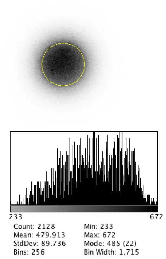
Petri dish was used to evaluate if platelets distribution is uniform throughout the gel, avoiding in-homogeneity in concentration of platelets, which would affect therapeutic effect of PRP. Images were acquired with ECAM Gamma Camera both with LEHR Collimator and with ultra High Resolution Collimator Pin Hole (See Figure 3 and Figure 4a and 4b), and then analyzed images with dedicated software, obtaining histogram distance to gray value and calculating the %variability value.
The linear capillary was used to assess the spatial distribution of the gel-PRP over time. Images of the capillary, immediately after its filling have been acquired, with a Gamma Camera Symbia S, evaluating longitudinal distribution, immediately after preparation, and after keeping the capillary in vertical position for 48 hours. (See Figure 5a and 5b).
16
Average = 60,542 [cts] StdDev = 17,274 [cts] Variability% = 12,8%
Figure 3: Syringe scintigraphic image of the 1 ml syringe with relative geometric distribution analysis for
17 Two-dimensional distribution: Average =44,888 [cts] StdDev = 8,927[cts] Variability%= 19.8%
Figure 4a: Ultra High Resolution scintigraphic image with relative geometric distribution analysis for
two-dimensional uniformity determination by distribution of gel-PRP labeled with 111In-Oxine distributed in Petri plate.
18
Two-dimensional distribution: Average = 479,913 [cts] StdDev = 89,736 [cts] Variability% = 18,7%
Figure 4b: Low Energy High Resolution scintigraphic image with relative geometric distribution analysis
for two-dimensional uniformity determination by distribution of gel-PRP labeled with 111In-Oxine distributed in Petri plate
19 Average = 50.751 [kcts] StdDev = 15.685 [kcts] Variability% = 13.2%
Figure 5a: Scintigraphic image immediately after preparation with relative geometric distribution analysis
for uniformity determination by Histogram Gray Scale / Distance to Longitudinal Profile of PRP gel marked with 111In-Oxine inserted into a glass capillary
Average = 56,871 [kcts] StdDev = 13,736 [kcts] Variability% = 14,8%
Figure 5b: Scintigraphic image after 48 hours in vertical position with relative geometric distribution
analysis for uniformity determination by Histogram Gray Scale / Distance to Longitudinal Profile of PRP gel marked with 111In-Oxine inserted into a glass capillary
20
Similar experiments were performed also by using 51Cr labelled platelets. The labeling yield of platelets by
51
Cr in the present work was about 12% in all experiments, similar to the values currently obtained with this technique 20,21.
In a second experiment the 51Cr labelled platelets, after washing with saline and centrifugation, were thoroughly mixed with 1 ml of the synthetic gel enclosed in the kit, then the mixture gel/51Cr labelled platelets was aspirated into a 1 ml syringe, and the syringe was kept in vertical position for 30 minutes. After this period the syringe was put in contact with the phosphor image screen for 3 minutes. The optical density profile plot of the radioactivity spot, obtained with ImageJ image analysis software on the image obtained on phosphor screen by the syringe, is shown in Figure 6; the grey rectangle represent the outline of the syringe. It can be seen that the gel has the capability of keep well suspended the platelets, with a standard deviation of + 5% respect to the mean integrated density measure.
In a third experiment the mixture gel/ 51Cr labelled platelets was applied by gently smearing the gel with a brush on a plastic film, then the film was placed on the phosphor screen for 3 minutes. The Figure 7 shows the obtained image; an optical density profile plot performed on the spot by ImageJ image analysis software demonstrated that the platelets were homogeneously distributed on the gel , with a standard deviation of + 7% respect to the mean integrated density measure. The result is shown in Figure 8; the rectangle represent the position of the mixture gel/ 51Cr labelled platelets spot on the film.
21
Figure 7
22
As above said, the technique of radiolabelled platelets represents a powerful tool for in-vivo clinical diagnostic in a wide class of disease, and a precious scientific investigation tool.
It was demonstrated that 111In labelled platelet scintigraphy is a highly accurate test for detecting acute untreated renal allograft rejection and it is shown that changes in platelet uptake can precede signs and symptoms of rejection by at least 48 hours 22.
Eleven patients with recent kidney transplants have undergone serial pelvic imaging after autologous platelet labelling with 111In. Rejection was accompanied by marked platelet deposition in the graft, whereas acute tubular necrosis was characterized by minimal platelet accumulation. The method has been quantified by comparing counts over the graft to counts from a similar area on the opposite site, and appears to distinguish between the two main causes of renal failure after transplantation. In addition, complications of transplant surgery may be demonstrated 23.
While splenectomy is an effective therapy for primary immune thrombocytopenia (ITP), possible complications and observed non-complete response (CR) in one-third of patients demonstrate the need for further research into potential pre-surgical predictors of outcomes. Past investigations into platelet sequestration studies, a hypothesized predictive test, have adopted heterogeneous methods and varied widely. A clinical study on a cohort of 256 patients with primary ITP demonstrated the utility of autologous 111 In-labelled platelet sequestration studies as an adjunct predictive instrument prior to splenectomy 24.
Forty-eight patients who had undergone surgical reduction of a fractured neck of femur or in whom deep vein thrombosis was suspected clinically were studied by ascending phlebography and imaging after injection of autologous 111In-labelled platelets to assess the accuracy and value of the radioisotopic technique in diagnosing deep vein thrombosis. Phlebography showed thrombi in 26 out of 54 limbs examined and a thrombus in the inferior vena cava of one patient; imaging the labelled platelets showed the thrombi in 24 of the 26 limbs and the thrombus in the inferior vena cava. The accumulation of 111In at sites corresponding to those at which venous thrombi have been shown phlebographically indicates that this radioisotopic technique is a useful addition to methods already available for the detection of deep vein thrombosis 25.
The recommended and commonly used methods for the isolation of platelets from whole blood do not harvest a representative platelet population. There is evidence that these methods may result in the loss of a functionally more active platelet subpopulation. In a clinical work a completely representative population of platelets was isolated from the whole blood of 28 normal human volunteers by repeated washing of platelets from the red-cell layer. This improved method allowed accurate quantification of organ 111In radioactivity. Following reinjection, the labelled platelets pooled in the spleen and the accumulated activity could be presented by a single exponential function. These technique of platelet labelling and measurement of the in vivo distribution of 111In-labelled platelets are relatively simply and highly accurate for the study of platelet kinetics in man 26.
23 Even though many scientists and hospital doctors now routinely use radiolabelled platelet as a diagnostic tool, there is as yet not a standardized labelling method. In addition to this, there are neither standardized image procedures for the different clinical applications nor an agreement about specificity and sensitivity of the method.
In the present paper an effort has been made on a standardization of separation protocol, and in the characterization of the features of platelets in regard of their activation characteristics.
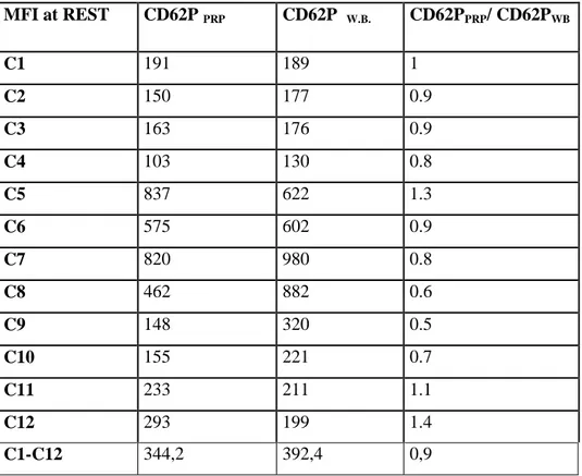
In Table 1 are represented the MFI values of PLT CD 62P of PRP at Rest and the ones of PLT CD 62P of W.B, and the MFI CD62P PRP / MFI CD62P WB ratio, representing the Activation at Rest parameter for each
sample. As can be seen a ratio ≈1 has been obtained, so demonstrating that the enrichment process is able to preserve the biological-biochemical functionalities of the PLT, as the process is not stressful or, more precisely, PLT in PRP shows a level of stress comparable to that of a blood draw.
In Table 2 are represented MFIs values of CD62P of PRP at REST and MFIs values of CD62P of PRP after STRESS of 12 PRP samples; as can be seen an activation ratio between 2 and 4 is obtained on examined samples, co confirming that the enrichment procedure does not alter the functional ability of the platelets and do not inhibits their ability to GF release.
Table 1 MFI at REST CD62P PRP CD62P W.B. CD62PPRP/ CD62PWB C1 191 189 1 C2 150 177 0.9 C3 163 176 0.9 C4 103 130 0.8 C5 837 622 1.3 C6 575 602 0.9 C7 820 980 0.8 C8 462 882 0.6 C9 148 320 0.5 C10 155 221 0.7 C11 233 211 1.1 C12 293 199 1.4 C1-C12 344,2 392,4 0,9
24 Table 2 MFI CD62P su PRP at REST CD62P su PRP after STRESS CD62PPRP REST/ CD62PPRP STRESS C1 191 850 4 C2 150 466 3 C3 163 253 1,7 C4 103 158 1,6 C5 837 2495 2 C6 575 1898 3 C7 820 3851 4 C8 462 781 1,9 C9 148 648 4 C10 155 766 4 C11 233 533 2 C12 293 690 2 C1-C12 344,17 1115,7 4
To demonstrate the ability of the gel to promote the full functionality of PRP without inhibiting the functional capabilities of the PLTs, it has been evaluated the release capacity of PLTs within the gel of the growth factor IGF-1. The IGF-1 immediately after the mixing formation of 111In-Oxine labelled PRP with CMC gel has been determined in three samples.
A fraction of the same samples of this gel-PRP have been activated with exogenous stimulus (Adrenaline and CaCl2) and then was determined the IGF-1 concentration. The normal values for GF IGF-1 for human
serum are between 55 and 327 ng / ml, while the sensitivity limit value of the method is nominally 11.94 ng / ml; therefore values of IGF-1 < 20 ng / ml were considered negative and values > 55 ng / ml were considered positive.
The obtained values are reported in Table 3; it can be seen that the GF is not present in sample immediately after separation and mixing with gel, so demonstrating that these proteins remain within the corresponding platelet granules.
Only when an agonist stimulus is administered to the mix gel-PRP, the PLTs release the GF, which is readily revealed by the RIA analysis.
25
Sample IGF-1 at REST IGF-1 after STRESS
C1 11,94 99,38
C2 11,94 118,43
C3 12,44 74,11
Table 3: Determination of IGF-1 in gel-PRP immediately after preparation (at Rest) and after
pharmacological activation (after Stress)
CONCLUSIONS
Our results indicate that a simple procedure can be used for an efficient separation of platelets, adapting to this use a disposable ready-to-use kit for PRP preparation. The technique is particularly attractive due to the fact that the separation process is performed in a closed system, and can be routinely performed in short times and without contamination danger.
The obtained platelets have been efficiently labelled by 51Cr and by 111In-Oxine, and used for further analytical characterization.
It has been demonstrated that the PLT mixed with a syntethic gel are homogeneously distributed and are stable also for a long period of time. This is important to warrant a homogeneous PLT coverage of the injured area in the surface application of the gel-PRP (typical dermatological / ophthalmic application). Scintigraphic images following the labeling process of PLTs have also demonstrated the maintenance of such uniformity for more than 48 hours, useful to increase the local residence time of PLTs and to prolong the therapeutic activity over time of the released GF.
Quantitative analysis of GF IGF-1 by RIA method demonstrates that gel-PRP mix maintain functional ability to GF release even in such a viscous environment, which is an important parameter in order to achieve an affordable activation of PLT and a stable and reproducible release of therapeutic GF.
26
Annex 1
patient 7 whole blood
27
patient 7
PRP - dilution factor 9x
platelet yield = 90%
platelet enrichment factor = 18 leucocytes = 36% whole blood erythrocytes = 0.008% whole blood
28
References
1. Becker W, Borst U, Krahe T, Borner W, Tc-99m-HMPAO labelled human platelets: in vitro and in vivo results, European Journal of Nuclear Medicine, June 1989, Volume 15, Issue 6, pp 296-301 2. Stratton JR, Ritchie JL,Hamilton GW,Hammermeister KE,Harker LA: Left ventricular thrombi:In
vivo detection by indium-111 platelet imaging and two-dimensional echocardiography. Am J Cardiol 1981;47:874-881
3. Ritchie JL, Stratton JR, Thiele B, Hamilton GW, Warrick LN, Huang TW, Harker LA, Indium-111 platelet imaging for detection of platelet deposition in abdominal aneurysms and prosthetic arterial grafts,The American Journal of Cardiology, Volume 47, Issue 4, April 1981, Pages 882-889
4. Mila TT, The significance of autologous platelets labelled with indium-111 oxinate in primary immune thrombocytopenia of adults and child, Rad Hrvatske Akademije Znanosti i Umjetnosti. Medicinske Znanosti . 2015, Vol. 524 Issue 42, p81-89
5. Wadenvik H, Kutti J, The in vivo kinetics of 111In- and 51Cr-labelled platelets: a comparative study using both stored and fresh platelets, Br J Haematol. 1991 Aug;78(4):523-8
6. Battaglia M, Guaraldi F, Vannini F, . Efficacy of ultrasound-guided intra-articular injections of platelet-rich plasma versus hyaluronic acid for hip osteoarthritis. Orthopedics. 2013;36:e1501-e1508. 7. Salem, A. and Tawfik,A.M. (2016) Role of Platelet Rich Plasma in Treatment of Diabetic Foot
Ulcers Surgical Science , 7 , 272-277
8. Cervelli, V., Gentile, P., Scioli, M.G., Grimaldi, M., Casciani, C.U et al . (2009) Application of Platelet -Rich Plasma in Plastic Surgery : Clinical and in Vitro Evaluation, Tissue Engineering Part C , Methods, 15 , 625 -663
9. Joshua A. Greenspoon, Samuel G. Moulton, Peter J. Millett, and Maximilian Petri, The Role of Platelet Rich Plasma (PRP) and Other Biologics for Rotator Cuff Repair,Open Orthop J. 2016; 10: 309–314
10. Khatu SS, More YE, Gokhale NR, Chavhan DC, Bendsure N. Platelet-rich plasma in androgenic alopecia: Myth or an effective tool. J Cutan Aesthet Surg 2014;7:107-10
11. Leo, M. S., Kumar, A. S., Kirit, R., Konathan, R. and Sivamani, R. K. (2015), Systematic review of the use of platelet-rich plasma in aesthetic dermatology. J Cosmet Dermatol, 14: 315–323
12. Kushida S, Kakudo N, Morimoto N, Ogawa T, Mitsui T, Kusumoto K, Platelet and growth factor concentrations in activated platelet-rich plasma: a comparison of seven commercial separation systems, J Artif Organs (2014) 17: 186
29 13. Mazzucco, L., Balbo, V., Cattana, E., Guaschino, R. and Borzini, P. (2009), Not every PRP-gel is born equal Evaluation of growth factor availability for tissues through four PRP-gel preparations: Fibrinet®, RegenPRP-Kit®, Plateltex® and one manual procedure. Vox Sanguinis, 97: 110–118 14. Danesh H, Ramsook R.R. Timing of Platelet Rich Plasma Injections During Antithrombotic Therapy
Pain Physician. 2016 Sep-Oct;19(7):E1055-61.
15. Rinder et all Activation in stored PLTCs: correlation between membrane expression of P-sectin, GPIIb/IIIa and BTG release Transfusion 1993 Jan;33(1):25-9
16. Cavallo C et all PRP: the choice of activation method affects the release of bioactive molecules BioMed Reserch International Vol 2016, Article ID 6591717
17. Frelinger A, Michelson A Immunophenotypic Analysis of Platelets Supplement 19 Current protocols in cytometry 2002 6.10.1-6.10.17
18. Jo CH, Roh YH, Kim JE, Shin S, Yoon KS, Optimizing platelet-rich plasma gel formation by varying time and gravitational forces during centrifugation, J Oral Implantol. 2013 Oct;39(5):525-32 19. Araki J, Jona M, Eto H, Aoi N, Kato H, Suga H, K, Yatomi Y, Yoshimura K,Optmized Preparation
Method of Platelet-Concentrated Plasma and Noncoagulating Platelet-Derived Factor Concentrates: Maximization of Platelet Concentration and Removal of Fibrinogen, Tissue Engineering: Part C, Volume 18, Number 3, 2012
20. Kiefel V, Becker T, Mueller-Eckhardt G, Grebe S, Mueller-Eckhardt C, Platelet survival determined with 51Cr versus 111In, Klin Wochenschr. 1985 Jan 15;63(2):84-9
21. Schmidt KG, Rasmussen JW, Rasmussen AD, Arendrup H, Lorentzen M, Comparative studies of the function and morphology of 111In- and 51Cr-labelled human platelets, Scand J Haematol. 1983 Jul;31(1):69-77
22. Desir, G.V., Bia, M., Lange, R.C., Smith, E.O., Flye, W., Kashgarian, M., Schiff, M., & Ezekowitz, M.D. Petrovici, J.-N. (Ed.). (1990). The clinical utility of indium-111 labelled platelet scintigraphy in the diagnoses of renal transplant rejection. Netherlands: Kluwer.
23. Fenech A, Nicholls A,Smith FW, Indium (111In)-labelled platelets in the diagnosis of renal transplant rejection: preliminary findings, The British Journal of Radiology 1981 54:640, 325-327 24. Sarpatwari, A., Provan, D., Erqou, S., Sobnack, R., David Tai, F. W. and Newland, A. C. (2010),
Autologous 111In-labelled platelet sequestration studies in patients with primary immune thrombocytopenia (ITP) prior to splenectomy: a report from the United Kingdom ITP Registry. British Journal of Haematology, 151: 477–487
25. Fenech A, Hussey JK, Smith FW, Dendy PP, Bennet B, Douglas AS, Diagnosis of deep vein thrombosis using autologous indium-111-labelled platelets, British Medical Journal, Vol 282, 28 March 1981, pp 1020-22
30
26. Wessels P, Heyns AP, Pieters H, Lötter MG, Badenhorst MG, An improved method for the quantification of the in vivo kinetics of a representative population of 111In-labelled human platelets, Eur J Nucl Med (1985) 10: 522.
ENEA
Servizio Promozione e Comunicazione
www.enea.it
Stampa: Laboratorio Tecnografico ENEA - C.R. Frascati gennaio 2018