LETTER TO THE EDITOR
OCCURRENCE OF
CANDIDA SPECIES COLONIZATION IN A POPULATION OF
DENTURE-WEARING IMMIGRANTS
R. CALCATERRAI, G. PASQUANTONI02, L.A. VITALP, M. NICOLETTI
4M. DI GIROLAM02, C. MIRISOLAI, M. PRENNA5,R. COND02 andL. BAGGP,2
'National InstituteforHealth, Migration and Poverty (NIHMP), Rome, Italy; 2Department of
Clinical Sciences and Translational Medicine, University ofRoma Tor Vergata, Rome, Italy;
3School ofPharmacy, Microbiology Unit, University ofCamerino, Camerino, Italy;
'Department
ofBiomedical Sciences, University "G. D 'Annunzio" Chieti, Italy;
'School ofBiosciences and
Biotechnology University ofCamerino, Camerino, Italy
Received September
2,2012 -Accepted November 21,2012
Infection of the oral cavity and dentures by Candida species are frequent in denture wearers.
C.albicans
is the most common pathogen; however, other emerging Candida species are also responsible
for this condition. Few data are available about the occurrence of Candida species in the oral cavities
of denture-wearing immigrants to Italy. In this study, we compare the Candida species found in the
oral mucosa and on dentures from a population of denture wearing immigrants to Italy to a matched
Italian group. Oral swabs were collected from dentures and the underlying mucosa of patients enrolled
in the study and were then cultured to test for the presence of Candida species in each sample. Out of
168 patients enrolled (73 Italians and 95 immigrants), 51 Italians (69.8%) and 75 immigrants (78.9%)
tested positive for the presence of Candida. Candida albicans was the most frequently observed species
overall; however, we found a higher occurrence of
C.glabrata
among immigrants than among Italians. In
addition, immigrants displayed a higher incidence of Candida - associated stomatitis and a lower mean
age than Candida-positive individuals from the Italian group. Immigrants are more prone to longer
colonization of the oral mucosa and dentures by Candida. In these patients, dentures must be checked
periodically to prevent the presence of Candida.
The alterations of the oral mucosa by dentures
might
result
from
mechanical
irritation
or
inflammatory responses induced by denture materials
(l,
2). In addition, biofilm formation on the denture
surface might contribute to the altered nature of the
oral micro-environment among denture wearers (3).
Candida
species, which comprise 25-50% of the
oral cavity microbiota from healthy individuals and
roughly 80% from denture wearers, are a primary
cause of microbial biofilm formation on medical
devices
(l).Until recently, Candida albicans was considered
the most important opportunistic pathogen in this
genus. However, other Candida species, such
as Candida glabrata, Candida krusei, Candida
parapsilosis
and Candida tropicalis, have also
emerged as causative agents of infection (1).
Candida
glabrata
is
an
emerging
fungal
pathogen that accounts for 15% of mucosal and
systemic candidoses and is associated with severe
Key words: Candida species, denture-related stomatitis, candidosis Mailing address: Dr Roberta Calcaterra,
National Institute for Health, Migration and Poverty (INMP) Via di San Gallicano 25/a 00153 Roma, Italy
Tel.: +39 06 58543782 Fax: +390658543686 e-mail: [email protected]
0394-6320 (2013) Copyright© by BIOLlFE, s.a.s. This publication and/or article is for individual use only and may not be further reproduced without written permission from the copyright holder. Unauthorized reproduction may result in financial and other penalties
239
DISCLOSURE: ALL AUTHORS REPORT NO CONFLICTS OF INTEREST RELEVANT TO THIS ARTICLE.240 R. CALCATERRA ET AL.
inflammation in denture wearers (1).
Candida dubliniensis
has recently been isolated
from the oral cavity ofhuman immunodeficiencyvirus
(HIV)-infected patients, leading to its consideration
as a novel, emerging, opportunistic pathogen (4).
Candidacolonization and biofilm formation on
dentures may depend on oral hygiene practices, such
as overnight denture removal, denture cleanser use,
smoking and specific denture characteristics (5).
Denture-related
stomatitis
(DRS)
is
an
inflammatory process of the mucosa underlying
a removable partial or total dental prosthesis or
appliance (6). DRS has been reported in more than
60% of denture wearers, and although it is typically
asymptomatic,
it
occasionally
associates
with
leukoplakia, pseudomembrane formation, erythema
and angular cheilitis (4, 6, 7).
Key factors that can dramatically increase the risk
of DRS are loose denture fit, poor denture hygiene
and
Candida albicanscolonization of the denture
surface and oral mucosa that contact the denture
fitting surfaces (5). Denture materials themselves
can contribute to the risk of denture stomatitis, as
areas of surface roughness and the hydrophobicity
of denture surfaces can promote the attachment of
microorganisms and biofilm development (8, 9).
The pathogenesis of the
Candida-associateddenture stomatitis includes local and systemic factors
related to the host and to the ability of
Candidato adhere and proliferate in the host epithelial
tissues (8).
Candida-associateddenture stomatitis
usually occurs when conditions of the oral
micro-environment are favorable for the growth and the
adhesion of yeast and when systemic factors lead to
a systemic immunodepression (10-12).
To date, no data are available about the
occurrence of
Candidaspecies in the oral cavity of
denture-wearing immigrants to Italy. In this
study,we compared the colonization of the oral mucosa
and dentures from a population of denture-wearing
immigrants to Italy with a matched Italian group.
MATERIALSAND METHODS
Patients
All denture-wearing patients attending the outpatient department of Social Dentistry Department at National Institutes of Health, Migration and Poverty during the period of June 20 l l-June 2012 were enrolled, A complete
medical and dental history was recorded for each patient, including age, gender, drug use, smoking habits, systemic diseases, DRS symptoms and hygienic habits (modality and frequency) (13).
Patients receiving antifungal or antibacterial treatment within 30 days of enrollment were excluded from the study as well as patients who had undergone immunosuppressive therapies or were affected by an immunosuppressive disease (diabetes, kidney failure, HIV infection). All patients affected with xerostosmy of idiopatic or iatrogenic origin were also excluded.
To identify and characterize the different presentations of DRS, we used the Newton Classification described by Budtz-Jorgensen and Bertram (14):
DRS type I - localised inflammation or hyperaemia points (pin point hyperaemia).
DRS type II - diffuse erythema.
DRS type III - pseudomembrane formation. Sample collection and isolation
After examination of the oral cavity, oral swabs were collected from the denture of each patient and from the underlying mucosa according to the procedure described by Marcos-Arias et al. (15). When denture-related stomatitis lesions were evident, a specimen was collected from the lesion. All oral swabs were cultured within 2 h of collection on CHROMagar Candida medium (Becton Dickinson GmbH, Germany) as well as on Sabouraud dextrose agar plates containing chloramphenicol (Becton Dickinson GmbH, Germany) and were incubated at 37° C for 48 h. We considered a Candida-associated denture stomatitis to be an isolation of> 10 Candida colonies (16). The plates were scored based on the number ofcolonies and then subcultured on the same chromogenic medium and Sabouraud dextrose agar to obtain pure cultures. Characterization ofCandida species
Isolates were identified by conventional mycological methods such as color formation in CHROMagar Candidamedium, germtube tests in calf serum at 37°C for 2 days, and microscopic morphology. Additionally, all yeast identified as Candida albicans were screened for their ability to grow at 45°C on Sabouraud dextrose agar for 3 days and for chlamydoconidia formation on Casein agar at 30°C for 10 days (16).
In these cases, isolates highly suspected to be Candida dublinensiswere definitively identified using polymerase chain reaction (PCR). PCR identification of Candida dublinensiswith the Candida dublinensis-specific primer pair DUBF and DUBR (17) was carried out in a 50-Ill final volume containing 10 pmol each of the forward and reverse primers, 2.5 mM MgCI2, 10 mM Tris-HCl (pH 9.0 at 25°C), 10 mM KCl, 0.1% (vol/vol) Triton X-I00, 2.5 U
ofTaq DNA polymerase (Promega), and 25 III of template DNA-containing cell supernatant. Cycling conditions consisted of 6 min at 95°C, followed by 30 cycles of 30 s at 94°C, 30 s at 58°C, and 30 s at
noc,
followed bynoc
for 10 min. Candida template DNA for use in PCR experiments with the Candida dubliniensis-specific primer pair DUBF (5'GTATTTGTCGTTCCCCTTTC-3') and DUBR (5'-GTGTTGTGTGCACTAACGTC-3') was prepared as described by Donnelly et al. (17).Statistical analysis
For the statistical analysis, SPSS software vers.13 was used(IBM,Armonk, NY, USA). A p< 0.05 was considered significant.
RESULTS
We enrolled a total of 190 patients with removable dentures, and 22 were excluded from the study for the following reasons: 5 were affected with diabetes mellitus, 1 was affected with kidney failure, 11 were suffering from xerostomy (2 because of Sjogren syndrome and 9 as consequence of anti-hypertensive or anti-depressive therapy) and 5 had received antimycotic or antibiotic therapy within 30 days of enrollment.
Of the remaining 168 patients, 73 were Italians (described as group 1) and 95 were immigrants (described as group 2), who were defined as people born in a country other than Italy that came to Italy within three years of enrollment in the study.
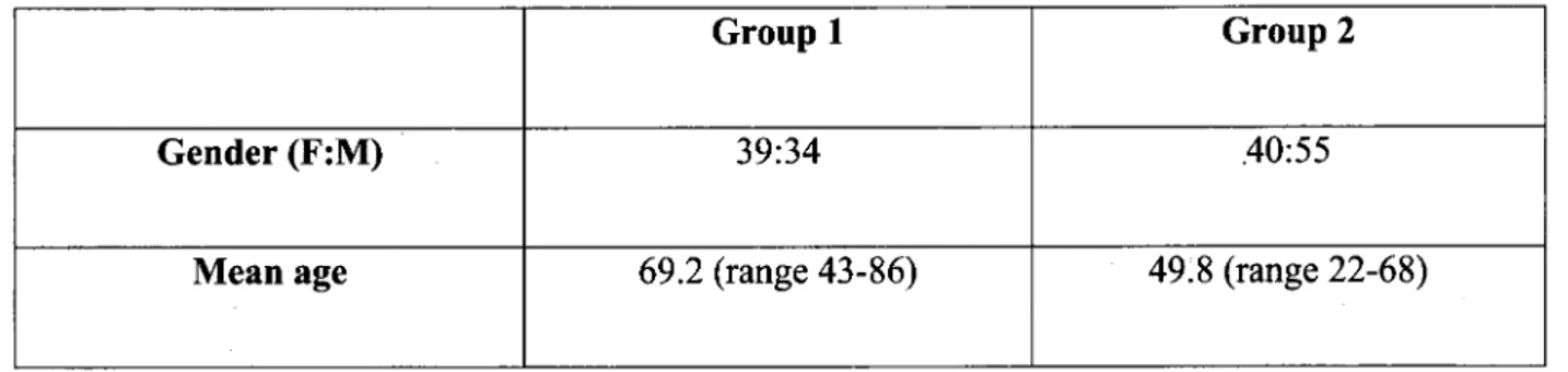
The demographics of the patients in group 1 (Italian patients) and group 2 (immigrant patients) are listed in TableI.
In total, 51 Italians (69.8%) and 75 immigrants (78.9%) tested positive for contamination by Candida
species (p=O.l2, chi-squared test). The patients displaying Candida-colonization of the oral mucosa were younger in group 2 than group 1 (71.5±11 vs 49.3±11.2 years ± ds p<0.05, Student's t-test).
Italian patients colonized by Candida species other than albicans had a younger mean age than those colonized by
C.
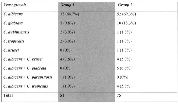
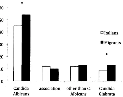
albicans (58±8.1 vs 75±9.2p<0.05 chi-squared test). Fig.l shows the percentage of patients positive for the colonization of dentures and oral mucosa, divided according to Candida species. Table II shows the frequency of Candida species isolated from Italian patients (group 1) and immigrant patients (group 2) based on culture and molecular methods. The most frequently isolated species from oral mucosa and dentures from group 1 were Candida albicans 33 (64.7%), Candida
glabrata 5 (9.8%), Candida dubliniensis 2 (3.9%), and Candida tropicalis 2 (3.9%). From group 2, the most frequently isolated species were Candida
albicans52 (69.3%), Candida glabrata 10 (13.3%),
Candida dubliniensis 1 (1.3%), Candida tropicalis 1 (1.3%) and Candida krusei 1 (1.3%).
Simultaneous colonization by two yeast species was found in 6 (11.7%) oral mucosa and denture samples from patients in group 1, particularly
Candida albicans and Candida krusei 4 (7.8%),
Candida albicans and Candida parapsilosis 1
(1.9%) and Candida albicans and Candida tropicalis 1 (1.9%). In group 2, we found simultaneous colonization in 13 (17.3%) oral mucosa and denture samples, particularly Candida albicans and Candida
glabrata 5 (6.6%), Candida albicans and Candida
krusei4 (5.3%) and Candida albicans and Candida
tropicalis4(5.3%).
The frequency of Candida albicans colonization
TableI.Demographic characteristic ofItalian (group1)and immigrant (group 2) patients.
Group 1
Group 2
Gender
(F:M) 39:34 .40:55242 R. CALCATERRA ET AL.
Table II. Presence of different species of Candida in positive oral and denture specimens in Italian (group 1)and immigrant ( group2)patients.
Yeast growth Group 1 Group2
C.albicans 33 (64 .7%) 52 (69 .3%) C.g/abrata 5 (9.8%) 10 (13.3%) C.dubliniensis 2(3.9%) 1( 1.3%) C.tropiealis 2(3.9%) I(1.3%) C.krusei 0(0%) I (1.3%) C.a/bieans +C.krusei 4 (7.8%) 4(5.3%) C.a/bieans +C.g/abrata 0(0%) 5 (6.6%) C.a/bieans +C.parapsilosis I(1.9%) 0(0%) C.a/bieans +C.tropiealis I(1.9%) 4(5.3%) Total 51 75
Table III. Gravity oflesion oforal mucosa in patients ofour series resulted positive to colonization by other species than Candida albicans alone and in association.
Other thanCa nd ida Albicans
lesion gravity I type II+III type Association lesion gravity I type II+III type
.*
p < 0.05 calculated by chi square.Italians 4 4+1 Italians 5 2+2 Immigrants 2 5+6* Immigrants 3* 3+4*
of the oral mucosa and dentures was significantly
(p<0.05) higher than that of other Candida species
in both groups I and 2. The colonization by Candida
glabrata
was more frequent among patients from
group 2 than those from group 1 (p<0.05).
Table III shows the severity of oral lesions
(according to the classification in degree 1-3 p<
0.05) observed from the two groups of patients.
Immigrants whose dentures and oral mucosa tested
positive for Candida species other than Candida,
both alone and in association, displayed oral lesions
that were clinically more severe (type II and III)
Table IV. Denture soaking and brushing habits (Panel A) andfrequency ofdenture cleaning (Panel B) in Italians (group 1) and immigrants (group 2).
A
Group 1 Group 2
POS NEG POS NEG
n=51 n=22 n=75 n=20
Brushing only 10 5 20 5
Soaking in solution 14 5 24 7
only
Soaking in water only 13 7 13 5
Brushing and soaking 13 4 16 2
Nothing 2 1 2 1
B
Group 1 Group 2
POS NEG POS NEG
n=51 n=22 n=75 n=20
<1 time / day 4 4 49 10
1 time / day 13 7 14 5
2 or>times / day 34 11 12 5
than those of Italian denture wearers with the same colonization.
In Table IV (A and B), we describe the oral and denture hygienic habits of the patients included in the study. No difference existed between the groups of patients divided according to the type and frequency of oral and denture cleaning (test U, p=O.l6) and the colonization by
Candida
species (Mann Whitney, p= 0.25).DISCUSSION
Our results show that the mean age of enrolled patients in group 2 was significantly lower than that of patients in group 1. One explanation for this finding is that, generally, immigrant populations
primarily consist of young individuals. Despite their younger age, the immigrant group had severe dental problems that could lead to tooth loss and denture placement. Early denture placement in immigrant patients may cause premature, chronic colonization of the denture by
Candida
species and an early onset of denture stomatitis.Our data corroborated that
Candida albicans
is the most frequently isolated species, followed byCandida glabrata, Candida dubliniensis
andCandida tropicalis
in the oral mucosa as well as on dentures in both group I and group 2. We also reported, in accordance with previous studies, that amongnon-albicans Candida
species, the most frequent yeast isolated from the oral mucosa and dentures isCandida glabrata;
however, we newly244
R. CALCATERRA ET AL.*
60
50
40
o
Italians30
-Migrants*
20
10
[RJ~
cI
0
Candida association other than C. Candida
Albicans Albicans Glabrata
Fig. 1. Percentage ofpatients positive to the different species ofyeast in our series, out of 51 Italians and 75 migrants with a contamination oforal mucosa and dentures. *p<0.05 Italians versus immigrants calculated by chi-square test}.
report that
Candida glabratais more frequently
found in immigrant patients than in native Italians.
Denture
use
has
been
demonstrated
as
a
predisposing condition for oral candidiasis (18, 19).
Compared with
Candida albicans, Candida glabratademonstrated a two-fold greater tendency to adhere
to denture acrylic surfaces
in vitro(20). With this
high propensity to adhere to denture surfaces, it is not
surprising that
Candida glabratahas been identified
as the predominant yeast isolated from dentures of
elderly persons with chronic atrophic candidiasis (21).
Due to the fact that
Candida glabratais frequently
co-isolated with other
Candidaspecies from oral lesions
(22), the role of this organism in pathogenesis is still
obscure.
Candida glabratais most frequently
co-isolated from mucosal lesions with
Candida albicans(23). It has been reported that mixed infections with
Candida glabrataand
Candida albicanscan cause
more severe symptoms and are more difficult to
treat (23, 24). Among the clinical manifestations
seen in our patients, we found a correlation of
Candidaspecies with the type of oral lesion;
Candida albicanscorrelated with the absence of lesions or
pin point heperemialerythema, whereas
Candidaglabata, Candida dublinesins
and
Candida tropicaliscorrelated
with
pseudomembrane
formation.
Pseudomembrane formation is likely to be linked to
the dramatic histopatological tissue alteration induced
by
Candida tropicalis, Candida dublinensisand
Candida glabrata,as reported previously (26).
Notably, the oral lesions of denture-stomatitis
related to
non-alMeans Candidaare clinically more
severe in immigrants than in Italians. In our opinion,
this finding is due to the fact that immigrants become
denture wearers at a younger age, and consequently,
the denture is exposed to potential pathogens for a
longer period of time. This leads immigrant patients
to have more severe denture stomatitis. Additionally,
denture materials are often not as reliable as those
available in Italy and thus are more prone to
colonization.
Moreover, immigrants have limited access to the
health care system, and thus, they rarely undergo
periodic clinical check-ups of the dentures and the
oral mucosa.
We also found that hygienic habits did not impact
Candidacolonization. This finding differs from
previous reports in the literature that describe a
correlation between hygienic habit and the frequency
of oral and denture colonization.
In our opinion, colonization mainly relies upon
the denture materials, particularly the micro-porosity
of the denture base and the micro-irregularity of
the denture surface. These features have a pivotal
role in the development of biofilm (26), although
hygienic habits might contribute to maintaining the
biofilm once established. Further studies evaluating
the resistance of different types of resins to different
Candida species colonization are required to confirm
this assumption.
Interestingly, we found simultaneous colonization
by two Candida species in the oldest Italian
patients. Therefore, we assumed that age-related
immunosuppression may play a major role in the
colonization and maintenance of Candida species in
the oral cavity as well as in dentures. Several factors,
in our opinion, can account for this, such as the
chronic use of dentures and the varied composition
of oral microbiota and diets.
Because biofilm formation is a risk factor for
Candida infection in denture wearers, it is advisable
to periodically screen denture wearers for Candida
presence. Nevertheless, a prompt treatment is strictly
required in patients with non-albicans Candida,
as these are the yeast with the highest tendency to
invade and destroy the underlying oral mucosa.
REFERENCES
1. Budtz-Jergensen E. Oral mucosal lesions associated with the wearing of removable dentures. J Oral Pathol 1981; 10:65-80.
2. Zomorodian K, Rahimi MJ, Pakshir K, Motamedi M, Ghiasi MR, Rezashah H. Determination of antifungal susceptibility patterns among the clinical isolates of Candida species. J Glob Infect Dis 2011; 3:357-60. 3. Dar-Odeh NS, Shehabi AA. Oral candidosis in patients
with removable dentures. Mycoses 2003; 46:187-91. 4. Mosca CO, Moragues MD, Llovo J, Al Mosaid A,
Coleman DC, Pont6n 1. Casein agar: a useful medium for differentiating Candida dubliniensis from Candida albicans. J Clin Microbiol2003; 41:1259-62.
5. Gendreau L, Loewy ZG. Epidemiology and etiology of denture stomatitis. J Prosthodont 2011; 20:251-60. 6. Dagistan S, Aktas AE, Caglayan F, Ayyildiz A,
Bilge M. Differential diagnosis of denture-induced
stomatitis, Candida, and their variations in patients using complete denture: a clinical and mycological study. Mycoses 2009; 52:266-71.
7. Fidel PL Jr, Vazquez JA, Sobel JD. Candida glabrata: review of epidemiology, pathogenesis, and clinical disease with comparison to C. albicans. Clin Microbiol Rev 1999; 12(1):80-96.
8. Ramage G, Tomsett K, Wickers BL, et al. Denture stomatitis: a role for Candida biofilms. Oral Surg Oral Med Oral Pathol Oral Radiol Endod 2004; 98:53-59. 9. Tari BF, Nalbant D, Dogruman Al F, Kustimur S.
Surface roughness and adherence of Candida albicans on soft lining materials as influenced by accelerated aging. J Contemp Dent Pract 2007; 8: 18-25.
10. Yuen HK, Wolf BJ, Bandyopadhyay D, Magruder KM, Salinas CF, London SD. Oral health knowledge and behavior among adults with diabetes. Diabetes Res Clin Pract 2009; 86:239-46.
11. Paillaud E, Merlier I, Dupeyron C, Scherman E, Poupon J, Bories PN. Oral candidiasis and nutritional deficiencies in elderly hospitalised patients. Br J Nutr 2004; 92:861-67.
12. Golecka M, Oldakowska-Jedynak D, Mierzwinska-Nastalska E, Adamczyk-Sosinska E. Candida-associated denture stomatitis in patients after immunosuppression therapy. Transplant Proc 2006; 38:155-56.
13. Kulak-Ozkan Y, Kazazoglu E, Arikan A. Oral hygiene habits, denture cleanliness, presence of yeasts and stomatitis in elderly people. J Oral Rehabil 2002; 29:300-04.
14. Budtz-Jorgensen E, Bertram D. Denture stomatitis. I. The etiology in relation to trauma and infection. Acta Odontol Scand 1970; 28(1):71-92.
15. Marcos-Arias C, Eraso E, Madariaga L, Carrillo-Mufioz.Al, Quind6s G. In vitro activities ofnewtriazole antifungal agents, posaconazole and voriconazole, against oral Candida isolates from patients suffering from denture stomatitis. Mycopathologia 2012; 173:35-46.
16. Marcos-Arias C, Vicente JL, Sahand IH, et al. Isolation of Candida dubliniensis in denture stomatitis. Arch Oral Bioi 2009; 54:127-31.
17. Donnelly SM, Sullivan DJ, Shanley DB, Coleman DC. Phylogenetic analysis and rapid identification of Candida dubliniensis based on analysis of ACTl
246
R.CALCATERRA ET AL. intron and exon sequences. Microbiology 1999;145:1871-82.
18. Ohman SC, Osterberg T, Dahlen G, Landahl S. The prevalence of Staphylococcus aureus, Enterobacteriaceae species, and Candida species and their relation to oral mucosal lesions in a group of 79-year-olds inGoteborg, Acta Odontol Scand 1995; 53:49-54.
19. Lockhart SR, Joly S, Vargas K, Swails-Wenger J, Enger L, Soll DR. Natural defenses against Candida colonization breakdown in the oral cavities of the elderly. J Dent Res 1999; 78:857-68.
20. Luo G, Samaranayake LP, YauJY. Candida species exhibit differential in vitro hemolytic activities. J Clin Microbiol2001; 39:2971-74.
21. Wilkieson C, Samaranayake LP, MacFarlane TW, Lamey PJ, MacKenzie D. Oral candidosis in the elderly in long term hospital care. J Oral Pathol Med
1991; 20:13-16.
22. Feng Z, Jiang B, Chandra J, Ghannoum M, Nelson S, Weinberg A. Human beta-defensins: differential activity against candidal species and regulation by Candida albicans. J Dent Res 2005; 84:445-50. 23. Redding Sw. The role of yeasts other than Candida
albicans in oropharyngeal candidiasis. Curr Opin Infect Dis 2001; 14:673-77.
24. Redding SW, Zellars RC, Kirkpatrick WR, et al. Epidemiology of oropharyngeal Candida colonization and infection in patients receiving radiation for head and neck cancer. J Clin Microbiol1999; 37:3896-900. 25. Sullivan DJ, Moran GP, Coleman DC. Candida dubliniensis: ten years on. FEMS Microbiol Lett 2005; 25:9-17.
26. Ramage G, Martinez JP, Lopez-Ribot JL. Candida biofilms on implanted biomaterials: a clinically significant problem. FEMS Yeast Res 2006; 6:979-86.