IDENTIFICATION OF MUTATIONS IN THE GLI2 GENE
IN CPHD (COMBINED PITUITARY HORMONE
DEFICIENCY) PATIENTS. FUNCTIONAL ANALYSIS
OF THE IDENTIFIED VARIANTS.
By
RANJITH MUNISWAMY
A thesis submitted to
The Department of Health Sciences of the University of Eastern Piedmont In Partial fulfilment of the requirements
For the degree of Doctor of Philosophy
In
Molecular Medicine
Supervisor: Prof. MARA GIORDANO Coordinator: Prof. EMANUELE ALBANO
YEAR 2016 SEPTEMBER XXVIII CYCLE
I
ACKNOWLEDGMENT
My scientific journey was a challenging and invaluable experience. Along the way, I had many people who guided my scientific training, supported my choices as well as helped me in my personal growth. First and Foremost, I would like to thank my supervisor, Prof. Mara Giordano who gave me opportunity to carry out this research. I appreciate all her contribution of time, advice, patience to make my Ph.D. possible. I would like to express my gratitude for St. Paul’s Society, Italy for funding my Ph.D.
I wish to express my sincere gratitude to all the members of my group, for providing a wonderful environment to work in. I gratefully acknowledge the contributions of Deepak Babu, Ileana Fusco, Simona Mellone, Antonella Fanelli, to this work. I would like to extend my sincere thanks to Prof. Avanzi, Prof. Lucia Corrado, Ileana Fusco and physician Dr. Giorda for their kindness and timely help in hospital, when I was ill and bed ridden. I would like to thank Nadia Barizzone, Miriam Zuccalà, and all the other present and past members of the laboratory for their kind gesture and moral support.
I am thankful to friends who made my time in Novara, enjoyable. I would like to thank Yogesh Shivakumar, Nausicaä Clemente and Simone Merlin for their friendship and timely help whenever I needed the most. Lastly, I would like to thank my family for all their love and encouragement. I am grateful to my parents for their unconditional support in all my pursuits, to whom I dedicate this thesis.
II
LIST OF CONTENTS
List of Tables III List of Figures V List of Abbreviations VI
I- INTRODUCTION 1
1. Pituitary Gland 2
1. Factors involved in embryogenesis of Pituitary Gland 7
2. Disorders and Deficiencies in Pituitary Gland hormones 11
3. Hedgehog Signaling Pathway 14
4. Release and Transport of Hh through Tissues 16
5. Receiving the Hh Signal 16
6. Transcriptional Targets of Hh Signaling 17
2. Human GLI2 gene 21
1. GLI2 in the development of Pituitary Gland 25
2. GLI2 in CPHD disease 26
II- AIM 27
III- SUBJECTS 29
1. CPHD Patients 29
IV- MATERIALS AND METHODS 32
1. Screening GLI2 gene 32
2. DNA Extraction 32
3. Polymerase Chain Reaction 33
4. Exo/SAP and Sequence Reaction 36
5. Site Directed Mutagenesis 37
6. Bacterial Transformation 40
7. Cell Culture and Transfection 41
V- RESULTS 42
1. Identified Mutations 43
2. In silico analysis 43
3. Details of Patients Carrying Mutations 45
4. Functional Analysis of Mutations 48
VI- DISCUSSION 53
VII- CONCLUSION 57
VIII- REFERENCES 60
IX- APPENCISE 68
III
LIST OF TABLES
Table 1: List of Pituitary Gland Hormones and their Target and Major Functions 4
Table 2: Genes involved in embryogenesis of Pituitary Gland with embryonic days 9
Table 3: Hedgehog pathway components in Drosophila and vertebrates 18
Table 4: GLI2 Isoforms with missing amino acid positions, length and mass 23
Table 5: GLI2 Zinc Finger positions and length of binding site 23
Table 6: To examine all coding regions of GLI2 gene 18 pairs of following primers designed 32 Table 7: Reagents used for Polymerase chain reaction fragments 1-18 except 4 and 13.2 33
Table 8: Thermal cycle conditions for fragments 1 – 18 except fragment 4 and 13.2 34
Table 9: Reagents used for Polymerase chain reaction fragments 4 and 13.2 35
Table 10: Thermal conditions for Fragment 4 and 13.2 35
Table 11: Reagents used for Exo/SAP purification 36
Table 12: Reagents used for Sequencing Reaction 36
Table 13: Reagents used for Mutagenesis for p.Y575H 37
Table 14: Thermal conditions for Mutagenesis of p.Y575H 38
Table 15: Reagents used for Mutagenesis for p.A593V 38
Table 16: Thermal conditions for Mutagenesis of p.A593V 39
Table 17: Reagents used for Mutagenesis for p.P386L 39
IV
Table 19: Reagents and thermal conditions for T4-Ligation 40
Table 20: Summary of GLI2 mutations Identified in this study, Allele Frequency Data from dbSNP 44 Table 21: Clinical characteristics of CPHD patients identified in this study (GLI2 Mutation) 47
V
LIST OF FIGURES
Figure 1: Schematic representation of Rathke’s pouch formation and Pituitary Gland
Development. 6
Figure 2: Embryonic days of Developing Pituitary Gland 7
Figure 3: Upstream regulation of the Gli transcription factors and their individual and combined roles in regulating Hh target gene expression 14
Figure 4: Regulatory domains in Gli Zinc Finger Family proteins 20
Figure 5: Schematic Representation of Gli2 Gene Full-Length with regulatory Domains 22
Figure 6: Schematic representation of variation’s found in this study on Gli2 Gene 43
Figure 7: Family tree of Patient B 45
Figure 8: pCS2-MT expression vectors bearing GLI2-cDNA full length and GLI2-∆N cDNA 48 Figure 9: Reporter plasmid with backbone of pδ51 LucII pGL4.10 bearing 8x3’- GLI-BS 49
Figure-10: Comparison of reporter activity between GLI2∆N and GLI2 Full length 50
Figure 11: Transcription activity of mutagenized and wild-type bearing the either full length GLI2-cDNA or ∆N constructs 51
Figure 12: Transcriptional activity of co-transfected mutated with wild type construct 52
Figure 13: (A) Sequence of the GLI zinc finger domain and the DNA-binding site. The five zinc fingers of GLI are align to show the conserved residues and secondary structures. The approximate position of the α-helix is underlined, and that of β-sheets is indicated by zig-zag lines. The position of mutations identified in our study is highlighted in color. (B) Sketch of DNA-Zn finger complex showing orientation of fingers 56
VI
LIST OF ABBREVATIONS
GH – Growth Hormone PRL – Prolactin Hormone
TSH – Thyroid Stimulating Hormone FSH – Follicle Stimulating Hormone LH – Luteinizing Hormone
ACTH – Adrenocorticotrophin POMC – Proopiomelanocortico
MSH – Melanocyte Stimulating Hormone AVP – Arginine Vasopressin
IGF1 – Insulin-Like Growth Factor 1
IGFBP3 – Insulin-Like Growth Factor Binding Protein 3 MC2R – Melanocortin 2 Receptor
SHH or Shh – Sonic Hedgehog Signaling BMPS – Bone Morphogenetic Protein FGF – Fibroblast Growth Factor WNT – Wingless Pathway TF – Transcription Factor
OTX2 – Orthodenticle Homeobox
HESX1 – Homeobox Gene Expressed in ES Cells PITX1 – Paired Like Homeodomain 1
PITX2 – Paired Like Homeodomain 2 CNS – Central Nervous System POU1F1 – Pou Class 1 Homeobox 1 SOX – Sry-Related HMG Box
NKX2 – NK2-Homeobox-Thyroid Transcript Factor CPHD – Combined Pituitary Hormone Deficiency
VII
HH – Hypogonadotropic Hypogonadism DI – Diabetes Insipidus
ONH – Optic Nerve Hypoplasia ACC – Agenesis of Corpus Callosum GHD – Growth Hormone Deficiency TRH – Thyrotropin-Releasing Hormone GnRH – Gonadotropin-Releasing Hormone IGHD – Isolated Growth Hormone Deficiency Hh – Hedgehog Pathway
DHH – Desert Hedgehog IHH – Indian Hedgehog Ttv – Tout Velu Family Sotv – Sister of Ttv Botv – Brother of Ttv Disp – Dispatched
Hip – Hh-Interacting Protein IHog – Interference Hedgehog Boi – Brother of Ihog
CDO – Cell Adhesion Associated Oncogene Regulated BOC – Brother of CDO
SMO – Smoothened Gene Ptc – Patched 1 Gene Wg – Wingless Proteins Col – Collagen Coding Gene
Gas1 – Growth Arrest Specific 1 Gene VEGF – Vascular Endothelial Growth Factor
PDGFRα – Platelet Derived Growth Factor Receptor Alpha HNFβ3 – Hepatocyte Nuclear Factor 3 Beta
VIII
SPOP – Speckle Type BTB/POZ Protein SuFu – Suppressor of Fused Homolog Ski – Skinny Hedgehog
Hhat – Hedgehog Acyltransferase Cos 2 – Costal 2 Gene
Kif7 – Kinase Family 7
STK36 – Serine/Threonine Kinase 36 Dlp – Dally Like Protein
GPC4 – Glypican 4 GPC6 – Glypican 6 Ext 1 – Exostosin 1 Ext2 – Exostosin 2 Ext3 – Exostosin 3 Shf – Shifted Gene
Wif – Wnt Inhibitory Factor PKA – Protein Kinase A CK1 – Casein Kinase 1 Sgg – Shaggy Gene
GSK3β – Glycogen Synthase Kinase 3 Beta Slimb – Supernumerary Limbs
β-TRCP – Beta-Transducin Repeat Containing. ORF – Open reading frame.
GLI2-FL – GLI2-full length. GLI2-∆N – GLI2-Delta N isoform.
1
2 PITUIARY GLAND
The pituitary gland is a central regulator of growth, reproduction, metabolism and stress responses, and functions to relay signals from the hypothalamus to peripheral organs. It is situated within the sella turcica, a recess in the sphenoid bone, at the base of the brain. The hypothalamus is the principal neural structure regulating homeostasis in vertebrates, coordinating complex signals from other regions of the brain and the periphery. The hypothalamus releases factors that control the endocrine activity of the pituitary cells. [1]
The pituitary gland is formed by the juxtaposition of the adenohypophysis (anterior and intermediate lobes) and the neurohypophysis (posterior lobe). The anterior pituitary consists of five different endocrine cell types secreting six hormones: somatotrophs that secrete growth hormone (GH), lactotrophs that secrete prolactin (PRL), thyrotrophs that secrete thyroid-stimulating hormone (TSH), gonadotrophs that secrete both gonadotrophins, follicle-thyroid-stimulating hormone (FSH) and luteinizing hormone (LH), and corticotrophs that secrete adrenocorticotrophin (ACTH). [2]
Somatotrophs are the majority of adenohypophyseal secretory cells comprising nearly 50% of all anterior pituitary cells. Lactotrophs embryologically arise from GH-producing cells, and constitute about 15-20% of the anterior pituitary cell population, although pregnancy and lactation alter the number of maternal lactotrophs. Corticotrophs and gonadotrophs represent 15-20% and 10-15% of anterior pituitary cells, respectively. The thyrotroph is the least common cell type in the anterior pituitary, accounting for less than 10% of the pituitary cell population [3, 4].
The intermediate lobe secretes proopiomelanocortin (POMC), a precursor to melanocyte-stimulating hormone (MSH), and involutes in the adult [2]. The neurohypophysis is formed from
3
axonal terminals, projecting from two discrete groups of magnocellular neurons in the hypothalamus, surrounded by modified astrocytes called pituicytes. The two hormones secreted by the posterior lobe of the pituitary gland, arginine vasopressin (AVP) and oxytocin, are synthesized in the paraventricular and supraoptic nuclei within the hypothalamus [2, 5].
The hypothalamus is positioned above the pituitary gland in the basal part of the forebrain. The magnocellular neurons, within the paraventricular and supraoptic nuclei in the hypothalamus, produce AVP and oxytocin. Their axons form the hypothalamo-hypophyseal tract, and the hormones are released from the posterior pituitary into the general circulation in response to electrical excitation. The adenohypophysis is anatomically distinct from the hypothalamus. However, parvocellular neurons of the hypothalamus secrete releasing factors that, via a system of hypophyseal portal vessels, act on the endocrine cells of the anterior lobe to stimulate or inhibit the synthesis and secretion of GH, prolactin, TSH, ACTH, and FSH and LH. The infundibulum (or pituitary stalk) carries both the portal blood delivering hypothalamic hormones to the anterior pituitary and the neural tract from the hypothalamic nuclei to the posterior pituitary. It is noteworthy that the optic chiasm lies above the hypophysis and anterior to the pituitary stalk. Thus, any mass lesion of sufficient size in the area of the pituitary gland will cause visual field defects [1, 2 and 4].
4
Table 1: List of Pituitary Gland Hormones and their Target and Major Functions.
ANTERIOR PITUITARY HORMONES PRIMARY TARGETS OF ANTERIOR PITUITARY HORMONES MAJOR FUNCTION OF ANTERIOR PITUITARY HORMONES Thyroid Stimulating Hormone (TSH, Thyrotropin)
Thyroid Gland Stimulates secretion of Thyroid Hormones
Follicle Stimulating Hormone (FSH,
Gonadotropin)
Ovaries and Testes Triggers ovulation, secretion of estrogen, progesterone and
testosterone Luteinizing Hormone (LH,
Gonadotropin)
Follicles and Testes Stimulation and maturation of Oocyte and sperm production AdrenoCortico Tropin
Hormone (ACTH, Corticotrophin)
Adrenal Cortex Secretion of Glucocorticoid
Growth Hormone (GH)
Most Tissues in Body Growth regulation, metabolism and protein biosynthesis and regulation of Blood Glucose level Prolactin (PRL) Mammary Glands Development and lactation of
mammary gland POSTERIOR PITUITARY HORMONES PRIMARY TARGETS OF POSTERIOR PITUITARY HORMONES MAJOR FUNCTIONS OF POSTERIOR PITUITARY HORMONES
Arginine Vasopressin (AVP) Vascular smooth muscles and kidney
Aquaporin and distal tubes development and water regulation
in kidney Oxytocin Mammary glands and
Uterus
Contraction of Uterus and Milk regulation in mammary gland
5
The hormones secreted from the anterior pituitary regulate growth, puberty, metabolism, response to stress, reproduction, and lactation, while those from the posterior pituitary are required during parturition and lactation, and regulate water balance see to (Tab.1) [2].
GH stimulates insulin-like growth factor 1 (IGF1) gene expression and IGF1 synthesis in liver and bone, amongst other tissues, acting on growth. GH also regulates the hepatic production of insulin-like growth factor binding protein 3 (IGFBP3), acting as a gluco-counterregulatory hormone in metabolism. In muscle, GH increases protein synthesis, while in the adipocyte, GH induces lipolysis [6, 7]. PRL is the major hormone that simulates milk production, it inhibits LH and FSH secretion inducing lactation-related amenorrhea in the postpartum period [5]. ACTH binds with high affinity to the melanocortin 2 receptor (MC2R) in the adrenal gland and regulates steroidogenesis [3].
In the male, LH binds to a receptor on testicular Leydig cells and increases the synthesis of testosterone [8], while FSH binds to testicular Sertoli cell and stimulates the production of proteins in the seminal fluid [9]. In females, LH binds to its receptor on ovarian theca cells and stimulates steroidogenesis; FSH stimulates ovarian follicular growth and facilitates generation of estrogen from thecal cells [10]. TSH binds to its receptor on thyrocytes, resulting in an increase in iodine transport, in the expression of thyroperoxidase and thyroglobulin, and ultimately in increased synthesis of thyroid hormones [2, 3].
AVP acts on the V2 receptor in the renal collecting duct and increases water permeability to facilitate water reabsorption, and on V1 receptor in endothelial cells to promote vasoconstriction [3]. Oxytocin acts through its receptor, inducing intracellular calcium release that, in turn, results
6
in smooth muscle contraction in the uterine myometrial cells and mammary gland myoepithelial cells to cause uterine contraction and milk ejection, respectively [11].
FACTORS INVOLVED IN EMBRYOGENESIS OF PITUITARY GLAND
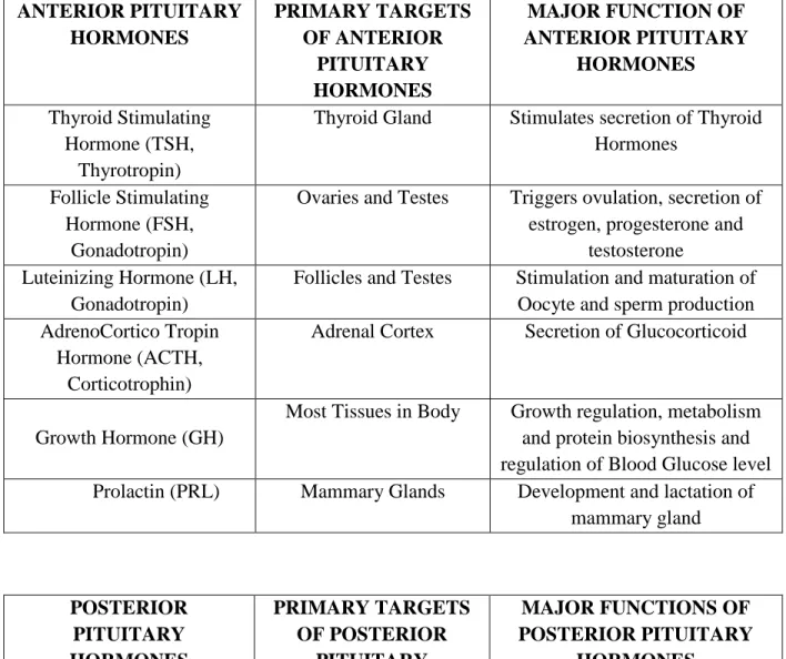
Pituitary gland is an amalgam of two tissues (Adenohypophysis and Neurohypophysis). Early in gestation a finger like projections of ectoderm grows upward from the upper portion of oropharynx. This protrusion is called Rathke's pouch and will develop into the anterior pituitary or adenohypophysis [13].
Figure 1: Schematic representation of Rathke’s pouch formation and Pituitary Gland Development.
At the same phase that Rathke's pouch is developing, another finger like projections of ectodermal tissue evaginates ventrally from the diencephalon of the developing brain. This extension of the ventral brain tissue will become the posterior pituitary or neurohypophysis. Finally, the two tissues grow into one another and become tightly apposed, but their structure remains distinctly different, reflecting their differing embryological origins (Fig.1) [12].
7
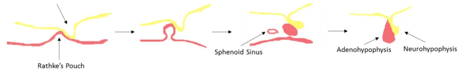
Pituitary cell proliferation and differentiation are regulated by different transcriptional activators and repressors and by signalling molecules from adjacent regions. The early development of pituitary gland in mammals are similar in embryonic stages, so here we considered mice pituitary gland development for explanation. In the early stage of pituitary development, which corresponds to embryonic days (E) 6.5–10.5 in mice, the extrinsic signalling pathways are activated, including the sonic hedgehog (Shh) [17], bone morphogenetic proteins (Bmps) [18], fibroblast growth factor (Fgf) [18] and wingless (Wnt) [19] pathways (Fig.2).
Figure 2: Embryonic days of developing Pituitary Gland.
Shh is not directly involved in Rathke’s pouch formation; however, it is required for midline
formation, forebrain development, brain lobe determination, eye formation [18, 20, 21 and 22] and BMP2 expression induction. Mouse embryos that lack Shh have pituitary hypoplasia and the optic
disc is absent [21]. The Shh pathway depends on zinc finger factors, such as Gli1, Gli2 and Gli3 [17]. Although Shh is not expressed in Rathke’s pouch, Gli factors are found in the precursor structures of the pituitary. Therefore, it is possible that in response to Shh signalling, Gli proteins
8
activate other target genes directly involved in pituitary organogenesis [20]. Otx2 is another TF that is not expressed in the pituitary tissues themselves [23, 24]. This is a bicoid protein that is important for eye and forebrain formation [24, 25]. Otx2 is also responsible for Hesx1 expression regulation [23]. Hesx1 is the first pituitary-specific TF to be expressed at or before E6.5 [26, 27]. Hesx1 expression begins in the rostral region and progresses dorsally; the restricted expression of
this TF is responsible for Rathke’s pouch formation. Hesx1 is important for midline formation and regulates the expression of other TFs [23, 26, 28 and 29].
The Pitx1 and Pitx2 genes are expressed at approximately E9 and participate in the different steps of central nervous system (CNS) organogenesis. Pitx1 is initially expressed in the first branchial arch, then in the oral cavity, and next in Rathke’s pouch [30]. Pitx1 continues to be expressed in the latter stages of pituitary embryogenesis and participates in cellular differentiation [30, 31]. Pitx2 is expressed in several organs, including the CNS, forelimbs, lungs, kidneys and tongue. In
addition to its role in CNS formation, Pitx2 appears to be important in the determination of the left–right axis. Similar to Pitx1, Pitx2 continues to be expressed during pituitary cell differentiation and acts synergistically with other TFs to determine pituitary cell types, primarily Pit1 (Pou1f1)-specific cells [30-32].
Similarly, other molecules play relevant roles in the development of the CNS, including the Soxb1 TFs (Sox1, Sox2 and Sox3) [33, 34]. Sox3 expression begins during early embryogenesis; recent studies have suggested that this gene must be expressed at a constant level because both increases and decreases in its expression are related to pituitary deficiencies and CNS malformations [34]. Some signalling molecules expressed in the infundibulum directly contribute to the induction of pouch invagination, among which Bmp4 [18] and Nkx2 are key [35].
9
Mutant animals lacking any of these factors may develop pituitary absence, malformation or even embryonic lethality [36, 37]. In parallel with the invagination of oral ectoderm, the pituitary precursor cells proliferate and migrate. The Wnt [33] and Shh [38] pathways are important for proliferation regulation, while the Bmp and Fgf pathways are required for proliferation and for determining cellular migration [39]. Rathke’s pouch formation is complete at approximately E10.5, and the pituitary precursor cells begin to express specific factors that determine their differentiation patterns (Tab.2) [33].
Table 2: Genes involved in embryogenesis of Pituitary Gland with embryonic days.
Pituitary embryogenesis Embryonic Day 6.5 – 7.0 Embryonic Day 8.0 Embryonic Day 9.0 Embryonic Day 10.5 Regulating Genes in Pituitary Gland Embryogenesis Sox3 Sox3, Shh,
Gli1,2,3, Six3, Otx2 and Hesx1 Sox3, Shh, Gli1,2,3, Six3, Otx2, Hesx1, Bmp4, Fgf8/10, Wnt, Nkx2, Pitx1/2 and Lhx4/3 Sox3, Shh, Gli1,2,3, Six3, Otx2, Hesx1, Bmp4/2, Prop1, Fgf8/10, Wnt, Nxk2, Pitx1/2, Lhx4/3
10
DISORDERS AND DEFICIENCIES IN PITUITARY GLAND HORMONES
Deficiency of one or multiple pituitary hormones is defined as Hypopituitarism. Congenital hypopituitarism is a syndrome with a wide variation in severity, age at presentation, from the early neonatal period to later in life (e.g. with abnormal pubertal development), and inheritance. It may manifest as isolated deficiency of GH, ACTH or TSH, hypogonadotropic hypogonadism (HH) or central diabetes insipidus (DI). Alternatively, several pituitary hormone axes may be defective, resulting in combined pituitary hormone deficiency (CPHD) syndromes. The hormonal deficits can be associated with extra-pituitary abnormalities, notably of the eye and midline forebrain, such as optic nerve hypoplasia (ONH), anophthalmia/microphthalmia, agenesis of corpus callosum (ACC) and absence of septum pellucidum [1, 2].
The endocrinopathy can evolve to include other hormonal deficits, necessitating ongoing assessment, as these conditions are often associated with significant morbidity and occasional mortality. Neonates with congenital hypopituitarism may present with nonspecific symptoms, such as hypoglycaemia, lethargy, seizures, failure to thrive, cholestasis and prolonged jaundice, with or without associated developmental defects. Alternatively, they may be initially asymptomatic but at risk of developing pituitary hormone deficiencies over time. Males may present with undescended testes and a micropenis. Growth failure in severe growth hormone deficiency (GHD) can occur early in infancy, while bone maturation may be delayed for the chronological age but this is usually evident later in life. Moreover, neonates with optic nerve hypoplasia and/or midline abnormalities or syndromes known to be associated with hypopituitarism will need, in the first instance, assessment of their endocrine status, as well as long term follow-up even if the initial endocrine investigations are normal. Early diagnosis of hypopituitarism in the neonatal period is difficult due to the immaturity of the hypothalamic-pituitary axis, and the contraindication for
11
some GH provocation tests at this age. More than 50% of patients with eye/forebrain and pituitary abnormalities have ACTH deficiency, and the resulting cortisol deficiency can be life threatening. Neonates with TSH deficiency may also present with temperature instability [14].
Investigations of hypopituitarism include the use of combined pituitary function and provocative testing of the hypothalamo-pituitary axis. GHD may be confirmed on the basis of low concentrations of IGF1 and IGFBP3 in combination with a poor growth rate, while GH provocation tests are contraindicated in children less than one year of age. The diagnosis of TSH deficiency is made in the presence of a low concentration of free thyroxine and basal TSH, and central hypothyroidism is associated with additional pituitary hormone deficiencies in 78% of cases. A thyrotropin-releasing hormone (TRH) test may be useful for the diagnosis of prolactin deficiency. A poor response to gonadotropin-releasing hormone (GnRH) stimulation within the first 12-18 months of life is suggestive of gonadotropin deficiency, which provides a window of opportunity for the early detection of HH, although patients will require repeat investigations at puberty. In neonates, multiple random cortisol measurements may point towards the integrity of the hypothalamo-pituitary-adrenal axis, but requires frequent blood sampling, while hypoglycaemia-inducing tests are contraindicated at this age. The cortisol response to an exogenous ACTH test is safe, but it has a sensitivity of 80%. Once the circadian rhythm has been established, an 08:00 am cortisol, a 24-hour plasma cortisol, and a mean cortisol may represent a more sensitive tool to confirm ACTH deficiency. Finally, early morning paired plasma and urine osmolarities point towards the diagnosis of DI [2, 14].
12
Neuroimaging also plays an important role in the diagnosis and monitoring of patients with congenital hypopituitarism, as there is a correlation between the neuroradiological abnormalities and the severity and evolution of the endocrinopathy. Signs to look for at the magnetic resonance imaging (MRI) of the brain and pituitary include the size of the anterior pituitary, the presence and location of the posterior pituitary (absent or ectopic/undescended), the presence and morphology of the infundibulum, the presence and morphology of the corpus callosum and septum pellucidum, the appearance of the optic nerves and chiasm, as well as associated brain abnormalities [15]. The risk of hypopituitarism is 27.2 times greater in patients with an undescended posterior pituitary as compared with those with a normally positioned posterior pituitary, and midline forebrain defects are up to 5.2 times more prevalent in patients with CPHD as compared with isolated growth hormone deficiency (IGHD) [14, 15]. The mainstay of treatment of hypopituitarism is replacement therapy with appropriate hormones, which entails the use of subcutaneously administered recombinant human growth hormone, oral hydrocortisone, thyroxine, and intramuscular or transdermal testosterone or estrogen [16].
13 HEDGEHOG SIGNALING PATHWAY
Hedgehog (Hh) signaling is mediated by a group of morphogen ligands: sonic hedgehog (SHH), Desert hedgehog (DHH) and Indian hedgehog (IHH). These are synthesized as precursor proteins that are then processed into two fragments, namely an amino-terminal peptide and a carboxy-terminal peptide. The amino-carboxy-terminal peptide is responsible for Hh signaling [40, 41]. Both the N- and C-termini of the amino-terminal Hh peptide are modified with lipid moieties, catalyzed in part by the carboxy-terminal peptide [42]. These lipid modifications must either be cleaved such that the secreted ligand is soluble or shielded in a transport mechanism through the bloodstream.
In Drosophila, it has been shown that heparin sulfate glycoproteins called glypicans play a role in the transport of Hh ligand [43]. These glypicans can recruit lipophorins, lipoproteins that transport the hydrophobic Hh ligand through the bloodstream.
Hh signaling requires intact primary cilium, a microtubule-containing organelle that extends from
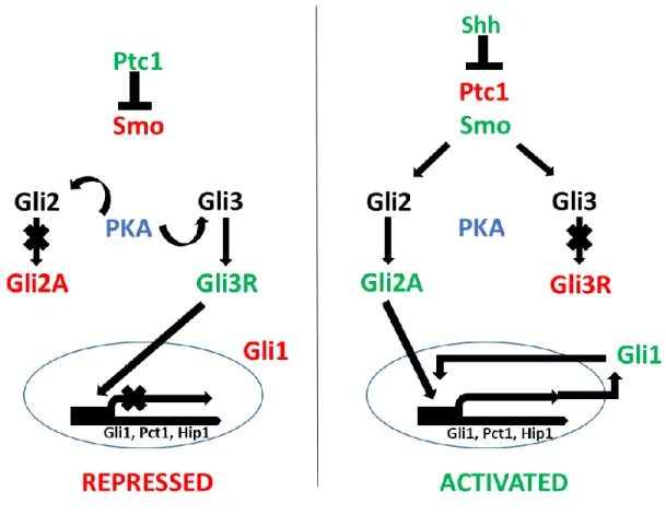
the surface of nearly all cells in mammalian tissues. Responding to mechanical and chemosensation, primary cilium are localization points for signaling receptors, ion channels and transporters. Acting at the primary cilium, Hh morphogens play essential roles in embryogenesis, cell proliferation and tissue development, and stem cell maintenance [44]. Specifically, at the activation of Hh signaling, Hh morphogens bind to the 12 pass-transmembrane receptor, Ptach (Ptch1) which is localized to the base of primary cilium, releasing its inhibition of Smoothened (Smo), a protein responsible for activating the downstream Hh pathway. Once Smo is activated, it binds to Sufu and induces nuclear translocation of Hh pathway transcription regulators, Gli1 (activator), Gli2 (activator and repressor) and Gli3 (repressor) [45, 46]. Gli1, Gli2 and Gli3
14
regulate the expression of downstream targets such as Gli1, Ptch1, cyclin D, and myc involved in cell survival, proliferation and differentiation (Figure 3) [47, 48].
Figure 3: Upstream regulation of the Gli transcription factors and their individual and combined roles in regulating Hh target gene expression. In the absence of the signal, Gli3 functions repressor of the Hh signaling pathway. When there is signaling, repression of Gli3 is
15
RELEASE AND TRANSPORT OF Hh THROUGH TISSUES
Despite its tight membrane association, Hh is able to affect patterning of distal tissues, acting directly over a long range in a time- and concentration-dependent manner [49]. The formation of the gradient of Hh activity emanating from the secreting cells is facilitated by multiple macromolecules, which control release, transport and sequestration of Hh. Hh is released from the secreting cell by Dispatched (Disp), a conserved protein that shares sequence similarity with transmembrane transporters [50, 51]. Subsequent transport of Hh through tissues requires heparin sulfate, as indicated by the failure of Hh transport in embryos lacking heparansulfate-synthesizing enzymes of the EXT/tout velu (ttv) family [51, 52]. The cholesterol modification of Hh also affects the range of Hh action by affecting its palmitoylation, stability, diffusion and/or transport [53-58]. Several other proteins that affect Hh transport and/or shape the Hh gradient have been described in different species. For example, in addition to the Hh receptor Patched (Ptc), which sequesters Hh and restricts its range of action in all species analyzed [51, 59], vertebrates have an additional
transmembrane protein, Hh-interacting protein (Hip), which binds to Hh proteins and reduces their range of movement [51, 60].
RECEIVING THE Hh SIGNAL
The binding of Hh to cells is facilitated by two classes of accessory receptor: the glypican-family of cell surface proteoglycans (e.g. dally-like in Drosophila) [61] and the transmembrane proteins iHog and Boi (CDO and BOC in vertebrates) [62, 63]. iHog and Boi also increase the binding
affinity of Hh for the signaling receptor Ptc, a 12- span transmembrane protein related to bacterial transmembrane transporters of the resistance-nodulation-division (RND) family. In the absence of Hh, Ptc catalytically inhibits the activity of the seven-transmembrane-span receptor-like protein
16
Smoothened (Smo) [64], potentially by affecting localization and/or concentration of a small
molecule. Smo activity can be modulated by many synthetic small molecules [65]. Of endogenous metabolites, oxysterol derivatives [66] and vitamin D3 derivatives [67] have been suggested to mediate the effects of Ptc on Smo. Binding of Hh to Ptc results in loss of Ptc activity, and consequent activation of Smo, which transduces the Hh signal to the cytoplasm [64, 68], ultimately leading to the activation of the Ci/GLI family of transcription factors [69, 70 and 71].
TRANSCRIPTIONAL TARGETS OF Hh SIGNALING
The Hh signaling response is mediated by the binding of the Ci/Gli1-3 transcription factors to a Gli-consensus binding sequence, ‘TGGGTGGTC’ [72, 73, 74 and 75], in promoter and enhancer
regions of target genes. The transcription factors act as both activators and repressors on the transcription of a number of genes that vary between organisms and tissues. There are several examples of graded responses to Hh signaling, often in conjunction with other signaling factors, including the establishment of the A/P boundary in wing disks and the tight segmental boundaries in Drosophila described above. In addition, several response elements and enhancers, in addition to the GLI consensus sequence, may regulate the expression of each specific gene target. In Drosophila, Hh targets genes include Dpp, Wg, Ptch, Col and En [76, 77]. Vertebrate targets
include components of the pathway, Ptch, Gli1 and Hip, as well as several proteins from various protein families including Bmp [78] Hox [78], Fgf [79], Myc [80], Cyclin [81], Vegf [82,83], Angiopoietin [83,84], and other proteins including Pdgfrα [85], Bcl-2 [86,87], Bmi1 [88], Wnt [89],
Hes1 [90], HNF-3b [75], Spop [91]. Gli-R represses target genes when Hh is absent, and while
17
transcriptional activation. In mice, a total of 42 genes have two or more Gli consensus binding sequences in the enhancer regions (Tab.3) [92].
18
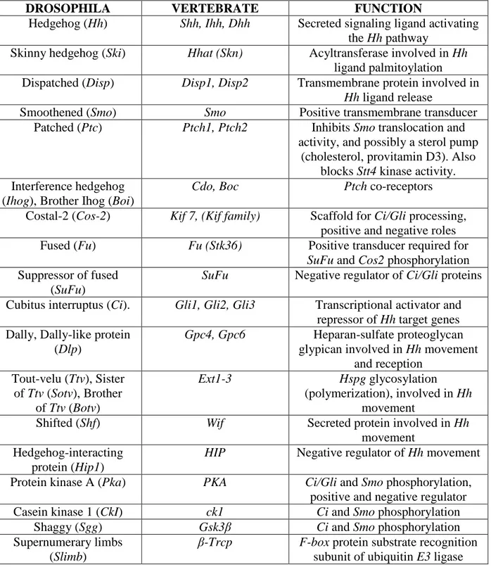
Table 3: Hedgehog pathway components in Drosophila and vertebrates
DROSOPHILA VERTEBRATE FUNCTION
Hedgehog (Hh) Shh, Ihh, Dhh Secreted signaling ligand activating the Hh pathway
Skinny hedgehog (Ski) Hhat (Skn) Acyltransferase involved in Hh ligand palmitoylation
Dispatched (Disp) Disp1, Disp2 Transmembrane protein involved in Hh ligand release
Smoothened (Smo) Smo Positive transmembrane transducer Patched (Ptc) Ptch1, Ptch2 Inhibits Smo translocation and
activity, and possibly a sterol pump (cholesterol, provitamin D3). Also
blocks Stt4 kinase activity. Interference hedgehog
(Ihog), Brother Ihog (Boi)
Cdo, Boc Ptch co-receptors
Costal-2 (Cos-2) Kif 7, (Kif family) Scaffold for Ci/Gli processing, positive and negative roles Fused (Fu) Fu (Stk36) Positive transducer required for
SuFu and Cos2 phosphorylation Suppressor of fused
(SuFu)
SuFu Negative regulator of Ci/Gli proteins
Cubitus interruptus (Ci). Gli1, Gli2, Gli3 Transcriptional activator and repressor of Hh target genes Dally, Dally-like protein
(Dlp)
Gpc4, Gpc6 Heparan-sulfate proteoglycan
glypican involved in Hh movement and reception Tout-velu (Ttv), Sister of Ttv (Sotv), Brother of Ttv (Botv) Ext1-3 Hspg glycosylation (polymerization), involved in Hh movement
Shifted (Shf) Wif Secreted protein involved in Hh movement
Hedgehog-interacting protein (Hip1)
HIP Negative regulator of Hh movement
Protein kinase A (Pka) PKA Ci/Gli and Smo phosphorylation, positive and negative regulator Casein kinase 1 (CkI) ck1 Ci and Smo phosphorylation
Shaggy (Sgg) Gsk3β Ci and Smo phosphorylation Supernumerary limbs
(Slimb)
β-Trcp F-box protein substrate recognition
19
There are Gli family of genes targets that act in feedback mechanisms on Hh pathway activity and the Hh protein. While Gli1 mediates an important positive feedback signal, the expression of Ptch and Hip reduce the movement of Hh ligands and retrains Hh signaling in a negative feedback loop. Factors involved in movement and reception of the Hh ligands, like Ihog/Boi, Cdo/Boc and Gas1 are down regulated in response to Hh signaling, also functioning as negative feedback to Hh pathway activation.
20 HUMAN GLI FAMILY
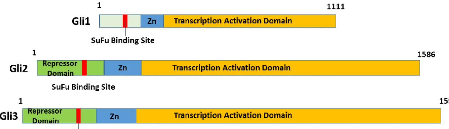
In vertebrates, there are three GLI (glioma-associated oncogene homolog) transcription factors, GLI1, GLI2 and GLI3, which mediate Hedgehog signalling [93-94]. The three isoforms contain
five zinc-finger DNA-binding domains, but their N-terminal domains exhibit important differences. GLI1 functions as a strong transcriptional activator. It lacks a repressor domain found in the N-terminus of GLI2 and GLI3. GLI1 expression also depends on GLI2 and/or GLI3-mediated transcription since it constitutes a direct target gene of the Hh pathway. Compared to GLI2, GLI1 exerts the largest part of activator functions by providing a positive feedback-loop. However, GLI2 represents the primary downstream activator and is indispensable to initiate activation of target genes of the Hedgehog pathway. Thereby, both factors display identical or very similar DNA binding specificities [95, 97-100]. By contrast, GLI3 exerts a role as a repressor (Fig.4) [96, 99].
21
GLI2 GENE
The GLI2 is a member of GLI zinc finger family of transcription factors along with GLI1 and GLI3. These zinc finger transcription factors are characterized by the consensus sequence
X3-Cys-X4-Cys-X12-His-X4-His-X3, where X is any amino acid. The zinc finger forms a compact globular structure that contains a β-sheet and an α-helix held together by a central Zinc ion. GLI2 is specifically recognized and binds to the 5’-GAACCACCCA-3’ motif in the target genes.
GLI2 is a 1586 – amino acid protein (197kDa) which is encoded by 13 exons on chromosome
2q14. In addition to the central zinc finger DNA binding domain consisting of 5 fingers, GLI2 proteins also contains an amino terminal (N-terminal) repressor domain and carboxyl terminal (C-terminal) transactivation domain (Fig.5).
In previous study showed that the region encoding GLI2 repressor domain is subject to alternative splicing in the gonadal tissues and different cell lines. The alternatively 5’ end of GLI2 mRNA splicing resulted in two major isoforms from skipping exon 3 (GLI2Δ3) or exons 4 and 5 (GLI2Δ4– 5 also known as GLI2-∆N). This both isoforms contain premature translational stop codons in the GLI2 open reading frame (ORF) starting from exon 2. Translation of GLI2Δ3 and GLI2Δ4–5
(GLI2-∆N) in vitro, initiated from downstream AUG codons, to produce N-terminally truncated proteins [119].
In GLI-dependent transactivation assay, expression of GLI2-Δ3 induced activation of the reporter gene similar to that of the GLI2-full-length construct containing complete ORF. However, expression of the GLI2Δ4–5 (GLI2-∆N) resulted in about 10-fold increase in activation, suggesting that deletion of the major part of repressor domain was responsible for the enhanced activation of GLI2 protein and study suggested that in addition to proteolytic processing, alternative splicing
22
may be another important regulatory mechanism for the modulation of repressor and activator properties of GLI2 protein. [119]
At least five different GLI2 isoforms are produced by alternative splicing of mRNA known as α (133kDa), β (131kDa), γ (88kDa), δ (86kDa) and GLI2 full-length [100, 107, 108-110] (Tab.4&5). The Gli2-α also known asGLI2-∆N variant which lacks the N-terminal repressor domain shows a 30-fold higher reporter activity compared with the full length protein in vitro.
23
Table 4: GLI2 Isoforms with missing amino acid positions, length and mass.
GLI2 ISOFORMS
NAME
MISSING AMINO ACID POSITIONS
LENGTH AND MASS OF ISOFORMS
GLI2 Alpha or GLI2-∆N 1-328 1258 amino acids & 133kDa
GLI2 Beta 1-328 & 394-410 1241 amino acids & 131kDa
GLI2 Gamma 1-328, 1149-1157 & 1158-1586 829 amino acids & 88kDa GLI2 Delta 1-328, 394-410, 1149-1157 &
1158-1586
812 amino acids & 86kDa
GLI2 Full-length No missing 1586 amino acids & 167kDa
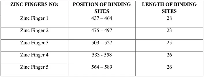
Table 5: GLI2 Zinc Finger positions and length of binding site.
ZINC FINGERS NO: POSITION OF BINDING SITES LENGTH OF BINDING SITES Zinc Finger 1 437 – 464 28 Zinc Finger 2 475 – 497 23 Zinc Finger 3 503 – 527 25 Zinc Finger 4 533 - 558 26 Zinc Finger 5 564 – 589 26
24
GLI2 IN THE DEVELOPMENT OF PITUITARY
As an effector molecule of the sonic hedgehog (SHH) signalling pathway, GLI2 has a fundamental role in the development. Sonic hedgehog is a morphogen expressed in the early steps of pituitary ontogenesis by exerting effects on both proliferation and cell-type determination. SHH is expressed in the ventral diencephalon and throughout the oral ectoderm except Rathke’s pouch [17, 101]. However, the patched receptor (PTCH1) as well as the GLI family of zinc finger transcription factors are expressed in the Rathke’s pouch, indicating that the developing gland is competent to receive and respond to SHH signalling [102].
The Hh pathway (Fig.3) is considered as the canonical pathway through which GLI2 activity is regulated. The Hedgehog ligands binds to and activate the transmembrane receptor called patched (PTCH). When the Hh ligand is absent, PTCH exerts a consistent inhibitory effect on transmembrane G-protein coupled receptor smoothened (SMO). When the Hh ligand is present and binds to PTCH, inhibition over SMO is released [59, 68]. The GLI2 transcription factors are bound with SuFu which keeps GLI2 tethered in the cytoplasm [103]. Activated SMO triggers the dissociation of SuFu/GLI2 complex and allowing the nuclear translocation and activation of GLI2. This translocation promotes the subsequent DNA binding and transcription of a series of Hh pathway target genes.
Multiple studies using knockout mice has been performed to study the importance of Gli2 in the development. Mice with homozygous loss of function Gli2 mutation resulted in lethal phenotype later in development while the heterozygous mice developed normally [102].
The phenotypic evaluation of abnormalities in the knock out mice showed severe skeletal abnormalities including absence of vertebral body and intervertebral disc, truncated mandibles
25
with absent incisors, shortened limbs and sternum, missing tympanic ring bones of the inner ear and severe cleft palate malformations [102]. Gli2 deficient mice also showed defects in the pituitary development including partial loss of anterior and complete loss of posterior pituitary [104, 105]. These defects were attributed by the loss of expression of Gli2 target genes Bmp4 and Fgf8 [104].
GLI2 IN CPHD DISEASES
SHH and to lower extent GLI2 mutations were initially reported in patients with
Holoprosencephaly (HPE), a severe neurological characterized by incomplete or failed forebrain separation, or HPE-like phenotypes with pituitary anomalies and postaxial polydactyly [118]. As the SHH pathway is also involved in pituitary development, mutations in SHH and GLI2 have been subsequently searched in CPHD patients. Franca et al [20] reported novel heterozygous frame-shift and nonsense GLI2 mutations and considerable frequency of missense GLI2 variants in patients with congenital hypopituitarism without HPE and most of these patients presented with CPHD and an ectopic posterior pituitary lobe.
More recently, individuals with truncating mutations in GLI2 were reported with the presence of typical pituitary anomalies, polydactyly and subtle facial features rather than HPE [115]. In all the patients so far identified carrying GLI2 mutations, the pattern of inheritance was dominant with incomplete penetrance and variable phenotype [20, 115].
It has to be considered that GLI2 is a large and highly polymorphic gene with several rare variations reported in the exome server database (http://evs.gs.washington.edu/EVS/). Thus especially for the missense variants it is quite difficult to assess the pathogenicity in the absence of functional studies.
26
27 AIM
The aim of this study was to determine the frequency of GLI2 mutations in a cohort of Italian CPHD patients that resulted negative for mutations in other causative genes encoding pituitary transcription factors (PIT1, PROP1, HESX1, LHX3, and LHX4). Moreover, in the case of missense mutations, to discern between polymorphic variants and causative mutation we settled a series of in-vitro functional study aimed to evaluate the modifications induced by the different variants on
28
29 SUBJECTS
One hundred and thirty-six CPHD patients were recruited based on the following criteria:
1) They presented with a clinical and hormonal evidence of childhood-onset GH deficiency combined with at least one other pituitary defect in the absence of an identified cause of hypopituitarism (e.g. cerebral tumors, cranial trauma, documented asphyxia, or other injuries at delivery).
2) Mutations in the coding sequences of genes associated with multiple pituitary hormone dysfunctions (PIT1, PROP1, HEXS1, LHX3, and LHX4) had been previously excluded.
Mean height SDS for chronological age was calculated using the criteria of Tanner-Whitehouse method [111]. The mean height of the patients at diagnosis was −2.26 SDS ± 2.3 sd. Morphological evaluation of the hypothalamus-pituitary area and/or of the central nervous system was performed in 136 patients by magnetic resonance imaging, using precontrast coronal spin echo T1-weighted images followed by postgadolinium T1-weighted imaging. Among the 136 CPHD index cases, 8 (5.8%) were the probands of pedigrees with more than one affected individual (familial cases). Four patients were born from consanguineous parents but they were considered as sporadic cases since they were the only affected subject in their families. The mean height of these patients at diagnosis was -2.81 ±1.83 SDS and the mean delay in bone age relative to chronological age was 2.57± 2.36 years. GHD was present in all the patients, TSH deficiency in 78.6% (107/136) and ACTH deficiency in 61% (83/136). Thirty-nine subjects were Prepubertal at the time of diagnosis. Among the remaining 97 subjects that could be evaluated in terms of pubertal age, 81 (83.5%) presented with FSH/LH deficiencies. Eight male patients presented neonatal micropenis and/or cryptorchidism. Five patients (3.5%) had diabetes insipidus. We obtained MRI data from 101
30
patients (74% of the total). Among these, abnormalities (ectopy of the neurohypophysis, pituitary hypoplasia and empty sella) were found in 81 (80%) subjects; in particular, anterior pituitary hypoplasia or aplasia was the most frequent abnormality and was present in 61 patients (60.4%), while pituitary stalk interruption and/or neuropediatric ectopia were observed in 35 patients (34.6%), 16 of them presenting both abnormalities. Eleven patients (10.8%) presented also extra-pituitary abnormalities such as SOD, other midline defects or cerebellar abnormalities.
Patients or parents of the patients under 18 years of age gave their written informed consent to participate to this study, which was approved by the local ethical committee of each contributing auxological center.
31
32 SCREENING GLI2 GENE
The entire coding region of GLI2 (13 exons and exon-intron boundaries) was PCR amplified from peripheral blood genomic DNA by 18 couple of primers designed for separate fragments (Tab.6). The PCR products were visualized on a 2% Agarose gel and purified using Exo/SAP-IT enzymatic PCR clean up system (Affymetrix). The Purified products were then sequenced with Big Dye Terminator kit (Applied Biosystems, Foster City, CA) and automatic sequencer ABI PRISM 3100 Genetic Analyzer (Applied Biosystems, Foster City, CA).
Table 6: To examine all coding regions of GLI2 gene 18 pairs of following primers designed.
EXONS FORWARD PRIMERS REVERSE PRIMERS
EXON 1 5’-TGGGTTTGGGCTCAGTGT-3’ 5’-CCTCTTCGCCCTCCATAAAC-3’ EXON 2 5’-TGGCTGCTCTTGCTATGAAA-3’ 5’-GCAGGAGATGTGGCTGAGG-3’ EXON 3 5’-CATGTTGGTTTTGGGGTCTT-3’ 5’-GACCAAGGCTGAGGAGTTGA-3’ EXON 4 5’-CCAGGTGTGCATTTCTCTCTG-3’ 5’-TTGTCCCCAAAAGAAACAGC-3’ EXON 5 5’-CCTTGCAGGCTCTTCCTATC-3’ 5’-TCTTTCTCCTCGGGTCAAAA-3’ EXON 6 5’-TGGGCAAGGTTCTCTCTGTC-3’ 5’-CTTAGCATGAGCTGGCAGTG-3’ EXON 7 5’-TGTGCGGAGAGATCCTAGAG-3’ 5’-TTCACCACCAAGGGTACAGC-3’ EXON 8 5’-TTCCCCACAGCACTTCGAT-3’ 5’-TCCAGCCCCTTCTGTCTAGT-3’ EXON 9 5’-GACAGCAGGGGGTGGTCT-3’ 5’-CCACCTCCAAACATGATCC-3’ EXON 10 5’-GGTTGGAGCAGAGCAGAGAA-3’ 5’-GGCACCTGGCTATCTACTGG-3’ EXON 11 5’- CGTGGGTAGCTTCAGGAGAA -3’ 5’-GATATCGCTGTGCCCCTAGA-3’ EXON 12 5’-GCCTGTGCAGGCCTAGAG-3’ 5’-GTGGGTGCCAGCCTAGTTG-3’ EXON 13.1 5’-GTGTTGCAAGCCCTCTTCTC-3’ 5’-AGTGGCTGCCGCGTACTT-3’ EXON 13.2 5’-AGCAGTACAGCCTGCGGGCCAAGTA-3’ 5’-CTCCATCGCCACGTTCTCGCT-3’ EXON 13.3 5’-CTTCCACAGCACCCACAAC-3’ 5’-CCTTGCGGACTGTAGCCC-3’ EXON 13.4 5’-GCAGTGGAATGAGGTGAGCT-3’ 5’-GATGGCTCTGCTGTGGGTAG-3’ EXON 13.5 5’-CCCTCAGCAGACAGAAGTGG-3’ 5’-GTACATGTGGATCTGGCCGT-3’ EXON 13.6 5’- CAGTCAGGAAACAGCAGAGG-3’ 5’-GGAAAAAGACAAGACAGCTGGA-3’
GENOMIC DNA EXTRACTION
Genomic DNA was extracted from whole blood samples using salting out method based on Miller et al. [112].
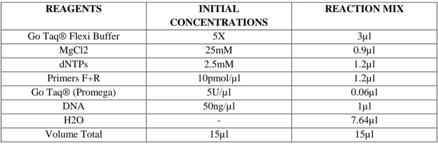
33 POLYMERASE CHAIN REACTION
The PCR reaction was carried out with the GoTaq Flexi DNA polymerase (Promega) in a 15µl reaction volume, with touchdown protocol from 650 C to 550 C annealing temperatures. The initial denaturation at 94°C for 5 min, 20 cycles consisting of 30s denaturation at 94° C, 30s annealing at higher temperature 65° C and 30s extension at 72° C, followed by second cycle consisting 25 cycles of denaturation at 940 C for 30s, annealing at lower temperature 550 C and extension at 720 C, followed by a final extension at 72°C for 7 minutes and cooled to 40C (Tab.7&8).
Table 7: Reagents used for Polymerase chain reaction fragments 1-18 except 4 and 13.2.
REAGENTS INITIAL
CONCENTRATIONS
REACTION MIX
Go Taq® Flexi Buffer 5X 3µl
MgCl2 25mM 0.9µl
dNTPs 2.5mM 1.2µl
Primers F+R 10pmol/µl 1.2µl
Go Taq® (Promega) 5U/µl 0.06µl
DNA 50ng/µl 1µl
H2O - 7.64µl
34
Table 8: Thermal cycle conditions for fragments 1 – 18 except fragment 4 and 13.2.
STEPS TEMPERATURES TIME NUMBER OF
CYCLES
Initial Denaturation 94°C 5 Minutes 1 Cycle
Denaturation 94°C 30 Seconds
20 cycles Annealing (High Temp.) 65°C 30 Seconds
Extension 72°C 30 Seconds
Denaturation 94°C 30 Seconds
25 cycles
Annealing (Low Temp) 55°C 30 Seconds
Extension 72°C 30 Seconds
Final Extension 72°C 7 Minutes ̶
Rest 4°C ∞ ̶
The exon 4 and exon 13.2 fragment which is GC rich region we modified the PCR reaction conditions by adding 5% DMSO and making final reaction volume to 20µl, with touchdown protocol from 640 C, 610 C, 580 C and 570 C annealing temperatures. The initial denaturation at 940 C for 2mins, 3 cycles consisting of 10s denaturation at 940 C, 10s annealing at higher temperature 640 C and 30s extension at 720 C, followed by second, third and fourth cycles consisting 3 cycles of denaturation, annealing and extension at 610 C, 580 C and 570 C, followed by final extension at 720 C for 5mins and cooled to 40 C (Tab.9&10).
35
Table 9: Reagents used for Polymerase chain reaction fragments 4 and 13.2.
REAGENTS INITIAL
CONCENTRATIONS
REACTION MIX
Go Taq® Flexi Buffer 5X 4µl
MgCl2 25mM 1.2µl
dNTPs 2.5mM 1.6µl
Primers F+R 10pmol/µl 2µl
DMSO 5% 1.2µl
Go Taq® (Promega) 5U/µl 0.06µl
DNA 50ng/µl 1µl
H2O - 8.94µl
Volume Total 20µl 20µl
Table 10: Thermal conditions for Fragment 4 and 13.2.
STEPS TEMPERATURES TIME NO OF CYCLES
Initial Denaturation 94°C 2 Minutes 1 Cycle
Denaturation 94°C 10 Seconds
3 Cycles Annealing (1st Temp) 64°C 10 Seconds
Extension 72°C 30 Seconds
Denaturation 94°C 10 Seconds
3 Cycles Annealing (2nd Temp) 61°C 10 Seconds
Extension 72°C 30 Seconds
Denaturation 94°C 10 Seconds
3 Cycles Annealing (3rd Temp) 58°C 10 Seconds
Extension 72°C 30 Seconds
Denaturation 94°C 10 Seconds
40 Cycles Annealing (4th Temp) 57°C 10 Seconds
Extension 72°C 30 Seconds
Final Extension And rest at 4°C.
36 EXO/SAP PURIFICATION
After the reaction all the PCR products were visualized on a 2% agarose gel and purified using Exo/SAP-IT enzymatic PCR clean up system (Affymetrix) (Tab.11).
Table 11: Reagents used for Exo/SAP purification.
REAGENTS THERMAL CONDITIONS VOLUMES
PCR Product 37⁰C – 15 Mins 80⁰C - 15 Mins 4⁰C - ∞ 1µl Exo/SAP mix 5µl Total Volume 6µl SEQUENCING
The purified products were directly sequenced in the forward or reverse direction with Big Dye Terminator kit (Applied Biosystems) and analyzed on an ABI PRISM 3100 Genetic Analyzer (Applied Biosystems) (Tab.12)
Table 12: Reagents used for Sequencing Reaction.
REAGNETS THERMAL CONDITIONS VOLUMES
Purified PCR products Steps Denaturation Annealing Extension Rest Temp 96⁰C 50⁰C 60⁰C 4⁰C Time 15 secs 5 secs 4 mins ∞ Cycles 25 2µl Primer (3.2µM) 1µl
Big Dye Terminator Mix 1µl
37 SITE DIRECTED MUTAGENESIS
The GLI2-cDNA incorporated pCS2-MT expression vectors used for site directed mutagenesis is commercial available. Size of pCS2-MT is 4.3KB and size of GLI2-cDNA is 4.8KB totally 9.1KB plasmid size. Two kinds of construct were used for study one pCS2-GLI2FL (Addgene plasmid #17648) and another one pCS2-GLI2∆N (Addgene plasmid #17649) [100]. GLI2∆N isoform is activated form of GLI2 gene and its expression is higher than that of GLI2-FL construct in Luciferase assay.
Variation p.Y575H:
We generated mutagenized GLI2-FL and GLI2-∆N constructs by using Stratagene QuikChange® Site-Directed Mutagenesis Kit and mismatch complementary primers containing desired mutation.
Primers used for Mutagenesis:
Forward GLI2_575HisFN 5’-CTGCACCAAGAGACACACAGACCCCAGCTC-3’
Reverse GLI2_575HisRN 5’-GAGCTGGGGTCTGTGTGTCTCTTGGTGCAG-3’
Table 13: Reagents used for Mutagenesis for p.Y575H
REAGENTS VOLUMES BUFFER (10x) 5µl dNTPs 6µl MgSO4 1µl Forward Primer (10mM) 1.5µl Reverse Primer (10mM) 1.5µl PCR Enhancer (10x) 5µl Pfx polymerase (2.5 U/µl) 1µl H2O 27µl Plasmid DNA 2µl (50ng/µl)
38
Table 14: Thermal conditions for Mutagenesis of p.Y575H
CYCLE STEPS TEMP. TIME NUMBER OF
CYCLES
Initial Denaturation 98⁰ C 5 mins 1
Denaturation 98⁰C 10 secs
25
Annealing 53⁰C 1 min
Extension 68⁰C 9 mins (1 min/kb)
Rest 10⁰C ∞ -
Variation p.A593V:
We generated mutagenized GLI2-FL and GLI2-∆N constructs by using Stratagene QuikChange® Site-Directed Mutagenesis Kit and mismatch complementary primers containing desired mutation.
Primers used for Mutagenesis:
Forward GLI2_593ValF 5’-GGTCCACGGCCCAGATGTCCACGTCACCAAGAAGC 3’
Reverse GLI2_593ValR 5’-GCTTCTTGGTGACGTGGACATCTGGGCCGTGGACC 3’
Table 15: Reagents used for Mutagenesis for p.A593V
REAGENTS VOLUMES BUFFER (10x) 5µl dNTPs 6µl MgSO4 1µl Forward Primer (10mM) 1.5µl Reverse Primer (10mM) 1.5µl PCR Enhancer (10x) 5µl Pfx polymerase (2.5 U/µl) 1µl H2O 27µl Plasmid DNA 2µl (50ng/µl)
39
Table 16: Thermal conditions for Mutagenesis of p.A593V
CYCLE STEPS TEMP. TIME NUMBER OF
CYCLES
Initial Denaturation 94⁰ C 5 mins 1
Denaturation 94⁰C 15 secs
25
Annealing 58⁰C 1 min
Extension 68⁰C 9 mins (1 min/kb)
Final Extension 68⁰C 2 mins 1
Rest 10⁰C ∞ -
Variation p.P386L:
We generated mutagenized GLI2-FL construct by using New England BioLabs® Q5 Site-Directed Mutagenesis Kit and mismatch complementary primers containing desired mutation.
Primers used for Mutagenesis: 5’ Phosphorylated Primers:
Forward GLI2-386Leu-FN 5’-GAGGGCCTGCGGCTGGCCTCCCCTCTGG-3’ Reverse GLI2-386Leu-RN 5’-AGGCTCGGTCTTGACCTTGCTGCGCTTG-3’
Table 17: Reagents used for Mutagenesis for p.P386L
REAGENTS VOLUMES Q5 BUFFER (5x) 5µl d-NTP’S (10mM) 0.5µl Forward Primer (10mM) 1.25µl Reverse Primer (10mM) 1.25µl GC Enhancer 5µl
Q5 Hot Start polymerase (0.02 U/µl) 0.25µl
H2O 10.75µl
40
Table 18: Thermal conditions for Mutagenesis of p.P386L
CYCLE STEPS TEMP. TIME NUMBER OF
CYCLES
Initial Denaturation 98⁰C 30 secs 1
Denaturation 98⁰C 10 secs
25
Annealing 72⁰C 45 secs
Extension 72⁰C 8 mins
Final Extension 72⁰C 5 mins 1
Rest 10⁰C ∞ -
The PCR product obtained by this site directed mutagenesis is linear product to circularize the PCR product we performed T4 ligation reaction.
Table 19: Reagents and thermal conditions for T4-Ligation.
REAGENTS VOLUMES INCUBATION TIME & TEMP.
T4 ligation buffer 2µl
16⁰C for Overnight
T4 ligase 1µl
H2O 12µl
Site directed mutagenesis product 5µl
Total Volume 20µl
BACTERIAL TRANSFORMATION:
After the reaction, the products were digested with DpnI. DH5α competent cells were transformed with the different mutagenized constructs and grown on Luria Broth/ampicillin media. After selecting the correct clones by colony PCR, the plasmid DNA was isolated using Miniprep and Maxiprep kit (QIAGEN). The desired mutations were confirmed by sequencing.
41 CELL CULTURE AND TRANSFECTION:
NIH-3T3 mouse fibroblast cell line was used for the transfection experiments [113, 114]. The stock culture was grown in DMEM High Glucose (Gibco-Life Technologies) each supplemented with 10% fetal calf serum and 1% Penicillin/Streptomycin in 5% CO2 at 37⁰C. A day before transfection 1x10⁵ cells were seeded into each well of a 24-well tissue culture plate in 500µl CGM (Complete Growth Medium). The wells were previously treated with 1:10 dilution Poly-L-lysine solution (Sigma Aldrich) to allow the cells to completely adhere to the plate surface. At 70%-90% confluency, cells were tranfected Mutagenized plasmid constructs, reporter plasmid, and as internal control pRL-TK renilla plasmids with Lipofectamin 2000 transfection reagent (Life Technologies). Green Fluorescent protein construct was used to test transfection efficiency. Whole cell lysate was collected for Luciferase assay after 48hrs of transfection and all assays were performed in triplicate.
CELL LYSATE
After 48 Hrs completely medium removed and washed with PBS solution then 1x PLB solution added to the respective wells and plate incubated at 37⁰C for 15 mins. The detached cells along with PLB collected and centrifuged at 13,000 rpm for 5 mins. Supernatant was transferred to another Eppendorf tube which is debris free used for assay.
42
43 RESULTS:
We identified 5 mutations in 5 subjects in a cohort of 136 patients with CPHD (3.6%). All the following mutations (Fig.6) were at the heterozygous state:
1) p.P386L (c.1157 C>T) falls within the repressor domain.
2) p.Y575H (c.1723 T>C) is within the Zinc Finger Binding domain.
3) p.A593V (c.1778 C>T) is located at the junction of Zinc Finger Binding Domain and Transactivation Domain.
4) p.V1111Gfs*19 (c.3332delT) is located in the Transactivation domain. It is a single nucleotide deletion of a T that causes a frameshift and the presence of a premature stop codon 19 amino acids downstream. This mutation removes 475 amino acids of the activator domain.
5) p.R1226* (c.3676 C>T)is located in the Transactivation domain. Also in this case the C-terminal part of the activator domain is removed [360 amino acids] (Fig.6 – Tab.20).
44
In table 20 are reported the characteristics of the identified mutation and the results of in-silico analysis performed by PolyPhen, PROVEAN and SIFT prediction tools for the missense mutations. All the three mutations are predicted as probably damaging.
Table 20: Summary of Gli2 mutation Identified in this study, Allele Frequency Data from dbSNP
Amino acid Position Nucleotide Position dbSNP ID Allele Frequency (%)
PolyPhen PROVEAN SIFT
P386L c.1157 C>T rs757467621 0.0001% Probably damaging Probably damaging NA Y575H c.1723 T>C rs763503195 NA Probably damaging Probably damaging Probably damaging A593V c.1778 C>T rs771880068 0.0001% Probably damaging Probably damaging NA R1226* c.3676 C>T NR - - - - V1111Gfs*19 c.3332delT NR - - - -
45 PATIENTS DETAILS:
Here are reported the data of the patients carrying a mutation at the time of the diagnosis (in parenthesis is indicated the mutation for each patient).
Patient A (p.P386L) – This patient was a 11.3 years old boy born at term with appropriate for
gestational age. The parents were non-consanguineous with normal stature. The height at the diagnosis was -2.6 SDS. The patient showed GH, TSH and ACTH deficiency and no extra pituitary features (Tabl.21).
Patient B (p.Y575H) - This patient was a 9.4 years old boy born at term with no perinatal
complications. The bone age was delayed by 1.8 years at diagnosis and the height was -2.2 SDS. The parents of the patient were non-consanguineous. The father was normal whereas the mother was short stature and presented with polydactyly but, not evaluated for pituitary hormone levels. The patient showed GH, TSH and ACTH deficiency. Patient’s Pituitary and cerebral imaging showed anterior pituitary hypoplasia. Extra pituitary manifestations were, polydactyly, cranio facial abnormalities and hypercholesterolemia. As expected the mutation was inherited from the affected mother (Fig.7 – Tab.21).
Mother +/- Father +/+
Patient B +/- Brother +/+
Wild type Mother and Mutated patient - Wild type Father and Brother - Figure 7: Family tree of Patient B.
46
Patient C (p.A593V) - This patient was a female born at term and appropriate for gestational age.
It was a sporadic case and was born from a non-consanguineous parents. The height at the time of the diagnosis was -2.1 SDS. The patient showed GH, TSH, ACTH, LH, and FSH deficiency. This patient did not show extra pituitary features. The DNA of the parents was not available (Tab.21).
Patient D (p.R1226*) - This patient was a 3 years male, born at term with appropriate for
gestational age, the parents were non-consanguineous with normal stature (their DNA was not available). The height at the diagnosis was -1.8 SDS. The patient showed GH, TSH, and ACTH deficiency. The pituitary and cerebral imaging showed anterior pituitary hypoplasia (Tab.21).
Patient E (p.V1111Gfs*19) - This case was a female born from non-consanguineous parents, and
was a sporadic case. The patient height was appropriate for gestational age and at term. The condition was identified at the age of 10.9. The height at the time of diagnosis was -1.6 SDS. The patient showed GH, TSH, ACTH, LH, and FSH deficiency. Pituitary and cerebral imaging of the patient showed anterior pituitary hypoplasia. Other pituitary manifestations included congenital malformation syndrome, myopia and intellectual disabilities The analysis on the parent’s DNA revealed that this mutation was “de novo” as none of them carried it (Tab.21).
47
Table 21: Clinical characteristics of CPHD patients identified in this study with GLI2 Mutation.
AGA – Appropriate for gestational age, NA – Not available, D- Deficiency of the evaluated pituitary axis, PP- Prepubertal status at diagnosis and APH – Anterior pituitary hypoplasia.
Patient ID Patient A Patient B Patient C Patient D Patient E
Mutation p.P386L p.Y575H p.A593V p.V1111Gfs*19 p.R1226*
Sex Male Male Female Female Male
Sporadic/familial Sporadic Familial Sporadic Sporadic Sporadic
Consanguinity Yes/No No No No No No
Birth data At term, AGA At term, AGA At term, AGA At term, AGA
Age at diagnosis, year 11.3 9.4 28 10.9 3
Height SDS at diagnosis
-2.6 -2.2 -2.1 -1.6 -1.8
Bone age delay at diagnosis, year 1.8 NA NA NA GH TSH ACTH LH, FSH PRL D D D D D D PP NA D D D D NA D D D D NA D D D PP NA Pituitary and cerebral
imaging
APH, EP NA APH, EP APH, EP
Other Clinical Characteristics Polydactyly, Craniofacial abnormalities and Hypercholesterolemia NA Congenital Poly-malformative syndrome, mental retardation and myopia.
48 FUNCTIONAL ANALYSIS:
To evaluate the role of the three missense mutations identified in CPHD patients a functional test (dual luciferase assay) was performed. For the nonsense and frameshift mutations no assay was settled as they are expected to produce C-terminal truncated non-functional protein. Two types of plasmids, one bearing the GLI2-cDNA and the other bearing the luciferase reporter gene, were used.
The plasmids bearing the GLI2-cDNA were of two types: one with the GLI2-Full length cDNA another with GLI2-∆N cDNA, lacking the repressor NH2-terminal domain, both inserted in the pCS2-MT vector (Fig.8). These two types of cDNA correspond to two different isoforms of the GLI2 protein as previously mentioned (see introduction). In vertebrate the GLI2 gene undergoes
alternative splicing to produce different isoforms, of which GLI2-∆N is the most important in the SHH pathway as it acts as an activator whereas the GLI2-full length gene (that maintains the
repressor domain) acts as a repressor.