R E S E A R C H A R T I C L E
Open Access
Arthroscopic debridement of the ankle for
mild to moderate osteoarthritis: a midterm
follow-up study in former professional
soccer players
Leonardo Osti
1, Angelo Del Buono
2and Nicola Maffulli
3,4*Abstract
Background: The aim of this study is to report the clinical and functional outcomes following arthroscopic management of anterior impingement, grade III–IV cartilage lesions, and mild to moderate osteoarthritis of the ankle in former soccer players.
Methods: The study included 15 former male professional soccer players with mild to moderate degenerative changes of the ankle who had undergone arthroscopic debridement and management of secondary injuries of the ankle. Preoperatively and at the last follow-up, at an average of 7.4 years, the American Orthopaedic Foot and Ankle Society (AOFAS) and the Kaikkonen scales and visual analogue scale (VAS) assessment were administered to all patients. Ankle osteoarthritis was assessed from weightbearing anteroposterior and lateral radiographs of both ankles. Results and discussion: At the last follow-up, the average AOFAS score had increased significantly from 48 (range, 29–69) to 86 (range, 63–94) (P < 0.0001), with good to excellent scores in 11 patients (74 %). The average Kaikkonen preoperative score of 43 (range, 28–70) had significantly improved to 85 (range, 61–95) (P < 0.0001), with good excellent scores in 11 patients (74 %). VAS values were also improved at the last follow-up. At the last appointment, only one (7 %) patient had abandoned altogether any sport, as he did not feel safe with his ankle and he felt too old to continue.
Conclusions: Anterior ankle arthroscopy for management of mild to moderate ankle arthritis is safe, effective, and low cost and allows former athletes to safely return to ordinary daily activities and recreational sport activities.
Background
Joint space narrowing, osteophytes, cartilage lesions, and loose bodies are all features of ankle arthritis. This con-dition is post-traumatic in 70–80 % of instances, espe-cially in athletes [1]. In soccer players, repetitive stresses and twisting injuries, even without frank traumatic in-sults, may cause it [2, 3], leading to “footballer ankle”, with anterior and posterior bone spurs, fibrous tissue impingement, and ligamentous instability [4].
At present, there is no consensus on the best manage-ment of this condition. Supramalleolar osteotomy, dis-traction arthroplasty, ankle arthrodesis, or total ankle replacement have all been performed successfully in pa-tients with end-stage osteoarthritis [5]. In the early stages, in young patients, ankle arthroscopy may well improve symptoms and function, and prevent or delay major surgery [6]. Even though arthroscopy may be indi-cated for the management of early arthritis, there is little information about its effectiveness in patients with frank osteoarthritis [7, 8]. This study reports the clinical and functional outcomes of former soccer players who had undergone arthroscopic management for anterior im-pingement, grade III–IV cartilage lesions, and mild to moderate osteoarthritis of the ankle.
* Correspondence:[email protected]
3Department of Musculoskeletal Disorders, Faculty of Medicine and Surgery,
University of Salerno, Salerno, Italy
4Centre for Sports and Exercise Medicine, Barts and The London School of
Medicine and Dentistry, Mile End Hospital, 275 Bancroft Road, London E1 4DG, UK
Full list of author information is available at the end of the article
© 2016 Osti et al. Open Access This article is distributed under the terms of the Creative Commons Attribution 4.0 International License (http://creativecommons.org/licenses/by/4.0/), which permits unrestricted use, distribution, and reproduction in any medium, provided you give appropriate credit to the original author(s) and the source, provide a link to the Creative Commons license, and indicate if changes were made. The Creative Commons Public Domain Dedication waiver (http://creativecommons.org/publicdomain/zero/1.0/) applies to the data made available in this article, unless otherwise stated.
Methods
This is a retrospective study including 15 male patients with mild to moderate degenerative changes of the ankle who had undergone arthroscopy from 2001 to 2010 at the Department of Arthroscopy and Minimally invasive surgery, Hesperia Hospital, Modena, Italy. The inves-tigation was performed after approval of the Ethics Committee of the Hesperia Hospital, Modena, Italy. Patients were selected according to the following cri-teria: arthritis of the ankle with osteophytes and nar-rowing with no disappearance of the joint space (grade I–II degenerative changes according to the classification by Krips et al. [9]), major anterior im-pingement, full sized cartilage lesion, loose bodies, limited motion, inability to practice loading sports, pain and discomfort in daily activities, and unrespon-sive to conservative treatment. Exclusion criteria in-cluded complete obliteration of the ankle joint space, reflex sympathetic dystrophy (RSD) or extensive area of bone marrow edema, posterior impingement re-quiring posterior arthroscopy of the ankle, body mass index (BMI) over 30, cavus/varus foot deformity, and history of ankle fracture.
Of 108 patients who underwent ankle arthroscopy in the period 2001–2010 at our institution, 15 patients met the inclusion criteria, and were enrolled in the study: they had all been professional soccer players who had competed at least at county level, and whose main source of income had derived from soccer. The first au-thor (LO) made the diagnosis in all patients by history, clinical examination, MRI findings, and confirmed it at arthroscopy in all instances. The anterior drawer test was evaluated bilaterally clinically, to exclude hypermo-bility. With the patients supine and the ankle in neutral position, a side-to-side difference in tibio-talar transla-tion less than 5 mm was considered normal (grade 0), a side-to-side difference 5 to 10 mm was classified as grade I, a side-to-side difference 10 to 15 mm was classi-fied as grade II, and a side-to-side difference greater than 15 mm was classified as grade III.
We considered the anterior drawer test as normal when a hard stop could be appreciated, and pathological when the stop was absent or soft. At anterior drawer testing, eight patients were normal, and seven presented grade 1 laxity. All patients underwent plain anteropos-terior and lateral radiographs of the ankle and foot and, for objective evaluation, the American Orthopaedic Foot and Ankle Society (AOFAS) ankle-hindfoot scale [10], the Kaikkonen et al. ankle scoring scale [11], and the vis-ual analogue scale (VAS) assessment were administered to each patient.
At the final assessment, a clinical research fellow (ADB) who had not been involved in the original man-agement of the patients performed all the examinations,
administered all the tests, and examined and classified all the images.
Surgery
The first author (LO) performed all surgical procedures, under general anesthesia, in the supine position, with manual or temporary body traction of the ankle, without a tourniquet. A standard knee arthroscope and two standard anteromedial and anterolateral portals were used, switching to a 2.7 mm 30° angled arthroscope if needed. At arthroscopy, cartilage lesions were classified according to Outerbridge (Fig. 1). All the structures were palpated with a probe to assess osteochondral and bony features, and the presence of soft tissue impingement.
Appropriate bony (Fig. 2) and soft tissue debridement (Fig. 3) was performed to address anterior impingement in all patients using both a mechanical shaver and a ra-diofrequency probe. Loose bodies were removed in the seven patients; microfractures were undertaken in all in-stances for management of grade III–IV cartilage lesions. Specifically, after preparation of the cartilage bed, the lesion was measured using a scaled probe. An awl (Condropick, Arthrex, Naples, FL, USA) was introduced with the tip perpendicular to the bed of the lesion (Fig. 4), and the holes were made as described by Steadman, 3 to 4 mm apart and about 2 to 4 mm deep, starting from the center of the lesion to proceed periph-erically. The residual stability of the cartilage was assessed, and the irrigation was temporarily stopped to ascertain that marrow fat droplets and blood came out from the holes (Fig. 5).
After surgery, all patients followed a homogeneous re-habilitation program. Specifically, active and passive mo-tion exercises were started the day after surgery; partial weightbearing with crutches was prescribed for 6 weeks.
Fig. 1 Arthroscopic view of grade IV cartilage lesion of the talus Osti et al. Journal of Orthopaedic Surgery and Research (2016) 11:37 Page 2 of 7
Assisted physiotherapy was started after 2 weeks, avoid-ing exercises which induced pain. Balance board weight-bearing exercises were allowed after 2 months, and loading activities were allowed after 3 months. Patients were allowed to return to sport after 6 months. Visco-supplementation of the ankle using medium weight hyaluronic acid was performed in all patients 2 to 4 months after surgery, based on the inflammatory status (synovitis) of the ankle joint. Patients were assessed thereafter at 6 month intervals for 2 years, and discharged at that stage.
Follow-up
At the last examination, a mean of 7.4 years after sur-gery (range, 4–13 years; standard deviation, 2.31 years), all patients underwent conventional anteroposterior and lateral radiographs. Immediately before the operation
and at the latest follow-up, all patients completed the AOFAS score, which was classified as excellent (90–100), good (80–89), fair (70–79), or poor (<70). We considered failure patients with an AOFAS score of <80. The Kaikko-nen scoring system was also used, and classified as excel-lent (85–100), good (70–80), fair (55–65), or poor (<50). A patient with a Kaikkonen score of <70 was considered as failure. The VAS assessment was also administered to all patients to assess ankle pain; patients were also asked about their activity level.
Ankle osteoarthritis (OA) was assessed from weight-bearing anteroposterior and lateral radiographs of both ankles, obtained preoperatively and at the final follow-up. The system described by Krips et al. [9] was used, which classifies the joint as grade 0 (normal), grade I (in the presence of osteophytes without joint space narrowing), grade II (when the joint space is narrowed),
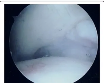
Fig. 2 Arthroscopic image showing debridement of bony impingement
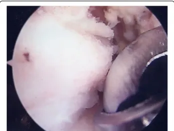
Fig. 3 Arthroscopic image showing debridement of scar tissue with the radiofrequency probe
Fig. 4 Arthroscopic image showing the position of the awl when performing microfractures. The tip of the awl is perpendicular to the bed of the lesion
and grade III (if the space is markedly reduced or de-formed). Radiographs were assessed by the clinical re-search fellow. In more demanding cases, the diagnosis was confirmed by a radiologist not involved in the study, and experienced in muscle, tendon, and ligament disorders in athletes.
Statistics
After the assessment of normality with the Kolmogorov-Smirnov test, the Wilcoxon signed-rank test was used to compare preoperative and postoperative AOFAS, Kaikkonen, and VAS values. A Pearson’s chi-square test was used to test the association between the status of postoperative sport activity and the grade of ankle OA. A P value of <0.05 was considered to be statistically signifi-cant. All analyses were performed using SPSS software, version 13.0 (SPSS, Chicago, Illinois).
Results
All 15 patients included in our original cohort returned to our clinic at the average final follow-up of 7.4 years. The average age at surgery was 42 (from 38 to 45; SD 2.3; median 44); the mean time of activity before the onset of symptoms had been 27 years (from 24 to 33; SD 2.7; median 26). Patients were operated on after an average of 8 months (from 6 to 12 months; SD 1.9; median 7) after the onset of symptoms. The right ankle was involved in 11 (67 %) patients, and the left in four (33 %) patients. The AOFAS significantly im-proved from an average preoperative score of 48 (range, 29–69; SD 11.6; median 48) to 86 (range, 63–94; SD 7.4; median 88) at the last follow-up (P < 0.0001). Postoperatively, the AOFAS scores were graded as ex-cellent in seven (47 %), good in four (27 %), fair in three (20 %), and poor in one (6 %) patients. The Kaikkonen score significantly improved from an aver-age preoperative score of 43 (range, 28–70; SD 12.5; median 40) to 85 (range, 61–95; SD 8.2; median 88) at the last follow-up (P < 0.0001). Postoperatively, the Kaikkonen score was graded as excellent in eight (53 %), good in three (20 %), fair in three (20 %), and poor in one (6 %) patient. VAS values also improved from a baseline average value of 7.1 (range, 5–10; SD 1.5; median 7) to an average of 2.9 (range, 0–6; SD 1.6; median 3) at the last follow-up.
At the last appointment, eight (53 %) patients prac-ticed recreational high-impact sport activities (soccer, tennis, and running) an average of two times per
week; six (40 %) practiced recreational
non-weightbearing sport activities (cycling and swimming) an average of two times per week; and 1 (7 %) aban-doned altogether sport, as he did not feel safe with his ankle and he felt too old to continue. No surgical complication was encountered.
Imaging outcomes
At the time of surgery, the ankle of all patients demon-strated radiographic signs of degenerative changes: eight patients exhibited grade I changes, and seven patients exhibited grade II changes. At the final follow-up, seven patients exhibited radiographic signs of grade I degen-erative changes of the ankle, and five had radiographic signs of grade II degenerative changes of the ankle, three had evolved to grade III osteoarthritis. One of these three patients (33 %) had abandoned sport activity, and two (67 %) practiced recreational low-impact sports such as swimming and cycling.
Discussion
The main finding of the present study is that ankle arth-roscopy and concomitant arthroscopic management of secondary injuries, when performed in selected patients with mild to moderate degenerative changes to the ankle, provide high rates of satisfaction and good func-tional results with positive impact on the quality of life.
There is strong evidence in support of ankle arthros-copy in patients with arthrofibrosis, synovitis, arthritis, fractures, and osteochondral defects [12]. Specifically, in osteochondral lesions, arthroscopic debridement and microfractures provide very satisfying results in the mid-term, in terms of AOFAS and VAS values [13]. In pa-tients with anterior bony impingement, at a mean follow-up of 9 years, the AOFAS scores were still signifi-cantly improved, but age at surgery, radiographic changes, and concomitant cartilage lesions were negative prognostic factors [14].
We performed microfractures in all patients for management of grade III–IV cartilage injuries. In appro-priately selected cases, microfractures produce stable regenerated fibrocartilage, reduce pain and crepitus, and improve the gliding of the articular surfaces. They are easy to perform, safe, effective in the short- and mid-term, but, in the long mid-term, outcomes decline [15]. Nevertheless, at a minimum follow-up of 8 years after arthroscopic microfractures, good-to-excellent results were observed in 78 % of patients, with no poor results, and low progression of osteoarthritis (33 % grade I pro-gression) [16].
A recent systematic review reported that, in patients with osteochondral lesions of the talus, arthroscopic debridement and microfractures provides 80 % of good to excellent results on average [17].
When evaluating prognostic factors, arthroscopy may be effective if cartilage lesions are small, and involve the lateral side of the talus [18].
In our study, all patients presented degenerative changes of the ankle. Ankle arthroscopy, though effective for isolated osteochondral lesions, provides less encour-aging results in patients with frank osteoarthritis [8, 19].
In the knee, arthroscopic debridement has no value in the management of arthritis [20]. In the ankle, the pattern and evolution of cartilage lesions is different compared to the knee [21], given the different microscopic and biomechan-ical properties, content of proteoglycan and water, and altered response to catabolic factors [22]. Therefore, selected groups of patients may benefit after ankle arth-roscopy, in terms of pain relief and functional improve-ment [6, 23]. More complex and invasive procedures such as supramalleolar osteotomy, ankle arthrodesis, or joint arthroplasty may be delayed or avoided [24].
In advanced osteoarthritis, in the midterm, microfrac-tures do not affect the outcomes [25]. Nevertheless, they decrease the risk of clinical failure, and improve the out-comes compared to debridement alone [26]. Therefore, it is essential to address associated intra-articular lesions, present in up to 72 % of patients [27, 28], and predictive of poor results.
There is no consensus on postoperative rehabilitation and time to return to activity after ankle arthroscopy [29]. We point out that all our patients returned to full weightbearing 6–8 weeks after the index surgery, to allow osteoblasts to form new woven bone and chondro-blasts to produce a matrix containing type II collagen and proteoglycans, all forming fibrocartilaginous tissue. After this time, a hyaline-like cartilage with a high com-ponent of type II collagen can be detected [30], and the osteochondral defects are completely filled with mostly hyaline-like tissue [31].
All our patients underwent viscosupplementation with hyaluronic acid. The most recent evidence on this mat-ter states that viscosupplementation does not influence the natural progression of osteoarthritis, but it reduces pain and improves function [32].
There are limitations to this study. First, it was a retro-spective and nonrandomized investigation. Although the
retrospective design of the study and the absence of a control group do not allow us to draw definitive conclu-sions, this is the first study to report on midterm out-comes in former professional soccer players who had undergone arthroscopic procedures for management of mild to moderate osteoarthritis of the ankle. We are aware that the AOFAS scale is not validated, and that
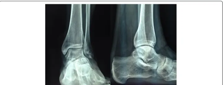
Fig. 6 Anteroposterior and lateral radiographs of the left ankle in a soccer player showing signs of moderate osteoarthritis of the ankle
Fig. 7 Lateral radiograph of the left ankle of the same patient at 7-year follow-up, showing no progression of degenerative changes
other scales are available to assess ankle function [33], but it was a common practice to administer it at the stage the study had been conceived, and we have suc-cessfully used it for the assessment of patients with ankle disorders [27, 28]. The strength of the study was that we enrolled a selected group of patients according to strict selection criteria, excluding all confounding factors of bias. Although the analysis was retrospective, data col-lection was longitudinal. The first author, a fellowship-trained surgeon, performed all surgical procedures, the rehabilitation protocol was homogeneous, an independ-ent investigator not involved at the index surgery exam-ined the patients at the last follow-up, and none of the patients was lost to the follow-up.
Conclusions
In former professional soccer players with mild to mod-erate ankle arthritis, microfractures, and concomitant arthroscopic management of associated injuries are safe, effective, and low cost, allowing them to return safely to ordinary daily and recreational sport activities. In this series, good excellent outcomes were observed in more than 70 % of patients, and radiographic degenerative changes progressed in two patients (13 %) (Figs. 6 and 7). Appropriately powered studies with longer follow-up, pos-sibly testing different treatment modalities and different rehabilitation protocols are nevertheless needed.
Competing interests
NM is the Editor-in-Chief for Journal of Orthopaedic Surgery and Research. The authors declare that they have no competing interests.
Authors’ contributions
LO, ADB, and NM conceived the study. LO and NM advised on the practicalities of the surgery. LO performed all the surgical procedures. LO and ADB were involved in the clinical assessment and administered scores. ADB performed the review of the literature and wrote the initial draft. LO and NM coordinated the work in all its phases. NM corrected the manuscript. All authors read and approved the final manuscript.
Author details
1Unit of Arthroscopy and Sports Trauma Surgery, Hesperia Hospital, Via
Arquà 80/b, Modena, Italy.2Department of Orthopaedics and Traumatology,
Hospital Vaio, Via Tincati, Fidenza, Parma, Italy.3Department of Musculoskeletal Disorders, Faculty of Medicine and Surgery, University of Salerno, Salerno, Italy.4Centre for Sports and Exercise Medicine, Barts and
The London School of Medicine and Dentistry, Mile End Hospital, 275 Bancroft Road, London E1 4DG, UK.
Received: 20 August 2015 Accepted: 15 March 2016
References
1. Conti SF, Wong YS. Complications of total ankle replacement. Clin Orthop Relat Res. 2001;391:105–14.
2. Battisti D, Oliva F, Tarantino U, Maffulli N. Pseudoaneurysm of peroneal artery after ankle arthroscopy. Muscles Ligaments Tendons J. 2014;4:269–72. 3. Ferran NA, Oliva F, Maffulli N. Ankle instability. Sports Med Arthrosc.
2009;17:139–45.
4. McDougall A. Footballer’s ankle. Lancet. 1955;269:1219–20. 5. Gougoulias N, Khanna A, Maffulli N. How successful are current ankle
replacements?: A systematic review of the literature. Clin Orthop Relat Res. 2010;468:199–208.
6. Cheng JC, Ferkel RD. The role of arthroscopy in ankle and subtalar degenerative joint disease. Clin Orthop Relat Res. 1998;349:65–72. 7. Amendola A, Petrik J, Webster-Bogaert S. Ankle arthroscopy: outcome
in 79 consecutive patients. Arthroscopy. 1996;12:565–73.
8. Phisitkul P, Tennant JN, Amendola A. Is there any value to arthroscopic debridement of ankle osteoarthritis and impingement? Foot Ankle Clin. 2013;18:449–58.
9. Krips R, van Dijk CN, Halasi PT, Lehtonen H, Corradini C, Moyen B, et al. Long-term outcome of anatomical reconstruction versus tenodesis for the treatment of chronic anterolateral instability of the ankle joint: a multicenter study. Foot Ankle Int. 2001;22:415–21.
10. Kitaoka HB, Alexander IJ, Adelaar RS, Nunley JA, Myerson MS, Sanders M. Clinical rating systems for the ankle-hindfoot, midfoot, hallux, and lesser toes. Foot Ankle Int. 1994;15:349–53.
11. Kaikkonen A, Kannus P, Jarvinen M. A performance test protocol and scoring scale for the evaluation of ankle injuries. Am J Sports Med. 1994;22:462–9.
12. Hsu AR, Gross CE, Lee S, Carreira DS. Extended indications for foot and ankle arthroscopy. J Am Acad Orthop Surg. 2014;22:10–9.
13. Morales J, García E, Asunción J, Poggio D. Medium-term results of arthroscopic debridement in osteochondral talar lesions. Acta Ortop Mex. 2013;27:319–23.
14. Parma A, Buda R, Vannini F, Ruffilli A, Cavallo M, Ferruzzi A, et al. Arthroscopic treatment of ankle anterior bony impingement: the long-term clinical outcome. Foot Ankle Int. 2014;35:148–55.
15. Murawski CD, Kennedy JG. Operative treatment of osteochondral lesions of the talus. J Bone Joint Surg Am. 2013;95:1045–54.
16. van Bergen CJ, Kox LS, Maas M, Sierevelt IN, Kerkhoffs GM, van Dijk CN. Arthroscopic treatment of osteochondral defects of the talus: outcomes at eight to twenty years of follow-up. J Bone Joint Surg Am. 2013;95: 519–25.
17. Donnenwerth MP, Roukis TS. Outcome of arthroscopic debridement and microfracture as the primary treatment for osteochondral lesions of the talar dome. Arthroscopy. 2012;28:1902–7.
18. Yoshimura I, Kanazawa K, Takeyama A, Angthong C, Ida T, Hagio T, et al. Arthroscopic bone marrow stimulation techniques for osteochondral lesions of the talus: prognostic factors for small lesions. Am J Sports Med. 2013;41:528–34.
19. Choi WJ, Choi GW, Kwon HM, Lee JW. Arthroscopic treatment in mild to moderate osteoarthritis of the ankle. Knee Surg Sports Traumatol Arthrosc. 2013;21:1338–44.
20. Kirkley A, Birmingham TB, Litchfield RB, Giffin JR, Willits KR, Wong CJ, et al. A randomized trial of arthroscopic surgery for osteoarthritis of the knee. N Engl J Med. 2008;359:1097–107.
21. Buckwalter JA, Brown TD. Joint injury, repair, and remodeling: roles in post-traumatic osteoarthritis. Clin Orthop Relat Res. 2004;423:7–16.
22. Cole AA, Kuettner KE. Molecular basis for differences between human joints. Cell Mol Life Sci. 2002;59:19–26.
23. Hassouna H, Kumar S, Bendall S. Arthroscopic ankle debridement: 5-year survival analysis. Acta Orthop Belg. 2007;73:737–40.
24. Giannini S, Buda R, Faldini C, Vannini F, Romagnoli M, Grandi G, et al. The treatment of severe posttraumatic arthritis of the ankle joint. J Bone Joint Surg Am. 2007;89 Suppl 3:15–28.
25. Spahn G, Mückley T, Kahl E, Hofmann GO. Factors affecting the outcome of arthroscopy in medial-compartment osteoarthritis of the knee. Arthroscopy. 2006;22:1233–40.
26. Osti L, Del Buono A, Maffulli N. Microfractures at the rotator cuff footprint: a randomised controlled study. Int Orthop. 2013;37:2165–71. 27. Maffulli N, Del Buono A, Maffulli GD, Oliva F, Testa V, Capasso G,
et al. Isolated anterior talofibular ligament Brostrom repair for chronic lateral ankle instability: 9-year follow-up. Am J Sports Med. 2013;41: 858–64.
28. Nery C, Raduan F, Del Buono A, Asaumi ID, Cohen M, Maffulli N. Arthroscopic-assisted Brostrom-Gould for chronic ankle instability: a long-term follow-up. Am J Sports Med. 2011;39:2381–8.
29. van Eekeren IC, Reilingh ML, van Dijk CN. Rehabilitation and return-to-sports activity after debridement and bone marrow stimulation of osteochondral talar defects. Sports Med. 2012;42:857–70.
30. Frisbie DD, Oxford JT, Southwood L, Trotter GW, Rodkey WG, Steadman JR, et al. Early events in cartilage repair after subchondral bone microfracture. Clin Orthop Relat Res. 2003;407:215–27.
31. Gill TJ, McCulloch PC, Glasson SS, Blanchet T, Morris EA. Chondral defect repair after the microfracture procedure: a nonhuman primate model. Am J Sports Med. 2005;33:680–5.
32. Liu C, Shi XG, Liu YJ, Wang ZG, Wei M. Analysis on arthroscopic debridement and visco supplement for the treatment of degenerative osteoarthropathy of ankle. Zhongguo Gu Shang. 2013;26:115–8.
33. Mann G, Nyska M, Hetsroni I, Karlsson J. Scoring systems for evaluating ankle function. Foot Ankle Clin. 2006;11:509–19.
• We accept pre-submission inquiries
• Our selector tool helps you to find the most relevant journal
• We provide round the clock customer support
• Convenient online submission
• Thorough peer review
• Inclusion in PubMed and all major indexing services
• Maximum visibility for your research Submit your manuscript at
www.biomedcentral.com/submit