O R I G I N A L A R T I C L E
SEPN1, an endoplasmic reticulum-localized
selenoprotein linked to skeletal muscle pathology,
counteracts hyperoxidation by means of
redox-regulating SERCA2 pump activity
Marianna Marino
1,
†
, Tatiana Stoilova
1,
†
, Carlotta Giorgi
2
, Angela Bachi
3
,
Angela Cattaneo
3
, Alberto Auricchio
4
, Paolo Pinton
2
, and Ester Zito
1,
*
1Dulbecco Telethon Institute at IRCCS-Istituto di Ricerche Farmacologiche Mario Negri, Milan, Italy,
2Section of
Pathology, Oncology and Experimental Biology, Laboratory for Technologies of Advanced Therapies (LTTA),
Department of Morphology, Surgery and Experimental Medicine, University of Ferrara, Ferrara, Italy,
3IFOM-FIRC
Institute of Molecular Oncology, Milan, Italy, and
4Telethon Institute of Genetics and Medicine (TIGEM), Naples,
and Medical Genetics, Department of Translational Medicine, Federico II University, Naples, Italy
*To whom correspondence should be addressed at: Dulbecco Telethon Assistant Scientist, IRCCS-Istituto di Ricerche Farmacologiche Mario Negri, Via La Masa 19, 20156 Milano, Italy. Tel: +39 0239014480; Fax: +39 023546277; Email: [email protected]
Abstract
Selenoprotein N (SEPN1) is a broadly expressed resident protein of the endoplasmic reticulum (ER) whose loss-of-function inexplicably leads to human muscle disease. We found that SEPN1 levels parallel those of endoplamic reticulum oxidoreductin 1 (ERO1), an ER protein thiol oxidase, and that SEPN1’s redox activity defends the ER from ERO1-generated peroxides. Moreover, we have defined the regulated interactome of SEPN1 and identified the ER calcium import SERCA2 pump as a redox-partner of SEPN1. SEPN1 enhances SERCA2 activity by reducing luminal cysteines that are hyperoxidized by ERO1-generated peroxides. Cells lacking SEPN1 are hypersensitive to ERO1 overexpression and conspicuously defective in ER calcium re-uptake. After being muscle-transduced with an adeno-associated virus driving ERO1α, SEPN1 knockout mice unmasks a myopathy that resembles the dense core disease due to human mutations in SEPN1, whereas the combined attenuation of ERO1α and SEPN1 enhances cellfitness. These observations reveal the involvement of SEPN1 in ER redox and calcium homeostasis and that an ERO1 inhibitor, restoring redox-dependent calcium homeostasis, may ameliorate the myopathy of SEPN1 deficiency.
Introduction
SEPN1, a member of the selenocysteine-containing protein fam-ily, is localized in the ER lumen, ubiquitously expressed through-out the body, and of unknown function (1,2). Despite its wide expression, SEPN1 mutations give rise to a selective muscle phenotype that suggests the interaction of SEPN1 with proteins involved in muscle physiology (3,4).
Many selenocysteine-containing proteins are enzymes in-volved in oxidation–reduction reactions whose selenocysteine residue is usually located in the catalytic site because its nucleo-philicity induces strong enzymatic activity (5). Like other members of the family, SEPN1 is also thought to have a redox function, and this idea is supported by the observation of a motif similar to the catalytic site of thioredoxin reductases (TRs) on the ER side of the
†These authors contributed equally to the study.
Received: October 7, 2014. Revised and Accepted: November 26, 2014
© The Author 2014. Published by Oxford University Press. All rights reserved. For Permissions, please email: [email protected] doi: 10.1093/hmg/ddu602
Advance Access Publication Date: 1 December 2014 Original Article
1843
at GOT (Consortium) on April 3, 2015
http://hmg.oxfordjournals.org/
SEPN1 sequence. In agreement, excessive protein oxidation in cells devoid of SEPN1 has been observed (1,6,7).
Calcium homeostasis has a crucial function in muscle and de-pends on the redox status of both cytoplasmic and luminal cy-steines in calcium-handling proteins (8,9). Among the calcium-handling proteins of the ER the Ca2+-ATPase (SERCA) pumps have the highest affinity for Ca2+removal from the cytosol and
therefore determine the resting cytosolic Ca2+concentration
which is important in the process of excitation–contraction coupling. Three differentially expressed genes encode at least five isoforms of the SERCA pump. SERCA1a and 1b are expressed in fast twitch muscle fibers of skeletal muscle, SERCA3 has limited expression in various non-muscle tissues, whereas SER-CA2a is expressed in slow-twitchfibers of skeletal muscle and in cardiac muscle. Its C-terminally extended isoform SERCA2b is ubiquitously expressed in muscle and non-muscle tissues (10). Interestingly, cysteine-dependent interactions between the oxi-doreductase ERp57 and the fourth luminal loop of the SERCA2b inhibit calcium reuptake into the ER by oxidizing two cysteines in the L4 of this ATP-driven pump (11).
ER oxidoreductin 1 (ERO1) is the main ER protein disulfide oxidase that channels electrons from PDI to the terminal acceptor of electrons in the reaction: i.e. molecular oxygen, which is reduced to H2O2(12–14). The oxidative activity of ERO1 may
there-fore burden the cell with potentially toxic reactive oxygen species (ROS) (15,16).
ERO1 transcription is activated by the unfolded protein re-sponse (UPR), which is one of the primary processes triggered by altered environmental cues in skeletal muscle, and contract-ing skeletal muscle produces ROS which, if they are not metabo-lized, can cause oxidative damage to the macromolecules in musclefibres (17–20).
Therefore, ROS production by increased ERO1 expression in a contracting muscle may be harmful and be responsible for the maladaptive branch of the UPR (21).
Consistent with this idea, a partial decrease in ERO1 activity is compatible with life and promotes resistance to the lethal effects of high ER stress levels, which suggests that less ERO1 may be an advantage under some unusual conditions (22–24).
Given that muscle activity begets ER stress and enhances ERO1 activity with an attendant ER-localized peroxide pro-duction, and that ER thiol redox plays a crucial role in calcium homeostasis, we examined the possibility that the loss of an SEPN1-mediated reductive function leads to myopathy exposing cells to an H2O2-mediated hyperoxidation of key molecules
involved in calcium handling.
We therefore established an unbiased method for uncovering redox-substrates of SEPN1 and found SERCA2 as one of them.
Results
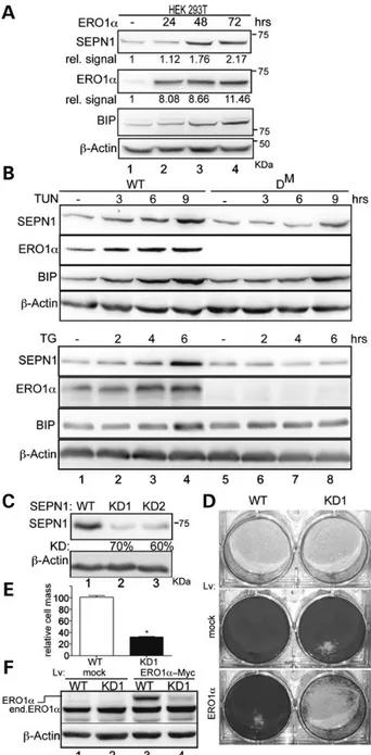
SEPN1 protects against the hyperoxidizing conditions elicited by ERO1
SEPN1 mutant cells are more sensitive to oxidative stress and show excessive protein oxidation (6), so we followed SEPN1 ex-pression in cells transfected with ERO1α, one of the main protein disulfide oxidases whose H2O2-generating activity can lead to ER
hyperoxidation/misoxidation. Immunoblotting of protein lysates from HEK 293T cells transfected with ERO1α and harvested at dif-ferent times showed that SEPN1 induction paralleled the level of ERO1α overexpression (Fig.1A, lanes 1–4).
The treatment of cells with tunicamycin and thapsigargin, two inducers of ER stress, elicits the adaptive UPR and the
Figure 1. SEPN1 protects against the hyperoxidizing conditions elicited by ERO1
induction. (A) Immunoblot of SEPN1, ERO1α, BIP and β-actin in extracts of
HEK-293T transfected with ERO1α and harvested at different time points. The relative amount (rel.signal) of SEPN1 and ERO1 is entered under the immunoblot and
expressed as arbitrary units (a.u.) of the ratio of SEPN1 and ERO1 to theβ-actin
signal. (B) Immunoblots of SEPN1, ERO1α, BIP and β-actin from WT and DM
(double mutant for ERO1-α and ERO1-β) MEFs treated with 0.5 ug/ml tunicamycin
(TUN) or 0.5 m thapsigargin (TG) for different times. (C) Immunoblot of SEPN1
andβ-actin in C2C12 7 days after transduction with a puror-marked lentivirus
carrying an irrelevant insert (WT) or two different short hairpin RNAs directed to mouse SEPN1: KD1 and KD2. The relative percentage of knockdown SEPN1 (KD) is
specified beneath the blot. (D) Photomicrographs of crystal violet-stained WT and
KD1 C2C12 cells untransduced (unt.) or transduced with a blasticidin
resistance-marked lentivirus expressing greenfluorescent protein (GFP, indicated as mock)
or ERO1α-Myc. (E) As in “D”: quantification of cell mass after the transduction of
WT and KD1 C2C12 cells with the mock GFP lentivirus (set at 100%) or the ERO1
α-Myc lentivirus followed by blasticidin selection for 7 days (mean values and
SEM, n = 3, *P < 0.01). (F) Immunoblot of ERO1α and β-actin from WT and KD1
C2C12 cells after transduction with a blasticidin resistance-marked lentivirus
expressing either GFP or ERO1α-Myc showing endogenous ERO1α (end. ERO1α)
and slowly migrating ERO1α-Myc (ERO1α).
at GOT (Consortium) on April 3, 2015
http://hmg.oxfordjournals.org/
consequent up-regulation of ERO1 in order to increase cell sur-vival (21). However, in certain circumstances of ER-stress a partial loss of function of the essential ERO1 gene enhances the ability of yeasts and worms to cope with severe stress as it burdens the cells with less H2O2(23,24). Gene duplication events in vertebrates have
generated two ERO1 isoforms, ERO1α and ERO1β, and mice with strong hypomorphic mutations in both ERO1s (DMmice) are viable
(25). SEPN1 induction by tunicamycin and thapsigargin was less in DMmouse embryonicfibroblasts (MEFs), thus indicating that SEPN1
induction is attenuated in cells lacking ERO1 (Fig.1B compare lanes 1–4 with 5–8) and suggesting a co-regulation of SEPN1 and ERO1.
In order to test the effect of ERO1 activity in SEPN1-deficient cells, we stably lowered SEPN1 gene products by means of lenti-viral RNAi in C2C12 cells, a myoblast mouse cell line that ex-presses high levels of SEPN1. The resulting pools of KD1- and KD2-infected cells had, respectively, 70 and 60% lower SEPN1 le-vels (Fig.1C, lanes 1–3), so we used the SEPN1 KD1 cells for the subsequent experiments. Lentiviral vectors carrying a dominant selection marker that imparts blasticidine resistance were used to transduce ERO1α-Myc in wild-type (WT) and KD1 cells, and led to the growth of a thick carpet of ERO1α-transduced WT cells, but the transduction of ERO1α in KD1 cells failed to elicit the growth of blasticidine-resistant colonies (Fig.1D and E), and the few cells recovered had a low level of transduced ERO1α (Fig.1F, lanes 3 and 4), indicating that low levels of SEPN1 are incompatible with high levels of ERO1α.
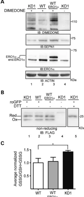
High cysteinyl sulfenic acid levels in SEPN1 knockdown cells
ERO1 is a key enzyme in oxidative protein folding whose oxida-tive activity is related to the build-up of H2O2(16).
Remarkably, the build-up of H2O2inside the ER exposes the
free thiols of new client proteins to competing H2O2-mediated
oxidation, which leads to an increase in sulfenylated proteins; these can be transient intermediates in the formation of more stable disulfides, or upon a further burst of H2O2, trapped in an
irreversible state of higher oxidized sulfur oxides (26).
In order to compare the burden of sulfenylated proteins in freshly lentiviral-infected C2C12 cells with different levels of SEPN1, we exposed the cells and lysates to dimedone, a chemical that selectively modifies sulfenylated cysteines, and detected the di-medone-modified proteins by means of immunoblotting using an antibody against the chemically modified group (27). In the absence of dimedone, the antibody gave a weak background signal (Fig.2A, lane 4) but, importantly, the sulfenic acid signal was considerably stronger in the KD1 cells and in cells overexpressing ERO1α, both of which showed a discrete band of∼110 kDa (Fig.2A, lanes 1 and 3). On the contrary, SEPN1 knockdown had no effect on the redox state of the sentinel ER-localized reduction–oxidation sensitive greenfluorescent protein (roGFP-iE) (Fig.2B lanes 1 and 2) (28,29). Glutathione is the main scavenger of cellular H2O2and acts to
protect the ER from hyperoxidizing conditions (30,31). Since it has been shown that hyperactive ERO1 and the direct exposure to H2O2
inside the ER increases luminal oxidized glutathione levels, we tested the level of total and oxidized glutathione in WT and KD1 cells (32,33). The 1.5-fold increase in the oxidized to total glutathi-one ratio in the KD1 cells was consistent with an H2O2-mediated
hyperoxidation that was not compensated by SEPN1 (Fig.2C).
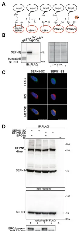
An SEPN1-trapping mutant engages protein-substrates in an ERO1-dependent manner
As human SEPN1 has a domain (CU: C427, U428) resembling the active site of TR, we took advantage of the electron transfer
mechanism of TR to identify the redox-mediated substrates of SEPN1. Mammalian TRs have a C-terminal redox-active tetrapep-tide (GCUG) in which the cysteine-selenocysteine dyad repre-sents the active site. The enzymatic mechanism of TR relies on an exchange of electrons with the thioredoxin (Trx) substrate: the nucleophilic selenolate anion of selenocysteine attacks a (sulfenylatable) cysteine on Trx to form a selenenylsulfide inter-mediate that is later resolved by the cysteine in the redox-active motif of TR (34,35).
Figure 2. High cysteinyl sulfenic acid levels in SEPN1 knockdown cells. (A)
Immunoblots of extracts from KD1, WT and WT transduced with ERO1α C2C12
cells exposed to the sulfenic acid-reactive probe dimedone and an antibody
reactive to dimedone-conjugated cysteine residues. The immunoblot ofβ-actin
is shown as the loading control. (B) Immunoblot of FLAG_M1_roGFP_iE
im-munopurified from the ER of cells with the indicated genotypes. The purified
proteins were resolved by SDS–PAGE under non-reducing conditions. The position of the reduced and oxidized FLAG_M1_roGFP_iE in a typical experiment is shown. (C) Bar diagram of the ratio between oxidized glutathione (GSSG) to total glutathione (GSH + GSSG) content in cells of the indicated genotypes. Results are normalized to the control WT and are the mean of three independent experi-ments (n = 3, *P < 0.01).
at GOT (Consortium) on April 3, 2015
http://hmg.oxfordjournals.org/
Based on this mechanism, we generated an SEPN1 mutant devoid of the resolving cysteine (C427, which was mutated to serine) in order to trap the redox substrates of SEPN1 (Fig.3A). To overcome the low level of SEPN1 expression due to the pres-ence of the selenocysteine (U428), this residue was converted to the similar cysteine amino acid (Fig.3B). This modification is
sup-ported by the fact that a previous study using a cysteine- or sele-nocysteine-based trapping mutant of TR did not reveal any difference between the interactors of the two enzyme forms (36). In order to establish the system of SEPN1-SC-pFLAG (the trap-ping mutant of SEPN1) and SEPN1-SS-pFLAG (a potential redox negative control lacking U and C), the proteins were expressed by means of the transient transfection of HEK 293T cells. The lo-calization of SEPN1-SC-pFLAG and SEPN1-SS-pFLAG wasfirst checked by means of indirect immunofluorescence which, as ex-pected, showed that both co-localize with PDI in the ER (Fig.3C). We then prepared protein lysates from SEPN1-SC-pFLAG- and SEPN1-SS-pFLAG-transfected cells in the presence of N-ethylma-leimide (NEM) in order to quench the free thiols, and the protein complexes were immunopurified using the FLAG-M2 tag. Immunoblotting of the non-reducing gel showed two main bands of∼70 and 140 kDa, probably corresponding to monomers and covalent homodimers of SEPN1, and additional bands in a high-molecular disulfide-bonded complex associated exclusively with SEPN1-SC-pFLAG and representing potential client proteins (Fig.3D, lanes 2 and 4). The simultaneous overexpression of ERO1α made the appearance of these bands more conspicuous, probably by enhancing the interaction between SEPN1-SC-pFLAG and the clients (Fig.3D, lanes 1 and 2). On the contrary, the delivery of a catalase–peroxidase to the ER made these bands less conspicuous (Supplementary Material, Fig. S1, com-pare lane 1 with 2). These observations confirmed that the trap-ping mutant was useful for identifying the redox-regulated interactors of SEPN1 and that SEPN1 interacts with these in an H2O2-dependent manner.
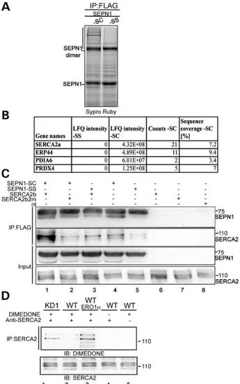
An SEPN1-trapping mutant forms a stable complex with SERCA2
In order to identify the protein(s) that may be associated with the trapping mutant of SEPN1 in a disulfide-bonded complex, we ex-cised the region of the non-reducing SDS–PAGE containing the trapped complexes. Following gel reduction, alkylation and di-gestion with trypsin, the peptides eluted from the gel slices were analyzed by means of LC–MS-MS (Fig.4A), and the corre-sponding human proteins were sorted and scored by means of label-free quantification (Fig.4B). Among the ER proteins discov-ered in a disulfide complex exclusively with the SEPN1-trapping mutant (and not with SEPN1-SS-pFLAG), we focused our atten-tion on the ER calcium pump SERCA2 because of its pivotal role in calcium signalling.
The excitation–contraction coupling that connects mem-brane depolarization with cytosolic calcium concentration and cell shortening is fundamental for muscle physiology. An action potential on neuromuscular junctions triggers a series of events that culminate in the SERCA-mediated re-uptake of Ca2+into the
ER/SR (sarcoplasmic reticulum), which brings cytosolic calcium to the resting level of muscle relaxation (37). The SERCA family includes various gene products and related splicing variants that are all involved in SR/ER calcium uptake. Importantly, SER-CA2b is ubiquitously expressed in muscle and non-muscle tis-sues and whose activity depends on the redox status of two cysteines exposed in the lumen of the ER (11). SERCA2a is the main isoform of SERCA in slow-twitch musclefibres (and models
Figure 3. An SEPN1-trapping mutant engages protein-substrates in an ERO1-dependent manner. (A) Proposed catalytic mechanism of mammalian SEPN1. The sulfenylated substrate (SOH) accepts electrons directly from the selenolate anion (U428) of SEPN1, and the intermediate selenenylsulfide bond formed between SEPN1 and the substrates is resolved by the cysteine (C427). The substrate could be trapped in a complex with a mutant SEPN1 (SEPN1-SU) lacking the resolving cysteine (C427) (that is mutated to serine) of its active C-terminal site. (B) Immunoblot of Flag-tagged SEPN1-SU and SEPN1-SC transfected in HEK-293T cells. The truncated SEPN1-SU is due to the UGA codon of the selenocysteine that is recognized as a stop codon. (C) Immunostaining of PDI and Flag-tagged SEPN1-SC and SEPN1-SS transfected in HEK-293T cells
(scale bar: 20μm). (D) Non-reducing and reducing Flag immunoblot of Flag-M2
immunopurified proteins from HEK-293T cells transfected as indicated. The low
mobility complex containing immunoreactive material in complex with
SEPN1-SC is indicated by a vertical line. Bottom: ERO1α reducing immunoblot of
proteins from transfected cells. Transfected ERO1α is distinguished from the endogenous form because of the Myc tag.
at GOT (Consortium) on April 3, 2015
http://hmg.oxfordjournals.org/
of SEPN1 loss of function show a selective hypotrophy of slow-twitch musclefibres (38)) and contains the two exposed cysteines (10).
A substantial amount of endogenous SERCA2 immunoreac-tivity was found complexed with SEPN1-SC-pFLAG in HEK 293T cells (Fig.4C, lane 4). In order to characterize the association be-tween SEPN1-SC-pFLAG and SERCA2, the cells were transfected with various combinations of SEPN1-SC-pFLAG, SEPN1-SS-pFLAG, pig SERCA2b and the mutant pig SERCA2b2m which lacks the two cysteines (C875 and C887) in the L4 ER domain. The protein extracts were quenched with NEM and immunopre-cipitated with FLAG M2; the immunocomplexes were resolved
under reducing conditions, and the blots were decorated with anti-FLAG and anti-SERCA2. An abundant band corresponding to SERCA2 was detected in the sample co-transfected with SEPN1-SC-pFLAG and SERCA2b (Fig.4C, lane 1). Of note, a much lower level of association was seen between SEPN1-SC-pFLAG and SERCA2b2 m (Fig.4C, lane 2). A weak band corresponding to SERCA2 was detected in the samples co-transfected with SEPN1-SS-pFLAG and SERCA2b, as well as in the sample trans-fected with SEPN1-SS-pFLAG alone, thus indicating a weak non-covalent association between SEPN1SS and SERCA2 (Fig.4C, compare lane 1 with 3 and lane 4 with 5). Because SER-CA2a also contains the two cysteines (C875 and C887) in the L4 ER domain, we tested whether SEPN1-SC-pFLAG associates with SERCA2a through the cysteines in the L4 domain. We co-expressed SEPN1-SC-pFLAG and SERCA2a or SERCA2a2m which lacks the two cysteines (C875 and C887) and found that SEPN1-SC covalently associates also with SERCA2a through the cy-steines in the L4 domain (Supplementary Material, Fig. S2, com-pare lane 1 with 2).
Hyperoxidized SERCA2 detected in cells devoid of SEPN1
As the molecular mass of all of the SERCA2 splicing variants is ∼110 kDa, we investigated whether the most abundant dime-done-reactive protein in the KD1 cells and the cells overexpressing ERO1 corresponded to SERCA2 (Fig.2A, lanes 1 and 3). To this end, protein lysates of C2C12 cells with different levels of SEPN1 and C2C12 cells overexpressing ERO1 were exposed to dimedone in order to quench the sulfenylated cysteines, and the proteins were immunopurified using an SERCA2 antibody. Immunoblotting SERCA2 and its related protein complex with an antibody against dimedone revealed a signal of∼110 kDa that suggested a sulfeny-lated SERCA2, the levels of which were higher in KD1 cells and in the cells overexpressing ERO1α (Fig.4D, lanes 1 and 3).
Redox-active SEPN1 increases SERCA-dependent Ca2+entry into the ER
Given the redox interaction between SEPN1 and SERCA2, and the redox-regulated activity of SERCA2, we analyzed the potential redox role of SEPN1 in SERCA-dependent Ca2+entry using cells
that were devoid of, or overexpressing SEPN1. Ca2+levels and
fluxes were monitored using an ER-targeted aequorin probe that allows the direct measurement of calcium in the ER, thus overcoming some of the uncertainties associated with indirect Ca2+ measurements and providing a significant advantage
when investigating Ca2+homeostasis in the ER. The reconstitu-tion of active aequorin with coelenterazine in the ER lumen requires the previous depletion of Ca2+from the organelle
fol-lowed by refilling (39). Figure5B shows that the re-addition of Ca2+to the medium rapidly increases [Ca2+] levels within the ER ([Ca2+]
ER) up to a steady-state level of∼500 μ (SEPN1: 508 ± 23 μ;
SEPN1-SS: 510 ± 20μ; n = 10) in HeLa cells transfected with the vector harbouring cDNA for SEPN1 (but not expressing the full-length SEPN1 protein because, in the absence of sodium selenite, the UGA codon of the selenocysteine is recognized as a stop codon, as shown in lane 2 of Fig.5A), or for the mutant SEPN1-SS protein. Under the same conditions, the steady-state ER level after sodium selenite treatment in cells overexpressing SEPN1 was significantly higher (586 ± 18 μ n = 10; P < 0.05), thus indicating potentiated SERCA activity. In contrast, the steady-state ER level in the sodium selenite-treated cells overexpressing the SEPN1SS mutant was slightly lower (432 ± 55μ, n = 10), thus suggesting the inability of the mutant to increase SERCA activity. A difference in intra-luminal [Ca2+] levels between the ER of
Figure 4. An SEPN1-trapping mutant forms a stable complex with SERCA2 in mammalian cells. (A) Sypro Ruby stained non-reducing SDS–PAGE of proteins
immunopurified in complex with FLAG-tagged SEPN1-SC and SEPN1-SS. The
vertical line demarcates the region analyzed by mass spectroscopy protein
analysis. (B) List of proteins identified by means of LC–MS-MS sequencing of the
tryptic peptide of endogenous proteins captured in disulfide-linked complex by a FLAG M2-tagged trapping mutant SEPN1-SC in HEK-293T cells. The ER proteins found only in SEPN1-SC were sorted against the total number of proteins using the Label Free Intensity parameter. (C) Anti-Flag and anti-SERCA2 immunoblots of proteins immunopurified with the FLAG-M2 antibody from lysates of HEK 293T cells that were untransfected or transfected with the indicated expression plasmids. The lower two panels show, respectively, 5 and 10% of the total
lysates before IP (“Input”). The proteins shown were resolved by means of
reducing SDS–PAGE. (D) Immunoblot using antibody reactive to
dimedone-conjugated cysteine residues of the protein samples shown in Figure2A
immunopurified with SERCA2 antibody. The immunoblot made using SERCA2 antibody is shown at the bottom.
at GOT (Consortium) on April 3, 2015
http://hmg.oxfordjournals.org/
the cells expressing SEPN1 or SEPN1-SS was not only observed at steady-state but also in the maximal rate of Ca2+accumulation in
the ER (SEPN1: 18.86 ± 2.42μ/s; SEPN1-SS: 13.96 ± 0.79 μ; n = 10) calculated at the beginning of ER Ca2+refilling, i.e. when
the rate is mainly dependent by the SERCA activity and neither influenced by ER Ca2+leak nor by ER Ca2+buffering.
We next examined SERCA activity in freshly isolated SEPN1 WT and SEPN1KO MEFs. As shown in Figure5D, and in line with the re-sults obtained in HeLa cells, the steady-state level of [Ca2+]
ERin the
SEPN1 WT cells was∼300 µ, whereas it was lower in SEPN1KO MEFs (WT: 292 ± 50μ; SEPN1KO: 189 ± 25 μ; n = 8; P < 0.05), and the rate of Ca2+accumulation was also lower (WT: 3.6 ± 0.7μ/s;
SEPN1KO: 2.6 ± 0.5μ/s; n = 8) despite comparable levels of SERCA2 (Fig.5C). This alteration can be attributed to the absence of SEPN1 because the re-introduction of SEPN1 protein in SEPN1KO MEFs restored [Ca2+]
ERlevels similar to those observed in WT cells
(312 ± 47μ; rate of accumulation 3.5 ± 0.7 μ/s; n = 8).
In line with a reductive function of SEPN1 that enhances SERCA activity by reducing the luminal H2O2-oxidized cysteines,
the delivery of a catalase–peroxidase, but not a cognate catalase mutated in the active site, to the ER restored [Ca2+]
ERlevels of
SEPN1KO MEFs (Supplementary Material, Fig. S3).
As Ca2+accumulation in the ER is determined by SERCA-de-pendent Ca2+entry and Ca2+efflux (most probably via inositol
1,4,5-trisphosphate receptors, IP3R1), we determined whether
the lack of SEPN1 has some impact on Ca2+efflux. Ca2+efflux
from the ER was compared in freshly isolated SEPN1KO MEFs and freshly isolated SEPN1WT MEFs after agonist stimulation of IP3R1. The SEPN1KO MEFs showed less SERCA activity, but there was no between-group difference in Ca2+efflux suggesting
that the activity of the major ER Ca2+release channels IP3R1 was
unaffected by SEPN1 protein (Supplementary Material, Fig. S4). These results indicate that a redox-active SEPN1 regulates the calcium level of the ER potentiating SERCA activity by reducing the luminal H2O2-oxidized cysteines.
An ERO1 surge in the protected SEPN1 KO muscle reveals a myopathic phenotype
Unlike human and zebrafish, in which SEPN1 loss of function gives rise to an overt muscle phenotype, SEPN1KO mice are somehow protected, showing no gross alterations in muscle histology (3,4). This protection may be provided by the activity of redundant pathways controlling redox balance in mouse mus-cle and/or by the limited musmus-cle activity.
We therefore decided to assess the effect of SEPN1 deficit in murine muscle forced into hyperoxidizing conditions by ERO1 overexpression.
In order to test the effects of ERO1 overexpression on the muscle phenotype of SEPN1KO mice, one-month-old WT and Figure 5. Reduced SERCA2 activity in freshly isolated SEPN1KO MEFs rescued by redox-active SEPN1. (A) Immunoblot of FLAG-tagged SEPN1 and SEPN1-SS transfected in HeLa cells in with or without Na-selenite. Note the truncated SEPN1 in the cells without Na-selenite. (B) HeLa cells were co-transfected with ER aequorin and SEPN1 or
SEPN1-SS with or without Na-selenite, and calcium refilling of the ER was recorded. The trace is representative of 10 independent experiments that led to similar results.
Bar graphs representing steady-state ER calcium levels are shown at the bottom (n = 10, *P < 0.05). (C) Immunoblot of SEPN1 and SERCA2 from freshly isolated WT and SEPN1KO MEFs. * Background band. (D) Freshly isolated WT and SEPN1KO MEFs were co-transfected with ER aequorin and SEPN1 in the presence of Na-selenite, and calcium refilling of the ER was recorded. The trace is representative of eight independent experiments that led to similar results. Bar graphs representing steady-state ER calcium levels are shown at the bottom (n = 8, *P < 0.05).
at GOT (Consortium) on April 3, 2015
http://hmg.oxfordjournals.org/
SEPN1KO mice were given direct intramuscular injections of an AAV2/1-ERO1α vector in three sites of the right gastrocnemius mus-cle, and AAV2/1-GFP vector or vehicle alone was injected into the contralateral muscle. The animals were sacrificed 3 weeks after in-jection in order to allow sustained vector expression. The levels of ERO1α expression analyzed by means of western blotting were more than 10-fold higher in the AAV2.1- ERO1α-injected muscles than in the controls (Fig.6A, compare lanes 1–4 with lanes 5–8).
In qualitative agreement with the cell-line results (Fig.2B), 2- and 3-fold increases in the ratio of oxidized to total glutathione were, respectively, detected in the mock- and ERO1α-injected gastrocnemii of SEPN1KO, and could be attributed to an ERO1-mediated oxidative burst not counteracted by SEPN1 (Fig.6B).
One of the most frequent pathologicalfindings in patients with SEPN1 mutations is the presence of minicores, which cor-respond to areas of muscle that are depleted in mitochondria and characterized by a lack of staining of the mitochondrial enzyme NADH dehydrogenase (4). The presence of cores was investigated using NADH staining. As expected, the WT and SEPN1KO mouse muscles injected with vehicle alone or AAV2.1-GFP showed no abnormalities, but core lesions were observed in the gastrocnemius sections of the ERO1α-injected SEPN1KO mice (Fig.6C). In order to quantify the loss of mitochondria, we isolated mitochondria from the gastrocnemii of the GFP-and ERO1α-injected SEPN1KO mice (Fig.6D right panel), and mea-sured the content of mitochondrial proteins. The ratio between the mitochondrial proteins and total protein content of the gastrocnemii indicated a 20% decrease in mitochondrial proteins in the ERO1α -injected SEPN1KO mice (Fig.6D).
These observations indicate a physiological link between the reductase activity of SEPN1 and the oxidative power of ERO1.
Increased resistance to ER stress in cells with simultaneously attenuated SEPN1 and ERO1
In line with an ERO1-mediated oxidative insult that needs com-pensation and is not counteracted in cells devoid of SEPN1, the pool of C2C12 lentivirally infected with SEPN1 RNAi (KD1) showed slightly slower metabolic activity than their mock-in-fected counterparts. However, after 10 passages, the KD1 cells adapted to the cell culture conditions as reflected by their in-creased metabolic rate, which was also accompanied by a 70% de-crease in ERO1 despite the normal levels of other ER markers (compare lanes 1 and 4 of (Fig.7A and B) with lanes 1 and 2 of Fig.1F). Moreover, the late-passage KD1 cells with less ERO1 were more resistant to the ER stress inducers tunicamycin and thapsigargin (Fig.7B).
In order to investigate whether the resistance to ER stress was due to the lack of ERO1 or a pleiotropic effect of cell adapta-tion, freshly isolated SEPN1KO MEFs were treated with low con-centrations of EN460, an ERO1 inhibitor. Remarkably, the cells devoid of SEPN1 and treated with EN460 (which traps ERO1 in a reduced state by blocking its enzymatic activity (Fig.7C bottom)) were protected from exposure to tunicamycin (Fig.7C). However, the protection was relatively modest as it was probably limited by toxicity due to the previously described non-selective reactivity of the ERO1 inhibitor (40). Nonetheless, these observations sug-gest the potentially protective role of ERO1 attenuation against the consequences of SEPN1 loss of function.
Discussion
Loss-of-function mutations in the human SEPN1 gene are in-volved in early-onset recessive neuromuscular disorders known as SEPN1-related myopathies. The mechanisms behind these pathologies are poorly understood as the function of SEPN1 is still not known. Hence, the characterization of SEPN1 function and interactome would open up avenues of investiga-tion for the design of suitable therapeutic approaches. The find-ings of this study show that SEPN1 can act as a reductase in the lumen of the ER (Fig.3). Among the potential redox-regulated SEPN1 substrates, we identify SERCA2 isoforms (SERCA2a and Figure 6. The intramuscular injection of AAV2.1-ERO1 in SEPN1KO mice promotes
a myophatic phenotype. (A) ERO1α and GAPDH immunoblot of proteins extracted from the gastrocnemii of two WT and two SEPN1KO (KO) mice transduced with AAV2.1-ERO1α or AAV2.1-GFP. (B) Bar diagram of the ratio between oxidized
glutathione (GSSG) to total glutathione (GSH+GSSG) content in ERO1α- and
GFP-injected gastrocnemii of WT and SEPN1KO mice (n = 5, *P < 0.05, **P < 0.01). Results are normalized to the GFP-injected gastrocnemii of WT. (C) NADH and H&E staining of ERO1α- and GFP-injected gastrocnemii of WT and SEPN1KO mice. Serial sections of the gastrocnemius muscle were stained with H&E and NADH. Images with each stain were taken from the same area of the section,
allowing the comparison of the samefibres with different staining methods
(scale bar 100μm). Red arrows indicate the myofibres that contain minicore
lesions in NADH staining. (D) Bar diagram of the ratio between mitochondrial proteins to total proteins content in ERO1α- and GFP-injected gastrocnemii of SEPN1 KO mice. The ratio of the GFP-injected gastrocnemii was arbitrarily set at 1 (n = 3, *P < 0.01). On the right: cythocrome c (cyt. c) (a mitochondrial protein), GAPDH (a cytosolic protein), SERCA2 (an SR protein), immunoblots of the total proteins (T) and enriched mitochondrial proteins (M).
at GOT (Consortium) on April 3, 2015
http://hmg.oxfordjournals.org/
SERCA2b) (Fig.4) andfind that SEPN1 redox-reduces and acti-vates this calcium pumps, consequently enhancing ER calcium loading (Fig.5).
Moreover, SEPN1 levels parallel those of the protein disulfide oxidase ERO1 suggesting a co-regulation of the two proteins (Fig.1). Although the underlying mechanisms have not been in-vestigated, this co-regulation suggests that SEPN1 expression may be tuned to attenuate the potentially harmful effects of ERO1 activity.
Indeed, SEPN1 function becomes essential in the case of a hy-peroxidized ER elicited by over-expressing ERO1 (Fig.1) and that, under such conditions, SERCA2 is H2O2-oxidized (Fig.4).
For reasons that are presently unclear, SEPN1KO mice are somehow protected from the effects of SEPN1 loss as they do not show an overt muscle phenotype under usual cage con-ditions (3,4). We show here that when the muscle level of ERO1 surges as a result of the injection of ERO1α-AAV in the gastrocnemius muscle, the fibers develop regions of mito-chondrial depletion, known as minicores, which are an hall-mark of the phenotype associated with the SEPN1-related myopathies; furthermore, the levels of oxidized glutathione in-crease. These observations indicate a physiological link be-tween the reductase activity of SEPN1 and the oxidative power of ERO1 (Fig.6).
In skeletal muscle, SERCA pumps are dominantly respon-sible for Ca2+reuptake into the sarcoplasmic reticulum (SR) during excitation contraction (EC) coupling. Interestingly, a def-icit in SERCA activity leads to a dystrophic muscle, raising cyto-plasmic calcium levels with consequent cellular necrosis through calpain activation and formation of mitochondrial per-meability transition pore, and adeno-associated virus–SERCA2a (AAV-SERCA2a) gene therapy in the gastrocnemius muscle of Sgcd–/– (δ-sarcoglycan–null) mice mitigated the dystrophic phenotype (41).
Ourfindings therefore suggest a simple scenario, in which the lack of SEPN1 redox activity in an oxidizing environment inhibits SERCA2 activity and thus adversely affects musclefitness by con-stitutively enfeebling the machinery for excitation–contraction coupling.
In line with a role of SEPN1 in the regulation of calcium signal-ling, SEPN1 was found to co-precipitate the calcium release channel RyR (ryanodine receptor) and function as a modifier of this calcium channel (38).
An important question that remains open regards the basis of the muscle-specific pathogenic effect of SEPN1 deficit. We propose that this restriction of damage to skeletal muscle may be linked to the properties of musclefibres to experience ER stress and produce ROS. Indeed, ER stress is one of the primary processes triggered by altered environmental cues in skeletal muscle. Long-distance run-ning or a simple dietary alteration can activate the ER stress path-way in skeletal muscle, and contracting skeletal muscle produces ROS which, if they are not metabolized, can cause oxidative dam-age to the macromolecules in musclefibres (42). Thus, the UPR-mediated up-regulation of ERO1 may cause production of excess of H2O2, further aggravating the situation, which must be
con-trolled by counteracting mechanisms (15).
ERO1 activity in mammals is normally counteracted by ER peroxidases such as GPX7, GPX8 and PRDX4, which may use the peroxide generated by ERO1 to introduce another disulfide in new nascent proteins and thus enhance the efficiency of oxidative protein folding (28,43–45). However, it is possible that under cer-tain circumstances and in contracting skeletal muscle the induc-tion of ER peroxidases does not match the excess of H2O2burden
produced by ERO1. Figure 7. Attenuated ERO1 activity is protective in SEPN1KO cells. (A)
MTT-dependent metabolic activity of control WT and KD1 cells at early passages (three passages after shRNA lentiviral infection) and late passages (10 passages after the lentiviral infection). The metabolic activity of the WT cells was arbitrarily set at 1 (n = 6, *P < 0.05). (B) MTT-dependent metabolic activity of control WT and KD1 cells treated with tunicamycin or thapsigargin 10 passages after the lentiviral infection of control or SEPN1-RNAi. Metabolic activity is expressed as the relative amount of MTT in the cells treated with tunicamycin or thapsigargin (the amount of MTT in the untreated cells was arbitrarily set at 100%) (n = 6, *P < 0.01). Bottom: immunoblots of endogenous ERO1α BIP and β-actin in lysates of the late passages of WT and KD1 cells exposed to tunicamycin (2.5 mg/ml). (C) Survival of SEPN1KO primary MEFs that were untreated or treated with an ERO1 inhibitor (EN460) and subsequently challenged with the indicated concentrations of tunicamycin for 24 h. Survival is expressed as the relative amount of MTT in the cells treated with tunicamycin and the unexposed cells (the amount of MTT in the untreated cells was arbitrarily set at 100%) (n = 6,
*P < 0.05). Bottom: non-reducing immunoblot of endogenous ERO1α in the lysates
of untreated MEFs or MEFs exposed to DTT (10 m, 30 min) or EN460 (50 μ, 30 min). ERO1ox: oxidized ERO1; ERO1 red: reduced ERO1.
at GOT (Consortium) on April 3, 2015
http://hmg.oxfordjournals.org/
At molecular level, the excess of H2O2converts the luminal
thiols of the SERCA2 pump into H2O2-mediated oxidized
deriva-tives, and the lack of SEPN1 reductase activity ( partially compen-sated by glutathione) traps these thiols in a hyperoxidized state by inhibiting pump activity with a consequent higher level of cytoplasmic calcium and cellular loss.
Thus, while in most circumstances, the transcriptional acti-vation of ERO1 by the UPR is an homeostatic response that in-creases fitness in some conditions, included the skeletal muscle of SEPN1KO, it may contribute to the maladaptive branch of the UPR. Accordingly, the attenuation of ERO1 activity with the consequent lower level of H2O2provides some protection against
the lethality of severe unfolded protein stress in the ER of cells in worms and yeasts and, as shown in this study, cells devoid of SEPN1 (Fig.7) (23,24).
In conclusion, we have identified SERCA2 isoforms as redox targets of SEPN1, and established that the two cysteines in the L4 domain are involved in the interaction with SEPN1. The interaction between SEPN1 and the isoform of SERCA2 ex-plains the selective hypotrophy of slow-twitch musclefibres in SEPN1 loss-of-function models, but we cannot exclude the possi-bility of interactions between SEPN1 and other SERCA. However, regardless of whether there are other redox targets of SEPN1, ourfindings suggest that the redox interaction between SERCA2 and SEPN1 (together with the previously shown interaction be-tween RyR and SEPN1) regulates calcium levels in the ER, and makes SEPN1 a key component of redox-regulated calcium me-tabolism. Moreover, our studies linking SEPN1 with ERO1 provide a rationale for using compounds that inhibit or regulate ERO1 ac-tivity as potential therapeutic agents in SEPN1-related myop-athies (Fig.8).
Materials and Methods
Animal experiments
All the procedures involving animals and their care carried out at the Mario Negri Institute were conducted as described by the in-stitutional guidelines that are in accordance with national (D.L. no. 116, G.U. suppl. 40, Feb. 18, 1992, No.8, G.U., 14 luglio 1994) and international laws and policies (EEC Council Directive 86/ 609, OJ L 358, 1 DEC.12,1987; NIH Guide for the Care and use of Laboratory Animals, U.S. National Research Council, 1996). The SEPN1KO mice were purchased from EMMA repository (Sepn1 < tm1.2Mred>/Orl). Genotyping at the SepN1 locus fol-lowed published procedures (3).
Production of AAV vectors
The human ERO1α coding with a Myc-tag at C terminal was ex-changed with EGFP in pAAV2.1 that contains the inverted termin-al repeats (ITRs) of AAV serotype 2 and the CMV promoter (46). pAAV2.1-CMV-ERO1α and -EGFP were used for AAV2/1 vector pro-duction. AAV2/1 vectors were produced by the TIGEM AAV Vector Core by triple transfection of HEK-293 cells followed by two rounds of CsCl2purification. For each viral preparation, physical
titers (genome copies-GC/ml) were determined by averaging the titer achieved by dot-blot analysis and by PCR quantification using TaqMan (Applied Biosystems, Carlsbad, CA, USA) (47).
Intramuscular injection of AAV-ERO1α, muscle staining
Six 1-month-old WT and six 1-month-old SEPN1KO mice were in-jected with a total dose of 1011GC of ERO1α-AAV2/1 vector
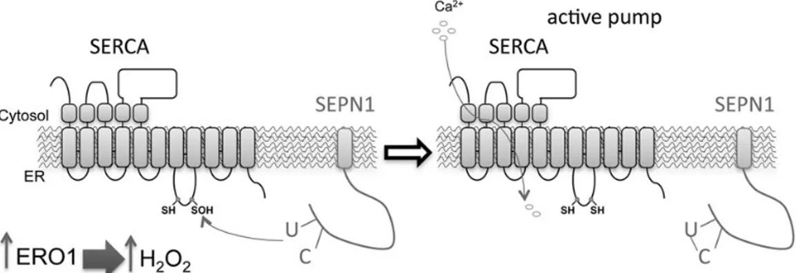
Figure 8. SEPN1 function: a working model. Within the ER, a burst of ERO1 activity (such as that occurring when UPR is elicited) increases the local concentration of
H2O2, which attacks the luminal thiols in SERCA2 and thus leads to cysteinyl sulfenic acid formation (–SOH). SEPN1 reduces the SOH back to free thiol (–SH), thus
restoring SERCA activity. In SEPN1 mutant cells, the unstable cysteinyl sulfenic acid can be further oxidized by one more molecule of H2O2to generate the oxidized
sulfenic (–SO2H), and by two more molecules of H2O2to generate the terminally oxidized sulfonic (–SO3H) SERCA2. In this oxidized state, SERCA2 is irreversibly inactivated.
at GOT (Consortium) on April 3, 2015
http://hmg.oxfordjournals.org/
preparation in three sites of the right gastrocnemius (three injec-tions of 30 µl each) using a Hamilton syringe. Equivalent doses of AAV2/1CMV-EGFP or equal volumes of PBS were injected into the contralateral muscle. The animals were sacrificed 3 weeks after being injected and perfused with PBS. Gastrocnemii, free from neighbouring muscles and connective tissue, were isolated and analyzed. One part of the sample was used to test the level of ERO1α expression by means of western blotting. The muscles were frozen in liquid nitrogen-cooled isopentane,fixed in forma-lin for morphological analysis and cross sections (8μm) of iso-pentane-frozen muscle were stained with hematoxylin and eosin (H&E) for histological evaluation. For the NADH staining, the frozen sections were incubated with nitro-blue tetrazolium (1 mg/ml) andβ-nicotinamide adenine dinucleotide (0.4 mg/ml) in 50 m Tris–HCl (pH 7.3) for 30 min at 37°C.
C2c12, SEPN1 knockdown and lentiviral transduction
C2C12 cells were cultured in DMEM supplemented with 25 m glucose and 10% fetal calf serum (FCS). SEPN1 was knocked down using Mission™ shRNA-encoding lentiviruses against mouse SEPN1 mRNA (SHCLND-NM_029100.2, Sigma) follow-ing the manufacturer’s instructions: knockdown pooled KD1 was targeted with shRNA TRCN000024786060 and KD2 with TRCN0000247862. Mock cells were obtained by means of a lenti-virus driving puro-resistance.
Blasticidin resistance-marked lentivirus, pLenti 6.3V5-TOPO (Sigma), which encodes human ERO1a was constructed from cDNA donated by Roberto Sitia (DIBIT, Milan, Italy). After trans-duction and selection with blasticidin at 1μg/ml for 7 days, the cells (in triplicate wells) werefixed and stained with crystal violet, and the relative cell mass was quantified by solubilising the dye in 0.2% Triton X-100 and measuring absorbance at 590 nm.
SEPN1 expression in eukaryotic cells
Expression plasmids encoding ER-localized and C-terminally FLAG-tagged human SEPN1 and the related mutants were constructed in pSel-Express vector (a kind gift from Vladimir Gladyshev). The SEPN1-SS and SEPN1-SC were made by means of QuiKChange site-directed mutagenesis (Stratagene). The cells transfected with SEPN1 were cultured in the presence of 0.25 m of sodium selenite in order to allow the expression of full-length SEPN1.
Detecting sulfenic acid-modified proteins in cultured cells
The sulfenic acid-modified proteins were detected using the pro-cedure of Seo and colleagues (27) and immunoblotting using a rabbit serum reactive to sulfenic acid-modified proteins (Millipore).
Isolation and analysis of control and SEPN1KO MEFs
Control and SEPN1 knockout MEFs, isolated at embryonic Day 13.5, were studied as I˚ MEFs. DMwere immortalized with SV-40
large T antigen and cultured in DMEM supplemented to 25 m glucose, 10% FCS, nonessential amino acids and, where indi-cated, were exposed to tunicamycin (0.5μg/ml) and thapsigargin (0.5 m) (Sigma).
The following I˚ immunochemical reagents were used: rabbit anti-ERO1α (25), rabbit anti-SEPN1 (Sigma), mouse anti-KDEL (Stressgen), mouse anti-PDI (Stressgen), mouse anti-β-actin
(Sigma), mouse anti-SERCA2 (Santa Cruz) and anti-GAPDH (Sigma).
Immunopurification of complexes trapped by SEPN1-FLAG and interaction with SERCA2
Expression plasmids encoding ER localized, C-terminally FLAG-tagged human SEPN1 were constructed in the pFLAG-CMV1 vec-tor (Sigma). Transfected HEK 293T cells from four confluent 100 mm plates were washed in PBS with 20 m N-ethyl malei-mide (NEM), lysed in 0.3% Triton X-100, 150 m NaCl, 20 m HEPES, pH 7.4, 20 m NEM and protease inhibitors. The FLAG-tagged proteins were immunopurified with Flag M2 affinity gel (Sigma) in an overnight incubation and eluted in Lemmli buffer. Ten percent of the eluted material was immunoblotted with anti-FLAG M2 (Sigma) following reducing and non-reducing SDS– PAGE. The co-immunoprecipitation of SEPN1 and SERCA2 was tested on the endogenous SERCA2 or transfected pig SERCA2b ( pcDNA3), pig SERCA2a ( pcDNA3) and the mutants pig SER-CA2b2m (C875S and C887S), pig SERCA2a2m (C875S and C887S) that lack the two cysteines in the L4 ER domain and was made by means of QuiKChange site direct mutagenesis (Stratagene). Expression plasmids encoding ER localized catalase–peroxidase from Magnaporthe grisea and the catalase mutated in the trypto-phan of the active site (W140F) and within frame a ro-GFP-KDEL at C terminal were constructed in pcDNA3 and were a kind gift from David Ron’s Laboratory.
Mass spectrometry
The remaining 90% of the eluted material was resolved by means of non-reducing SDS–PAGE and lightly stained with Sypro Ruby. The regions of the gel containing complexes larger than the SEPN1 dimer bait were excised, reduced in 10 m DTT, alkylated with 20 m NEM and analyzed by means of mass spectrometry. Briefly, LC–ESI–MS-MS of 5 µl of each sample was performed on a quadrupole Orbitrap Q-exactive mass spectrometer (Thermo Scientific). Peptides separation was achieved on a linear gradient from 88% solvent A (2% ACN, 0.1% formic acid) to 50% solvent B (80% acetonitrile, 0.1% formic acid) over 20 min and from 50 to 100% solvent B in 2 min at a constantflow rate of 0.25 µl/min on UHPLC Easy-nLC 1000 (Thermo Scientific) where the LC sys-tem was connected to a 23-cm fused-silica emitter of 75 µm inner diameter (New Objective, Inc. Woburn, MA, USA), packed in-house with ReproSil-Pur C18-AQ 1.9 µm beads (Dr Maisch Gmbh, Ammerbuch, Germany) using a high-pressure bomb load-er (Proxeon, Odense, Denmark).
MS data were acquired using a data-dependent top 10 method for HCD fragmentation. Survey full scan MS spectra (300–1750 Th) were acquired in the Orbitrap with 70000 resolution, AGC target 1e6, IT 120 ms.
For HCD spectra, resolution was set to 17 500 at m/z 200, AGC target 1e5, IT 120 ms; Normalized Collision energy 25%
and isolation with 3.0 m/z. Technical replicates were conducted on the LC–MS-MS part of the analysis. Raw data were processed with MaxQuant version 1.4.05. Peptides were identified from the MS-MS spectra searched against the uniprot_cp_human_ 2013_11 database using the Andromeda search engine. Cysteine N-ethylmaleimide was used asfixed modification, methionine oxidation and protein N-terminal acetylation as variable modifi-cations. Mass deviation for MS-MS peaks was set at 20 ppm and a maximum of two missed cleavages was allowed. The peptides and protein false discovery rates (FDR) were set to 0.01; the minimal length required for a peptide was six amino acids; a
at GOT (Consortium) on April 3, 2015
http://hmg.oxfordjournals.org/
minimum of two peptides and at least one unique peptide were required for high-confidence protein identification. The lists of identified proteins were filtered to eliminate reverse hits and known contaminants.
LFQ Intensities of the Ratios SC/SS was normalized by SEPN1 intensity for every conditions.
Statistical t-test analysis were done using Perseus program (version 1.4.0.20) in the MaxQuant environment. For all the stat-istical analysis was applied an FDR 0.05 using a Permutation Test (500 randomizations).
Glutathione content
Tissue and cell levels of total and oxidized glutathione were measuredfluorimetrically using the DTNB glutathione reductase recycling assay as described previously (48).
Calcium measurements
HeLa and MEFs cells were grown in Dulbecco’s modified Eagle’s medium (DMEM) supplemented with 10% FCS in 75 cm2Falcon
flasks, seeded onto 13 mm glass coverslips and allowed to grow to 50% confluence. At this stage, they were transfected with vari-ous constructs using the Ca2+phosphate technique for the HeLa cells or a MicroPorator (Digital Bio) for the MEFs.
In order to obtain efficient reconstitution of the aequorin needed to produce the functional Ca2+-sensitive luminescent
protein, the [Ca2+] in the lumen of the store was reduced 36 h after transfection by incubating the cells for 1 h at 4°C in a Krebs–Ringer buffer (KRB: 125 m NaCl, 5 m KCl, 1 m Na3PO4,
1 m MgSO4, 5.5 m glucose, 20 m HEPES, pH 7.4) containing
5μ coelenterazine, the Ca2+ionophore ionomycin and 600 µ
EGT. After this incubation, the cells were extensively washed with KRB supplemented with 2% bovine serum albumin (BSA) before measuring the luminescence. After the reconstitution step, the cells were placed in a perfused, thermostated chamber in close proximity to a low-noise photomultiplier with a built-in amplifier/discriminator. The output of the discriminator was captured using a Thorn-EMI photon counting board and stored in an IBM-compatible computer for subsequent analysis. Aequorin photon emission was calibrated off line into [Ca2+]
values using a computer algorithm based on the Ca2+response
curve of aequorins, as previously described (39,49).
Protein lysate from mouse muscles and mitochondria isolation
The muscles were mechanically disrupted using an Ultra Turrax homogenizer in RIPA buffer. The insoluble material was isolated by means of clarification at maximum speed for 5′ and the lysate was recovered and quantified using the BCA method. The mus-cles were resuspended in 8 ml mitochondria buffer (70 m su-crose, 1 m EGTA, 210 m sorbitol, 10 m MOPS, pH 7.4), and crude mitochondria were purified as described in (50). The total proteins and the mitochondrial proteins were quantified using the BCA method.
Statistics
All results are expressed as mean values ± SEM. Two-tailed Stu-dent t-tests were used to determine P-values for paired samples in the case of the experiments involving more than one inde-pendent variable.
Supplementary Material
Supplementary Material is available at HMG online.
Acknowledgements
We are indebted to David Ron as some reagents were developed in his lab and for the fruitful discussion on the results. We thank Nica Borgese for the comments to our manuscript, Roberto Sitia (Dibit, Milan, Italy) for the human ERO1 plasmid, Vladimir Gladyshev (Harvard Medical School, Boston, USA) for the psel-express vector, the EMMA repository and Alain Lescure as the provider of the SEPN1 KO mice. The help of Monica Doria (TIGEM AAV Vector Core, Napoli, Italy) for AAV vector production and Sonia Missiroli for carrying out some preliminary experi-ments is gratefully acknowledged.
Conflict of Interest statement. None declared.
Funding
Supported by a Telethon career award (TDEZ00112T) to E.Z., the Italian Association for Cancer Research (AIRC) and local funds from the University of Ferrara to P.P. and C.G., and from Telethon (GGP11139B) and the Italian Ministry of Education, Universities and Research (COFIN, FIRB and Futuro in Ricerca) to P.P.
References
1. Petit, N., Lescure, A., Rederstorff, M., Krol, A., Moghadasza-deh, B., Wewer, U.M. and Guicheney, P. (2003) Selenoprotein N: an endoplasmic reticulum glycoprotein with an early de-velopmental expression pattern. Hum. Mol. Genet., 12, 1045– 1053.
2. Castets, P., Maugenre, S., Gartioux, C., Rederstorff, M., Krol, A., Lescure, A., Tajbakhsh, S., Allamand, V. and Guicheney, P. (2009) Selenoprotein N is dynamically expressed during mouse development and detected early in muscle precur-sors. BMC Dev. Biol., 9, 46.
3. Rederstorff, M., Castets, P., Arbogast, S., Laine, J., Vassilopou-los, S., Beuvin, M., Dubourg, O., Vignaud, A., Ferry, A., Krol, A. et al. (2011) Increased muscle stress-sensitivity induced by se-lenoprotein N inactivation in mouse: a mammalian model for SEPN1-related myopathy. PLoS ONE, 6, e23094.
4. Moghadaszadeh, B., Rider, B.E., Lawlor, M.W., Childers, M.K., Grange, R.W., Gupta, K., Boukedes, S.S., Owen, C.A. and Beggs, A.H. (2013) Selenoprotein N deficiency in mice is associated with abnormal lung development. FASEB J., 27, 1585–1599.
5. Lacourciere, G.M. and Stadtman, T.C. (1999) Catalytic proper-ties of selenophosphate synthetases: comparison of the selenocysteine-containing enzyme from Haemophilus influ-enzae with the corresponding cysteine-containing enzyme from Escherichia coli. Proc. Natl. Acad. Sci. USA, 96, 44–48. 6. Arbogast, S., Beuvin, M., Fraysse, B., Zhou, H., Muntoni, F. and
Ferreiro, A. (2009) Oxidative stress in SEPN1-related myop-athy: from pathophysiology to treatment. Ann. Neurol., 65, 677–686.
7. Lescure, A., Rederstorff, M., Krol, A., Guicheney, P. and Allamand, V. (2009) Selenoprotein function and muscle dis-ease. Biochim. Biophys. Acta, 1790, 1569–1574.
8. Ashby, M.C. and Tepikin, A.V. (2001) ER calcium and the functions of intracellular organelles. Semin. Cell Dev. Biol., 12, 11–17.
at GOT (Consortium) on April 3, 2015
http://hmg.oxfordjournals.org/
9. Higo, T., Hattori, M., Nakamura, T., Natsume, T., Michikawa, T. and Mikoshiba, K. (2005) Subtype-specific and ER lumenal en-vironment-dependent regulation of inositol 1,4,5-trispho-sphate receptor type 1 by ERp44. Cell, 120, 85–98.
10. Baba-Aissa, F., Raeymaekers, L., Wuytack, F., Dode, L. and Casteels, R. (1998) Distribution and isoform diversity of the organellar Ca2+ pumps in the brain. Mol. Chem. Neuropathol., 33, 199–208.
11. Li, Y. and Camacho, P. (2004) Ca2+-dependent redox modula-tion of SERCA 2b by ERp57. J. Cell Biol., 164, 35–46.
12. Frand, A.R. and Kaiser, C.A. (1998) The ERO1 gene of yeast is required for oxidation of protein dithiols in the endoplasmic reticulum. Mol. Cell, 1, 161–170.
13. Pollard, M.G., Travers, K.J. and Weissman, J.S. (1998) Ero1p: a novel and ubiquitous protein with an essential role in oxida-tive protein folding in the endoplasmic reticulum. Mol. Cell, 1, 171–182.
14. Sevier, C.S., Qu, H., Heldman, N., Gross, E., Fass, D. and Kaiser, C.A. (2007) Modulation of cellular disulfide-bond formation and the ER redox environment by feedback regulation of Ero1. Cell, 129, 333–344.
15. Gross, E., Kastner, D.B., Kaiser, C.A. and Fass, D. (2004) Struc-ture of Ero1p, source of disulfide bonds for oxidative protein folding in the cell. Cell, 117, 601–610.
16. Tu, B.P. and Weissman, J.S. (2002) The FAD- and O(2)-dependent reaction cycle of Ero1-mediated oxidative protein folding in the endoplasmic reticulum. Mol. Cell, 10, 983–994.
17. Harding, H.P., Zhang, Y., Zeng, H., Novoa, I., Lu, P.D., Calfon, M., Sadri, N., Yun, C., Popko, B., Paules, R. et al. (2003) An inte-grated stress response regulates amino acid metabolism and resistance to oxidative stress. Mol. Cell, 11, 619–633.
18. Powers, S.K., Nelson, W.B. and Hudson, M.B. (2011) Exercise-induced oxidative stress in humans: cause and conse-quences. Free Radic. Biol. Med., 51, 942–950.
19. Kim, H.J., Jamart, C., Deldicque, L., An, G.L., Lee, Y.H., Kim, C.K., Raymackers, J.M. and Francaux, M. (2011) Endoplasmic reticu-lum stress markers and ubiquitin-proteasome pathway activity in response to a 200-km run. Med. Sci. Sports Exerc., 43, 18–25. 20. Wu, J., Ruas, J.L., Estall, J.L., Rasbach, K.A., Choi, J.H., Ye, L.,
Bostrom, P., Tyra, H.M., Crawford, R.W., Campbell, K.P. et al. (2011) The unfolded protein response mediates adaptation to exercise in skeletal muscle through a PGC-1alpha/ATF6alpha complex. Cell Metab., 13, 160–169.
21. Ron, D. and Walter, P. (2007) Signal integration in the endo-plasmic reticulum unfolded protein response. Nat. Rev. Mol. Cell Biol., 8, 519–529.
22. Curran, S.P. and Ruvkun, G. (2007) Lifespan regulation by evo-lutionarily conserved genes essential for viability. PLoS Genet., 3, e56.
23. Haynes, C.M., Titus, E.A. and Cooper, A.A. (2004) Degradation of misfolded proteins prevents ER-derived oxidative stress and cell death. Mol. Cell, 15, 767–776.
24. Marciniak, S.J., Yun, C.Y., Oyadomari, S., Novoa, I., Zhang, Y., Jungreis, R., Nagata, K., Harding, H.P. and Ron, D. (2004) CHOP induces death by promoting protein synthesis and oxidation in the stressed endoplasmic reticulum. Genes Dev., 18, 3066– 3077.
25. Zito, E., Chin, K.T., Blais, J., Harding, H.P. and Ron, D. (2010) ERO1-beta, a pancreas-specific disulfide oxidase, promotes insulin biogenesis and glucose homeostasis. J. Cell Biol., 188, 821–832.
26. Kettenhofen, N.J. and Wood, M.J. (2010) Formation, reactivity, and detection of protein sulfenic acids. Chem. Res. Toxicol., 23, 1633–1646.
27. Seo, Y.H. and Carroll, K.S. (2009) Profiling protein thiol oxida-tion in tumor cells using sulfenic acid-specific antibodies. Proc. Natl. Acad. Sci. USA, 106, 16163–16168.
28. Zito, E., Melo, E.P., Yang, Y., Wahlander, A., Neubert, T.A. and Ron, D. (2010) Oxidative protein folding by an endoplasmic re-ticulum-localized peroxiredoxin. Mol. Cell, 40, 787–797. 29. Lohman, J.R. and Remington, S.J. (2008) Development of a
family of redox-sensitive greenfluorescent protein indicators for use in relatively oxidizing subcellular environments. Bio-chemistry, 47, 8678–8688.
30. Cuozzo, J.W. and Kaiser, C.A. (1999) Competition between glutathione and protein thiols for disulphide-bond forma-tion. Nat. Cell Biol., 1, 130–135.
31. Hayes, J.D. and McLellan, L.I. (1999) Glutathione and glutathi-one-dependent enzymes represent a co-ordinately regulated defence against oxidative stress. Free Radic. Res., 31, 273–300. 32. Appenzeller-Herzog, C., Riemer, J., Christensen, B., Sorensen, E.S. and Ellgaard, L. (2008) A novel disulphide switch mechan-ism in Ero1alpha balances ER oxidation in human cells. EMBO J., 27, 2977–2987.
33. Margittai, E., Low, P., Stiller, I., Greco, A., Garcia-Manteiga, J. M., Pengo, N., Benedetti, A., Sitia, R. and Banhegyi, G. (2012) Production of H(2)O(2) in the endoplasmic reticulum promotes in vivo disulfide bond formation. Antioxid. Redox. Signal., 16, 1088–1099.
34. Lu, J., Berndt, C. and Holmgren, A. (2009) Metabolism of selenium compounds catalyzed by the mammalian seleno-protein thioredoxin reductase. Biochim. Biophys. Acta, 1790, 1513–1519.
35. Lu, J. and Holmgren, A. (2009) Selenoproteins. J. Biol. Chem., 284, 723–727.
36. Turanov, A.A., Kehr, S., Marino, S.M., Yoo, M.H., Carlson, B.A., Hatfield, D.L. and Gladyshev, V.N. (2010) Mammalian thiore-doxin reductase 1: roles in redox homoeostasis and charac-terization of cellular targets. Biochem. J., 430, 285–293. 37. Sandow, A. (1952) Excitation-contraction coupling in
muscu-lar response. Yale J. Biol. Med., 25, 176–201.
38. Jurynec, M.J., Xia, R., Mackrill, J.J., Gunther, D., Crawford, T., Flanigan, K.M., Abramson, J.J., Howard, M.T. and Grunwald, D.J. (2008) Selenoprotein N is required for ryanodine receptor calcium release channel activity in human and zebrafish muscle. Proc. Natl. Acad. Sci. USA, 105, 12485–12490.
39. Bonora, M., Giorgi, C., Bononi, A., Marchi, S., Patergnani, S., Ri-messi, A., Rizzuto, R. and Pinton, P. (2013) Subcellular calcium measurements in mammalian cells using jellyfish photopro-tein aequorin-based probes. Nat. Protoc., 8, 2105–2118. 40. Blais, J.D., Chin, K.T., Zito, E., Zhang, Y., Heldman, N., Harding, H.
P., Fass, D., Thorpe, C. and Ron, D. (2010) A small molecule in-hibitor of endoplasmic reticulum oxidation 1 (ERO1) with select-ively reversible thiol reactivity. J. Biol. Chem., 285, 20993–21003. 41. Goonasekera, S.A., Lam, C.K., Millay, D.P., Sargent, M.A.,
Haj-jar, R.J., Kranias, E.G. and Molkentin, J.D. (2011) Mitigation of muscular dystrophy in mice by SERCA overexpression in skel-etal muscle. J. Clin. Invest., 121, 1044–1052.
42. Rayavarapu, S., Coley, W. and Nagaraju, K. (2012) Endoplas-mic reticulum stress in skeletal muscle homeostasis and dis-ease. Curr. Rheumatol. Rep., 14, 238–243.
43. Tavender, T.J., Springate, J.J. and Bulleid, N.J. (2010) Recycling of peroxiredoxin IV provides a novel pathway for disulphide for-mation in the endoplasmic reticulum. EMBO J., 29, 4185–4197. 44. Zito, E., Hansen, H.G., Yeo, G.S., Fujii, J. and Ron, D. (2012) Endoplasmic reticulum thiol oxidase deficiency leads to as-corbic acid depletion and noncanonical scurvy in mice. Mol. Cell, 48, 39–51.
at GOT (Consortium) on April 3, 2015
http://hmg.oxfordjournals.org/
45. Ramming, T., Hansen, H.G., Nagata, K., Ellgaard, L. and Ap-penzeller-Herzog, C. (2014) GPx8 peroxidase prevents leakage of H2O2 from the endoplasmic reticulum. Free Radic. Biol. Med., 70, 106–116.
46. Auricchio, A., Hildinger, M., O’Connor, E., Gao, G.P. and Wil-son, J.M. (2001) Isolation of highly infectious and pure adeno-associated virus type 2 vectors with a single-step grav-ity-flow column. Hum. Gene Ther., 12, 71–76.
47. Colella, P., Trapani, I., Cesi, G., Sommella, A., Manfredi, A., Puppo, A., Iodice, C., Rossi, S., Simonelli, F., Giunti, M. et al. (2014) Efficient gene delivery to the cone-enriched pig retina by dual AAV vectors. Gene Ther., 21, 450–456.
48. Griffith, O.W. (1980) Determination of glutathione and gluta-thione disulfide using glutagluta-thione reductase and 2-vinylpyr-idine. Anal. Biochem., 106, 207–212.
49. Pinton, P., Pozzan, T. and Rizzuto, R. (1998) The Golgi appar-atus is an inositol 1,4,5-trisphosphate-sensitive Ca2+ store, with functional properties distinct from those of the endo-plasmic reticulum. EMBO J., 17, 5298–5308.
50. Jonassen, T., Marbois, B.N., Faull, K.F., Clarke, C.F. and Larsen, P.L. (2002) Development and fertility in Caenor-habditis elegans clk-1 mutants depend upon transport of dietary coenzyme Q8 to mitochondria. J. Biol. Chem., 277, 45020–45027.
at GOT (Consortium) on April 3, 2015
http://hmg.oxfordjournals.org/