Università del Piemonte Orientale “Amedeo Avogadro” Novara
Doctoral Program in Medical Sciences and Biotechnology
XXX Cycle
Chair: Prof. Marisa Gariglio
Clinical and instrumental evaluation of Botulinum Toxin type A
safety profile in post stroke spasticity rehabilitation treatment
Doctoral Thesis
Author:
Dott. Alessio Baricich Tutor:
Prof. Carlo Cisari Prof. Claudio Molinari SSD: MED/34
Reviewers: Prof. Thierry Deltombe Prof. Cristina Tassorelli
Index
Page
Abstract 3
Introduction 5
Post stroke spasticity 7
Botulinum Toxin and post stroke spasticity 8
Botulinum Toxin type A doses, spread and adverse
events in post stroke spasticity treatment 15
Personal contributions 22
Discussion 52
Conclusions and future perspectives 55
Bibliography Appendix
56 63
Abstract
Background
Post stroke spasticity (PSS) occurs approximately in 30% of stroke survivors. Spasticity varies from a subtle neurological sign to a gross increase in tone causing immobility of joints. PSS is associated with several complications, increasing care needs and utilisation of healthcare resources.
Botulinum toxin type A (BoNT-A) has been considered as an effective and safe treatment for focal spasticity in stroke survivors, with low prevalence of complications, reversibility of effect, and efficacy in reducing spastic hypertonia. Recent studies estimated that a significant percentage of patients affected by PSS could benefit from higher doses than those permitted by current country directives. However, at present time, there is no general consensus on the maximum dose of BoNT-A in terms of safety and clinical interchangeability among the three commercially approved products (abobotulinumtoxinA, onabotulinumtoxinA, incobotulinumtoxinA).
In light of these considerations, the aim of this thesis is to investigate the safety profile of BoNT-A high doses in the treatment of post stroke spasticity.
In our research activity we investigated the clinical effect of this treatment in severely affected patients, focusing on both clinical and instrumental assessment of systemic effects of BoNT-A.
Results
Although systemic BoNT-A toxicity is a rare event and as such not necessarily fatal, fear of systemic toxicity is still the most vigorous concern against application of increased BoNT-A doses.
Current evidence coming from published literature, considering both clinical and instrumental analysis of BoNT-A systemic diffusion, suggests that higher doses of BoNT-A are efficacious in reducing spasticity of the upper and lower limbs after stroke, with rare occurence of mild adverse effect.
Conclusions
The evidence coming from published studies suggests that use of doses of BoNT-A higher than those reported in product labels could be considered as a safe therapeutic option to reduce multifocal or generalized post stroke spasticity in selected patients. The clinicians have to carefully define the clinical goal before starting with BoNT-A treatment, considering all the factors which could affect the safety profile of BoNT-A.
Further evidence is mandatory to confirm higher doses of BoNT-A as a safe and effective therapeutic option for the treatment of post stroke spasticity. In particular, it should be pointed out the potential role of higher doses of BoNT-A in order to improve the functional outcome of these patients.
Riassunto
Introduzione
La spasticità post ictus (PSS) è osservata in circa il 30% dei soggetti con esiti di stroke.
La presentazione può variare da un lieve incremento del tono muscolare ad una immobilizzazione di segmenti articolari, ed è causa di significative complicanze e di incremento di costi assistenziali e sanitari.
La tossina botulinica di tipo A (BoNT-A) è un trattamento sicuro ed efficace nel trattamento della PSS focale. Da studi recenti emerge che una significativa percentuale di pazienti potrebbe trarre benefici dall’utilizzo di dosi di BoNT-A superiori a quelle indicate in scheda tecnica. Tuttavia, non vi sono attualmente pareri unanimi in merito alla massima dose utilizzabile ed alla intercambiabilità fra le BoNT-A in commercio (abobotulinumtoxinA, onabotulinumtoxinA, incobotulinumtoxinA). Obiettivo della tesi è la valutazione del profilo di sicurezza delle alte dosi di BoNT-A nel trattamento della PSS. Nella nostra attività di ricerca abbiamo analizzato l’effetto clinico di questo trattamento in pazienti affetti da PSS severa multifocale; particolare attenzione è stata rivolta alla valutazione clinica e strumentale degli effetti sistemici di BoNT-A.
Risultati
La tossicità sistemica è un evento raro e non sempre fatale, ma il timore della sua insorgenza è tuttora la maggiore criticità legata all’applicazione di alte dosi di BoNT-A.
Le attuali evidenze, che considerano sia una valutazione clinica che una valutazione strumentale della diffusione sistemica di BoNT-A, suggeriscono che alte dosi di BoNT-A sono efficaci nella riduzione della PSS all’arto superiore ed inferiore, con una bassa incidenza di effetti avversi.
Conclusioni
Le attuali evidenze suggeriscono che l’utilizzo di dosi di BoNT-A superiori a quelle indicate in scheda tecnica possono essere considerate come un’opzione terapeutica sicura ed efficace nel trattamento di PSS in pazienti selezionati. I clinici devono definire con accuratezza gli obiettivi terapeutici, considerando tutti i fattori che possono influenzare il profilo di sicurezza del farmaco. Ulteriori evidenze sono necessarie per confermare il profilo di efficacia e sicurezza delle alte dosi di BoNT-A nel trattamento della spasticità focale post ictus. Particolare attenzione deve essere rivolta a definire il ruolo delle alte dosi di BoNT-A nel miglioramento dell’outcome funzionale del paziente.
Introduction
Stroke-related disability is a significant health problem with relevant socioeconomic consequences for patients as well as society with long-lasting effects.
More than two-thirds of stroke survivors develop poststroke sequelae, including impaired motor function and spasticity. These impairments have a significant impact on a stroke survivor’s daily life, such as eating, walking and self-care. In addition, these disabilities involve a significant burden on caregivers of these patients [Wissel et al, 2013].
Post stroke spasticity (PSS) occurs approximately in 30% of stroke survivors. Spasticity varies from a subtle neurological sign to a gross increase in tone causing immobility of joints. PSS is associated with several complications, increasing care needs and utilisation of healthcare resources [Lundstrom et al 2010], and carers of patients with spasticity are more likely to experience anxiety and depression [Denno et al, 2013].
Management of spasticity requires a balanced approach, weighing the benefits of treatment against the side effects.
Botulinum toxin type A (BoNT-A) has been considered as an effective and safe treatment for focal spasticity in stroke survivors, with low prevalence of complications, reversibility, and efficacy in reducing spastic hypertonia [Santamato et al, 2015], with the approval of the U.S. Food and Drug Administration and the European regulatory agencies for this indication. However, at present time, there is no general consensus on the maximum dose of BoNT-A in terms of safety and clinical interchangeability among the three commercially approved products (abobotulinumtoxinA, onabotulinumtoxinA, incobotulinumtoxinA).
A recent survey [Picelli A, Baricich A et al, 2017] suggested that there is a need to reconsider the maximum dose administered per single treatment in order to improve the clinical outcome of treated patients. In fact, the use of high doses of BoNT-A is an established practice that, moreover, addresses the very real need to improve the quality of life of patients with post-stroke spasticity [Baricich et al., 2015]. It has been reported that a high percentage of patients (up to about 62%) needs a combined BoNT-A treatment of upper and lower limb, while only a very low proportion (< 25% on average) requires treatment in the upper or lower limb alone in a single session; in addition, anecdotal, unpublished, 10- year follow-up observations showed that a tendency to increase BoNT-A doses over time was paralleled by a tendency of patients to be more satisfied [Picelli A, Baricich A et al, 2017]. However, the most important adverse effect of BoNT-A is the systemic diffusion of the toxin and it has been suggested [Lange et al, 1987] a potential relationship with BoNT-A dose, causing a possible increase of adverse events after injection.
Aim of the thesis
In light of these considerations, the aim of this thesis is to investigate the safety profile of BoNT-A high doses in the rehabilitation treatment of post stroke spasticity, focusing on both clinical and instrumental assessment of systemic effects of BoNT-A.
Presentation of data derived from published studies will be integrated with the findings obtained by our group.
Post stroke spasticity
The term spasticity as a clinical entity was proposed by J.W. Lance in the 1980s as “a motor disorder characterized by a velocity-dependent increase in tonic stretch reflex (muscle tone) with exaggerated tendon jerks, resulting from hyperexcitability of the stretch reflex as one component of the upper motor neuron syndrome (UMNS)”. In clinical practice, spasticity describes a combination of symptoms and clinical signs after lesion formation in sensorimotor brain areas and tracts in the Central Nervous System (CNS), resulting from impaired reflex function; In addition, spasticity induces changes in rheological muscle properties like stiffness, fibrosis and atrophy [Dietz and Sinkjaer, 2007].
There is still no consensus for the definition of spasticity and this reflects the complexity and the diversity of the phenomena [Baricich et al, 2016; Picelli A, Vallies G et al, 2017]. This is especially true for post-stroke motor disorders, which can show a considerable variety of symptoms (e.g. clonus, dystonia, muscle weakness, abnormal reflex responses).
In general, the ‘upper motor neuron syndrome’ can be defined by the presence of positive and negative signs [Young, 1994]. Spasticity is part of the positive signs among other motor symptoms which occur after lesions in the descending corticospinal system such as spastic dystonia (muscle constriction in the absence of any voluntary movement), spastic co-contraction (contraction of both the agonist and antagonist muscles resulting from an abnormal pattern of commands in the descending supraspinal pathway), extensor or flexor spasms, clonus, exaggerated deep tendon reflexes and associated reaction [McComas, 1994; Sommerfeld et al, 1994]. On the other hand, negative signs are muscle weakness, loss of dexterity and fatigue.
Prevalence estimates of post stroke spasticity (PSS) were highly variable, ranging from 4% to 42.6%, with the prevalence of disabling spasticity ranging from 2% to 13%. Data on phases of the PSS continuum revealed evidence of PSS in 4% to 27% of those in the early time course (1–4 weeks post stroke), 19% to 26.7% of those in the postacute phase (1–3 months post stroke), and 17% to 42.6% of those in the chronic phase (>3 months post stroke) [Wissel et al, 2013].
In the upper limbs, the most frequently observed pattern is internal rotation and adduction of the shoulder coupled with flexion at the elbow, the wrist and the fingers; in the lower limbs, adduction and extension of the knee with equinovarus foot [Thibaut et al, 2013].
can limit the success of rehabilitation. Spasticity can also affect quality of life and be highly detrimental to daily function [Duncan et al, 2005; Langhorne, 2011; Chae and Celnik, 2015]. However, there is currently a lack of specific guidelines for the stratification and individualization of rehabilitation programmes [Thibaut, 2013; Picelli A, Baricich A et al, 2017].
Therapeutic interventions include physical therapy, occupational therapy, self-rehabilitation, orthoses equipment and assistive devices, pharmacological treatment, orthopaedic surgery and neurosurgery [Thibaut et al, 2013; Deltombe et al, 2017].
Botulinum Toxin and post stroke spasticity
Botulinum Toxin type A (BoNT-A) administered by intramuscular injection, is the gold standard for the treatment of focal spasticity, with low prevalence of complications, reversibility, and efficacy in reducing spastic hypertonia with the approval of the U.S. Food and Drug Administration and the European regulatory agencies for this indication.
BoNT-A showed to increase patient’s ability to actively mobilize their upper and lower limbs and improve their autonomy (e.g. self-care, walking) [Thibaut et al, 2013; Simpson et al, 2016].
Pharmacology and immunology of Botulinum Toxin
Botulinum neurotoxin (BoNT) is a microbial protein which exists in seven different serotypes, designated A through G. Although the individual serotypes are immunologically distinct, all members of the group present similar subunit structures, act on the same target organs, and produce similar functional outcomes [Lacy and Stevens, 1999; Johnson and Bradshaw, 2009]. Each molecule is typically released from bacteria as part of a noncovalent complex with other associated proteins. These auxiliary proteins do not play an active role in the therapeutic actions of the toxin, even if it was hypothesized a possible involvement in undesirable effects.
BoNT is an enzyme which acts in the cytosol of nerve endings: it cleaves three polypeptides governing exocytosis. Serotypes A and E cleave synaptosomal-associated protein (SNAP)-25, serotypes B, D, F, and G cleave vesicle-associated membrane protein (VAMP), and serotype C cleaves both syntaxin and SNAP-25 [Simpson, 2004; Humeau et al, 2000].
The block of acetylcholine release at neuro-muscular junctions is the mechanism involved in the therapeutic effect of BoNT to relieve dystonia, spasticity, and related disorders [Tassorelli et al, 2006].
BoNT showed additional therapeutic benefits, not necessarily related to neuromuscular transmission, including blockade of acetilcholine release at autonomic nerve endings and blockade of transmitter release at peripheral nerve endings that use other mediators.
In addition to peripheral effects of BoNT, indirect effects on CNS have been observed, probably resulting from changes in the normal balance of efferent and afferent signals. Interestingly, both the direct and indirect actions of the toxin are largely or completely reversible [Simpson et al, 2008]. At the present time, BoNT is commercially available in 2 serotypes, A and B. In United States, Food and Drug Administration approved four preparations of BoNT: onabotulinumtoxinA (Botox®, Allergan, Inc., United States), abobotulinumtoxinA (Dysport®, Ipsen, France), incobotulinumtoxinA (Xeomin®, Merz Pharmaceuticals GmbH, Germany), and rimabotulinumtoxinB (Myobloc/Neurobloc® (US WorldMeds/Solstice Neurosciences, United States). [Table 1]
Table 1: Botulinum toxins and FDA-approved indications (modified from Simpson et al, 2016)
BoNT preparation Brand name (manufacturer) FDA approved indications
OnabotulinumtoxinA Botox (Allergan, Inc., Irvine, CA)
Blepharospasm, cervical dystonia, upper extremity spasticity, lower extremity spasticity, chronic migraine, treatment of urinary incontinence due to detrusor overactivity, axillary hyperhidrosis, strabismus AbobotulinumtoxinA Dysport (Ipsen Ltd., Paris, France) Cervical dystonia, spasticity
IncobotulinumtoxinA Xeomin (Merz Pharmaceuticals, Frankfurt, Germany)
Blepharospasm, cervical dystonia, upper extremity spasticity
RimabotulinumtoxinB
Myobloc/Neurobloc (US WorldMeds/Solstice
Neurosciences, Louisville, KY)
BoNT-A is approved for cervical dystonia and spasticity, whereas BoNT-B was approved for cervical dystonia only.
In nature, BoNT-A is synthesized as macromolecular protein complexes [Aoki and Guyer, 2001]. These protein complexes are referred to as progenitor toxins and consist of nontoxic accessory proteins (NAPs) bonded to the 150-kD active neurotoxin.
The BoNT-A progenitor toxins vary in molecular weight (300–900 kD) depending on the composition of NAPs and the manufacturing process [Dressler and Benecke, 2007]. The 150-kD neurotoxin must dissociate from NAPs in order to exert its pharmacologic effects. This dissociation occurs in physiologic pH conditions.
Although no clear differences in effectiveness between the various formulations were demonstrated, their comparability is still intensely debated, focusing on several issues such as potency, dose equivalence, immunogenicity, spread and systemic diffusion.
Potency
Although the various BoNT-A products differ in NAP composition, the 150-kD neurotoxin is the active part inhibiting acetylcholine release.
Since the toxin moiety is the same in all pharmaceutical preparations, differences in potency could depend of the amount of active toxin available. To become fully activated, the single chain 150-kD neurotoxin must be cleaved from the protein complex. All of the commercially available BoNT-A formulations are composed of the 150-kD neurotoxin with NAPs; the only exception is incobotulinumtoxinA, which contains only the 150-kD neurotoxin.
However, also the manufacturing process may affect the amount of active toxin; for instance, enzymes added to increase the percentage of cleaved active toxin may denature the neurotoxic protein itself.
BoNT-A formulations contain different percentages of inactive toxin which contribute to the overall protein load. For this reason, the potency is expressed in biological units. Potency is related to the quantity of toxin (in ng of protein content, i.e., 150 kD neurotoxin including NAPs) required to achieve a median lethal dose (LD50) unit [Sesardic et al, 2003; McLellan et al, 1996]. However, many factors affect the mouse LD50 bioassay including mouse strain, sex, age, volume and route of injection, time of examination after injection, and delivery vehicle or reconstituting buffer. Moreover, the LD50 units of BoNT products are not standardized across manufacturers.
Due to the lack of LD50 bioassay harmonization, the unit potencies of BoNT formulations cannot easily be compared. For this reason, it is mandatory that physicians consider that, even if the active
molecule is botulinum neurotoxin type A, different forms of the complex can affect the therapeutic profiles. In fact, as prevously described, there are several BoNT-A products on the market: onabotulinumtoxinA (ONA), incobotulinumtoxinA (INCO) and abobotulinumtoxinA (ABO) [Albanese, 2011].
However, despite the difficulties related to the biologic units, the most informative comparisons of BoNT-A products have been made in clinical studies.
Dose equivalence
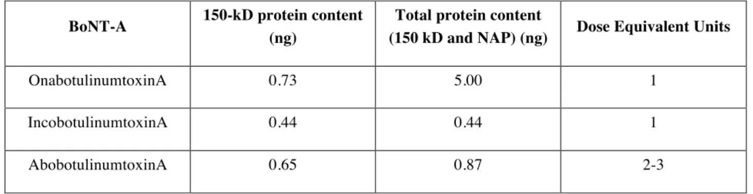
Each BoNT-A formulation contains different amounts of the 150-kD toxin (and NAPs)/LD50 unit (Table 2). However, although there are some difficulties establishing the comparative potencies, the equivalence ratio of the dose should be established.
There are several reasons for identifying a conversion factor: medical (i.e., patients may need to switch to another formulation) as well as economical (an incorrect conversion factor may negatively impact the real cost of treatment) [Chen and Dashtipour, 2013; Frevert, 2015].
Table 2: Botulinum toxin products and protein content/100 units (Adapted from Scaglione, 2016)
BoNT-A 150-kD protein content (ng)
Total protein content
(150 kD and NAP) (ng) Dose Equivalent Units
OnabotulinumtoxinA 0.73 5.00 1
IncobotulinumtoxinA 0.44 0.44 1
AbobotulinumtoxinA 0.65 0.87 2-3
NAP: nontoxic accessory proteins
INCO has been shown to be as effective as ONA with a comparable adverse event profile with a clinical conversion ratio of 1:1 or 1:1.2 [Benecke et al, 2005; Roggenkamper et al, 2006; Jost et al, 2005; Park et al, 2011; Zoons et al, 2012]. Clinical results are consistent with preclinical comparability data [Dressler and Benecke, 2007; Dressler et al, 2012]. Thus, both clinical and preclinical analyses have demonstrated a clinical conversion ratio between ONA and INCO very close to 1:1.
In contrast, the conversion ratio between ONA (or INCO, consequently) and ABO is highly debated. Even if the most commonly used conversion ratios are 1:3 or 1:4 [Aoki et al, 2006], they ranged from 1:1 [Wohlfarth et al, 2007] to as high as 1:11 [Marchetti et al, 2005]. This wide conversion ratio range
reflects real-life clinical practice; the treating physician determines the number of muscles to be treated and the empiric dose based on each patient’s conditions, their clinical pattern, and treatment goals.
Although the various BoNT products differ in NAP composition, the toxins ultimately inhibit acetylcholine release. Since the active toxin content is established for each product, a conversion rate should be defined. More precise estimation of conversion ratios should also ensure the development of comparable clinical data on the efficacy and safety of currently available BoNT-A formulations since they have qualitatively and quantitatively similar clinical efficacies and side effects at equipotent doses.
A large number of studies have reported an ONA:ABO conversion factor of 1:3 with clinical equivalence [Marion et al, 1995; Whurr et al, 1995; Kollewe et al, 2010; Odergren et al, 1998; Shin et al, 2009]. Moreover, when the conversion factor is close to 1:3, ABO showed higher efficacy [Wohlfarth et al, 2008; Mohammadi et l, 2009; Rystedt et al, 2012], indicating that the conversion factor could be rather lower than equal to 1:3. Interestingly, studies where the conversion ratio was higher than 1:3 showed higher efficacy and longer duration of action of ABO compared to ONA, but with more adverse events, supposing an overdose of ABO determined by this conversion ratio [Sampaio et al, 1997; Nussgens et al, 1997; Ranoux et al, 2002; Bentivoglio et al, 2012].
These clinical data are consistent with preclinical data where a conversion ratio for ONA/ABO of 1:3 or lower has been found [Van den Berg an Lison, 1998; Rosales et al, 2006; Wohlfarth et al, 2009; Keren-Capelovitch et al, 2010, Brockmann et al, 2012; Kollewe et l, 2015; Rystedt et al, 2015; Yun et al, 2015; Hambleton and Pickett, 1994].
In conclusion, current data suggest that a conversion ratio ONA/ABO of 1:3—or even lower—is appropriate for treating spasticity, cervical dystonia, and blepharospasm or hemifacial spasm. A higher conversion ratio may lead to an excessive ABO dose (with the potential for an increased incidence of adverse events) or underdosing when switching ABO to ONA [Scaglione, 2016].
Immunogenicity
A possible reason for secondary treatment failure of any therapeutic protein is its neutralization [Kromminga and Schellekens, 2005]. Antibodies that block its pharmacological effects are termed neutralizing antibodies, addressed against the active toxin. In this case, the clinical effect may wane gradually, eventually leading to complete treatment failure.
In a study of 27 patients with complete treatment failure due to neutralizing antibodies, the majority (81%) of patients had previously experienced partial antibody-induced treatment failure [Dressler, 2002]. Most patients in this study developed treatment failure within 40 months of starting BoNT
treatment.
However, another study reported a high mean clinical benefit similar for ABO and ONA and <2% of the patients developed neutralizing antibodies [Mohammadi et al, 2009].
However, in more recent investigations, BoNT-A antibodies were not detected [Bakheit et al, 2012; Wissel et al, 2017].
The debate regarding immunogenicity includes the role of the non-toxic proteins, collectively referred to as complexing proteins or neurotoxin-associated proteins (NAPs). Under physiological pH conditions, the complexing proteins dissociate from the neurotoxin after constitution with saline and even before injection [Eisele et al, 2011; Benecke, 2012]. Complexing proteins are not expected to modify clinical outcomes, and specific antibodies generated against the complexing proteins are termed non-neutralizing and should not affect the secondary response. However, it has been argued that complexing proteins may increase the bacterial protein load and could potentially increase the immunogenic risk of neutralizing antibody formation [Kukreja et al, 2009]. Even if several studies have been conducted, there are no clear demonstrations that NAPs modify the immunogenicity of the active toxin [Atassi, 2004; Atassi, 2006; Bigalke, 2009]. However, these studies revealed that the toxoid complex is more immunogenic than the purified neurotoxin. This could be relevant considering that cross-reactivity may occur between the toxoid and toxin. However, despite the considerations mentioned above, the risk of immunogenicity of BoNT-A is very low in clinical practice as reported by a large data review [Jankovic et al, 2004; Wissel et al, 2017].
BoNT and systemic diffusion
BoNT ability to remain relatively localized at the site of injection is largely responsible for its remarkable safety profile. In general, spread and diffusion are supposed to underlie most of the local, distal, and systemic effects of BoNT [Ramirez-Castaneda et al, 2013].
BoNT spread (also called diffusion) describes the toxin’s effect on areas away from the injection site. The potential risk for adverse effects due to toxin spread is described in the labeling for each BoNT-A product [Scaglione, 2016]. However, the mechanism of this phenomenon is not completely understood.
Spread to contiguous areas could increase the risk of adverse effects. For example, spread from injections in the cervical or craniofacial musculature may induce diplopia, dysarthria, or dysphagia, whereas injections in extremities could induce weakness in non-treated, close muscles.
Although uncommon, distant spread can occur, causing unintended neuromuscular blockade remote to the injection site. For example, systemic botulism symptoms such as dysphagia can occur when the toxin is injected at a distant site (e.g., lower extremities for spasticity). Even if generalized
weakness is a rare occurrence after BoNT injections, Bhatia and colleagues reported the case of three patients who developed generalized muscle weakness, drawing attention to the rare possibility of mild botulism after treatment with BoNT-A for dystonia [Bhatia et al, 1999]. Moreover, flu-like symptoms (another possible sign of distant spread or a systemic immune response) vary widely in different studies: 1.7% to 20% of patients treated with various preparations of BoNT-A, and 5% to 55% of patients receiving BoNT-B, reported mild to moderate flu-like symptoms [Baizabal-Carvallo et al, 2011].
In addition, although BoNT probably does not cross the blood–brain barrier [Truong et al, 2009], Hristova et al reported three cases of encephalitic clinical features that occurred shortly after treatment of focal dystonia with BoNT-A [Hristova et al, 2012].
It must be highlighted that any potential differences in toxin spread characteristics among the different BoNT would be clinically relevant.
Differences in the potential for contiguous spread among the BoNT products have been studied, but at the moment there is no clear evidence that differentiates the various products. It has been hypothesized that diffusion of neurotoxin into adjacent tissue is slower with the high molecular weight complex compared with the lower molecular weight or free neurotoxin [Dressler et al, 2012]. Therefore, theoretically, ONA with the highest complex size of 900 kD should be less diffusible, whereas INCO containing only the 150-kD neurotoxin (without NAPs) should be the most diffusible, with a higher rate of side effects related to toxin spread. However, this has not been demonstrated. On the other hand, progenitor toxin size may be irrelevant with regard to toxin diffusion, because all BoNT progenitor complexes immediately dissociate following injection [Wagman and Bateman, 1953]; in addition, dissociation probably occurs in the vial on reconstitution with normal saline [Eisele et al, 2011]. This is consistent with data from an animal model, in which there were no significant differences in the field of effect among ABO, INCO, and ONA [Carli et al, 2009]. However, several factors other than the pharmaceutical preparation such as dose, dilution, injection technique, target site, location of injection within the muscle, level of muscle hyperactivity, depth of injection, and post-injection rehabilitation could influence the potential for spread [Roche et al, 2008; Pickett, 2009; Brodsky et al 2012; Baricich et al, 2015].
BoNT-A doses, spread and adverse events in Post stroke spasticity treatment
As reported above, in case of focal post stroke spasticity, BoNT-A injection is the gold standard therapy, with low prevalence of complications, reversibility, and efficacy in reducing spastic hypertonia [Simpson et al, 2016], with the approval of the U.S. Food and Drug Administration and the European regulatory agencies for this indication.
Current guidelines suggest the employment of a dose up to 600 units (U) of onabotulinumtoxinA (Botox®, Allergan, Inc., United States) and incobotulinumtoxinA (Xeomin®, Merz Pharmaceuticals GmbH, Germany) or up to 1500 U of abobotulinumtoxinA (Dysport®, Ipsen, France) per injection session to treat spasticity after stroke [Wissel et al, 2009]. However, in recent years, higher doses were used, especially in case of upper and lower limb severe spasticity. It is known that low doses of BoNT-A can be used to increase motor function in those patients affect by spasticity graded 1 or 2 as measured by Modified Ashworth Scale (MAS) [Bohannon and Smith, 1987], whereas in the case of severe spasticity elevated doses of BONT-A may be useful to improve limb posture, to apply splinting, to consent hygiene, to increase passive articular range of motion, to walk and stand in patients with spastic equino-varus foot deformities, to improve joint range of motion and muscle extensibility or to reduce spasticity-related pain [Aoki, 2005]. However, many clinicians suggested that higher doses of BoNT-A may cause generalized, adverse effects [Hesse et al, 1995; Mancini et al, 2005; Varghese-Kroll and Elovic, 2009; Crowner et al, 2010; Thomas and Simpson, 2012]. In particular, Lange and colleagues (1987) hypothesized a possible relationship between BoNT-A dose and systemic effects, even if a clear relationship between dose and severity of symptoms was observed.
In light of these considerations, the assessment of patients treated with high doses of BoNT-A should include a systematic evaluation of the presence of undesired, adverse events. Moreover, in association with clinical assessment, a non-invasive, instrumental evaluation should be considered to detect also subclinical diffusion of BoNT-A, in order to better undestand the pathophysiological mechanisms possibly involved in systemic side effects.
Clinical assessment
Hesse and colleagues [1995] published one of the first studies focused on high doses of BoNT-A. All patients of the group treated with 2000 U of AbobotulinumtoxinA (n=5) completed the study. Four weeks after injection they reported a muscle tone reduction, improved gait velocity, stride length, stance- and swing-symmetry without adverse effects, whereas a patient of the other group (treated with 1500 U) developed a bladder paresis, requiring catheterization for 14 days.
In a randomised, double-blind, dose-ranging study, Mancini and colleagues [2005] treated 45 patients with three different doses of OnabotulinumtoxinA, on the basis of suggestions in the literature. All the groups showed significant improvements after treatment. Group II (mean BoNT-A total dose: 322 U) and Group III (mean dose: 540 U) showed a greater and more prolonged response than Group I (mean dose: 167 U). However, Group III showed the highest rate of adverse effects 4 weeks post-treatment (prolonged weakness of the treated limb, flu-like syndrome and oedema of the injected leg). Varghese-Kroll and Elovic (2009) reported the first known case of repeated, contralateral weakness and fatigue after high-dose BoNT-A injection. A 53-yr-old woman developed contralateral weakness and fatigue, without autonomic symptoms, 2 weeks after receiving an injection with 800 units of onabotulinumtoxinA for management of poststroke spasticity. The patient reported resolution 4 weeks later. The patient experienced the same, contralateral symptoms more than a year later, after a 500 U injection, which took a similar length of time to resolve. Interestingly, three previous injections of onabotulinumtoxinA of 700 U, 500 U, and 600 U that were spaced three months apart were well tolerated.
In a case series, Crowner and colleagues (2010) described the adverse effect (difficulty getting on/off his bus) of a 16-year old male treated with 640 U of onabotulinumtoxinA into the left flexor carpi radialis, flexor carpi ulnaris, pronator teres, flexor digitorum superficialis, biceps, brachioradialis, and quadriceps muscles. These symptoms lasted only one month. Interestingly, also in this study the Authors reported a previous, well tolerated injection of 635–640 U of BoNT-A. After a re-injection of 650 U of onabotulinumtoxinA into the same muscles, the patient presented weakness in both upper and lower extremities, dysarthria, and increased falls and gait instability after the injection. Twelve weeks post-injection, he had continued difficulty ascending stairs but was no longer falling and had regained full strength in his upper extremities.
Thomas and Simpson also described [2012] contralateral weakness following repetitive onabotulinumtoxinA administrations in two patients affected by post-stroke spasticity.
In the first case report, a 43-year-old woman, treated for more than one year with 575–700 U of OnabotulinumtoxinA into the upper and lower limb muscles without adverse effects, developed after a re-injection of 700 U total dose, contralateral weakness in the shoulder girdle and distal arm; generalized weakness, bulbar, respiratory, sphincter, pain, sensory symptoms, or other systemic symptoms were not reported.
In the second case report, a 21-year-old woman with post-stroke spasticity and dystonia did not report adverse effects with total doses ranging from 550 to 700 U of onabotulinumtoxinA into the proximal upper limb muscles. However, after a new treatment in the same muscles with a total dose of 700 U, she reported weakness of her non-treated right arm, starting within days after the last injection. She
did not report neck pain, radiating symptoms to the right upper extremity, sensory disturbances, diplopia, dysphagia, or shortness of breath. The same symptoms were reported after the injection of 600 U. However, no adverse effects were described with 500 U of onabotulinumtoxinA, avoiding any muscles proximal to the elbow. In this case, the Authors hypothesized the development of contralateral limb weakness for diffusion of BoNT-A through tissue planes from proximal upper extremity muscles, across the midline, to contralateral muscles.
On the other hand, in recent years, many experienced clinicians investigated the effects of higher BoNT-A doses. In fact, as evidenced in expert consensus panel reviews [Wissel et al, 2009; Santamato et al, 2015] the recommended doses of BoNT-A in the product label reflect older clinical trials, whereas the clinical management of the patients could require higher doses in order to improve patients’ clinical outcome and quality of life [Picelli A, Baricich A et al, 2017]. Interestingly, in a recent survey, Bensmail and colleagues [2014] estimated that 24.6% of the patients could benefit from higher doses than those permitted by current country directives.
In a prospective, non-randomized, open-label study, Santamato and colleagues [2013] described the safety and efficacy of higher doses (ranged from 750 to 840 U) of incobotulinumtoxinA in 25 subjects with upper and lower limb spasticity after stroke. The patients were treated under ultrasound guide in several muscles of the upper and lower limbs, reporting after 30 days of follow-up, a substantial improvement in functional disability, spasticity-related pain, and muscle tone. Only 16% of patients experienced treatment-emergent, mild adverse events (injection site pain, muscular weakness), resolved in a few days.
Intiso and colleagues [2014] reported the effectiveness of high doses (up to 840 U) of incobotulinumtoxinA to treat spasticity due to brain injury or cerebral palsy. A significant reduction of muscle hypertone and pain was observed, but global functionality and arm dexterity were unchanged. Three patients (13.6%) complained of adverse events: of these, 2 subjects had local side effects consisting of injection site hematoma and one subject complained of weakness and reduction of active motility of the injected arm lasting for 2 weeks. No generalized side effects were observed. Dressler and colleagues [2015] demonstrated that high doses of incobotulinumtoxinA (minimum 400 U and maximum 1200 U), injected into fifty-four patients suffering from spasticity of several etiologies, did not cause any generalized effects which could be attributed to BoNT therapy or complete secondary therapy failure. The Authors concluded that generalised weakness, being bedridden, feeling of residual urine and constipation were caused by the underlying tetra- or
paraparesis, blurred vision by presbyopia. Neurologic examination, serum chemistry and full blood count did not indicate any systemic adverse effects.
In a retrospective analysis [Baricich et al, 2015], we evaluated the efficacy and safety of high doses of onabotulinumtoxinA (from 600 to 800 units) in 26 patients affected by upper and/or lower limb post-stroke spasticity. They were assessed before, 30 and 90 days after treatment. We observed a significant muscle tone reduction and a significant functional improvement. No adverse events were reported.
In a recent study, Wissel and colleagues [2017] evaluated safety (primary objective) and efficacy of increasing doses (400 U up to 800 U) of incobotulinumtoxinA for patients with limb spasticity. In this prospective, single-arm, dose-titration study, patients (18-80 years) with spasticity due to cerebral causes, who were clinically deemed to require total doses of 800 U incobotulinumtoxinA, received 3 consecutive injection cycles with 400 U, 600 U, and 800 U incobotulinumtoxinA, respectively, each followed by 12-16 weeks' observation. In total, 155 patients were enrolled. IncobotulinumtoxinA dose escalation did not lead to an increased incidence of treatment-related AEs. No treatment-related serious AEs occurred. The Authors concluded that escalating incobotulinumtoxinA doses (400 U up to 800 U) did not compromise safety or tolerability, enabling treatment in a greater number of muscles/spasticity patterns with increased treatment efficacy.
However, the available evidence mainly referred to a single set of injections evaluating the efficacy and safety of BoNT-A. Interestingly, in a recent prospective, non-randomized, open-label study, Santamato and colleagues [2017] studied the safety of repeated higher doses of incobotulinumtoxinA in post-stroke upper and lower limb spasticity. Two years after the first set of injections, they evaluated in 20 stroke survivors with upper and lower limb spasticity the long-term safety of repeated high doses of incobotulinumtoxinA (up to 840 U) for a total of eight sets of injections. In a two-year follow-up, repeated high doses of incobotulinumtoxinA, administered for eight sets of injections, appeared to be safe in patients with upper and lower limb spasticity after stroke without general adverse effects.
Instrumental assessment
As previously stated, the clinical manifestations of the systemic spread of BoNT-A, or remote effects, can be detected in various forms [Castaneda-Ramirez et al, 2013].
In the past years, several studies analyzed the subclinical impairment of endplate function in non-injected muscles by the use of neurophysiologic studies.
neuromuscular transmission in muscles remote from the site of BoNT injections [Sanders, 2002]. A double-blind, placebo- controlled study of SFEMG changes in 42 patients assessed the efficacy of BoNT injections for cervical dystonia [Lange et al, 1991]. SFEMG was performed in a limb muscle before treatment and 2 weeks and 12 weeks after the injection of placebo or BoNT. Before and after treatment, the mean jitter was unchanged in the placebo group, whereas the mean jitter had a maximal increase after two weeks of BoNTA treatment and was still elevated after 12 weeks. The fiber density did not change in any patient during the study, and there were no remote clinical effects of BoNT. Girlanda and colleagues [1992] evaluated the distal effects of BoNT on neuromuscular transmission and on autonomic function in five patients who received BoNT-A injections for craniocervical dystonia and hemifacial spasm. Detection of increased neuromuscular jitter by single-fiber electromyography (SFEMG) on the extensor digitorum communis muscle and six tests of cardiovascular reflexes were performed The Authors reported that BoNT-A injections induced an increase in mean jitter value above normal limits in all patients as well as an increase of fiber density recorded six weeks after the treatment. However, the Authors conducted this study with BoNT-A Oculinum, which is significantly different from ONA [Borodic et al, 1996].
Garner and colleagues [1993] analyzed repeated SFEMG in the extensor digitorum brevis muscle of eight patients who received a small dose of BoNT-A as therapy for focal dystonias in the head/neck region. They observed an increase of jitter and blocking in six of those patients. Fiber density of the extensor digitorum communis muscle on alternating sides showed a tendency to increase after BoNT-A injection [Garner et al 1993]
In addition to SFEMG, quantitative electromyography (EMG) has been described as a measure for distant effects of BoNT. A group of 27 patients with cervical dystonia was followed over an average of 31 months [Erdal et al, 1999]. They received repeated, unilateral BoNT-A injections of the sternocleidomastoid muscle (SCM), and quantitative EMG at rest and at maximal contraction were recorded. The study demonstrated no cumulative chemodenervation by repeated BoNT injections of the SCM measured by quantitative EMG. However, the contralateral, non-injected SCM showed significant reduction of quantitative EMG parameters, suggesting a functional weakening after long-term treatment.
Interestingly, since a frequent target of BoNT-A during botulism is the autonomic nervous system [Vita et al, 1987], several researchers have investigated autonomic function in patients with cervical dystonia receiving BoNT-A and BoNT- B, showing controversial evidence about the development of signs of subclinical diffusion.
In particular, heart rate variability (HRV), a simple and non-invasive electrocardiographic (ECG) derived measure, can provide detailed information about the control exerted by the autonomic nervous system (ANS) on cardiovascular activities including vagal and sympathetic components [Akselrod et al, 1981; Pomeranz et al, 1985; Kleiger et al, 1991; Tsuji et al, 1996; EuroAmerican Task Force, 1996]. Interest in these measures has recently increased in the light of predictive associations between reduced HRV and increased mortality after an acute myocardial infarction and between HRV and the incidence of coronary heart disease [Wichterle et al, 2004]. HRV has been categorised into high frequency (HF), low frequency (LF) and very low frequency (VLF) power ranges according to its frequency. HF is equivalent to the well-known respiratory sinus arrhythmia and is considered to represent vagal control of heart rate. LF is jointly contributed by both vagal and sympathetic nerves [Wichterle et al, 2004]. Because of its accessibility and non-invasiveness, frequency domain analysis of HRV has gained its popularity with broad clinical and research applications as a functional indicator of the ANS activity.
In previously published literature, there are few works investigating HRV modifications after BoNT-A injection, with contrasting results.
In a previously cited study, Girlanda et al [1992] observed in patients affected by cervical dystonia and hemifacial spasm significant differences in autonomic cardiac drive. As descrived above, this study was mainly an EMG study, conducted with BoNT-A Oculinum, which is significantly different from onabotulinumtoxinA [Borodic et al, 1996], and methods used to monitor the autonomic effects at cardiac level are not clearly described.
Nebe and colleagues [1996] showed that abobotulinumtoxinA did not modify significantly HRV in patients treated for cervical dystonia.
Meichsner et al [2005] evaluated the effect of BoNT-A on HRV in a quite large sample of patients affected by different diseases (cervical dystonia, spasticity and hyperhidrosis). They showed a reduction in the very low frequency domain and in the high frequencies in those treated with abobotulinumtoxin A, and a very marked reduction in the low frequencies in those treated with rimabotulinumtoxin B.
A short-term power spectral analysis of heart rate and systolic blood pressure variability, high-frequency and low-high-frequency oscillations of heart rate variability, low high-frequency/high high-frequency ratio, and baroreflex sensitivity were measured in 12 patients with cervical dystonia before and 2 to 4 weeks after onabotulinumtoxinA injection and were compared with normative data [Tiple et al, 2008]. Their data demonstrated a dose-dependent effect on neuromuscular transmission in distal muscles, possibly owing to hematogenous spread of BoNT-A through the bloodstream. Overall, these results suggest that the effect of locally injected, intramuscular BoNT-A on autonomic cardiovascular
innervation cumulates over time. None of these findings, however, were noticed by the patient, nor were they clinically relevant.
However, it must be pointed out that in cervical dystonia doses are largely inferior to those utilized in spasticity.
Personal contributions
In the last years, our research group deeply investigated the possible effect on autonomic nervous system of high doses of BoNT-A.
In a case control study [Invernizzi et al, 2015], we evaluated the changes in autonomic heart drive induced by high doses (higher than 600 units) of IncobotulinumtoxinA injection in patients affected by post stroke spasticity. Moreover, we considered the treatment safety by monitoring adverse events. Each patient underwent an ECG recording before injection and 10 days after treatment. Linear and non-linear HRV measures were derived from ECGs with a dedicated software. None of the variable considered showed statistically significant changes after BoNT-A injection.
More recently, in order to confirm these results, we evaluated changes in HRV induced by high doses (>600 U) of IncobotulinumtoxinA or OnabotulinumtoxinA [Baricich et al, 2017]. We recruited patients affected by post stroke spasticity in a single blind, randomized controlled crossover study. In the first part of the study, patients in the first group were injected with incobotulinumtoxinA while patients in the second group with onabotulinumtoxinA; after 6 months, a crossover intervention was performed. All patients were blinded to BoNT-A type, and performed an ECG registration in the 24 h before injection (t0) and 10 days after treatment (t1), both in the first and in the second part of the study. Functional status was also evaluated. In this study HRV analysis showed no significant changes after each BoNT-A injection in both groups at any evaluation time. Moreover, no statistically significant differences were found regarding each variable between the two groups.
Related articles
• Invernizzi M, Carda S, Molinari C, Stagno D, Cisari C, Baricich A. Heart Rate Variability (HRV) modifications in adult hemiplegic patients after botulinum toxin type A (NT-201) injection. Eur J Phys Rehabil Med. 2015 Aug;51(4):353-9. PubMed PMID: 25051207.
• Baricich A, Grana E, Carda S, Santamato A, Cisari C, Invernizzi M. High doses of onabotulinumtoxinA in post-stroke spasticity: a retrospective analysis. J Neural Transm (Vienna). 2015 Sep;122(9):1283-7. doi: 10.1007/s00702-015-1384-6.PubMed PMID: 25724294.
• Baricich A, Picelli A, Molteni F, Guanziroli E, Santamato A. Post-stroke spasticity as a condition: a new perspective on patient evaluation. Funct Neurol. 2016 Jul-Sep;31(3):179-80. PubMed PMID: 27678212; PubMed Central PMCID:PMC5115233.
• Baricich A, Grana E, Carda S, Santamato A, Molinari C, Cisari C, Invernizzi M. Heart Rate Variability modifications induced by high doses of incobotulinumtoxinA and onabotulinumtoxinA in hemiplegic chronic stroke patients: A single blind randomized controlled, crossover pilot study. Toxicon. 2017 Nov;138:145-150. doi:10.1016/j.toxicon.2017.08.027.
Heart Rate Variability (HRV) Modifications in adult hemiplegic patients after
botulinum toxin type A (NT-201) injection
Marco Invernizzi1,2, Stefano Carda3, Claudio Molinari4, Davide Stagno5, Carlo Cisari1,2, Alessio Baricich1,2
1 Physical and Rehabilitative Medicine Department of Health Sciences University of Eastern Piedmont “A. Avogadro”
Novara, Italy
2
Department of Physical Medicine & Rehabilitation, University Hospital «Maggiore della Carità», Novara, Italy
3
Department of Neuropsychology and Neurorehabilitation, Centre Hospitalier Universitaire Vaudois (CHUV) Lausanne, Switzerland.
4
Human Physiology, Department of Translational Medicine, University of Eastern Piedmont “A. Avogadro” Novara, Italy
5
Department of Neurosciences, University of Genoa, Genoa, Italy
Eur J Phys Rehabil Med. 2015 Aug;51(4):353-9.PubMed PMID: 25051207.
Abstract
Background. The most important adverse effect of BoNT-A is the systemic diffusion of the toxin.
There is some evidence that the administration of high doses can increase the risk of systemic diffusion and the development of clinically evident adverse effects, however an international consensus does not exist about its maximum dose.
Aim. The aim of this study was to evaluate changes in autonomic heart drive induced by high doses
(higher than 600 units) of incobotulinumtoxinA injection in spastic stroke patients. Moreover, the treatment safe- ty by monitoring adverse events occurrence was as- sessed.
Design. Case control study.
Population. Eleven stroke survivors with spastic hemiplegia.
Methods. Patients were treated with intramuscular focal injections of IncobotulinumtoxinA (NT 201;
Xeomin®, Merz Pharmaceuticals GmbH, Frankfurt, Germany). Doses were below 12 units/Kg. Each patient underwent an ECG recording before injection and 10 days after treatment. Linear and non-linear Heart Rate variability (HRV) measures were derived from ECGs with a dedicated software.
Results. None of the variable considered showed statistically significant changes after BoNT-A
injection.
Conclusion. The use of incobotulinumtoxinA in adult patients at doses up to 12 units/kg seems to be
safe regarding autonomic heart drive.
Clinical Rehabilitation Impact. The use of IncobotulinumtoxinA up to 600 units could be a safe
therapeutic option in spastic hemiplegic stroke survivors.
Key words: Botulinum toxins, Type A, Stroke; Muscle spasticity; Heart rate.
Introduction
Botulinum toxin type A (BoNT-A) has been utilized from more than twenty years to treat focal spasticity in different pathologies such as stroke, traumatic brain injury, cerebral palsy and multiple sclerosis. In Europe and in the USA BoNT-A is available with three established drug names: abobotulinumtoxinA, incobotulinumtoxinA and onabotulinumtoxinA. Even if these three products are not interchangeable, the suggested dose ratio is 1:1 for ona- and incobotulinumtoxinA,1-3 while for the conversion ratio between ona- and incobotulinumtoxinA or abobotulinumtoxinA the reported ratio ranges are from 1:3 4 to 1:4 5 or 1:5.6, 7 At present time, despite it has been clinically utilized for several years, a clear international consensus does not exist about recommended and maximum BoNT-A dose. Current guidelines suggest maximal doses ranging from 360 to 400 units of onabotulinumtoxinA and 1000 units of abobotulinumtoxinA.8 However, in some countries, the use of up to 600 units of onabotulinumtoxinA or incobotulinumtoxinA is admitted,9 and the safe administration of even higher doses of incobotulinumtoxinA in the adult patient if medically indicated is reported as well.10, 11
BoNT-A has shown to be effective,12 safe 13 and well tolerated,14, 15 but some adverse effects can occur, and the most important is the local and systemic diffusion of the toxin.16 There is some evidence that the administration of higher doses can increase the risk of systemic diffusion and the development of clinically evident adverse effects 17 that can resemble, to a lesser extent, those seen during botulism. Since a frequent target of BoNT-A during botulism is the autonomic nervous system,18 several researchers have investigated autonomic function in patients with cervical dystonia receiving BoNT-A and BoNT- B, showing controversial evidence about the development of signs of subclinical diffusion.19-23 However, in cervical dystonia doses are largely inferior to those utilized in spasticity, and, to our knowledge, nobody at present time has investigated the possible effect on
autonomic nervous system of high doses of BoNT-A.
Heart rate variability (HRV), a simple and non-invasive electrocardiographic (ECG) derived measure, can provide detailed information about the control exerted by the autonomic nervous system (ANS) on cardiovascular activities including vagal and sympathetic components.24-28 Interest in these measures has recently increased in the light of predictive associations between reduced HRV and increased mortality after an acute myocardial infarction and between HRV and the incidence of coronary heart disease.29 HRV has been categorized into high frequency (HF), low frequency (LF), and very low frequency (VLF) power ranges according to its frequency. HF is equivalent to the well-known respiratory sinus arrhythmia and is considered to represent vagal control of heart rate. LF is jointly contributed by both vagal and sympathetic nerves.29 Because of its accessibility and non-invasiveness, frequency domain analysis of HRV has gained its popularity with broad clinical and research applications as a functional indicator of the ANS activity.
The aim of this study was to evaluate changes in HRV induced by high doses (higher than 600 units) of incobotulinumtoxinA injection in spastic stroke patients. Moreover, we assessed the treatment safety by monitoring adverse events occurrence.
Matherials and methods
We recruited 11 stroke survivors with spastic hemiplegia (5 male and 6 female) aged from 44 to 72 years at the Rehabilitation Unit of the Maggiore Hospital in Novara. Demographic data are resumed in Table 1.
The inclusion criteria were: 1) focal spasticity graded >= 2 on Modified Ashworth Scale at upper an lower limb muscles, requiring at least 600 incobotulinumtoxinA units; 2) hemiplegia after ischemic or hemorrhagic stroke documented by CT scan and/ or available case history; 3) age >18 years. Exclusion criteria were: 1) heart failure with NYHA>=3; 2) previous diagnosis of cardiac arrhythmia; 3) concomitant use of beta-blockers; 4) pace-maker implant; 5) presence of fixed contractures at BoNT-A target muscles.
After enrollment patients were injected with IncobotulinumtoxinA (NT 201; Xeomin®, Merz Pharmaceuticals GmbH, Frankfurt, Germany) with a dilution of 100 units/2 mL of 0.9 % sterile saline). All patients received doses below 12 units/Kg. The study protocol consisted in two ECG recordings of 30 minutes each, the first one has been performed in the 24h before incobotulinumtoxinA injection (Baseline) and the second one 10 days after the treatment. Each ECG recording was performed in a quiet room with a constant temperature of 24° C with an analogical ECG recorder. Participants were instructed to avoid a heavy meal, to abstain from smoking, caffeine beverages and alcohol and to avoid physical activity for at least two hours prior to ECG measurement. All examinations were performed from 11 to 15 h to limit circadian influences on cardiac rhythm. Treatment safety was assessed by monitoring adverse events occurrence of any degree. Lastly, the following functional measures were recorded at baseline: Barthel Index,30 Motricity Index (MI) for upper and lower limb,31 Functional ambulation category (FAC).32
Data obtained from analogic ECG were processed with an A/D converter (micro 1401 CED© Cambridge Electronic Design, Cambridge, UK) and recorded on a PC by means of a data acquisition system (Spike2 v.5, CED) with a sampling rate of 3000 Hz. Guidelines were followed for time recordings, sampling rate and HRV analysis of electrocardiograms.27 Artifacts and noise regions were removed and in case of premature beats they have been manually corrected. Only recordings that contained <1% of premature beats were considered.
HRV outcome measures
ECG’s have been analyzed by means of a software called “Kubios HRV analysis”, the evolution of the software “HRV analysis” originally created by Niskanen et al.,33 which can perform a wide range of measurements of HRV.
Linear variables
The so called time-domain variables simply calculate the intervals and standard deviations between each consecutive RR interval and are the easiest and simplest among all the HRV variables.
1. The Standard Deviation of RR intervals (SDNN) reflects the overall (both short-term and long-term) variation within the RR interval series, whereas the standard deviation of successive RR interval dif-ferences (SDSD), can be used as a measure of the short-term variability. These measurements of short- term variation estimate high frequency variations in heart rate and thus are highly correlated. 2. Geometrical Indexes (TINN and RR tri-index) express overall HRV measured over 24 h and are more influenced by the lower than by the higher frequencies. The major disadvantage is the need for a reasonable number of NN intervals to construct the geometric pattern. In practice, recordings of at least 20 min (but preferably 24 h) should be used to ensure the correct performance of the geometric methods.27, 33
The other category in this group are frequency-domain variables based on spectral analysis which provide direct information about the vagal and sympathetic activity driven to the heart. The power ranges commonly suggested by guidelines and used in this study are the following: very low frequency (VLF, <0.04 Hz), low frequency (LF, rang- ing from 0.04 to 0.15 Hz) and high frequency (HF, ranging from 0.15 to 0.4 Hz).27 Frequency Domain variables are rather complex compared to time do- main, however they are commonly and extensively used for both clinical (Holter recordings) and re- search purposes.27
Non-linear Variables
In the non-linear group are included variables based on complex mathematical fractal algorithms, which are able to investigate the deep correlations between ANS, Central nervous system, hemodynamic and cardiac electrophysiology.34 However, whereas these algorithms have been demonstrated as powerful tools in describing and predicting the behavior of complex systems in different science fields, at present time their use in large cohorts of patients has not been performed, so the evidence of their reliability in the medicine is scarce. Notwithstanding, since they are not affected by non-stationarity, as it happens for linear HRV indexes, they are a promising technique in light of the vision of physiological processes in the human body as the result of a complex interaction between multiple systems. Among these variables we used the one provided by the HRV software: the Poincaré plot, Approximate Entropy (ApEn), Sample Entropy (SampEn), the Detrended fluctuation analysis (DFA) with short term and long term fluctuation slope (α1 and α2), correlation dimension (D2), and lastly the recurrence plot (RP) with the following variables: mean line length (Lmean), Maximum line length (Lmax), recurrence rate (REC), determinism (DET) and Shannon
Entropy (ShanEn). A more detailed description is available in the Annex I.
Statistical analysis
Statistical analysis was performed using the GraphPad 4 package, version 4.0 (GraphPad Software, Inc., San Diego, CA, USA). Due to the small sample size we supposed a non-gaussian distribution of variables. Thus, differences between single-variable measurements in each group were evaluated with Wilcoxon’s signed-rank test. A type I error level of 0.05 was chosen. In order to obtain comparable and univocal data, for frequency domain variables only the power ranges expressed in normalized units were statistically analysed.27
Results
Demographic data of patients enrolled, mean doses of incobotulinumtoxinA used and injection sites are resumed in Table I. Details of injected muscles and single doses of incobotulinumtoxinA used for each patient are resumed in Table II. As shown in T able III and Table IV, none of the variable con- sidered for time, frequency domain and non linear domain showed statistically signi cant changes after BoNT-A injection. Moreover, none of the patients enrolled in the study experienced adverse event after injection.
Discussion
Our data show that high doses of incobotulinumtoxin A do not influence the autonomic drive directed to the heart in stroke survivors with spasticity. Moreover, no clinical adverse events of any kind occurred in anyone of our patients.
The absence of relevant effect on autonomic drive directed to the heart may have a clinical relevance when deciding to treat with high doses of BoNT-A stroke survivors affected by cardiovascular comorbidities, in which it is known that a reduction in HRV is able to increase the incidence of cardiovascular events29.
Moreover, even in patients without known cardiac diseases, our results can limit the potential concerns about the use of incobotulinumtoxinA at a dosage greater than 600 units. 17 Actually, dose limitations often influence the clinician’s decision about which muscles should be injected. There are few previous works investigating HRV modifications after BoNT-A injection with contrasting results. Wissel et al. showed that BoNT-A did not modify significantly HRV;21 however, this study was performed on patients with cervical dystonia treated with abobotulinumtoxin A at lower dosages than those utilized in our study. In a more recent paper, Tiple et al.23 showed mild, subclinical abnormalities in autonomic cardiovascular regulation after treatment with onabotulinumtoxin A in
patients with cervical dystonia. However, this study was conducted with a different toxin and at doses far lower than in our study. Meichsner et al. 35 evaluated the effect of BoNT-A on HRV in a quite large sample of patients affected by different diseases (cervical dystonia, spasticity and hyperhidrosis). They showed a reduction in the very low frequency domain and in the high frequencies in those treated with abobotulinumtoxin A, and a very marked reduction in the low frequencies in those treated with rimabotulinumtoxin B. Again, it should be noted that the used doses were much lower than in our study. Lastly, Girlanda et al. 19 found in patients affected by cervical dystonia and hemifacial spasm significant differences in autonomic cardiac drive. This study was mainly an EMG study, con ducted with Oculinum, which is signi cantly different from onabotulinumtoxinA,36 and methods used to monitor the autonomic effects at cardiac level are not clearly described.
These effects, even if inconsistent, can be explained by the diffusion of the toxin far from the injection side, via the blood circulation or via the retrograde transport and transcytosis to the central nervous system.37 These mechanisms could explain the reduction in vagal control of the heart,38 that has been observed also in botulism.18, 39, 40 It should be remembered that there are some evidence that, in both animals 41 and humans, 42, 43 BoNT-A can spread far from the injection site. However, the clinical relevance of these phenomena has never been clarified and they probably remain observations without consequences for patients.
Conclusions
In conclusion, our work, even if it has been carried out on a relatively small sample, confirms that the use of incobotulinumtoxin A in adult patients at doses up to 12 units/kg seems to be safe regarding autonomic heart drive.
References
1. Jost WH, Kohl A, Brinkmann S, Comes G. Ef cacy and tolerability of a botulinum toxin type A free of complexing proteins (NT 201) compared with commercially available botulinum toxin type A (BOTOX) in healthy volunteers. J Neural Transm 2005;112:905-13.
2. Benecke R, Jost WH, Kanovsky P, Ruzicka E, Comes G, Grafe S. A new botulinum toxin type A free of complexing proteins for treatment of cervical dystonia. Neurology 2005;64:1949-51.
3. Dressler D, Mander G, Fink K. Measuring the potency labelling of onabotulinumtoxinA (Botox®) and
incobotulinumtoxinA (Xeomin®) in an LD50 assay. J Neural Transm 2012;119:13- 5.
4. Ranoux D, Gury C, Fondarai J, Mas JL, Zuber M. Respective potencies of Botox and Dysport: a double blind, randomised, crossover study in cervical dystonia. J Neurol Neurosurg Psychiatry 2002;72:459-62. 5. Sampaio C, Ferreira JJ, Simoes F, Rosas MJ, Magalhães M, Correia AP et al. DYSBOT: a single-blind,
randomized parallel study to determine whether any differences can be detected in the ef cacy and tolerability of two formulations of botulinum toxin type A--Dysport and Botox--assuming a ratio of 4:1. Mov Disord 1997;12:1013-8.
6. Summary of Product Characteristics Dysport 500 U. [Internet]. Available from: http://www.medicines.org.uk/emc/medicine/870/SPC/Dysport+300+units%2c+Dysport+500+units/ [cited on 2013, November].
7.Summary of Product Characteristics Xeomin 100 U. [Internet]. Available from: http://www.medicines.org.uk/emc/medicine/20666/SPC/Xeomin+100+Units/ [cited on 2013, November]. 8. Physicians RCo. Spasticity in adults: management using botulinum toxin. National Guidelines. RCP 2009. 9. Wissel J, Kempf F. Botulinum toxin in clinical neurology. Fortschr Neurol Psychiatr 2012;80:167-78; quiz 179.
10. Dressler D, Saberi FA. Botulinum toxin: from drug to poison. Fortschr Neurol Psychiatr 2009;77 Suppl 1:S49-54.
11. Santamato A, Panza F, Ranieri M, Frisardi V, Micello MF, Filoni S et al. Efficacy and safety of higher doses of botulinum toxin type A NT 201 free from complexing proteins in the upper and lower limb spasticity after stroke. J Neural Transm 2012;120:469-76.
12. Brashear A, Gordon MF, Elovic E, Kassicieh VD, Marciniak C, Do M et al. Intramuscular injection of botulinum toxin for the treatment of wrist and finger spasticity after a stroke. N Engl J Med 2002;347:395-400.
13. Naumann M, Jankovic J. Safety of botulinum toxin type A: a systematic review and meta-analysis. Curr Med Res Opin 2004;20:981-90.
14. Bakheit AM, Pittock S, Moore AP, Wurker M, Otto S, Erbguth F et al. A randomized, double-blind, placebo-controlled study of the efficacy and safety of botulinum toxin type A in upper limb spasticity in patients with stroke. Eur J Neurol 2001;8:559-65.
15. Simpson DM, Alexander DN, O’Brien CF, Tagliati M, Aswad AS, Leon JM et al. Botulinum toxin type A in the treatment of upper extremity spasticity: a randomized, double-blind, placebo-controlled trial. Neurology 1996;46:1306-10.
16. Sheean G, Lannin NA, Turner-Stokes L, Rawicki B, Snow BJ. Botulinum toxin assessment, intervention and after-care for upper limb hypertonicity in adults: international consensus statement. Eur J Neurol 2010;17 Suppl 2:74-93.
17. Varghese-Kroll E, Elovic EP. Contralateral weakness and fatigue after high-dose botulinum toxin injection for management of poststroke spasticity. Am J Phys Med Rehabil 2009;88:495-9.
18. Vita G, Girlanda P, Puglisi RM, Marabello L, Messina C. Cardiovascular-reflex testing and single-fiber electromyography in botulism. A longitudinal study. Arch Neurol 1987;44:202-6.
19. Girlanda P, Vita G, Nicolosi C, Milone S, Messina C. Botulinum toxin therapy: distant effects on neuromuscular transmission and autonomic nervous system. J Neurol Neurosurg Psychiatry 1992;55:844-5. 20. Claus D, Druschky A, Erbguth F. Botulinum toxin: influence on respiratory heart rate variation. Mov Disord 1995;10:574-9.