Oleoylethanolamide in the homeostatic and
non-homeostatic control of eating
A Dissertation in Fulfillment of the Requirements for the Degree of Doctor of Philosophy in Pharmacology and Toxicology
Cristina Anna Gallelli
Department of Physiology and Pharmacology “Vittorio Erspamer” Sapienza University of Rome
Director of the Ph.D Program: Thesis Committee:
Prof. Maura Palmery Prof. Paola Casolini
Thesis Advisor: Prof. Grazia Graziani
Prof. Silvana Gaetani Prof. Luigia Trabace
Index
1. Chapter I: General Introduction
1.1. Obesity and eating-related disorders ………..2
1.1.1. The binge eating disorder (BED) ……….3
1.2. Neurobiological mechanisms regulating energy homeostasis ………4
1.2.1. Hormonal Signals ……….9
1.2.2. Neuropeptidergic system ………...13
1.2.2.1. Oxytocinergic system ……….15
1.2.3. Neurotransmitter systems ………...17
1.3. Neurobiological mechanisms regulating stress ………23
1.4. Neurobiological mechanisms regulating food addiction ………..25
1.5. Pharmacological treatment for obesity and BED ……….27
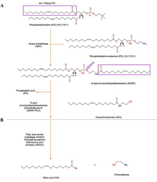
1.6. Oleoylethanolamide and N-acylethanolamides as lipid mediators ………..32
1.6.1. Synthesis and metabolism ………..33
1.6.2. Receptors ………35
1.6.3. OEA and the control of food intake ………...36
1.6.4. OEA effects in the central nervous system ………37
1.7. Aim of the thesis ………..40
1.8. References ………43
2. Chapter II: Role of the area postrema in the hypophagic effects of oleoylethanolamide Abstract ………66
2.1. Introduction ………..67
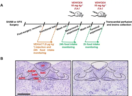
2.2. Materials and Methods ……….69
2.2.1. Animals and housing ………..69
2.2.2. Area postrema lesion surgery ……….70
2.2.3. Drugs and treatments ……….71
2.2.4. Food intake experiment ………..72
2.2.5. Terminal experiment ………..72
2.2.6. Immunohistochemistry study ……….72
2.2.8. Statistical analyses ……….75
2.3. Results ………..75
2.3.1. APX prevents the hypophagic effect of OEA ………75
2.3.2. OEA induces Fos in DBH neurons of the AP ………77
2.3.3. APX prevents OEA-induced DBH and Fos expression in the NST ………..79
2.3.4. APX prevents OEA induced Fos in the PVN and TMN ………82
2.3.5. APX prevents OEA-induced DBH and OXY expression in the PVN ………...84
2.3.6. PPAR-alpha receptors are expressed at AP level ………..86
2.4. Discussion ………87
2.5. Conclusions ………..91
2.6. References ………92
3. Chapter III: Evaluation of the brain distribution of oleoylethanolamide and its analogues after acute systemic administration in rats Abstract ………..97
3.1. Introduction ………98
3.2. Materials and Methods ………..100
3.2.1. Animals and housing ………...100
3.2.2. Drugs and treatments ………...100
3.2.3. Tissue collection ………..100
3.2.4. N-acylethanolamides extraction ………...101
3.2.5. UPLC-MS/MS analyses ………...101
3.2.6. Statistical analyses ………...103
3.3. Results ………103
3.3.1. Effects of OEA systemic administration on the levels of OEA and its analogues in selected brain areas ……….103
3.3.2. Effects of OEA systemic administration on the levels of OEA and its analogues in the plasma ………...109
3.4. Discussion ………..111
3.5. References ………..118
4. Chapter IV: Satiety factor oleoylethanolamide prevents binge-like palatable food consumption induced by stress in female rats with a history of food restriction Abstract ………..123
4.1. Introduction ………125
4.2. Materials and Methods ………...128
4.2.1. Experimental procedure for Binge-eating induction ………128
4.2.2. Experiment 1: Effects of OEA on stress-induced binge-eating ………...129
4.2.3. Experiment 2: Effects of OEA on the pattern of c-fos expression and on monoamine turnover in selected brain areas ………...131
4.2.4. Experiment 3: Effects of OEA on dopamine transmission in the AcbSh …………133
4.2.5. Experiment 4: effects of OEA on frustration stress exposure ………..134
4.2.6. Statistical Analyses ………..135
4.3. Results ………136
4.3.1. The combination of caloric restriction and stress exposure induced BED ………..136
4.3.2. OEA treatment selectively prevented binge-like eating in a dose dependent manner………..137
4.3.3. OEA treatment affected the brain pattern of c-Fos expression in bingeing rats …..138
4.3.4. OEA treatment dampened AcbSh DA release induced by stress exposure or amphetamine challenge ………...142
4.3.5. OEA treatment affected monoaminergic system in bingeing rats ………...143
4.3.6. OEA treatment affected CRF mRNA level in the AMY and oxy expression in the PVN of bingeing rats ………...147 4.4. Discussion ………..150 4.5. Conclusions ………156 4.6. References ………..157 5. Chapter V: Conclusions 5.1. Conclusions ………165 5.2. References ………..167
1
Chapter I
2
1.1.
Obesity and eating-related disorders
Obesity and overweight, defined as abnormal or excessive fat accumulation, are currently considered as major worldwide public health issues 1. According to the World Health Organization (WHO), obesity is recognized as a global epidemic that has been spreading all over the world contributing to the higher incidence of major health problems, thus increasing mortality. The high levels of intra-abdominal or visceral fat that are associated with this clinical condition are the most responsible for the development of the main obesity comorbidities such as hypertension, dyslipidemia, type 2 diabetes, osteoarthritis, and changes in the reproductive system 2.
Obesity is a multifactorial disease and among the several conditions that predispose to the weight gain and consequently to obesity, it is well known that genetic, environmental and physiologic factors play a key role 3.
Moreover, some medications can contribute to the development of this complex disease by increasing appetite, reducing the basal metabolic rate and by stimulating adipose cells proliferation as well. Among the pharmacological classes that might be more commonly involved in this process there are antidepressants and corticosteroids 4,5.
More rarely obesity is the consequence of other medical conditions including Prader Willi 6 and Cushing syndrome 7, hypothyroidism or hypothalamic dysfunctions 8.
The current most commonly used method for classifying obesity is the body mass index (BMI), calculated as weight (kg) divided by the square of the height (m2), which ranges between 25 and 29.9 in overweight individuals whereas it is equal to or more than 30 in obese-classified patients 9,10. Furthermore, waist circumference, another marker of
excess body fat, has becoming discriminating as a measure of overweight/obesity 11. The WHO estimated that in 2016 more than 1.9 billion adults, over the age of 18 years old, were overweight, of these over 650 million were obese 12.
Since over the past 40 years it has been registered a huge increase in the percentage of overweight and obese individuals worldwide, it has been estimated that by 2030 the 38% of the world’s adult population will be overweight and another 20% will be obese
13.
Moreover, changes in body weight are frequently accompanied by psychological and psychosocial problems thus leading to persistent disrupted eating behaviours, which are defined as eating disorders (EDs).
3
EDs are currently considered, likewise obesity, major health problems worldwide and they are characterized by altered dietary habits and impaired physical health. The 5th
edition of the Diagnostic and Statistical Manual of Mental Disorders (DSM-V) recognizes three different EDs: anorexia nervosa (AN), bulimia nervosa (BN), and binge eating disorder (BED) 14. The latter will be abundantly discussed in the following
paragraph since it is one of the main topic of this experimental dissertation.
Although EDs can occur in adults or young individuals indiscriminately, adolescence represents the most vulnerable period for the onset of these diseases 15.
The main criteria for classifying an individual affected by AN are represented by: a persistent caloric restriction followed by a considerable body weight loss with a BMI of 17.5 or even less 16; a strong fear to gain weight along with a distorted body image perception 14.
AN occurs more commonly among women than men (80-90% of patients with AN are female) and it reaches a peak incidence in adolescents between 14 and 17 years 15.
Moreover, AN is often linked with depressive disorder 17, obsessive compulsive disorder and autism spectrum disorder 18, counting the highest mortality of any psychiatric disease 19.
BN is primarily characterized by recurrent overeating episodes followed by compensatory purging behaviours such as vomiting, use of laxative or excessive physical activity whose purpose is avoiding body weight gain 17. Likewise the AN, BN
is also more common in females than males occurring in 1-2% of women mainly between 20 and 30 years 20. BN can also take place along with other psychological problems such as affective disorders, impulse control disorders, attention deficit/hyperactivity disorder (ADHD), drug dependence, anxiety and dissociative disorders 17.
1.1.1. The binge eating disorder (BED)
BED is the most frequent eating-related maladaptive disorder, characterized by compulsive and distressing overeating of high palatable food (HPF), a food with high fat or sugar content and consequently rich in kilocalories.
The above-delineated situation is called “binge eating episode”, which is defined, following the description of the DSM-V, by two main features: 1) the individual consumes a huge quantity of food in a short period of time, 2) along with a strong sense of loss of control, feeling of shame, guilt, disgust and anxiety 15.
4
Binge eating episodes occur at least once a week for three months. BED is classified as moderate when the weekly binge eating episodes are 1-3, while it is considered as severe when they become more frequent rising to 14 or even more 14.
It is possible to distinguish three different feeding behaviours, which can be at the bases of binge eating episodes:
-
Restrictive eating: food consumption is predominantly driven by the appearance,smell and brand of the food; this particular behaviour causes loss of control and overeating episodes more frequently;
-
Emotional eating: feeding as an escape strategy to fight negative emotions such asanxiety, sadness and stress;
- External eating: the feeding behaviour is mainly triggered by external stimuli including the sight and smell of the food 21.
Due to its critical features, BED leads to reduced social and emotional functioning, health impairments and worsens the quality of life 15.
BED differs from the other eating disorders, since it is not characterized by extreme compensatory strategies such as vomiting, use of laxative or increased physical activity
14,22.
It has been observed that the excessive intake of certain foods under specified conditions is able to alter not only eating behaviour but also it induces changes in the brain that resemble an addiction-like state. In particular, growing evidence found that one of the central mechanisms disrupted in BED conditions is represented by the dopamine (DA) rewarding circuitry.
BED is not only linked with the DA system, on the contrary it has been also demonstrated that an impairment in brain serotonin or 5-hydroxytryptamine (5-HT) signalling might play a role in the pathophysiology of BED in humans 23–26.
1.2. Neurobiological mechanisms regulating energy homeostasis
Overweight and obesity derive by an imbalance of the energetic metabolic rate. Our body, in fact, uses the food ingested-derived energy to manage the complete functioning of the main physiological systems at basal or active conditions, process that is called energetic balance. The excess energy quantity is normally stored and it is the primary responsible for fat accumulation leading to obesity development.
5
Several mechanisms partake in the control of energy balance by sending pro-satiety or pro-appetite signals to the brain in order to regulate energy consumption. Among these, white adipose tissue and liver play a key role in storing energy in the form of fat (energy storing) and in regulating the energy storage and consumption cycle, respectively 27.
The central nervous system (CNS) exerts the most crucial role in controlling feeding behaviour and energy homeostasis by interacting with several other systems. Most of the individuals, in fact, are able to maintain a stable body weight 28 thanks to a homeostatic regulation involving hypothalamic nuclei which integrate orexiant and anorexiant signals coming from other brain regions or from the periphery 29. The hypothalamus (HYPO) is a structure of the CNS located on the sides of the third ventricle 30 that regulates the energy metabolism by producing several neuropeptides 29. Moreover, it also partakes in neuronal networks that consist of non-peptidergic neurotransmitters including noradrenaline (NA), DA, 5-HT, histamine and endocannabinoids 29. The hypothalamic system is also connected with
extra-hypothalamic areas primarily located in the brainstem such as the dorsal motor nucleus of the vagus (DMV), the nucleus of solitary tract (NST), the area postrema (AP) and the parabrachial nucleus (PB) 31. In this regard, it has been demonstrated that hindbrain neurons from the NST and the AP project to the HYPO by sending signals and building integrative networks 29.
Among the several nuclei of the HYPO, the arcuate (ARC), paraventricular (PVN), supraoptic (SON), and tuberomamillary (TMN) nuclei are the most studied nuclei for their pivotal role in integrating appetite-regulating signals.
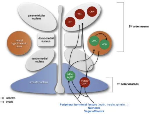
The ARC is situated in close contact with the median eminence (ME), a circumventricular organ with fenestered capillaries and a weak blood brain barrier (BBB) 32. Therefore, the ARC, via the ME, can represent a receptive region for circulating signals regulating energy balance such as leptin and insulin 33. It consists of two distinct neuronal populations which are called “first order” neurons and exert opposite effects in regulating energy homeostasis: 1) the agouti-related protein (AgRP)/neuropeptide Y (NPY) neurons that are responsible for stimulating food intake, and 2) those co-expressing the pro-opiomelanocortin (POMC) and cocaine-and amphetamine regulated transcript (CART) exerting anorexigenic effects 34 (Fig. 1.1). Both the two neuronal populations, POMC/CART and NPY/AgRP neurons, project to a variety of hypothalamic nuclei such as PVN, the lateral hypothalamic area (LHA) and
6
the perifornical area (PFA), each containing ‘‘second-order’’ neurons that integrate the information (Fig. 1.1).
Fig. 1.1: schematic representation of the neuropeptidergic hypothalamic networks involved in the homeostatic control of food intake. Abbreviations: CRH, corticotropin-releasing hormone; MCH,
melanin-concentrating hormone; NPY/AgRP, neuropeptide Y/agouti-related protein; ORX, neurons producing orexigenic peptides orexin; OT, oxytocin; POMC/CART, pro-opiomelanocortin/cocaine-and amphetamine regulated transcript; TRH, thyrotropin-releasing hormone 42.
The PVN represents a crucial hypothalamic nucleus involved in the control of feeding behaviour 35–37. It is composed of magnocellular and parvocellular divisions. The
magnocellular neurons are oxytocinergic and vasopressinergic neurons, which release oxytocin (OXY) and vasopressin (AVP) in the neurohypophysis 38. On the other hand, the medial parvocellular neurons contain corticotropin-releasing hormone (CRH) and thyrotropin-releasing hormone (TRH) which are secreted into the hypophysial portal vessels in the ME 39, whereas the dorsal, ventral and lateral subdivisions of parvocellular neurons mainly project to the periaqueductal grey matter, PB and NST 38–
41. The SON neurons, likewise those located in the PVN, have been demonstrated to
produce either OXY or AVP and they all project to the neurohypophysis. Furthermore, these neurons can also co-release a variety of other peptides such as cholecystokinin (CCK) and CRH, among others.
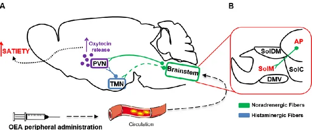
Several lines of evidence suggest that the TMN is the main histaminergic cell group in the rat brain which projects to a number of brain regions such as the nucleus accumbens (Acb), the prefrontal cortex (PFC) and other hypothalamic nuclei 43. Thanks to its
7
widespread innervation, the TMN exerts a control in many physiological functions by regulating the daily food consumption, the glucose absorption as well as emotional and memory processes 44,45.
The above-described complex neuronal network is responsible for the long-term regulation of energy balance and is primarily involved in the maintaining of a stable body weight.
On the other hand, a short-term regulation modulates the daily consumption of food, the number and duration of meals and it is modulated by the physico-chemical signals that are generated in the gastrointestinal tract upon the meal ingestion 46. Therefore, the short-term regulation of feeding behaviour occurs at the level of mesencephalic nuclei such as DMV and NST, that project to the PB, that, in turn, sends innervation to the PVN 46 (Fig. 1.2).
The NST is considered as an entry point for a variety of peripheral signals that are responsible for lowering the quantity of food consumed upon a meal 47. As already
mentioned in the text, projections from the NST, in particular noradrenergic neurons deriving from the A2 region, reach the hypothalamic neurons in the PVN 47. Moreover, growing evidence showed that the ablation of these noradrenergic projections prevents the anorexigenic effects of many pro-satiety endogenous signals, such as the CCK. The NST consisted of distinct neuronal populations that, depending on their specific localization within different NST subnuclei, react in different ways to the central and visceral stimuli 48. These subnuclei are innervated in a selective and specific manner by
afferents fibres arising from specific segments of the gastrointestinal tract 48. For instance, the gustatory nerves end in the most rostral portion of the NST 49, whereas the vagal afferents mainly project to the caudal part of the nucleus 50. Furthermore, the neurons of the dorsomedial subnucleus (SolDM) of the NST are particularly sensitive to gastric distention 51, on the contrary the neurons of the commissural part (SolC) mostly respond to the duodenal signals and those located in the medial portion (SolM) are responsive to both gastric and duodenal signals 51.
Moreover, it has been demonstrated that the SolM responds to leptin that acts directly on SolM neurons to reduce food intake 52. In 1986, Edwards et al., demonstrated that the lesions that extensively damage both medial and commissural subnuclei of NST markedly attenuate CCK-suppression of food intake 53.
An additional brain region crucially involved in food intake regulation is the AP, a circumventricular organ highly vascularized that displays a weak BBB by virtue of its
8
lack of tight junctions and the presence of fenestrated capillaries 54, features that give to the AP the appearance of a sponge.
The AP is the most caudal of the sensory circumventricular organs 54, located at the
apex of the calamus scriptorius in the dorso-medial medulla oblongata, at the base of the fourth ventricle and it appears as a hump of tissue surrounded by the NST 31.
The AP consists of specialized ependymal cells that make up a unique capillary architecture in the ventricles and central canal 55 and it can be subdivided into several zones depending on cell type location and projection patterns 54. The central and mantle zones predominantly contain neuronal cell bodies and axons which are in close contact with the ependymal cells, while in the ventral zone mainly reside glia 56.
Although the AP has been considered, for many years, for its function as the “chemoreceptor trigger zone” 57,58, it is now well recognized also its role in the
regulation of cardiovascular system, metabolism, immune function as well as in the control of cerebrospinal fluid (CSF) balance 54. In this context, recent studies have
suggested that the AP expresses a variety of receptors for many hormones involved in the control of feeding behaviour including amylin, glucagon-like peptide-1 (GLP-1), ghrelin, CCK, NPY, AVP, substance P and insulin 59–68. Moreover, through its efferent projections, the AP can in turn signal to important autonomic control centres behind the BBB and regulate the autonomic nervous system 54. Studies conducted in rats in which
the AP has been lesioned, recognise the important role of this hindbrain region in the homeostatic control of food intake and body weight as well as in the modulation of peripheral signals 69. The AP is therefore considered as an entry and integration point in the CNS for a variety of blood-borne substances 54.
Because of its anatomical location, the AP is in close contact with the NST, mostly at -13,80 mm from the bregma where it reaches the maximum extent 31. Several evidence
70,71 suggests that AP sends major and minor efferents to several nuclei in both the
brainstem and the HYPO.
AP primarily projects to the lateral parabrachial nucleus (LPBN) and to the subnuclei of the NST, among which the most innervated is the SolM 70–76 (Fig. 1.2). Furthermore, minor projections innervate the nucleus ambiguus, DMV, dorsal regions of the tegmental nucleus, cerebellar vermis, paratrigeminal nucleus, ventrolateral catecholaminergic column in the medulla, and the spinal trigeminal 70,71 (Fig. 1.2). Moreover, the AP also receives afferents from the NST and LPBN but also from functionally distinct regions of the brain such as the hypothalamic PVN and
9
dorsomedial nucleus (DMH) 70,71,77. Additionally to this central input, the AP receives also afferents from the periphery, in particular from the vagus nerve 70,71 (Fig. 1.2).
Fig. 1.2: Neuronal projections to and from the area postrema in the central nervous system (afferents (blue) and efferents (red)). 4V = 4th ventricle; AP = area postrema; DMH = dorsomedial
hypothalamus; DMV = dorsal motor nucleus of the vagus; DTN = dorsal tegmental nucleus; NA = nucleus ambiguus; NST = nucleus of the solitary tract; PB = parabrachial nucleus; PVN = paraventricular nucleus of the hypothalamus; SON = supraoptic nucleus. Adapted from 54.
In addition to catecholaminergic and serotonergic neurons 78,79, AP has been shown to
abundantly express also noradrenergic fibres 76 as well as neurons containing GABA,
substance P, encephalin, neurotensin, and CCK 78,80–83. Moreover, catecholaminergic and CCK neurons, which are located in the central zone of the AP, have been found to project to the NST and to modulate arousal and the emetic reflex 84,85. While A2 neurons project to oxytocinergic hypothalamic neurons of the SON and have been shown to play a role during experimental fear conditioning 86.
1.2.1. Hormonal signals
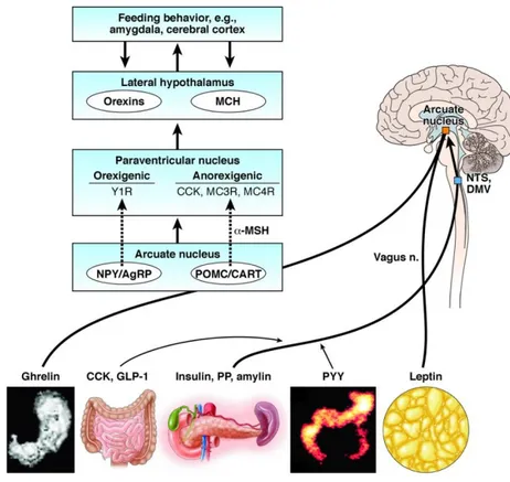
A variety of circulating hormones, mainly originating from fat cells, gastrointestinal tract, and endocrine pancreas, partake in the control of energy homeostasis through their actions on the HYPO, brainstem, or afferent autonomic nerves 87 (Fig. 1.3).
Leptin is a 146 amino acids peptidic hormone, produced by adipocytes 88 and involved in the regulation of energy homeostasis, neuroendocrine and immune functions 89. It has
10
also been found in several other tissues including placenta, mammary gland, ovary, skeletal muscle, stomach, pituitary gland, and lymphoid tissue 90.
Fig. 1.3: Role of circulating hormones in the central control of energy homeostasis. Gastrointestinal
and fat-derived hormones modulate food intake through stimulating specific brain areas such as the arcuate nucleus (ARC) in the hypothalamus and the hindbrain nucleus of solitary tract (NST) and dorsal motor nucleus of the vagus (DMV) 104.
Leptin is transported across the BBB by a saturable transport system 91 and exerts its anorexigenic effect in the ARC by activating POMC/CART 92,93, and inhibiting
NPY/AgRP neurons 92,93, thereby reducing food consumption.
Moreover, Leptin is also implicated in stimulating the secretion of neuropeptides that play a role in energy homeostasis such as CRH, TRH and the brain-derived neurotrophic factor (BDNF) 91,94–96. Beyond interacting with these central neuronal processes it became evident an interplay between leptin and CCK 97. In particular, leptin appeared to increase the CCK-induced pro-satiety effect 98.
Interestingly, hematic leptin concentration changes in respect to the body fat mass 99. Leptin is also able to signal rapid negative variations of energetic balance, although they occur independently of body weight fluctuations; during fasting, for example, circulating leptin drastically decreases while body weight does not significantly change
11
membrane receptor (Ob-R) thathas a single transmembrane domain and is a member of cytokine receptor family 101.
Finally, it has been demonstrated that the direct leptin administration in the ARC inhibits feeding 102; moreover, ARC lesions dampen the hypophagic effect of
peripherally administered leptin 103, thus suggesting the ARC crucial role in mediating
leptin effects on energy homeostasis. The gastrointestinal tract also produces other hormones that are involved in the short-term regulation of appetite by predominantly interacting with the HYPO.
In this context, although ghrelin has been originally identified as the main responsible for growth hormone secretion 105, it is now recognised as one of the peripheral orexigenic factor implicated in sending to the CNS short-term modifications of energy balance.
Ghrelin is a peptide hormone of 28 amino acids secreted by stomach and duodenum 105, which exerts, compared to leptin, opposite effects at hypothalamic level. In fact, it activates the anabolic pathway increasing dietary intake and reducing energy expenditure. Moreover, it has been shown that circulating ghrelin levels increase immediately before eating thus promoting food intake 99,100. By contrast, in a post-prandial phase, hematic ghrelin levels significantly decrease 99,100.
In the HYPO ghrelin-containing neurons are mainly located in the ARC, which is also a leptin target 106. From here, ghrelin neurons project to NPY/AgRP expressing fibres in
order to prevent leptin-induced inhibition of food intake and stimulate orexigenic peptides release; whereas by contacting POMC neurons, ghrelin fibres suppress anorexigenic peptides production 107.
Among other intestinal hormones that play a role in feeding behaviour regulation, the most studied are the CCK and peptide tyrosine-tyrosine (PYY) 108. CCK is considered as a physiological pro-satiety factor that is produced at both peripheral (intestine) and central level upon food consumption 109. It is normally released in response to long-chain fatty acids, amino acids and small peptides deriving by proteins ingestion 110,111. CCK binds two different receptors: CCK receptor type 1 (CCK1) and 2 (CCK2). The former has been identified in peripheral tissues including pancreas, gall bladder, vagal afferents innervating intestine 112 as well as in CNS areas involved in food intake regulation such as NST, AP, and DMH 63. While the latter has a different distribution, being mostly located in the cortex, HYPO, vagal afferents and gastric mucosa.
12
Intravenous injection of physiological CCK doses in humans has been demonstrated to increase appetite sensation and reduce food consumption 113.
PYY is a 36 amino acids peptide mostly containing tyrosine, produced by enteroendocrine L-cells of the distal gastrointestinal tract, whose plasmatic levels increase within 30 minutes after a meal consumption 114. PYY consists of 2 different
isoforms: a 36 amino acids isoform (PYY1-36) and another who misses the first two
amino acids (PYY3-36). A line of evidence showed that circulating PYY3-36 levels are
particularly high after meal consumption 114 thus suggesting the anorexic effect of this hormone peptide. The hypophagic effects of PYY3-36 appeared to be mediated by the
ARC. Moreover, both the two isoforms exert their action by binding NPY receptors family 115; in particular, PYY1-36 shows the same affinity for all the receptor subtypes,
while PYY3-36 displays a higher selective affinity for Y2 receptor 116.
Finally, other two peripheral hormones regulating energy homeostasis are represented by insulin and amylin.
Insulin is a metabolic hormone produced by the β-cells of the pancreatic islets of Langerhans and consists of two polypeptide chains which are linked together by two disulfide bonds: an A chain of 21 amino acids and a B chain of 30 amino acids.
Insulin is known to act on hypothalamic neurons located in the ARC, where insulin receptors are abundantly expressed, thus exerting a pivotal role in regulating glucose homeostasis and reducing energy intake 117. It has been observed that insulin
intracerebroventricular (i.c.v.) infusion or systemic injection induce a dose-dependent reduction of food intake 118 through NPY inhibition and POMC stimulation 119.
Both insulin and leptin are able to activate POMC neurons, however they appeared to differently regulate AgRP, since leptin is responsible for its inhibition while insulin is important for its stimulation 120.
Amylin is a polypeptide of 37 amino acids which is co-secreted, along with insulin, from pancreatic mammalian β-cells in response to food consumption, in a ratio of 1:100 amylin:insulin 121. Likewise insulin, amylin displays a role in glucose metabolism regulation; moreover, it also appears to lessen digestion through reducing digestive secretions (bile, enteric and pancreatic fluids) and to inhibit plasmatic glucagon production 121.
Amylin represents a pro-satiety physiological signal 122; in fact, it has been demonstrated to exert an anorexic effect even during fasting conditions 27, through both central and peripheral mechanisms. Amylin inhibitory effect on food intake seems to be
13
mediated by the activation of hindbrain regions including the AP 123, as well as the NST and LPB 124.
A large body of evidence demonstrated amylin ability to exert its hypophagic effect also through serotonergic, histaminergic, and dopaminergic systems 124. Moreover, amylin
appeared to communicate, at the level of brainstem, with other signals involved in short-term control of food intake such as CCK, GLP-1 and PYY 121.
1.2.2. Neuropeptidergic system
A major player in energy homeostasis regulation in the CNS is represented by the hypothalamic neuropeptidergic system that consists of a variety of orexigenic and anorexigenic peptides, which are released at both synaptic and soma-dendritic level and modulate the neuronal networks by binding specific receptor targets 125. As already mentioned in the text, the neurons populating the HYPO are classified in “first-order” and “second-order” neurons. The former are represented by the neurons co-expressing POMC/CART and those co-expressing NPY/AgRP both located in the ARC, while the latter consist of the PVN neurons releasing TRH, CRH and OXY (Fig. 1.1).
Among the neuropeptides stimulating appetite NPY, AgRP, orexins and the melanin-concentrating hormone (MCH) are the most studied.
Although the NPY displays different actions in the control of feeding behaviour, its most evident effect is the stimulation of food intake after its central administration 126. Furthermore, it has been observed that acute food deprivation 127 or chronic food restriction 128,129 induced the increase of hypothalamic NPY mRNA levels, which returned to normal levels after refeeding 130.
NPY synthesis in the ARC and its release at the level of PVN are regulated by inhibitory afferent signals including leptin and insulin, and by stimulatory signals such as ghrelin 126.
5 different G protein-coupled receptors (GPCRs) for NPY have been identified: Y1, Y2, Y4, Y5 and Y6 131,132 and they are widely distributed throughout the brain, including cortex, hippocampus (Hipp), amygdala (AMY), and HYPO.
Likewise NPY, AgRP is another orexigenic peptide of 132 amino acids which induces a sensation of appetite when i.c.v. injected in the PVN or in the DMH 133.
In vitro and in vivo studies have demonstrated that both POMC and AgRP neurons
express leptin and insulin receptors and are targeted by the respective hormones to increase POMC mRNA expression and decrease NPY and AgRP mRNA levels 134,135.
14
Orexin-A and orexin-B are appetite stimulant neuropeptides which are produced in the PFA, DMH and LHA 136,137 and bind two distinct GPCRs: orexin receptor 1 (OxR1),
mainly expressed in the ventromedial region of the HYPO (VMH), and orexin receptor 2 (OxR2) mostly present in the PVN 138. Evidence showed that after being i.c.v.
administered, orexin-A and orexin-B caused increased food intake in rats, while fasting led to upregulation of prepro-orexin mRNA 137. Orexin secreting neurons project their axons broadly throughout the brain and, particularly, to the NPY neurons of the ARC, which express OxR1 139.
The MCH is an orexigenic neuropeptide of 19 amino acids abundantly expressed in the LHA 140 and targeting two central receptors: MCHR1 and 2 141,142. The crucial role of MCH in controlling feeding is demonstrated by the observation that its injection into the rat lateral ventricle induces increased food intake 143.
Besides the orexigenic signals, growing interest is given to a number of anorexigenic peptides.
CART is a pro-satiety neuropeptide located in several regions of the HYPO including the DMH, PFA, PVN, and the ARC 94 as well as in the periphery. It has been observed that CART mRNA co-localizes with AVP and CRH containing neurons 94 in the PVN and with POMC in the ARC 144.
Several evidence supported the anorexic effect of CART, for instance its i.c.v. administration showed to inhibit food intake in rats 145, result that has been further
sustained by the injection of an antibody against CART peptide 82–103 which has been demonstrated to stimulate feeding 145.
Melanocortins are bioactive lipids deriving from the tissue-specific post-translational cleavage of the precursor molecule POMC 95. The POMC coding gene is physiologically expressed in several tissues including pituitary gland, skin, immune system, and hypothalamic neurons 96.
The long-term regulation of energy homeostasis mediated by POMC is a consequent of αmelanocytestimulating hormone (α-MSH) release, which occurs after POMC neurons stimulation 34. α-MSH, in turn, induces satiety by binding to downstream melanocortin-3 and melanocortin-4 receptors (MC3 and MC4) in the PVN 34. Consistently, it has been found that the ability of POMC neurons to suppress feeding is consecutive to melanocortin receptors activity 146.
On the contrary, it has been found that AgRP is a MC3/4-R competitive antagonist of α – MSH and lowers food intake by reducing α –MSH signaling 147.
15
CRH is the main physiological regulator of the adrenocorticotropic hormone (ACTH) secretion by pituitary gland, and growing evidence suggests its involvement in energy balance regulation 148. It is a 41-amino acid neuropeptide largely expressed in PVN
neurons and, in mammalian brain, it is also abundantly present in extrahypothalamic regions, including the olfactory bulb, bed nucleus of the stria terminalis (BNST), medial preoptic area, PVN, LHA, and central amygdala (CeA).
Moreover, when centrally injected, CRH inhibits food consumption and reduces body weight in rats through stimulating POMC-related peptides synthesis and secretion in the hypophysis 149. It has been observed that, when peripherally administered in human, CRH significantly increases energetic expenditure and fat oxidation 118. Furthermore, leptin infusion appeared to stimulate CRH expression, whereas the pre-treatment with a CRH antagonist dampens leptin-induced food intake and body weight reduction 118.
1.2.2.1. Oxytocinergic system
Among the satiety stimulating neuropeptides, a great deal of attention has been attracted by OXY, a 9-amino acid peptide crucially involved in feeding behaviour and energy homeostasis regulation 119–121. OXY is synthesized as a part of a precursor protein, preprohormone, which is cleaved with the consequent release of a nonapeptide, the neurophysin 150. Neurophysin appeared to be non-biologically active, however it seems
to protect OXY against enzymatic damage and to mediate OXY inclusion into neurosecretory vesicles 151,152.
The major OXY neurosecretory system is represented by the hypothalamic-neurohypophysial system whose neurons, having the cell body in the PVN and SON, project their axons to the posterior lobe of the pituitary gland (neurohypophysis) as well as to the ARC, ME, lateral septum (LS), and medial amygdala nucleus (MeA) 153.
Different observations suggest that OXY is primarily synthesized into the magnocellular neuronal population of the PVN and SON 154 that, once activated, are responsible of OXY release in the neurohypophysis. OXY is then secreted from the posterior lobe of the pituitary gland into the blood circulation where it can exert its effects by binding OXY receptors (OXTR), which are located throughout the body 155. Furthermore, it has been observed that after being secreted into the blood stream, or after its peripheral stimulation (during milk suckling or vaginal dilatation), cerebral OXY levels do not change, thus suggesting that OXY does not readily permeate the BBB 156.
16
Several evidence suggests that in the PVN and SON OXY can be released at both axonal and dendritic level 157.
The observation that hypothalamic oxytocinergic neurons project, among other destinations, also to the anterior part of the pituitary gland 158, suggested OXY ability to
act as a hypothalamic regulation factor able to physiologically modulate the adenohypophysial hormones including prolactin adrenocorticotropic hormone 159, and gonadotropins 160.
OXTR, is a GPCR coupled to a Gq/11α protein that has been identified not only in the CNS but also in the periphery 156. Interestingly, its distribution changes according to the species. For instance in the rat brain, in addition to PVN and SON, OXTR is also widely expressed in the olfactory system, cortex, thalamus, basal ganglia, VMH, BNST, CeA, ventral subiculum (vSUB), Hipp, Acb, brainstem, and spinal cord 155; while in human brain it is abundantly distributed in pars compacta of substantia nigra (SN), globus pallidus, anterior cingulate and medial insula 155.
Interestingly in the periphery OXTR is present in a variety of organs including uterus, mammary gland, ovary, kidney, heart, bone, and endothelial cells 155.
Thanks to its wide distribution in the body, the oxytocinergic system plays a crucial role in many physiological functions. For instance, it has an uterotonic action and therefore it is involved in the induction of labor 161,162. Growing evidence suggests an
involvement of OXY in the regulation of social behaviour 163,164 that is dependent on the
context: when the social cues are considered as “safe” OXY is able to improve sociality; whereas if they are interpreted as “unsafe” OXY induces defensive behaviour 165,166. A large body of evidence indicates that OXY is also released during stressful conditions including conditioned fear, pain, electric footshock, exposure to novel environments, and restraint stress 156. Moreover, concerning OXY capability to dampen the stress-induced hypothalamic-pituitary-adrenal (HPA) axis activation 167, it has been shown an interplay between the hypothalamic oxytocinergic system and the corticotrophin-releasing factor (CRF) system 168.
Furthermore, OXY has been shown to participate in the control of autonomic and somatic effects, such as sexual behaviour in both rodents 163 and humans 163,169, the cardiovascular system 170 and analgesia 171,172.
Interestingly, due to its effects in the control of social behaviour, OXY appeared to improve social cognition, functioning and repetitive behaviour in autism spectrum disorders 156.
17
A large body of evidence suggests the pivotal role of OXY in the regulation of feeding behaviour and energy homeostasis, being OXY particularly involved in stimulating appetite suppression 119–121. Intravenous infusion of both OXY and OXTR receptor
agonists has been demonstrated to induce a dose-dependent reduction of food intake
121,173,174. Subsequent studies revealed that OXY acts as “homeostatic” inhibitor of
feeding behaviour. In fact, it plays a role in gastric motility and in the response of stomach distension, which occurs upon food consumption 175. Olszewski and collaborators found increased oxytocinergic neurons activity in concomitance with the meal termination 175, thus suggesting their function in mediating satiety.
Moreover, studies conducted in rodents revealed that OXY plays a role in the conditioned taste aversion (CTA) mechanisms 156.
Several studies have been conducted in order to investigate a possible interaction between OXY and other feeding related peptides. In this context, Olszewski demonstrated that α-MSH injection in the PVN, reduces food intake because of a concomitant increase of the percentage of neurons co-expressing c-Fos and OXY 176. Furthermore, CART 177 and CCK 178 injection determined increased oxytocinergic neuronal activity in PVN and SON and/or increased OXY release, while a cerebral injection of OXTR antagonists has been demonstrated to prevent CRH-induced hypophagic effect 179 and to attenuate the pro-satiety leptin action 180.
OXY is not only involved in the control of energy homeostasis but it also regulates body weight and fat 181. Finally, McCann and Rogers found that oxytocinergic neurons
modulate gastric motility by increasing the excitability of sensorial NST fibres and DMV motoneurons 182.
1.2.3. Neurotransmitter systems
It is now well recognised the existence of an interplay between the monoaminergic neurotransmitters (such as DA, 5-HT and NA) along with neuropeptidergic and hormonal signals in regulating eating behaviour. Also the neurotransmitter histamine participates in the regulation of feeding consumption and it is considered a neural satiety signal; in fact it has been demonstrated that pharmacological treatments that increase the brain histamine availability are able to suppress eating, whereas administration of histamine antagonists increases food consumption and body weight
18 - Dopaminergic system
Knowledge accumulated during the last years investigated DA role in the control of food intake, particularly in the rewarding aspects of food 184–186.
DA is the predominant catecholamine neurotransmitter, which is synthetized within the ventral tegmental area (VTA) and the SN of the midbrain, from here DA neuronal cell bodies project to various brain structures.
We distinguish three different circuitries: 1) a nigrostriatal pathway originating in the SN and projecting to the dorsal part of the striatum such as the caudate putamen (CPu), involved in the regulation of movements; 2) a mesocorticolimbic pathway where dopaminergic cell bodies, residing in the VTA, project to both limbic (such as Acb and AMY) and cortical (such as PFC and medial prefrontal cortex (mPFC)) regions, thus controlling reward, and reinforcement behaviours as well as emotional and motivational responses; and 3) a tuberoinfundibular circuit which connects the ARC, where reside the cell bodies, and the ME 187 (Fig. 1.4).
Fig. 1.4: Dopaminergic pathways in the rat brain. The mesocorticolimbic circuit connects the VTA
and both limbic (Acb, black‐dotted line) and cortical (PFC, dark gray line) areas; the nigrostriatal circuit connects the SN to the CPu and others brain regions including the PFC, Acb and AMY (light gray line). The tuberoinfundibular pathway connects the ARC and the ME (dark gray dotted line).
Acb, nucleus accumbens; AMY, amygdala; ARC, arcuate nucleus; CPu, caudato putamen; ME, median eminence; MPOA, medial preoptic nucleus; PFC, prefrontal cortex; PVN, paraventricular nucleus; SC, spinal cord; SON, supraoptic nucleus; VTA, ventral tegmental area; ZI, zona incerta. Adapted from 187.
It has been demonstrated that DA effects in the regulation of feeding vary depending on the brain sites of action and on the variety of receptor subtypes 35. For instance, mesolimbic dopaminergic circuitry is responsible of the rewarding aspects of consuming HPF 188, whereas DA release in the HYPO suppresses food intake by
19
Growing evidence showed that DA release is linked with both short-term and long-term control of feeding 189,190. Moreover, several findings suggested that a different
expression in the HYPO of dopaminergic receptors in obese and lean phenotypes is directly involved in the regulation of meal size and meal number 189.
Among the different neurotransmitters implicated in the rewarding effects of food, DA represents the best characterized, in fact several evidence revealed that the ingestion of HPF induces DA release in the striatum of both humans and rodents 191,192. In particular, the first exposure to HPF stimulates an increase in the firing of DA neurons, whose cell bodies are located in the VTA, with the consequent DA release in the Acb 193. On the other hand, DA response habituates following situations of repeated HPF exposures 194. The involvement of dopaminergic circuitry in regulating food reward is strictly correlated to the motivational aspects driving the desire for the food, referred to as “wanting” 195. The dopaminergic brain regions involved in this rewarding aspect of food
are represented by the striatal nuclei such as CPu and Acb 196.
This concept is opposed to the mechanism involved in the hedonic properties of food, referred as “liking” which are predominantly regulated by opioid and cannabinoid systems 195. Conversely, in the hedonic aspects of food are implicated the LHA, ventral pallidum (VP), orbitofrontal cortex (OFC), Acb 197–199 and insula 200.
Brain-imaging studies revealed that the food cue-induced increase of DA release in humans is associated with the motivational salience of food (wanting) 201, while opioid
or cannabinoid receptors activation enhances the liking of the food, thereby stimulating appetite.
- Serotonergic system
5-HT is a monoamine neurotransmitter derived from the amino acid tryptophan and it is synthesized both peripherally (in the gastrointestinal tract) and centrally 202–204.
Among the variety of functions that 5-HT exerts including reproduction, mood, sleep, and cognition, a very important physiological role appeared to be the control of feeding behaviour 189.
In this context, it has been demonstrated that peripheral administration of 5-HT inhibits food intake by reducing meal size in rats 205. This effect seems to be associated to the indirect activation of the CNS, which occurs through the recruitment of ascending vagal afferent fibres 206–208. 5-HT, in fact, does not cross the BBB.
20
Central 5-HT is released by the 5-HT-synthesizing neurons, whose cell bodies are located in the brainstem, particularly in the caudal and rostral raphe nuclei, and densely innervate many regions of the brain 189.
More specifically, the 5-HT neurons of the caudal raphe project to the cerebellum, midbrain, pons, medulla, and most segments of the spinal cord, while 5-HT neurons whose cell bodies reside in the rostral raphe project in regions including cortex, Hipp, thalamus, HYPO, striatum, and AMY 209,210.
In particular, it has been observed that 5-HT fibres synaptically contact POMC and AgRP ARC neurons thereby affecting melanocortin neuronal activity 211,212.
The role of central 5-HT in eliciting satiety has been demonstrated by the observations that the administration of different compounds acting as direct or indirect agonists at central 5-HT synapses cause a significant reduction of food intake 189.
Moreover, it has been found that during feeding, 5-HT release increased in the HYPO
213.
A large body of evidence shows the existence of an interplay between 5-HT and DA, in particular it has been observed that DA release is partially regulated by 5-HT release
214–216.
To this regard, it is important to mention that meal size and meal number are independently regulated by distinct brain regions. In particular, several evidence demonstrated that the meal size is influenced by the intra-LHA DA and 5-HT, whereas the meal number is modulated by DA and 5-HT interaction within the VMH 217–219.
Thanks to 5-HT properties in inducing satiety, a number of drugs enhancing 5-HT transmission have been used for obesity treatment. In this context, several evidence supported an important role for 5-HT2C receptors in the inhibitory control of eating.
For example, mice lacking these receptors have been found to be hyperphagic thus developing obesity 220–222. Moreover, they showed a lower responsiveness to dexfenfluramine 223, GLP-1, and CCK 224. This aspect will be further discussed in the next sessions of the text.
- Noradrenergic system
NA is a catecholamine neurotransmitter synthesized in the dorsal vagal complex and in the locus coeruleus (LC) of the brainstem. In particular, Dalhström and Fuxe have recognised in mammalian brainstem several distinct groups of NA neurons from A1 to A7 225.
21
These regions contain the caudal ventrolateral medulla (A1), the NST (A2) the reticular regions around inferior olivary nuclei (A3) and the ependyma of the fourth ventricle (A4), the ventrolateral pons (A5), the LC (A6) and the rostral caudal pons (A7) 225,226.
From these hindbrain regions, noradrenergic fibres have been found to project both caudally to the spinal cord and rostrally to the HYPO, thalamus and cortex 93. In
particular, it has been shown that the NA neurons innervating the HYPO are those originating from A1, A2, and A6 regions 40,227.
Several observations demonstrated the crucial involvement of the noradrenergic neurotransmission in controlling food intake 93. For instance, particularly in the noradrenergic neurons projecting to the PVN, NA has been found to co-localize with NPY 93. Moreover, likewise NPY, intra-PVN injection of NA in rats appeared to strongly increase feeding and body weight 228 through the activation of the α2-receptor 229. Conversely, NA by binding to α
1- and β2-receptors exert opposite effects on food
intake 229.
The observation that elevated NA signaling in the PVN or other hypothalamic areas produced an increase of leptin deficiency-induced hyperphagia, suggests that NA acts as an anabolic effector in the control of energy homeostasis of CNS 93.
Further evidence confirming the role of noradrenergic tone in feeding behaviour regulation is represented by the observation that A2 neurons are activated when food intake is inhibited 230.
Moreover, in order to investigate the role of noradrenergic neurons located in the NST in mediating anorexic responses to gastric stimulation and in conveying gastric sensory signals to the HYPO and AMY, these hindbrain noradrenergic fibres have been lesioned through the microinjection of saporin, a toxin conjugated to an antibody against the enzyme responsible for NA synthesis, dopamine-β-hydroxylase (DBH) 231.
These experiments demonstrated the involvement of noradrenergic neurons of the NST in mediating CCK-anorexia and the hyphotalamic responses to gastric sensory stimulation 231.
- Histaminergic system
Histamine is an organic nitrogenous compound that is produced from the amino acid L-histidine by the enzyme L-histidine decarboxylase (HDC) and it has been isolated for the first time by Sir Henry Dale and colleagues from ergot in 1910 232. The first evidence of histamine presence in the brain dates back to 1919 233, however its role as a
22
neurotransmitter became evident only several decades later with the morphologic characterization of histamine-synthesizing neurons 234.
The central location of histaminergic neurons is represented by the above-mentioned TMN of the HYPO, from here these neurons project to the whole CNS through three major pathways: two ascending fibre groups reaching the forebrain structures and one ascending way that innervates the spinal cord 235–237 (Fig. 1.5).
Fig. 1.5: The histaminergic pathways in the rat brain 234.
Histamine is known to be involved in local immune responses and, as a neurotransmitter, it regulates many physiological functions 234 including arousal 238, sleep-wake cycle 239, seizure activity 239, release of hypothalamic hormones (AVP, OXY, prolactin, ACTH and β-endorphin) 240–243, thermoregulation 244, glucose and lipid metabolism 245, blood pressure 239 and food intake suppression 44,246.
Although histamine is able to activate both G protein-coupled receptors 247 and ligand-gated ion channels 239, its physiological functions are mainly mediated by the binding of the former referred to as H1R 248, H2R 249, H3R 250, H4R 251.
Among histamine biological functions, a crucial role is represented by the regulation of eating behaviour. Histamine, in fact induces satiety being released during the appetitive phase of food intake 252. Moreover, the observation of increased hypothalamic histamine levels during the feeding state of rats that have been fasted for 24 hours demonstrated a role for histamine also in regulating the consummatory phase of eating behaviour 253,254. Preclinical data collected over the years concerning the administration of histamine within the brain of laboratory rats 255,256 or cats 257 revealed histamine ability to induce a
23
Moreover, additional evidence of the pro-satiety action of histamine comes from the observation that both the activation of H1R within the PVN and VMH and the blockade
of brain H3R autoreceptors (that increases histamine release) are able to suppress food
consumption 258. These receptors have been recognized to regulate energy intake and
expenditure and their activation has been demonstrated to modulate feeding circadian rhythms 44,259.
Furthermore, there is consistent evidence indicating that a variety of regulatory peptides involved in the homeostatic control of food intake, including orexin, leptin, GLP-1, TRH, amylin and the emerging gut-derived pro-satiety compound oleoylethanolamide (OEA), require an intact histaminergic system to exert their orexigenic or anorexigenic effect 45,260.
1.3. Neurobiological mechanisms regulating stress
The way by which our body reacts to the exposure of stressful agents (physical, chemical or emotional stress) consists of an adaptation response whose objective is the homeostasis maintenance.
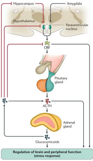
The mechanisms accounting for this process consider both the catecholamines release from adrenal medulla and the activation of the HPA axis that is responsible for higher cortisol levels 261. As previously described in the text, medial parvocellular neurons of
PVN containing CRF, project to the ME where they release this hormone into the hypophysial portal vessels 39. From here, CRF reaches corticotropic cells of anterior pituitary gland 261 where it binds to its receptor, thus leading to ACTH secretion within
the systemic circulation (Fig. 1.6). ACTH, in turn, binds to specific receptors expressed by the adrenal cortex in order to stimulate synthesis and release of corticosterone (CORT) in the general circulation, a hormone crucially involved in the body adaptation mechanisms to the stress response 262.
Among the stress factors characterizing the stressful conditions, both systemic and psychological factors result in HPA activation.
In particular, systemic stress factors are also associated with an activation of brainstem and circumventricular organs that directly project to the PVN neurons. Moreover, it is also possible that HPA axis hyperactivation, occurring during stressful conditions, is dependent on catecholaminergic ascendant input 261.
On the other hand, psychological stress factors evoke excitement conditions leading to the so-called “fight-or-flight” homeostatic stress response, which is linked to
24
glucocorticoids and neuro-hormones secretion and to the activation of the sympathetic part of the autonomic nervous system.
Both systemic and emotional stress factors are elaborated in a variety of limbic structures, including AMY, Hipp, and PFC regions of the brain receiving projections from sub-cortical and cortical areas and involved in regulating emotional or memory processes and conditioned responses 263.
The above-mentioned limbic regions also modulate HPA axis activation and collaborate in the autonomic stress responses 264.
Fig. 1.6: Regulation of the hypothalamic–pituitary–adrenal axis (HPA) 265.
Limbic modulation of the stress responses is primary mediated by oligosynaptic inputs to the PVN and other preautonomic brain regions. In this context, the PVN is considered as the principal integrator of stress signals 264.
In particular, the vSUB provides glutamatergic input to primarily inhibitory PVN relays, thus limiting psychogenic stress responses 264.
25
On the other hand, AMY, that is a structurally complex brain region, differently regulates systemic and psychogenic stressor responses. For instance, GABAergic projections from the CeA are involved in the regulation of responses to systemic stressors, while those from the MeA are responsible for the modulation of psychogenic stressor responses. Finally, the basolateral amygdala (BLA) via glutamatergic projections, within and outside the AMY, regulates both the acute and the chronic psychogenic stress response 264.
As far as the cortex, the prelimbic cortex (PL) via glutamatergic projections directed to inhibitory PVN relays suppresses the responses to emotional stress, whereas the infralimbic cortex (IL) appeared to activate autonomic responses to psychogenic stress, via direct (NST) or indirect (CeA) projections.
CRF effects on physiological and behavioural stress responses are mediated by binding to its receptors: CRF receptor type 1 (CRF1) and 2 (CRF2). Between the two receptors, the CRF1 represents the most abundant and it is predominantly expressed within HYPO, VTA, AMY, and cortical structures; while CRF2 distribution is restricted to the HYPO, dorsal raphe (DR), and LS 266–268.
It has been reported that, CRF1 antagonists reduce stress-induce food-seeking in rats 269. Moreover, an interplay between CRF and the dopaminergic and oxytocinergic systems has been predominantly studied in the last years 270,271.
For instance, the HPA stimulation leads to the activation of the mesolimbic dopaminergic pathway, which is associated to the reward circuitry. In fact, it has been demonstrated that the high CRF levels followed stressful events exert a role on DA neurons of the VTA, which projects to both limbic and prefrontal regions 272.
Moreover, experiments conducted in rodents reported that a variety of stressful stimuli including pain, fear conditioning and the exposure to novel environments induce an increase of plasmatic OXY concentration 161, which leads to the decrease of the stress-induced HPA axis activation 167. OXY represents, in fact, an important regulator of the anxiety disturbances related to the physiological stress response 164.
1.4. Neurobiological mechanisms regulating food addiction
Growing evidence considers obesity not only as a metabolic disease but also as the consequence of persistent disrupted eating behaviours thus affecting not only the physical but also the psychosocial health 273.
26
In this context, it is current knowledge that overeating is linked to neurobiological and psychological aspects, such as mood disturbances, altered reward perception and motivation, and addictive behaviour 274.
From this point of view, it is now prevailing the tendency to consume certain foods, particularly those high in sugar or in fat and therefore highly palatable, for pleasure and not to maintain energy homeostasis. Due to its intrinsic features, the HPF has an important impact on mood regulation so that it is becoming frequent the activity to overconsume these nutrients to self-medicate from negative emotional conditions such as anxiety, depression, mental fatigue 275.
Based on this background, it is now clear that there are many similarities between HPF consumption and reinforce behaviours related to the use of abuse drugs 276–279. In fact, HPF has been demonstrated to induce CNS modifications similarly to those evoked by drugs of abuse 276–279. In this context, it has been defined the concept of food addiction, suggested for the first time by Randolph in 1956 280.
The first indirect evidence showing affinities between excessive food consumption and drugs of abuse comes from the pharmacological data, since common therapies that are known to influence the intake of abuse drugs have been also shown to attenuate compulsive overeating occurring, for example, during binge eating episodes. Among these drugs, we recognize naloxone 281, naltrexone, baclofen 282,283, topiramate 284,
Rhodiola rosea and hypericum perforatum extracts 285,286, adenosine A
2A receptor
agonists 287, orexin 1 288 and CRF1 receptor antagonists 289.
Moreover, additional molecular and neurochemical evidence suggests that uncontrollable HPF consumption stimulates reward dopaminergic and opioid systems
273, which appear deeply readapted in people suffering from BED and obesity, thus
leading to a reward hyposensitivity state 290–293. Moreover, this readaptation seems to be the consequence of disrupted striatal D2 receptors signalling 294–297.
Interestingly, another evidence supporting the link between obesity and drug addiction comes from the observation that the disruption of the reward dopaminergic circuitry occurring during food overconsumption is similar to that observed after intravenous cocaine or heroin self-administration 298–300.
Human brain imaging data showed an increase in DA levels in the brain of both addicted individuals and obese people with BED 301,302. This effect appeared to be parallel to a reduced expression of DA receptors observed during obesity and drug dependence conditions 290.
27
In particular, in BED patients the higher DA release, which occurs upon exposure to food cues, has been described as an index to classify the severity of the disorder 302.
Moreover, the symptoms described during food withdrawal including headaches, sweats, irritability, and panic 303, are also similar to those found in drugs addicted
individuals such as symptoms resembling tolerance, withdrawal, and craving 291.
The aversive emotional state that triggers the negative reinforcement of addiction such as stress and negative moods (depression, anxiety), is also predominant in food addiction and can cause uncontrollable food consumption in humans through the involvement of a three-step cycle characterized by binge/intoxication, withdrawal/negative affect and preoccupation/anticipation 304–306.
It is important to mention that obesity and BED are not synonyms of food addiction, which is neither necessary nor sufficient for obesity 277,307.
To conclude, the inclusion of food addiction in the DSM-V suggests the similarities between people suffering from EDs and drug addicted individuals 308.
This observation indicates that the neuronal circuitry engaged in the two pathological conditions are the same 273.
1.5. Pharmacological treatment for obesity and BED
- Obesity
The growing prevalence of obesity highlights the need to deal with this public health issue by using specific anti-obesity treatments as an adjunct to a complete weight-loss program including diet and physical activity. Moreover, for patients with a BMI of 40 kg/m2 or greater, who failed an adequate exercise and diet program (with or without
therapy), the most effective anti-obesity approach is represented by a bariatric surgery, which reduces the size of the stomach 309.
The European Medicines Agency (EMA) recommends that, in order to be considered efficient against obesity, a drug needs to induce a response that is more than 10% weight loss at the end of a 12-month period 310.
The anorexiant drugs that have been used over the last years are numerous, however most of them has been withdrawn from the market due to their several side effects. Some examples are represented by d-fenfluramine, a 5-HT reuptake inhibitor, that was withdrawn from the U.S. market in 1997 after reports of heart valve disease and pulmonary hypertension 311; rimonabant, a cannabinoid receptor type 1 (CB1)