UNIVERSITÀ DEGLI STUDI DI FERRARA
Dottorato di Ricerca in
Biochimica, Biologia Molecolare e Biotecnologie
Ciclo XXIII
Coordinatore Prof. Francesco Bernardi
NEW INSIGHTS ON PML TUMOR SUPPRESSOR
DEGRADATION IN CANCER
Settore Scientifico Disciplinare MED/04
INDEX
ABSTRACT... 4
1. INTRODUCTION... 6
1.1 The promyelocytic leukemia PML... 6
1.2 The proteasome-ubiquitination/SUMOylation machinery... 13
1.3 The Casein Kinase 2 CK2... 21
2. RESULTS... 22
2.1 PML undergoes ubiquitin/proteasome-mediated degradation... 22
2.2 CK2 phosphorylates the PML degron directly... 22
2.3 p38 MAPK activation is required for PML degradation... 24
2.4 CK2 is required for PML degradation... 25
2.5 Mutations at S517 affect PML stability and tumor suppressive function in vitro and in vivo... 25
2.6 CK2-dependent degradation of PML in tumor derived cell lines and in human NSCLC... 26
2.7 Pharmacologic inhibition of CK2 leads to a significant anti-tumor effect in vivo... 26
2.8 PML interacts with PIAS1... 27
3. DISCUSSION... 30
4. EXPERIMENTAL PROCEDURES... 33
4.1 Recombinant Retroviruses, Transductions, and Drug Selection... 33
4.2 Immunoblotting, Immunoprecipitation, and Chemicals... 33
4.4 In Vitro CK2 Kinase Assays... 34
4.5 In Vitro Phosphorylation-Site Mapping by Mass Spectrometry... 35
4.6 Determination of Apoptosis, Replicative Senescence, and Growth
Kinetics... 36 4.7 Yeast two-hybrid system... 36 5. REFERENCES... 39
ABSTRACT
The promyelocytic tumor suppressor PML controls growth suppression, induction of apoptosis, and cellular senescence. It was originally identified as a component of the PML-RARα oncoprotein of acute promyelocytic leukemia (APL). Moreover, PML protein is completely or partially lost in a large fraction of human cancers and that loss correlates with tumor progression. PML localizes to nuclear matrix-associated macromolecular structures known as PML nuclear bodies (PML-NBs), which are dependent on PML for assembly. Recently it was discovered that PML is enriched at the endoplasmic reticulum (ER) and at the mitochondria-associated membranes, signaling domains involved in ER-to-mitochondria calcium ion (Ca2+) transport and in induction of apoptosis.
PML undergoes ubiquitin/proteasome-mediated degradation in immortalized and tumor derived cell lines. PML degradation depends on direct CK2 phosphorylation of PML Ser517.
CK2 is a stress-activated serine/threonine protein kinase that is oncogenic and frequently overexpressed in human tumor of multiple histological origins. In addition, CK2 overexpression due to gene amplification has been reported to be an adverse prognostic factor in non-small cell lung cancer.
Interestingly, PML mutants that are resistant to CK2 phosphorylation display increased tumor suppressive functions in assays measuring apoptosis, replicative senescence, and in xenograft models.
NBs contain components of the SUMOylation machinery and both PML and PML-RARα undergo SUMOylation. However, the biological significance of these events is not completely understood.
To determine the significance of PML SUMOylation we sought to identify the PML E3 SUMO ligase. In particular, we identified PIAS1 as a PML E3 SUMO ligase that mediates ubiquitin/proteasomal degradation of PML in cancer cells promoting tumorigenesis.
1. INTRODUCTION
1.1 The promyelocytic leukemia PML
The promyelocytic leukemia gene (PML), originally identified at the breakpoint of the t(15;17) translocation in acute promyeolocytic leukemia (APL), is the essential component of the PML nuclear body (PML-NB), a nuclear-matrix-associated macromolecular structure where multiple tumor suppressor proteins colocalize (Bernardi and Pandolfi, 2007). PML deficiency occurs commonly in human cancers and its inactivation in mice leads to cancer susceptibility (Trotman et al., 2006; Wang et al., 1998). Moreover, the oncogenic activity of the PML-RARα oncoprotein in APL is, at least in part, due to its ability to disrupt PML-dependent tumor suppressive pathways (Scaglioni and Pandolfi, 2007; Matsushita et al., 2006; Rego et al., 2001). Thus, PML is considered a tumor suppressor.
PML is a member of the TRIM/RBCC family of proteins, many members of which are ubiquitin ligases that generate subcellular structures through autoassembly (Reymond et al., 2001; Meroni and Diez-Roux, 2005). Transcription of the PML gene is tightly controlled by interferons α,β or γ, but also by p53 (Stadler et al., 1995; de Stanchina et al., 2004), which both yield a dramatic increase in the number and the size of the PML-NBs. PML harbors an amino-terminal RING finger that directly binds the SUMO E2 ligase UBC9 (Duprez et al., 1999), two RING-like domains, the B boxes (Tao et al., 2008), and a coiled-coil mediating homodimerization (Kastner et al., 1992). For other RBCC/TRIM family members, partner binding-specificity often relies on the carboxyl terminus. For PML, a variety of carboxy-terminal domains generated by alternative splicing yield isoforms (Jensen et al., 2001). When expressing single PML isoform in Pml -/- cells, distinct types of PML-NBs were observed, implying that isoform-specific sequences contact different nuclear constituents
that influence morphogenesis (Beech et al., 2005; Condemine et al., 2006; Weidtkamp-Peters et al., 2008). Yet, because of the coiled-coil, all endogenous isoforms colocalize. The most abundant (but perhaps least studied) isoform, PML-I, harbors an exonuclease-III domain, that targets PML to nucleolar caps in stressed or senescent cells (Condemine et al., 2007). In addition to the nuclear localization signal (NLS) present in all PML isoforms, PML-I harbors a nuclear export signal (NES) that allows shuttling of all isoforms between the two compartments through heterodimer formation (Henderson and Eleftheriou, 2000; Beech et al., 2005; Condemine et al., 2006). The most extensively studied isoform, PML-IV, induces senescence in primary human fibroblasts (Bischof et al., 2002) and apoptosis in many other cellular settings, at least in part through p53 activation (Guo et al., 2000).
PML undergoes several critical post-translational modifications, notably phosphorylation and SUMOylation. PML SUMOylation has been implicated in NB-morphogenesis. DNA damage- or stress-activated kinases like ATM, ATR, CHK2, HIPK2, CK2, or ERK phosphorylate PML, possibly regulating PML stability, NB biogenesis and partner association (Engelhardt et al., 2003; Hayakawa and Privalsky, 2004; Scaglioni et al., 2008; Scaglioni et al., 2006; Gresko et al., 2009) and contributing to DNA repair or apoptosis control.
PML is ubiquitously expressed, albeit at very low levels, and PML-NBs are detected in almost any cell of the developing embryo or the adult organism. Nonetheless, PML is markedly up-regulated upon a number of cellular stresses including inflammation, oncogenic transformation and proapoptotic stimuli such as for instance ionizing radiation (Terris et al., 1995; Ferbeyre et al., 2000; Pearson et al., 2000; Carbone et al., 2002). Under these conditions, the number and the size of the PML- NBs (and hence the matrix-associated PML fraction), as well as the soluble non matrix PML nuclear and cytosolic fractions,
increase (Salomoni, Bernardi and Pandolfi, unpublished observation). These stimuli also trigger a dynamic reorganization of the PML-NB with transient recruitment and release of proteins from these nuclear organelles, as we will thoroughly discuss throughout the following paragraphs. The PML-NB can be therefore regarded as a multiprotein stress response machinery.
Among the functions attributed to PML and PML-NBs are the regulation of transcription, neo-angiogenesis, DNA damage responses, cellular senescence and apoptosis (Zhong et al., 2000; Bernardi and Pandolfi, 2007; Borden, 2002).
As soon as a KO model for Pml was created, it became evident that Pml -/- mice and cells are protected from multiple and diverse apoptotic stimuli (Fig. 2), such as γ-irradiation and CD95/Fas (Wang et al., 1998). In addition, primary cells from Pml -/- mice, such as splenocytes, thymocytes, mouse embryonic fibroblasts (MEFs) and hematopoietic cells, are resistant to apoptosis induced by CD95/Fas or γ-irradiation, and many other stimuli, including ceramide, TNF, IFN, UV light and chemotherapeutic drugs (Wang et al., 1998; Louria-Hayon et al., 2003; Wu et al., 2003; Bernardi et al., 2004). In agreement with the dominant negative role of PML-RARα on PML, hematopoietic progenitors from PML-RARα transgenic mice are resistant to similar apoptotic stimuli (Wang et al., 1998).
The reason why Pml -/- cells are resistant to many apoptotic stimuli is in part because PML acts as a pleiotropic factor that regulates the function of several pro- and anti-apoptotic factors such as p53, Daxx and c-Jun among others (Bernardi et al., 2008).
PML is believed to interact with a large number of cellular proteins. However, it is unclear how many of these interactions are functional and which of them modulate PML function in in vivo settings. This remains one of the key questions in the PML field.
The available literature suggests that PML is functioning as part of a complex tumor-suppressive network. It is well established that PML is an important factor in the regulation of both p53- dependent and independent apoptotic pathways (Bernardi et al., 2008; Takahashi et al., 2004). PML activates p53 by several means: by recruiting p53 to PML-NBs by promoting its acetylation and phosphorylation and by binding and inhibiting Mdm2, the main negative regulator of p53 (Bernardi and Pandolfi, 2003; Dellaire and Bazett-Jones, 2004).
In detail, PML was first implicated in the control of p53 activity as a regulator of p53 acetylation in the context of cellular senescence, a condition induced by DNA damage caused by specific agents or by over-expression of oncogenes (Halazonetis et al., 2008). It was shown that, upon expression of a strong oncogene such as RasV12, the full activation of
p53 depends on the presence of PML, which recruits p53 to the PML-NBs alongside the acetyl transferase p300 and in so doing facilitates p53 acetylation (Ferbeyre et al., 2000; Pearson et al., 2000).
Later on, it became clear that PML regulates not only p53 acetylation but also p53 stabilization, by interfering with the ubiquitination of p53 by Mdm2 (Kurki et al., 2003). PML can bind to both p53 and Mdm2 (Wei et al., 2003; Zhu et al., 2003) and it has been suggested that PML inhibits Mdm2-mediated ubiquitination of p53 either by forming a trimeric complex with Mdm2 and p53 (Kurki et al., 2003), or by sequestering Mdm2 away from p53 (Bernardi et al., 2004), or yet by promoting p53 de-ubiquitination by the ubiquitin protease HAUSP (Everett et al., 1997; Li et al., 2002). In addition, PML blocks p53-Mdm2 interaction also by promoting p53 phosphorylation by Chk2 and CK1 (Louria-Hayon et al., 2003; Alsheich-Bartok et al., 2008). PML may additionally stimulate p53 phosphorylation by Chk2 also indirectly, by promoting Chk2 activity. In fact, it was recently shown that,
upon DNA damage, PML facilitates Chk2 autophosphorylation and activation (Yang et al., 2006). Finally, PML promotes the phosphorylation of p53 by HIPK2 (Moller et al., 2003), a modification that increases p53-mediated transcription of a subset of promoters and is associated with UV-induced apoptosis (D'Orazi et al., 2002; Hofmann et al., 2002).
Despite the large number of publications addressing the issue of the regulation of p53 by PML, caution needs to be raised about the fact that the majority of these studies have been conducted in vitro. Since p53 is a protein regulated by many factors and co-factors, the physiological relevance of these data remains to be established in vivo. Furthermore, it is also important to understand if tumors that have lost PML expression but retain wt p53 can be treated with PML-inducing drugs to reestablish sensitivity to pro-apoptotic chemotherapy.
On a final note, most of the functions of PML described so far do depend not only on the ability of PML to recruit p53 and/or p53 modifying proteins but also on their translocation to the PML-NBs. Consistent with this notion, it was recently reported that cytoplasmic PML mutants inhibit p53 activity by acting in a dominant-negative fashion on wt nuclear PML (Bellodi et al., 2006). Mutations introducing a stop codon upstream the NLS of PML have been identified in patients with aggressive forms of APL, where mutations are found in the allele of Pml not involved in the translocation (Gurrieri et al., 2004). In cells expressing wt PML, mutant PML accumulates in the cytoplasm, delocalizes nuclear PML into cytoplasmic aggregates and inhibits p53 transcriptional activation (Bellodi et al., 2006). It must be emphasized that wt cytoplasmic PML isoforms have been characterized and found to be critical in regulating TGF-β signaling and anti-viral responses (McNally et al., 2008; Lin et al., 2004).
isoforms in the modulation of cytoplasmic p53 function, they are nonetheless important because they indicate that patients bearing these or similar PML “nuclear exclusion” mutations could prove insensitive to pro-apoptotic drugs acting through p53.
However, PML can also induce apoptosis in a p53-independent manner. After DNA damage, for example, PML can induce apoptosis by mediating Chk2 autophosphorylation and activation (Yang et al., 2006), although the interaction between PML and Chk2 is probably more complicated than this simple scenario. On the one hand, Chk2 localizes to PML-NBs in undamaged cells and is subsequently released after DNA damage (Yang et al., 2002). On the other hand, PML promotes Chk2 autophosphorylation and activation after DNA damage. It is unclear, therefore, whether Chk2 autophosphorylation is occurring in the PML-NBs or if, perhaps, it is indirectly mediated by PML.
Another protein that can explain the role of PML in apoptosis is Daxx (Bernardi et al., 2008). However, the role of Daxx in apoptosis is still not completely understood because Daxx has been reported both to enhance and to suppress apoptosis (Salomoni and Khelifi, 2006). Daxx localization to PML-NBs correlates with its ability to sensitize cells to Fas- and splenocyte activation-induced apoptosis (Torii et al., 1999; Zhong et al., 2000). It has therefore been proposed that Daxx exerts anti-apoptotic functions outside PML-NBs (Chen and Chen, 2003), whereas the pro-apoptotic functions of Daxx, such as the repression of anti-apoptotic genes (Croxton et al., 2006) may require its localization to PML-NBs. This notion is now challenged by a recent study showing that, in rheumatoid arthritis fibroblasts, increased expression of sumo and increased recruitment of Daxx to PML-NBs contributes to resistance to Fas-induced apoptosis, thus indicating that Daxx pro-apoptotic or anti-apoptotic activity in the PML-NBs may be cell-type specific. In this respect, Daxx could regulate gene expression from the PML-NBs differentially in distinct cell types. Further
work is therefore necessary to elucidate the role of Daxx and its localization to PML-NBs in opposing and favoring programmed cell death. By contrast, the role of PML is more coherently pro-apoptotic irrespective of the cell type.
Finally, PML can also contribute to Fas-induced apoptosis by another mechanism. The protein FLICE-associated huge protein (FLASH), which functions as a positive regulator of Fas-induced apoptosis, localizes to the PML-NBs under steady-state conditions. In response to Fas activation, FLASH is released from PML-NBs and accumulates in mitochondria where it promotes the activation of caspase-8 (Milovic-Holm et al., 2007).
New findings indicate that PML can act as a suppressor of major oncogenic pathways such as the PI3K/Akt pathway. In particular, the Pandolfi’s laboratory has shown that PML, through its ability to interact with the protein phosphatase PP2A, inhibits the nuclear function of Akt, thus leading to suppression of its prosurvival and promitogenic functions (Trotman et al., 2006).
In Pten +/- animals, reduction of PML gene dosage results in transition to invasive carcinoma which is accompanied by increased Akt phosphorylation (Trotman et al., 2006); this suggests a genetic interaction between the two pathways. Nonetheless, it is not clear whether PP2A is delocalized also in PML tumors. The same group has demonstrated that another component of the PI3K/Akt pathway, the mTOR (mammalian target of rapamycin) kinase can associate with PML-NBs (Bernardi et al., 2006). This, in turn, results in inhibition of mTOR-dependent HIF1α translation and suppression of tumor angiogenesis in vivo. However, it is still unclear to which extent mTOR sequestration acts as a regulatory mechanism in vivo. A further contribution from Pandolfi’s group provided evidence that PML regulates the function of the phosphatase PTEN, which is a tumor suppressor itself and the main inhibitor of the PI3K pathway. This occurs mainly through
inhibition of PTEN deubiquitination by HAUSP and its nuclear retention (Trotman et al., 2007; Wang et al., 2007). In this respect, nuclear exclusion of PTEN has been associated with increased aggressiveness of colon cancer and is predominant in a small number of Cowden disease cases due to a mutation affecting PTEN ubiquitination. It would be, therefore, interesting to determine whether PML loss in colon and prostate carcinomas correlates with PTEN nuclear exclusion. In summary, it appears that PML is able to affect the PI3K pathway at multiple levels. However, it is quite difficult to understand how PML could act on the PI3K pathway at so many different levels. One of the potential explanations would be that PML action is dependent on the cell or tumor type. This would imply that cell/tumor-specific modifications of PML or of its targets, within the PI3K pathway exist to allow for selective interactions. In general, this remains one of the key questions regarding the multifaceted function of PML.
One clear concept that is emerging from the current knowledge about the role of PML in apoptosis is that although Pml -/- mice and cells are resistant to a wide variety of apoptotic stimuli both in vitro and in vivo, there appears to be no common mechanism to explain the general resistance of these cells to apoptosis. Instead, PML-NBs emerge as signaling centers that regulate the availability, post-translational modification and activation of many and diverse proteins implicated in apoptotic pathways. Most of these proteins are upstream regulators of apoptosis, in other words they are “decision makers” that evaluate if the conditions are right to trigger apoptosis. Given the general and important role of PML as a mediator of apoptosis, our goal was to investigate whether or not PML might also be involved more directly in the execution of apoptosis.
The proteasome is composed of a core (20S) particle containing multiple proteolytic sites and a 19S regulatory particle that governs access to the core. To enter the core, substrates must be amenable to unfolding by a hexamer of ATPases associated with the base of the regulatory particle. Other constituents of the regulatory particle are implicated in the recruitment of substrates (Finley, 2009). Rpn10 and Rpn13 interact with ubiquitinated substrates through UIM (ubiquitin-interacting motif) domains and a Pru (pleckstrin-like receptor for ubiquitin) domain, respectively. The UBL/UBA family of proteins are substoichiometric components of purified proteasomes that bind ubiquitin via their UBA (ubiquitin-associated) domain and the proteasome regulatory particle through its UBL (ubiquitin-like) domain. They are proposed to remotely scavenge ubiquitinated substrates and present them to the proteasome (Figure 1). Figure 1. Depiction of the ‘ubiquitin
receptors’ associated with each degradative pathway. The domain structures shown are for the human representatives of each protein family, except for yeast Ddi1, the human ortholog of which does not contain a UBA domain. CB: clathrin binding motif; CC: coiled coil; ESCRT: endosomal sorting complex required for transport; GGA: golgi associated, gamma adaptin ear containing, ARFbinding protein; GAE: gamma adaptin ear; GAT: GGA and TOM1; GLUE: GRAMlike ubiquitinbinding in Eap45; HRS: HGF receptor tyrosine kinase substrate; LIR: LC3 interacting region; PB1: Phox and Bem1; PRU: Pleckstrinlike receptor for ubiquitin; SH3: Src homology domain 3; STAM: signal transducing adaptor molecule; TOM1: target of myb1; TSG101: tumor susceptibility gene 101; UBA: ubiquitinassociated domain; UBL: ubiquitinlike domain; UEV: ubiquitin E2 variant domain; UIM: ubiquitininteracting motif; VHS: Vps27, HRS, and STAM; VPS36: vacuolar protein sorting 36; vWFA: von Willebrand Factor type A; ZZ: zinc finger. Note the following gene names and commonly used alternative names also apply: p62; SQSTM1 (sequestosome), NDP52; CALCOCO2, UBQLN1; PLIC1; DSK2. Domain annotation based on PFAM and UNIPROT.
Particular proteasome-associated ubiquitin receptors have been linked with the degradation of specific substrates (reviewed in Finley, 2009).
Substrate proteins are selected for modification of lysine residues by ubiquitin through interaction with an E3 ligase protein that recruits an E2-enzyme charged with ubiquitin. This can result in transfer of a single ubiquitin molecule (monoubiquitination) or coupling of further ubiquitin molecules, through integral lysine residues, to form a chain. The seven lysines of ubiquitin provide for the formation of different isopeptide chain linkages, which adopt different three-dimensional structures, and all of which are represented in eukaryotic cells (Xu et al., 2009). The specific combination of E2 and E3 enzymes recruited to a substrate dictates the chain linkage type. The human genome encodes more than 20 different types of ubiquitin-binding domains, and proof of principle for linkage specificity of binding has been established. One means to achieve this is through the spatial arrangement of tandem ubiquitin-binding domains (UBDs) either encoded in a single protein or by combining domains within a multimolecular complex, such that simultaneous occupancy of two binding sites is restricted to particular chain configurations.
Currently, there are two families of ubiquitin-protein ligases: the HECT domain (homologous to the E6-accessory protein [AP] carboxyl-terminus) enzymes (which form thioesters with ubiquitin) and the RING finger ligases. The E6 protein is encoded in malignant forms of papilloma viruses and through recruitment of the cellular E6-AP ubiquitin-protein ligase causes the degradation of p53. The RING finger proteins are either in complexes with other proteins essential for ligase activity (the SCF and APC/cyclosome complexes) or are associated with putative substrate proteins. The RING finger ubiquitin-protein ligases bind ubiquitin-conjugating enzymes to facilitate ubiquitinylation of target proteins. The latter group of RING finger ligases includes c-Cbl, which is an adaptor for
receptor protein tyrosine kinases. The c-Cbl protein binds to phosphorylated tyrosine residues in activated receptors via SH2 domains and triggers ubiquitinylation via the associated ubiquitin-conjugating enzyme (Joazeiro et al., 1999). Other RING finger proteins may act as ubiquitin-protein ligases. For example, the protein product of the breast cancer 1 gene (BRCA1) has a RING finger and is a ubiquitin-protein ligase (Lorick et al., 1999). At least 7 other RING finger proteins have demonstrated ubiquitin-protein ligase activity.
Ubiquitin molecules, which are linked together in chains to a protein as a degradation signal, are covalently coupled via an isopeptide bond as described earlier utilising the lysine48 (K48) residue of each ubiquitin. However, chains have also been shown to be linked via four of the other six lysines in ubiquitin (K6,K11, K29, and K63). The K63-linked polyubiquitin chains appear to play a role in DNA repair. The formation of K63-linked chains is not a signal for degradation, which means that attachment of K63-linked chains to proteins is not for degradation but for some other purpose, probably in the nucleus in DNA repair. The generation of K63-linked chains is through a heterodimer composed of an ubiquitin-conjugating enzyme variant (UEV) and a specific ubiquitin-ubiquitin-conjugating enzyme, ubp13p (Hofmann and Pickart, 1999). The UEV proteins are homologous to ubiquitin-conjugating enzymes but lack the critical catalytic cysteine residue. The UEV proteins have been implicated in cell transformation and tumour suppression. Again a protein, the ubiquitin-conjugating enzyme variant, activates an ubiquitin-ubiquitin-conjugating enzyme.
The SUMO conjugation pathway has a lot in common with the ubiquitination pathway. Both processes involve the use of three enzymes: E1: activating enzyme, E2: conjugating enzyme and E3: ligase (Figure 2) (Takahashi et al., 2001). SUMO is bound to its target protein via an isopeptide bond formed between an ε-amino group on the lysine residue on the target protein and the C terminal carboxyl group on the SUMO protein (Desterro et al., 1997). The
pro-form of SUMO needs to be cleaved prior to protein conjugation. This is carried out by isopeptidases, also known as the SENP SUMO deconjugating enzymes (Mukhopadhyay and Dasso, 2007). The SUMO activating enzyme (E1), SAE1/2, commences the reaction process by interacting with SUMO (activated by SENP enzymes—Figure 2), to form a high energy thiolester bond. The SUMO conjugating enzyme (E2) then binds SUMO via its cysteine residue in its active site. This intermediate provides a highly reactive species, important in the final conjugation, usually facilitated by an E3 ligase (Kroetz, 2005). SUMO E3 ligases act to either activate Ubc9 or bring Ubc9 and the target protein within close proximity of each other, thus enhancing SUMOylation (Ulrich, 2009). They can be regarded as E3 enzymes as they are able to bind to the E2 and the substrate and facilitate the formation of the bond formed between SUMO and the target protein. It has also been shown that a large number of proteins ( 40%) can be SUMOylated without the presence of the∼ consensus sequence (ψKxE), demonstrating differences in substrate specificity (Ulrich, 2009).
Figure 2. The SUMO conjugation and deconjugation pathway. The SUMO conjugation
pathway requires the activity of four enzymes: SENP the SUMO specific protease family; E1 composed of 2 subunits Uba2/Aos1; E2—Ubc9 and the E3 ligases. The reaction is initiated by cleaving the proform of SUMO at its C terminus, to its active state, by the SUMO specific proteases (SENPs). Following which, the E1 enzyme, a heterodimer of Uba2/Aos1, binds SUMO in an ATP dependant reaction, before the transfer to the E2 conjugating enzyme Ubc9. Ubc9 forms a reactive bond between itself and SUMO. Subsequently, the E3 ligase facilitates the transfer and covalent attachment of SUMO, via an isopeptide bond, to the target protein. This process is in equilibrium with the SUMO deconjugation (SENPs)
Unlike the ubiquitin (Ub) E1, the SUMO E1 exists as a heterodimer; with each monomer corresponding to a particular region of the Ub E1. The SAE subunit Aos1 (SAE1) shares similarity with the N terminus of the Ub E1, whilst Uba2 (SAE2), the second component of the SAE complex, is similar to the C terminus of the Ub E1 (Johnson et al., 1997). The monomers are never found individually and hence it is assumed that they are unable to function independently (Azuma et al., 2001). The SAE complex is responsible for preparing SUMO for transfer to the SUMO conjugating enzyme, Ubc9 (Walden et al., 2003). Ubc9 is the only known SUMO conjugating enzyme, unlike the ubiquitination pathway where each E2 has a specific set of target proteins (Hayashi et al., 2002). Ubc9 contains an active site with a cysteine residue which is responsible for binding the SUMO molecule directly to the ψKxE sequence found on the target protein (Sternsdorf et al., 1999).
In contrast to SUMO E2s, a larger number of SUMO E3 ligases have been discovered and have been categorized into three types: the protein inhibitor of activated STAT—signal transducer and activator of transcription (PIAS) family (Hochstrasser, 2001), the nuclear pore proteins Ran binding protein 2 and nucleoporin 358 (RanBP2/Nup358) (Pichler et al., 2002) and the polycomb group protein Pc2 (Kagey et al., 2003). E3 ligases are usually substrate specific with little redundancy found within the system.
The largest group of E3 ligases are the PIAS proteins with four genes in mammals: PIAS1, PIAS3, PIASx and PIASγ (Liu et al., 1998). The PIAS E3s have a conserved region consisting of a SAP domain responsible for binding AT rich DNA sequences and an SP-RING domain which binds to Ubc9 and promotes SUMOylation (Schmidt and Müller, 2002). They also contain SUMO interaction motifs (SIMs) that are able to directly bind SUMO (Rytinki et al., 2009). It has been found that the different PIAS proteins SUMOylate distinct sets of substrates, with occasional overlap (Schmidt and Müller, 2002). The second
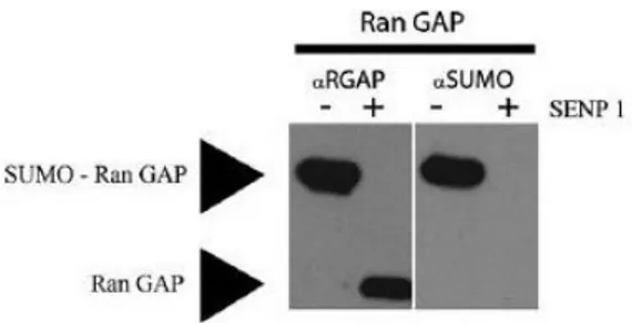
group of E3 SUMO ligases consists of the nuclear pore protein RanBP2 (Nup358) with only one known substrate, RanGAP1, a GTPase activating protein important in nuclear transport of proteins (Nishimoto, 1999; Saitoh et al., 1997). RanGAP can be SUMO modified in vitro, as displayed in Figure 3. The final family SUMO E3 ligase identified so far is the PC2 protein part of the polycomb group (Kagey et al., 2003). Pc2 has been shown to SUMOylate the transcriptional co-repressor CtBP, localising it to the nucleus (Lin et al., 2003), and to co-localise with PcG bodies (Kagey et al., 2003).
SUMO modification is a dynamic process involving both conjugation and deconjugation enzymes. The deconjugation enzymes function by cleaving the isopeptide bond between SUMO and the modified protein (Melchior et al., 2003). There are seven isoforms of these
Figure 3. In vitro SUMOylation of RanGAP. An in vitro SUMO conjugation
assay was carried out to SUMOylate the RanGAP protein. RanGAP was incubated with the SUMO conjugation machinery; SUMO1, SAE1/2 and Ubc9 under the following conditions: 10 µl containing 50 mM Tris pH 7.5, 5 mM MgCl2 , 2 mM ATP, 5 mM DTT, 100 ng SAE2/1 (E1), 100–600 ng UBc9 (E2), Sumo 2.5 µg and substrate 1–3 µg. The solution was incubated at 37 C for 2.5 h and the reaction was stopped by the addition of LDS ◦ sample buffer. An additional control was added where the SUMO conjugated RanGAP is deconjugated using SENP 1. The conditions for the reaction are as follows: iodoacetamide was added to the conjugated RanGAP solution at 10 mM and incubated at room temperature for 30 min. βMercaptoethanol or DTT was added at 20 mM and left for 15 min at room temperature. SENP1 was finally added at 10 nM and incubated for 1 h at 37 C. The reaction was stopped by the addition of LDS buffer. ◦ The samples were run on an SDSPAGE gel and were then used to detect SUMO conjugation using Western blotting. The membranes were first probed for RanGAP (1:500) and as seen in this figure, an upper band is observed, a 10 kDa shift upwards; where free RanGAP is approximately 32 kDa and SUMOylated Ran GAP is approximately 42 kDa. In the control lane with the addition of SENP1, the upper band disappears; suggesting RanGAP is SUMO modified in vitro. The same membrane was stripped and probed for SUMO 1 (1:1000), as shown in this figure; the upper band is also SUMO positive.
isopeptidases, including SENP1, SENP2, SENP3, SENP6 and SENP7 (Mukhopadhyay and Dasso, 2007). The SENPs contain a Ulp domain at their C terminus responsible for cleaving the isopeptide bond and distinct N terminal domains that regulate their cellular localisation, suggesting each SENP has a distinct set of substrates (Mukhopadhyay and Dasso, 2007). In addition to their deconjugation role, the SENPs also play an essential role in maintaining the levels of free SUMO within the cell (Ulrich, 2009). Other forms of SUMO regulation include the E3 ligases and the presence of the consensus motif on target proteins. It has previously been stated that 40% of proteins modified by SUMO do not have the typical consensus sequence; as such this could also be regarded as another form of regulation. Over the last decade a number of groups have investigated how the SUMO pathway is regulated in response to different stimuli. In response to heat shock, erythroleukemia cells induce transcription of heat shock factor 1 (HSF1). After its translation, HSF1 is phosphorylated prior to its SUMOylation, which enhances its DNA binding ability (Hong et al., 2001). It is also widely recognised that SUMO alters protein activity by modulating other PTMs, such as phosphorylation and ubiquitination. For example, SUMOylation of IκBα, an important factor in the inflammatory response, prevents its ubiquitination, and therefore inhibits its degradation and subsequent NF-κB activation and nuclear translocation (Desterro et al., 1998). SUMO can also regulate protein activity by modulating its interactions with other macromolecules or proteins. Various models have been proposed such as the addition of SUMO by altering protein configuration, creating a new interaction motif affecting its function (Johnson, 2004). An interesting example of interaction motifs is arsenic induced RNF4 mediated degradation of promyelocytic leukemia (PML) bodies. In the presence of arsenic, PML is polysumoylated, and following the recruitment of RNF4, an E3 Ub ligase, PML is ubiquitinated and degraded (Tatham et al., 2008).
1.3 The Casein Kinase 2 CK2
Casein Kinase 2 (CK2) is a nuclear-matrix-associated, highly conserved, and ubiquitous serine/threonine kinase that consists of two catalytic (αα, α'α' or αα') and two β regulatory subunits (Pinna, 2002). The regulation and function of CK2 are not well defined, and, traditionally, CK2 has been considered a constitutively nonregulated protein kinase (Allende and Allende, 1995). However, it has been shown recently that CK2 is a stress-activated protein kinase implicated in prosurvival functions through the phosphorylation of substrates such as IκBα. (Ahmed et al., 2002; Kato et al., 2003; Litchfield, 2003). Importantly, CK2 is frequently activated in human cancers and can induce mammary tumors and lymphomas when expressed in transgenic mice (Landesman-Bollag et al., 2001; Seldin and Leder, 1995). Traditionally, CK2 has been regarded as a constitutively active, ubiquitous serine/threonine protein kinase in search of specific physiological functions (Pinna, 2002). However, several studies have indicated that CK2 plays a critical role in the regulation of cell proliferation and survival (Ahmed et al., 2002). The molecular pathways modulating the prosurvival properties of CK2 have remained largely unknown, with the exception of its role in cellular UV response. In this setting, CK2 is activated by UV radiation in a p38 MAPK-dependent manner, leading to phosphorylation and degradation of the NF-κB inhibitor IκBα (Kato et al., 2003). Furthermore, upon UV irradiation, CK2 complexes and phosphorylates p53 at Ser389. MEF cells and mice carrying the p53 S389A mutant in the p53 locus have defects in the induction of p53 target genes and apoptosis and exhibit increased skin tumorigenesis upon UV irradiation (Bruins et al., 2004; Keller et al., 2001). Conversely, wild-type p53 inhibits CK2 protein kinase (Schuster et al., 2001). These observations support the notion that p53 and CK2 functions are interconnected in a tightly regulated
2. RESULTS
2.1 PML undergoes ubiquitin/proteasome-mediated degradation
We determined whether PML undergoes degradation through an ubiquitin-dependent proteasome pathway. Treatment of HEK293 cells with proteasome inhibitor such as MG132 (data not shown) led to increased PML levels, indicating that PML undergoes degradation. Moreover we expressed Flag-tagged PML in HEK293 cells together with HA-ubiquitin and treated them with MG132. Immunoprecipitation (IP) followed by Western blot (WB) analysis detected a ladder of HA-marked PML polypeptides, indicating that PML undergoes polyubiquitination. We concluded that regulation of PML turnover involves an ubiquitin-dependent proteasome pathway.
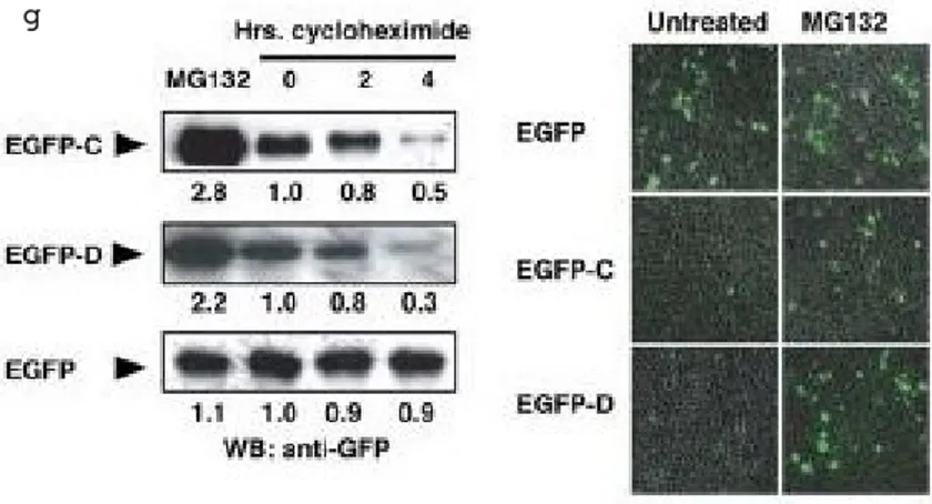
To map the protein sequence necessary and sufficient to direct PML ubiquitinylation, we generated a large series of PML deletion mutants that we screened upon transfection with HA-ubiquitin in HEK293 cells for the ability to be conjugated to polyubiquitin chains. This analysis led to the discovery that a discrete C-terminal PML protein sequence (PML 498– 525) is necessary to allow PML protein polyubiquitination and degradation. Moreover, PML 498–524 is sufficient to direct polyubiquitinylation of a heterologous protein, such as GFP. A summary of these results is provided in Figure 4. We concluded that this sequence is the PML degron.
2.2 CK2 phosphorylates the PML degron directly
Protein sequence analysis of the PML degron with the Scansite (http://scansite.mit.edu) and ELM (Eukaryotic Linear Motif, http://elm.eu.org) prediction algorithms revealed the presence of multiple phosphorylation consensus sites for CK2. We found that 6His-tagged
full-length CK2α produced in baculovirus readily phosphorylates bacterially expressed PML by an immunocomplex kinase assay. Site directed mutagenesis experiments followed by in vitro kinase assays performed in the presence of specific CK2 inhibitors, such as TBB and DMAT indicated that PML S517 is the primary CK2 phosphorylation site and suggested that phosphorylation of this serine primes phosphorylation of S514. These conclusions were confirmed by MALDI-re-TOF mass spectrometry experiments on recombinant PML proteins phosphorylated in vitro (Scaglioni et al., 2008; Scaglioni et al., 2006). These experiments led to the conclusion that PML S517 is the major CK2 acceptor site.
a b c
d
2.3 p38 MAPK activation is required for PML degradation
Since CK2 activation depends on p38 MAPK during cellular stress, we investigated whether p38 MAPK activation leads to PML polyubiquitinylation and degradation (Kato et al., 2003; Sayed et al., 2000). We found that several treatments that activate p38 MAPK, such as osmotic shock, anisomycin, and UV radiation, lead to PML protein polyubiquitination and degradation in NIH 3T3 cells. Next, we found that in the context of osmotic shock,
g
Figure 4. a) PML undergoes degradation in HEK293 cells. Endogenous PML and
actin proteins were detected by Western blot (WB) in cycloheximidetreated cells. b) Proteasome inhibition leads to PML upregulation. HEK293 cells were treated with MG132 as indicated. Endogenous PML and actin were detected by WB. c) PML is polyubiquitylated. HEK293 cells were transfected and analyzed by immunoprecipitation (IP) and WB as indicated (upper panel). Note that the membrane was not stripped in between hybridizations. Ten percent of the input lysate was analyzed by WB (lower panel). Ubn = polyubiquitin chains. d) Schematic representation of informative constructs used in this study. PML modular organization is represented along with its major domains. Boxes in light blue and light brown represent PML domains. R: RING finger; B1 and B2: B boxes: CC: coiled coil; D: degron; PEST: PEST domain. The column on the right indicates whether mutant PML proteins undergo polyubiquitylation. e) Deletion of a critical PML Cterminal protein sequence abrogates PML ubiquitylation. HEK293 cells were transfected as indicated, and their lysates were analyzed by IP and WB. Ten percent of the input lysate was analyzed by WB with an antiHA antibody (lower panel). * = background bands. f) A critical PML Cterminal protein sequence is sufficient to cause ubiquitylation of EGFP. HEK293 cells were transiently transfected with a vector expressing EGFPC, EGFPD, or wildtype EGFP. HAubiquitin was cotransfected as indicated and analyzed by IP and WB. Note that the membrane was not stripped in between hybridizations. Intensity of the polyubiquitin chains is expressed as a ratio between polyubiquitylated EGFP proteins and EGFP protein input. g) A critical PML Cterminal sequence is sufficient to cause proteasomemediated degradation of EGFP. HEK293 cells were transiently transfected and treated as indicated and were analyzed by WB. Representative fields of EGFPpositive cells obtained from the same transfection are presented inthe right panels. Note that MG132 upregulates EGFPC and EGFP D, but not EGFP.
inhibition of p38 MAPK with p38AF, a dominant negative mutant, or the specific inhibitor SB202190 efficiently blocks PML degradation in NIH 3T3 cells. Thus, we concluded that PML degradation induced by osmotic shock is dependent on p38 MAPK activity.
2.4 CK2 is required for PML degradation
We found that TBB and TBCA (two specific CK2 inhibitors) abrogate osmotic shock induced PML degradation and that two specific siRNA oligonucleotides caused a 75% reduction in CK2α protein in NIH 3T3 cells. In this setting, endogenous PML protein was more than twice the level observed in cells treated with scrambled siRNA. These experiments confirm that CK2 kinase is required for PML degradation. Furthermore, we found that abrogation of PML S517 resulted in complete resistance to sorbitol-induced degradation, These experiments further validate that PML phosphorylation by CK2 of PML S517 is essential for its degradation (Scaglioni et al., 2008; Scaglioni et al., 2006).
2.5 Mutations at S517 affect PML stability and tumor suppressive function in vitro and in vivo
PML is a tumor suppressor protein capable of inducing growth arrest and apoptosis (Bernardi and Pandolfi, 2007). Therefore, we hypothesized that PML S517A, the mutant refractory to CK2-mediated phosphorylation and ubiquitin-mediated degradation, acts as a super-tumor suppressor due to its resistance to CK2-mediated degradation. In line with this hypothesis, we found that the PML S517A mutant induces replicative senescence in WI38 human primary fibroblasts and UV induced apoptosis in MEFs unlike wild-type PML. We then examined the tumor suppressive role of PML S517A as compared to wild-type PML in vivo. To this end, we utilized Colo320DM cells, where PML is degraded in a CK2
dependent manner, that stably expressing either wild-type or PML S517A proteins. Transduced Colo320DM cells were injected subcutaneously into athymic nude mice and xenograft growth quantified. These experiments indicated that growth of Colo320DM cells expressing PML S517A is reduced by more than 50% when compared to cells expressing wild-type PML (Scaglioni et al., 2008; Scaglioni et al., 2006). These data demonstrate that PML S517A behaves as a super active PML mutant due to defective CK2 mediated degradation.
2.6 CK2-dependent degradation of PML in tumor derived cell lines and in human NSCLC PML is often partially or completely lost in non-small cell lung cancer (NSCLC) (Scaglioni et al., 2008; Scaglioni et al., 2006; Gurrieri et al., 2004; Zhang et al., 2000), while CK2 is overexpressed and amplified in NSCLC (O-charoenrat et al., 2004). Therefore, we tested for an inverse correlation between the two in a panel of NSCLC cell lines and primary human NSCLC specimens. We found that PML protein was barely detectable in A549, H1299, and H322 cells. On the contrary, PML protein was easily detected in H2030, H157, H1975, H1650, and H358 cells. We performed a CK2 kinase assay on H1299 and H322 and on H1650 and H358 (representative of cells with high and low PML protein levels, respectively) cells. CK2 kinase activity was strikingly elevated in H322 and H1299 as compared to H1650 and H358 cell lines. We also found an inverse correlation between PML protein levels and CK2 kinase activity in primary NSCC specimens. We evaluated CK2 kinase activity and PML protein levels in 18 primary NSCLC specimens and their unaffected counterpart tissue that were snap frozen at the time of their surgical resection. PML protein was reduced by at least 50% in 10 out of the 18 tumors analyzed, as compared to the unaffected tissue. CK2 kinase activity was increased by at least 50% in nine of these
tumors. Therefore, we found a strong association between elevated CK2 kinase activity and decreased PML protein level (P = 0.002). These observations strongly suggest that elevated CK2 kinase activity leads to PML degradation in primary human NSCLC.
2.7 Pharmacologic inhibition of CK2 leads to a significant anti-tumor effect in vivo
We tested whether CK2 pharmacologic inhibition led to significant anti-tumor effects. For these experiments, we used emodin, a specific CK2 inhibitor, to treat nude mice bearing Colo320DM xenografts (Yamada et al., 2005). As expected, emodin upregulated PML, but not PML S517A in cultured Colo320DM cells. Treatment with emodin in vivo reduced the tumor burden by more than 50%. These results suggest that pharmacologic inhibitors may have anti-tumor properties (Scaglioni et al., 2008; Scaglioni et al., 2006).
2.8 PML interacts with PIAS1
Yeast two-hybrid screening was carried out according to a standard protocol (Clontech) to identify proteins that interact with PML. The bait plasmid pBJKT7-PML4 and the p8op-LacZ reporter gene plasmid were first transformed in AH109 yeast cells followed by transformationof the library of rat lung cDNA plasmids. Approximately 3.5 × 10⁶ yeast cells
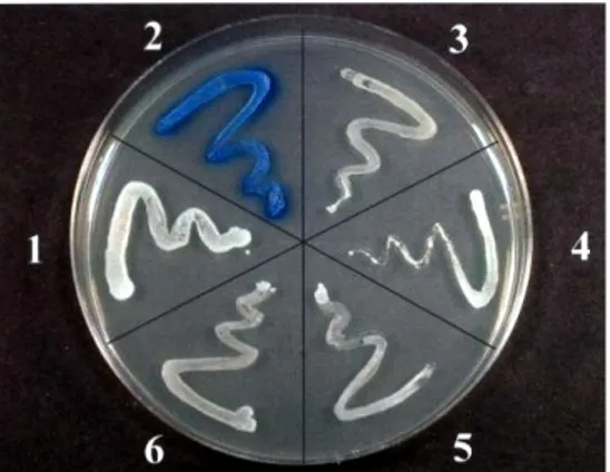
cotransformed with the bait and cDNAs from a premade rat lung cDNA Matchmaker library (Clontech) were screened. About 120 positive clones were selected for their ability to grow on plates lacking leucine, tryptophan, histidine, and adenine (Figure 5a) and assayed for β-galactosidase activityon media supplemented with X-gal (Figure 5b).
The X-gal staining assay kit is designed for sensitive and low-background in situ visualization of cells carrying the lacZ gene. The β-galactosidase enzyme catalyzes the hydrolysis of X-gal (5-bromo-3-indoyl-β-D- galactopyranoside), a β-galactoside.
Following fixation and incubation with the X-gal substrate, cells transfected with a β-galactosidase-expressing plasmid will appear blue. These blue cells can be easily visualized also by microscopy. Plasmids containing the rat lung cDNAs were isolated from positive yeast cells, and their nucleotide sequences were determined by DNA sequencing. The plasmid containing the positive cDNA (Figure 6) was isolated from yeast by miniprep protocol: the cells of positive clone were resuspended in SE medium (1 M Sorbitol, 50 mM EDTA) and 2 mg/ml of the yeast lytic enzime (ICN Biomedical). Following 30 min at 37 °C the yeast cells were centrifuged and the pellet was dissolved in HIRT’S solution (10 mM Tris-Cl pH 7.5, 50 mM EDTA, 0.2 % SDS) added with 0.5 mg/ml proteinase K (Invitrogen) and incubate at 50 °C for more than 6 h. The cDNA plasmid was extract by phenol/chloroform/isoamyl alcohol (25:24:1) protocol. The aqueous phase was precipitated
a
b
Figure 5. a) Yeast growth on
meduim selection. b) Xgal staining of colonies.
with the same volume of 20% PEG and 2.5 M NaCl, the nucleic acid was pelleted and washed in 70% ethanol and resuspended in 10µl TE (10mM Tris-HCl, pH 7.5, 1 mM EDTA). The plasmid obtained was transformed into E. coli strainDH5α by electroporation. In future experiments we will focus on PIAS1 since it was already known by literature being an E3 SUMO ligase.
Figure 6. Yeast colony #2
3. DISCUSSION
SUMOylation is emerging as a critical mechanism regulating protein localization and function. This post-translational modification relies on an enzymatic cascade that leads to covalent conjugation of SUMO1 and/or SUMO2/3 to target proteins substrates (Geiss-Friedlander and Melchior, 2007).
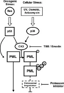
NBs contain components of the SUMOylation machinery and both PML and PML-RARα undergo SUMOylation (Kamitani et al., 1998; Lallemand-Breitenbach et al., 2001). However, the biological significance of these events is not completely understood. For example, it has been reported that SUMOylation promotes both PML tumor suppressive function and PML degradation (Lallemand-Breitenbach et al., 2001; Zhong et al., 2000; Ishov et al., 1999; Shen et al., 2006). Moreover, the striking anti-leukemic activity of arsenic trioxide (ATO), a drug commonly used in the treatment of APL, has been attributed to its ability to induce PML-RARα SUMOylation followed by ubiquitin mediated proteasomal degradation (Lallemand-Breitenbach et al., 2001; Tatham et al., 2008; Lallemand-Breitenbach et al., 2008). However, it has also been reported that SUMOylation of PML-RARα is essential for its leukemogenic activity, a seemingly opposed biological outcome (Zhu et al., 2005). Our work defines a functional network between p38 MAPK, p53, CK2, and PML (Figure 7). In addition to its well-established role as a p53 coactivator during genotoxic stress (Bernardi et al., 2004; Guo et al., 2000; Salomoni and Pandolfi, 2002), PML regulates UV response by inducing apoptosis or cell-cycle arrest in a p53-independent manner (Salomoni et al., 2005). In this context, the phosphorylation of PML by CK2 (and the concomitant phosphorylation of IκBα by CK2) is part of a cellular circuitry
that attenuates apoptosis, allowing cells to recover from noxious stimuli.
Under conditions of oncogenic stress, such as the ones triggered by oncogenic Ras, PML is activated and exerts its tumor-suppressive function in concert with several partners, including p53. In this context, p53 may inhibit CK2 to achieve maximal PML activity and tumor-suppressive effects. On the contrary, when CK2 kinase activity is upregulated (as often happens in human cancers), PML is polyubiquitylated and degraded (Figure 7). This scenario may be particularly relevant for the pathogenesis of NSCLC, in which increased CK2 kinase activity may occur because of either p38 MAPK activation or CK2α gene amplification, a marker of poor prognosis in this disease (O-charoenrat et al., 2004).
Figure 7. Molecular mechanisms controlling PML
polyubiquitylation. During the cellular response to stress, CK2 kinase controls PML protein levels through
integration of upstream p53 and p38 MAPK signals. Therapy with CK2 or proteasome inhibitors will abrogate aberrant PML protein degradation, lead ing to restoration of PML tumorsuppressive properties.
To determine the significance of PML SUMOylation we sought to identify the PML E3 SUMO ligase. We found that the E3 SUMO ligases protein inhibitor of activated STAT PIAS1and PIASxα, interact with PML in a yeast two-hybrid screening using a cDNA library generated from adult human lung tissue as prey.
Subsequently, we will investigate the PML protein sequence required for interacting with PIAS1 by performing co-immunoprecipitation assays with a series of PML deletion mutants in HEK293T cells. Moreover, we will characterize whether the presence of PIAS1 affects levels of PML-RARα SUMOylation.
4. EXPERIMENTAL PROCEDURES
4.1 Recombinant Retroviruses, Transductions, and Drug Selection
Recombinant retroviruses were generated by transient transfection of pWzl and/or pBabe puro based vectors in amphotropic Phoenix packaging cell lines. 48 to 72 hours after transfection, the retroviral supernatants were collected and filtered through a 0.45 μm filter and used for transduction. Three to five milliliters of freshly made retroviral supernatants containing 4 μg/ml polybrene were added to exponentially growing cells for each 10 cm diameter plate culture. After 12 hours fresh medium was added. Generally cells were selected in 150 μg/ml of hygromycin and/or 2 μg/ml of puromycin 48 hours after infection. In order to favor a higher number of viral integrations, we selected doubly transduced cells in 400 μg/ml of hygromycin 5 μg/ml of puromycin.
4.2 Immunoblotting, Immunoprecipitation, and Chemicals
Cells were harvested in RIPA lysis buffer (150 mM NaCl, 10 mM Tris [pH 7.5], 1% NP40, 1% deoxycholate, 0.1% SDS, protease inhibitor cocktail [Roche], phosphatase inhibitor cocktail [Sigma]). For immunoprecipitation, lysates were precleared with IgG and Protein G agarose beads and then incubated with the precipitating antibody for one hour to overnight, followed by one hour incubation with protein G agarose beads (Amersham Biosciences). Immune complexes were then washed three times in lysis buffer and boiled in gel loading buffer. Proteins from total cell lysates or immunoprecipitates were resolved by SDS-poly-acrylamide gel electrophoresis (SDS-PAGE), transferred to nitrocellulose membrane, blocked in 5% nonfat milk or BSA, and blotted with the appropriate antibody.
Biosciences); Xpress (Invitrogen); HA (Covance); phospho-p38 MAPK, anti-p38 MAPK (Cell Signaling Technology); anti-CK2α (Upstate Biotechnology); anti-GFP (Clontech); anti-human PML antibodies PG-M3 and H-238 (Santa Cruz) and AB1370 (Chemicon); anti mouse p16INK4 (sc-1207) and anti p21 (F-5) (Santa Cruz), anti-murine PML S36 (gift of Dr. Scott Lowe, Cold Spring Harbor Laboratories); anti-phosphoserine/threonine (BD Bioscience); anti-Phospho-PML S517 was generated by immunizing rabbits with the peptide: CVISSSED(P)SDAENSSSR, the serum was affinity purified. The chemicals were either from Calbiochem or Sigma. SB202190 was used at 10 μM, MG132 at 10 μM, 4,5,6,7-tetrabromobenzotriazole (TBB) at 50 μM, cycloheximide at 10 μM, polybrene at 4mg/ml, sorbitol at 0.5 M, 3-methyl-1,6,8-trihydroxyanthraquinone (emodin) at 50μg/ml in tissue culture cells and at 20 mg/kg daily in vivo. 2-dimethylamino- 4,5,6,7-tetrabromo-1H-benzimidazole (K25) was a gift of Dr. Lorenzo Pinna (Padua University) and was used at 5 μM. Hygromycin and puromycin were from Invitrogen.
4.3 Immunofluorescence and Immunohistochemistry Microscopy
Cell cultures were fixed in 4% paraformaldehyde solution for 10 minutes at room temperature, permeabilized with 0.1% Triton X-100 and incubated with the appropriate antibodies in 10% goat serum in PBS. For immunohistochemistry, tissue were fixed in 10% formalin and embedded in paraffin according to standard procedures.
4.4 In Vitro CK2 Kinase Assays
Bacterially expressed Histidine-tagged wild-type PML, S512-514A, S517A, S512-517A mutants or GST-CS proteins were incubated with Ni-NTA agarose beads for 30 minutes, then they were washedtwice in kinase buffer (4mM MOPS, pH 7.2, 5mM β-glycerol
phosphate, 1mM EGTA, 200nM sodium orthovanadate, 200nM dithiothreitol and 50 μM ATP [pH 7.5]) and incubated with 0.05 μg purified CK2 (Upstate Cell Signaling Solutions) or 20-50 μg of cellular lysate and 2 μCi [γ-32P]-ATP in 40 μl kinase buffer for 30 min at 30°C. Reactions were stopped by washing twice in kinase buffer and boiling in gel loading buffer. Proteins were resolved by 10% SDS-PAGE, stained with coomassie blue and dried on Whatman paper. Radiolabeled 32P incorporation was detected by autoradiography.
4.5 In Vitro Phosphorylation-Site Mapping by Mass Spectrometry
Gel-resolved proteins from in-vitro phosphorylation reactions were digested with trypsin, batch purified on a reversed-phase micro-tip, and analyzed by matrix-assisted laser desorption/ionization reflection time-of-flight (MALDI-reTOF) mass spectrometry (MS) (UltraFlex TOF/TOF; BRUKER; Bremen, Germany) for peptide mass fingerprinting, as described (Winkler et al., 2002). This served to confirm the identity of the proteins and to locate differences between the tryptic peptide maps of the phosphorylated and unphosphorylated forms. Differential peak m/z values were matched to the fusion protein sequence, allowing for the likely presence of one or more phosphate groups. Massspectrometric sequencing of the putative phosphopeptides was then carried out by MALDI-TOF/TOF MS/MS analysis using the UltraFlex instrument in “LIFT” mode. Fragment ion spectra, derived from averaging 2,000 laser shots, were inspected for the partial loss of phosphate (98 Da), and for a′′, b′′ and y′′ ions to compare with the computer-generated fragment ion series of the predicted tryptic peptides to locate the exact or approximate position of phosphoamino acids.
4.6 Determination of Apoptosis, Replicative Senescence, and Growth Kinetics
Hypodiploid events were evaluated by FACS analysis using propidium iodide staining. For growth curves, WI38 cells were plated in triplicate at 2.0 x 10⁴ per well in 12-well plates. Population doublings and senescence-associated ß-galactosidase (SA-ß-Gal) was detected as described previously (Bischof et al., 2002). Growth kinetics of retrovirally transduced H1299, H322, H2030 and Colo320DM cells were performed by plating in triplicate 3.0 X 10³ cells per well in 12 well-plates. Cells were fixed in formalin and stained with 1% crystal violet.
4.7 Yeast two-hybrid system
The key to the two-hybrid screen is that in most eukaryotic transcription factors, the activating and binding domains are modular and can function in close proximity to each other without direct binding. This means that even though the transcription factor is split into two fragments, it can still activate transcription when the two fragments are indirectly connected.
The most common screening approach is the yeast two-hybrid assay. This system often utilizes a genetically engineered strain of yeast in which the biosynthesis of certain nutrients (usually amino acids or nucleic acids) is lacking. When grown on media that lacks these nutrients, the yeast fail to survive. This mutant yeast strain can be made to incorporate foreign DNA in the form of plasmids. In yeast two-hybrid screening, separate bait and prey plasmids are simultaneously introduced into the mutant yeast strain.
Plasmids are engineered to produce a protein product in which the DNA-binding domain (BD) fragment is fused onto a protein while another plasmid is engineered to produce a protein product in which the activation domain (AD) fragment is fused onto another protein.
The protein fused to the BD may be referred to as the bait protein, and is typically a known protein the investigator is using to identify new binding partners. The protein fused to the AD may be referred to as the prey protein and can be either a single known protein or a library of known or unknown proteins. In this context, a library may consist of a collection of protein-encoding sequences that represent all the proteins expressed in a particular organism or tissue, or may be generated by synthesising random DNA sequences. Regardless of the source, they are subsequently incorporated into the protein-encoding sequence of a plasmid, which is then transfected into the cells chosen for the screening method. This technique, when using a library, assumes that each cell is transfected with no more than a single plasmid and that, therefore, each cell ultimately expresses no more than a single member from the protein library.
If the bait and prey proteins interact (i.e., bind), then the AD and BD of the transcription factor are indirectly connected, bringing the AD in proximity to the transcription start site and transcription of reporter gene(s) can occur. If the two proteins do not interact, there is no transcription of the reporter gene. In this way, a successful interaction between the fused protein is linked to a change in the cell phenotype.
To link the interaction to a change in observable phenotype, a reporter gene is provided with the upstream activation sequence (UAS) the binding domain binds to, resulting in gene expression in successful cases of interaction. Since its inception in 1989, the technique has been combined with a number of different reporter genes that allow selection through a simple colour change or through automatic death of cells in which the interaction does or does not take place.
interaction takes place. β-galactosidase, the protein product of the lacZ gene produces a blue colouration through the metabolism of X-gal (5-bromo-4-chloro-3-indolyl-β-D-galactoside), which allows the experimenter to manually choose the individuals that host proteins displaying the required interaction.
5. REFERENCES
Ahmed K, Gerber DA, Cochet C (2002) Joining the cell survival squad: an emerging role for protein kinase CK2. Trends Cell Biol 12 226
Allende JE, Allende CC (1995) Protein kinases. 4. Protein kinase CK2: an enzyme with multiple substrates and a puzzling regulation. FASEB J 9 313
Alsheich-Bartok O, Haupt S, Alkalay-Snir I, Saito S, Appella E, Haupt Y (2008) PML enhances the regulation of p53 by CK1 in response to DNA damage. Oncogene 27 3653 Azuma Y, Tan SH, Cavenagh MM, Ainsztein AM, Saitoh H, Dasso M (2001) Expression
and regulation of the mammalian SUMO-1 E1 enzyme. FASEB J 15 1825
Beech SJ, Lethbridge KJ, Killick N, McGlincy N, Leppard KN (2005) Isoforms of the promyelocytic leukemia protein differ in their effects on ND10 organization. Exp Cell Res 307 109
Bellodi C, Kindle K, Bernassola F, Cossarizza A, Dinsdale D, Melino G, et al. (2006) A cytoplasmic PML mutant inhibits p53 function. Cell Cycle 5 2688
Bernardi R, Papa A, Pandolfi PP (2008) Regulation of apoptosis by PML and the PML-NBs. Oncogene 27 6299
Bernardi R, Pandolfi PP (2007) Structure, dynamics and functions of promyelocytic leukaemia nuclear bodies. Nat Rev Mol Cell Biol 8 1006
Bernardi R, Guernah I, Jin D, Grisendi S, Alimonti A, Teruya-Feldstein J, et al. (2006) PML inhibits HIF-1alpha translation and neoangiogenesis through repression of mTOR. Nature 442 779
Bernardi R, Scaglioni PP, Bergmann S, Horn HF, Vousden KH, Pandolfi PP (2004) PML regulates p53 stability by sequestering Mdm2 to the nucleolus. Nat Cell Biol 6 665
Bernardi R, Pandolfi PP (2003) Role of PML and the PML-nuclear body in the control of programmed cell death. Oncogene 22 9048
Bischof O, Kirsh O, Pearson M, Itahana K, Pelicci PG, Dejean A (2002) Deconstructing PML-induced premature senescence. EMBO J 21 3358
Borden KL (2002) Pondering the promyelocytic leukemia protein (PML) puzzle: possible functions for PML nuclear bodies. Mol Cell Biol 22 5259
Bruins W, Zwart E, Attardi LD, Iwakuma T, Hoogervorst EM, Beems RB, Miranda B, van Oostrom CT, van den Berg J, van den Aardweg GJ, et al. (2004) Increased sensitivity to UV radiation in mice with a p53 point mutation at Ser389. Mol Cell Biol 24 8884
Carbone R, Pearson M, Minucci S, Pelicci PG (2002) PML NBs associate with the hMre11 complex and p53 at sites of irradiation induced DNA damage. Oncogene 21 1633
Chen LY, Chen JD (2003) Daxx silencing sensitizes cells to multiple apoptotic pathways. Mol Cell Biol 23 7108
Condemine W, Takahashi Y, Le Bras M, de Thé H (2007) A nucleolar targeting signal in PML-I addresses PML to nucleolar caps in stressed or senescent cells. J Cell Sci 120 3219
Condemine W, Takahashi Y, Zhu J, Puvion-Dutilleul F, Guegan S, Janin A, de Thé H (2006) Characterization of endogenous human promyelocytic leukemia isoforms. Cancer Res 66 6192
Croxton R, Puto LA, de Belle I, Thomas M, Torii S, Hanaii F, et al. (2006) Daxx represses expression of a subset of antiapoptotic genes regulated by nuclear factor-kappaB. Cancer Res 66 9026
de Stanchina E, Querido E, Narita M, Davuluri RV, Pandolfi PP, Ferbeyre G, Lowe SW (2004) PML is a direct p53 target that modulates p53 effector functions. Mol Cell 13 523
D'Orazi G, Cecchinelli B, Bruno T, Manni I, Higashimoto Y, Saito S, et al. (2002) Homeodomain-interacting protein kinase-2 phosphorylates p53 at Ser 46 and mediates apoptosis. Nat Cell Biol 4 11
Dellaire G, Bazett-Jones DP (2004) PML nuclear bodies: dynamic sensors of DNA damage and cellular stress. Bioessays 26 963
Desterro JM, Rodriguez MS, Hay RT, (1998) SUMO-1 modification of IkappaBalpha inhibits NF-kappaB activation. Mol Cell 2 233
Desterro JM, Thomson J, Hay RT (1997) Ubch9 conjugates SUMO but not ubiquitin. FEBS Lett 417 297
Duprez E, Saurin AJ, Desterro JM, Lallemand-Breitenbach V, Howe K, Boddy MN, Solomon E, de Thé H, Hay RT, Freemont PS (1999) SUMO-1 modification of the acute promyelocytic leukaemia protein PML: Implications for nuclear localisation. J Cell Sci 112 381
Engelhardt OG, Boutell C, Orr A, Ullrich E, Haller O, Everett RD (2003) The homeodomain-interacting kinase PKM (HIPK-2) modifies ND10 through both its kinase domain and a SUMO-1 interaction motif and alters the posttranslational modification of PML. Exp Cell Res 283 36
Everett RD, Meredith M, Orr A, Cross A, Kathoria M, Parkinson J (1997) A novel ubiquitin-specific protease is dynamically associated with the PML nuclear domain and binds to a herpesvirus regulatory protein. EMBO J 16 1519
Ferbeyre G, de Stanchina E, Querido E, Baptiste N, Prives C, Lowe SW (2000) PML is induced by oncogenic ras and promotes premature senescence. Genes Dev 14 2015
Finley D (2009) Recognition and processing of ubiquitin-protein conjugates by the proteasome. Annu Rev Biochem 78 477
Geiss-Friedlander R, Melchior F (2007) Concepts in sumoylation: a decade on. Nat Rev Mol Cell Biol 8 947
Gresko E, Ritterhoff S, Sevilla-Perez J, Roscic A, Frobius K, Kotevic I, Vichalkovski A, Hess D, Hemmings BA, Schmitz ML (2009) PML tumor suppressor is regulated by HIPK2-mediated phosphorylation in response to DNA damage. Oncogene 28 698
Guo A, Salomoni P, Luo J, Shih A, Zhong S, Gu W, Pandolfi PP (2000) The function of PML in p53-dependent apoptosis. Nat Cell Biol 2 730
Gurrieri C, Capodieci P, Bernardi R, Scaglioni PP, Nafa K, Rush LJ, et al. (2004) Loss of the tumor suppressor PML in human cancers of multiple histologic origins. J Natl Cancer Inst 96 269
Halazonetis TD, Gorgoulis VG, Bartek J (2008) An oncogene-induced DNA damage model for cancer development. Science 319 1352
Hayakawa F, Privalsky ML (2004) Phosphorylation of PML by mitogen-activated protein kinases plays a key role in arsenic trioxide-mediated apoptosis. Cancer Cell 5 389
Hayashi T, Seki M, Maeda D, Wang W, Kawabe YI, Seki T, Saitoh H, Fukagawa T, Yagi H, Enomoto T (2002) Ubc9 is essential for viability of higher eukaryotic cells. Exp Cell Res 280 212
Henderson BR, Eleftheriou A (2000) A comparison of the activity, sequence specificity, and CRM1-dependence of different nuclear export signals. Exp Cell Res 256 213
Hochstrasser M (2001) SP-RING for SUMO: new functions bloom for a ubiquitin-likeprotein. Cell 107 5
Hofmann TG, Moller A, Sirma H, Zentgraf H, Taya Y, Droge W, et al. (2002) Regulation of p53 activity by its interaction with homeodomain-interacting protein kinase-2. Nat Cell Biol 4 1