UNIVERSITÀ
DEGLI
STUDI
DEL
MOLISE
Department of Agricultural, Environmental and Food Sciences
PhD Course in:
A
GRICULTURET
ECHNOLOGY ANDB
IOTECHNOLOGY(CURRICULUM: FOOD SCIENCE, TECHNOLOGY AND BIOTECHNOLOGY )
(C
YCLEXXXI)
Related disciplinary scientific section: AGR/16 (Microbiologia Agraria)
PhD thesis
Adhesion properties to human cell lines and
other features of probiotic interest in
Lactobacillus rhamnosus and Akkermansia
muciniphila strains
Coordinator of the PhD Course: Prof. Giuseppe Maiorano
Supervisor: Prof. Elena Sorrentino
Co-Supervisor: Prof. Mariantonietta Succi
PhD Student: Autilia Cozzolino
Matr:155927
Summary
Abstract ... 1
Riassunto ... 3
Chapter 1 ... 5
1.1 The microbiota ... 5
1.2 Development and Composition ... 8
1.3. Functions of the Gut Microbiota ... 9
1.3.1. Metabolism. ... 9
1.3.2 Host Protection and Immune-system Development ... 9
1.3.3 The Gut–Brain Axis ... 10
1.3.4. Gut Microbiota in Disease ... 11
1.3.4.1. Irritable Bowel Syndrome ... 11
1.3.4.2. Inflammatory Bowel Disease ... 12
1.3.4.3. Systemic Metabolic Diseases ... 13
1.3.4.4. Atopic Eczema and Other Allergic Disease ... 13
1.4. Manipulating the gut microbiota through diet ... 14
References ... 16 Chapter 2 ... 19 2.1 Probiotics ... 19 2.1.1. Mechanism of action ... 18 2.1.2. Lactobacillus genus ... 20 2.1.3. Lactobacillus rhamnosus ... 21 2.2. Prebiotics ... 22
2.3. Hydrophobicity, auto-aggregation and co-aggregation activity ... 24
2.4. Research aim ... 27
2.5. Materials and Methods ... 28
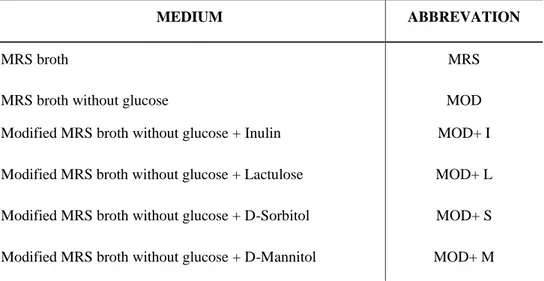
2.5.1. Bacterial Strains and Prebiotics ... 28
2.5.2. Growth of Lb. rhamnosus in presence of prebiotics ... 29
2.5.3 Physical-chemical properties of bacterial cell surface ... 31
2.5.3.1. Hydrophobicity assays ... 31
2.5.3.2. Auto-aggregation assays ... 32
2.5.3.3. Co-aggregation assays ... 32
2.5.4. Antimicrobial activity ... 33
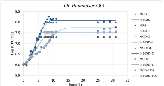
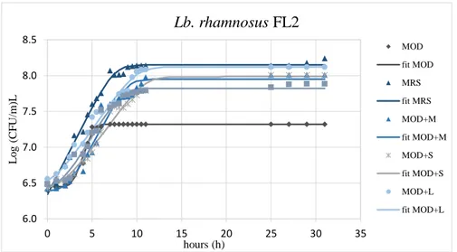
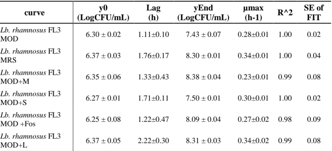
2.6. Results and Discussion ... 34
2.6.2. Hydrophobicity and Aggregation activity in presence of prebiotics . 46
2.6.3 Antimicrobial activity ... 49
2.7. Conclusion ... 51
References ... 53
Chapter 3 ... 57
3.1 Akkermansia muciniphila: new potential probiotic strain ... 57
3.2. Antibiotic susceptibility ... 58
3.3 Survival at the gastrointestinal transit ... 59
3.4. Adhesion ... 61
3.4.1 Cell lines used in vitro model adhesion ... 62
3.4.2. Biofilm ... 63
3.5. Research aim ... 66
3.6. Materials and Methods ... 67
3.6.1. Antimicrobial Susceptibility Test ... 67
3.6.2. Co-culture Lb. rhamnosus and A. muciniphila ... 67
3.6.3. Survival in the gastrointestinal transit... 68
3.6.4. Adhesion assay... 69
3.6.5. Cell surface hydrophobicity ... 70
3.6.6. Auto-aggregation assay ... 71
3.6.7. Biofilm formation ... 71
3.6.8. Statistical analysis ... 72
3.7 Results and Discussion ... 73
3.7.1. Antimicrobial Susceptibility ... 73
3.7.2. Co-culture of A. muciniphila and Lb. rhamnosus ... 75
3.7.3. Survival at simulated gastrointestinal transit ... 78
3.7.4. In vitro adhesion to different cell lines of human origin ... 86
3.7.5. Auto-aggregation ... 92
3.7.6 Adhesion of bacterial cells to liquid hydrocarbons... 92
3.7.7. Correlation between hydrophobicity and adhesion capability ... 95
3.7.8. Correlation between auto-aggregation and adhesion capability ... 97
3.7.9. Biofilm ... 98
References ... 100
Abstract
In recent years, the market for probiotics has evolved rapidly, thanks mainly to more interest that consumers give to those foods that are marketed primarily for their health benefits. Although many potential benefits of probiotics on human health have been hypothesized, only for few strains there is scientific evidence that demonstrate their beneficial effect. For this reason, it is necessary intensive research aimed at investigating new probiotic strains, their mechanisms of action, and their health properties. The aim of this study is to investigate new potential probiotic strains and to test criteria for their screening.
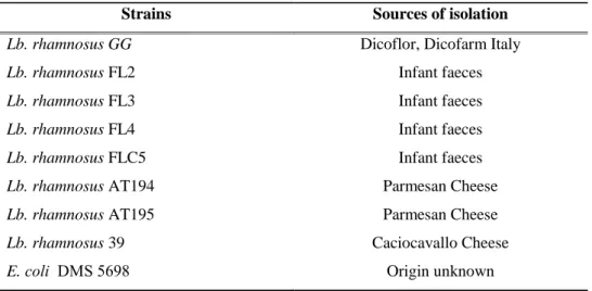
Eight Lactobacillus rhamnosus strains of different origin (infant faeces or cheese), and
Akkermansia muciniphila of human origin, were tested in this study.
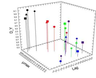
In the first step of this research, the interactions between lactobacilli and prebiotics were investigated, considering eight different strains belonging to Lb. rhamnosus and different prebiotics often found in commercial symbiotic products, such as FOS, lactulose, inulin, mannitol, and sorbitol. The survival and the growth kinetic parameters of Lb. rhamnosus strains cultivated in presence of the different prebiotics as a unique carbon source were evaluated. Moreover, the influence of pre-cultivation in the presence of different prebiotics on the cell surface properties of strains (hydrophobicity, auto-aggregation, co-aggregation) was estimated. Results showed that the conbination of some prebiotics with specific strains can be a stress factor that significantly affects the growth of these strains. In detail, most of the prebiotics used as unique carbon source caused a growth delay compared with glucose, as evidenced by increased values of the lag phase and decreased values of the μmax. Moreover, the results showed significant differences in the cell surface properties (hydrophobicity, auto- and co-aggregation) in the different combinations of strain and prebiotics. Therefore when formulating combinations of prebiotics with probiotics, growth should not be the only parameter taken in to account, since cell surface properties essential to the probiotic function of the strain are also affected.
The second part of the research assessed the safety and some probiotic characteristics of the Lb. rhamnosus and A. muciniphila strains for use in humans. Therefore, it was important to test whether the strains could survive in the acidic environment of the stomach, multiply in the intestine even in the presence of bile salts, and be able to
adhere to intestinal cells. Therefore, experiments were performed to evaluate the survival at the GI transit, and the adhesion features, such as auto-aggregation, hydrophobicity and adhesion to human cell lines of the tested strains. Another objective of this step of the study was to evaluate whether aggregation capacity and hydrophobicity can be used as a preliminary and rapid screening of the adhesion properties of strains.
The results of survival to simulated GI transit evidenced significant differences in the responses of the strains to the stresses encountered. In addition, most Lb. rhamnosus strains have shown a greater sensitivity to exposure to low pH and bile salts, compared to A. muciniphila. Furthermore, this study demonstrates that the exposure to the low pH of the stomach inhibits the survival of Lb. rhamnosus strains to bile-salt induced stress. The results of the adhesion tests to Caco-2, HT29, and MiaPaca-2 cell lines have shown that in the strains tested adhesion is a strain-specific trait, little or not at all influenced by the isolation matrix. The intensity of adhesion to the Caco-2 cell line was the highest using an inoculum of up to 105 CFU/mL in Lb. rhamnosus strains. While a similar adhesion was observed for A. muciniphila in all cell lines.
The results obtained indicate, with sufficient degree of reliability, that the characteristics of hydrophobicity and auto-aggregation of the strains do not correlate to their ability to adhere to intestinal tissues.
Riassunto
Negli ultimi anni, il mercato dei probiotici si è evoluto rapidamente, grazie al crescente interesse dei consumatori nei confronti dei prodotti funzionali. Sebbene siano molti i benefici dei probiotici sulla salute umana ad oggi ipotizzati, solo per pochi ceppi l’effetto benefico è stato dimostrato attraverso studi in vivo. Il presente lavoro, inserendosi nel complesso filone della probiosi, ha inteso individuare nuovi ceppi potenzialmente probiotici definendone anche nuovi criteri di selezione.
A tal proposito sono stati individuati 8 ceppi di diversa origine appartenenti a
Lactobacillus rhamnosus ed un ceppo di Akkermansia muciniphila di origine umana.
Nelle prime fasi della ricerca, sono state studiate le interazioni tra lattobacilli e differenti sostanze prebiotiche spesso impiegate in prodotti simbiotici commerciali, quali FOS, lattulosio, inulina, mannitolo e sorbitolo. In particolare è stata valutata la capacità di crescita dei lattobacilli e l'influenza della pre-coltivazione con i diversi prebiotici sulle caratteristiche della superficie cellulare dei ceppi (idrofobicità, auto-aggregazione, co-aggregazione). I risultati hanno mostrato che le combinazioni lattobacillo-prebiotico influenzano significativamente il comportamento dei ceppi condizionandone la capacità di sviluppo, le proprietà della superficie cellulare nonché l’attività antagonista. Pertanto, si evince in maniera chiara che nella progettazione di un prodotto simbiotico occorre indagare il rapporto tra il ceppo di interesse salutistico e la sostanza prebiotica valutando non solo la capacità di sviluppo ma anche l’espressione dei principali caratteri probiotici.
Nel successivo periodo di dottorato l’attenzione è stata rivolta alla valutazione di alcuni caratteri funzionali dei ceppi di Lb. rhamnosus, confrontandoli con quelli esibiti dal ceppo tipo di A. muciniphila, ritenuto un probiotico emergente.
I benefici dei probiotici sono strettamente connessi all’abilità di questi batteri di superare le condizioni avverse incontrate nel tratto gastro-intestinale e di permanere nell’intestino, che rappresenta il sito target dove esplicano i loro effetti positivi. Pertanto, sono stati condotti esperimenti per valutare la sopravvivenza al transito GI e le caratteristiche di adesione, come l'auto-aggregazione, l'idrofobicità e l'adesione a linee cellulari umane. Inoltre, è stata valuta la correlazione tra le proprietà della superficie cellulare (auto-aggregazione e idrofobicità) e l’adesione a linee cellulari. Dall'analisi dei dati ottenuti è stato riscontrato che i ceppi esibiscono una differente sensibilità alle condizioni di stress simulato nel passaggio gastrointestinale. La maggior parte dei ceppi
di Lb. rhamnosus ha mostrato una maggiore sensibilità agli stress rispetto al ceppo riferibile a A. muciniphila. Inoltre, è emerso che la sopravvivenza dei ceppi di Lb.
rhamnosus è fortemente inibita in presenza di sali biliari come conseguenza del danno
determinato dalla precedente esposizione a pH bassi.
I risultati dei test di adesione a linee cellulari Caco2, HT29 e MiaPaca2 hanno evidenziato che tutti i ceppi saggiati hanno un comportamento ceppo-specifico, poco o per nulla influenzato dalla matrice di isolamento.
Per quanto riguarda l’intensità di adesione, i ceppi di Lb. rhamnosus hanno mostrato una percentuale di adesione più elevata alle cariche microbiche comprese tra 3 e 5 log CFU/mL solo nel caso della linea cellulare Caco-2, mentre A. muciniphila ha evidenziato il medesimo comportamento su tutte le linee cellulari utilizzate.
Infine, è importante sottolineare che i risultati ottenuti indicano, con sufficiente grado di affidabilità, che le caratteristiche di idrofobicità e auto-aggregazione dei ceppi non consentono di presagire la loro capacità di aderire ai tessuti intestinali.
Chapter 1
1.1 The microbiota
The famous statement by Hippocrates “all disease begins in the gut” not only seems to be supported by many evidences but is becoming increasingly important nowadays. With these words the father of the modern medicine suggested the essential role played by gut and diet in many vital homeostatic functions of the human body (Savel & Munro, 2014).
In the recent decade, our understanding of the role of the human gut microbiome has been revolutionized by enormous advances in investigative methods (especially high throughput DNA sequencing). Based on this knowledge, the gut microbiome is concerned as the most densely populated and diverse microbial consortium (Konturek
et al., 2015).
The human microbiota is a dynamic ecosystem established after birth and composed by all the microorganisms living on the human body surface or inside our body in naturally symbiotic relationship within it. (van Best et al., 2015).
The intestinal microbiota has the highest microbial diversity of the human body, with more than 1000 different bacterial species mainly belonging to Firmicutes,
Bacteroidetes, Actinobacteria, and Proteobacteria, these four microbial phyla account
for 98% of the intestinal microbiota (D'Argenio & Salvatore, 2015).
Microbial colonization is a process that begins at birth, when the baby encounters microorganisms from the urogenital tract of the mother. After birth, the neonatal intestine becomes rapidly colonized by maternal and environmental microorganisms and colonization continues during lactation, increasing complexity and microbial diversity (Koenig et al., 2011). The microbiota composition is influenced by many factors such as: the type of diet, the body temperature, the use of drugs, the quantity of ingested food and other physiological characteristics; accordingly, it is subject to variations in relation to the changes that occur in the life of an individual.
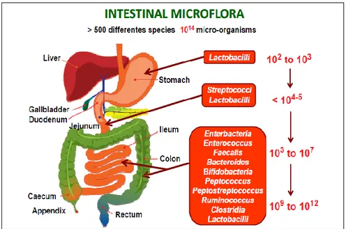
Any portion of the gastrointestinal tract is colonized by specific bacteria that adapt to local conditions. The number of bacterial cells present in the gastrointestinal tract of a
mammal shows a continuum increasing, varying from 103 bacteria/g in the stomach and duodenum, to 104- 107 in jejunum and ileum, to over 1012 cells/g in the cecum and in the colon (Figure 1.1).
Figure 1.1 Spatial and longitudinal variations in microbial numbers and composition across the length of the gastrointestinal tract (Konturek et al., 2015).
The oral cavity due to its characteristics of temperature, pH and nutrient availability is a favorable environment for the growth of many microorganisms. Bacteria are the main inhabitants of the oral cavity. In healthy adults, most species belong to the bacterial
phyla Firmicutes, Proteobacteria, Actinobacteria, Bacteroidetes, and Fusobacteria
(Human Microbiome Project Consortium 2012). In addition, archaea, protozoa, viruses, and fungi are present. Of the 700+ species of identified oral bacteria, a healthy person is colonized by 100 - 200+ microbial species (Rosier et al., 2018).
Thick mucus layer, acidic gastric juice and peristaltic movement in the stomach have raised the dogma that “the stomach is a sterile organ” (Massarrat et al., 2016). However, the dogma quickly changed after the discovery of Helicobacter pylori (Marshall & Warren, 1984). The detection of the bacterial genera such as
Streptococcus, Lactobacillus, Propionibacterium, and Staphylococcus in the stomach of
for certain microorganisms despite the occurrence of acidic pH and digestive enzymes (Delgado et al., 2013).
Recently, the use of culture-independent molecular technologies has revealed that five microbial genera, other than H. pylori, reside in the stomach: Neisseria, Haemophilus,
Prevotella, Streptococcus, and Porphyromonas (Iizasa et al., 2015; Massarrat et al.,
2016).
Most of the bacteria reside in the lower part of the digestive system, especially in the large intestine, also because in the most proximal tract bile and pancreatic secretions are toxic or not favorable for the growth of most microorganisms. Although there is considerable diversity in the intestinal microbiota, a few phyla predominate:
Bacteroidetes, Firmicutes, Actinobacteria, Proteobacteria, Verrucomicrobia, Fusobacteria, and a limited number of Archaea, mainly methanogens. In addition,
despite the consistency of these major phyla, their relative proportions and the species present can vary dramatically between individuals (Shortt et al., 2018).
These microbial populations are normally not pathogenic and play a major role in the breakdown, absorption, and metabolism of key dietary constituents, contributing, for example, to the breakdown of indigestible substances, the biosynthesis of vitamin and bioactive compounds, the degradation of potentially harmful substances and allergenic proteins (Kailasapathy & Chin, 2000; Shortt et al., 2018).
Microbial richness, intended as high bacterial diversity, is usually considered an indicator of a healthy status and makes the host less prone to a number of diseases. Low richness is associated with several life-style related non-communicable diseases such as obesity, metabolic syndrome, immune-related, and inflammatory diseases (Jordan et al., 2015).
The number and diversity of bacterial species within an individual’s gastrointestinal tract remain relatively constant throughout life, as mentioned previously, but it is possible to stimulate the proliferation of specific microorganisms with beneficial health effects by manipulating the host diet (Petschow et al., 2013). More recently, groups of bacterial families have been classified into enterotypes based on their functions (Sun & Chang, 2014). This classification is based on metabolism of dietary components and ability to tolerate and metabolize drugs which should help to further understand the role of enteric microbiota in health and disease.
1.2 Development and Composition
Microbial colonization of the human gut begins at birth. The infant’s intestines are sterile or contain a very low level of microbes at birth, but the GIT is quickly colonized during and after delivery. As a neonate passes through the birth canal, he or she is exposed to the microbial population of the mother’s vagina. This process influences the development of an infant’s intestinal microbiota, which shows similarities to the vaginal microbiota of his or her mother. Infants who were delivered through cesarean section showed reduced microbial numbers in the gut at 1 month when compared with those who were delivered vaginally, although these differences do not remain detectable at 6 months of age (Bull & Plummer, 2014). Breast milk contains living bacteria in a concentration of 102 to 104 CFU/mL, prebiotic nutrients and bioactive components, playing an important role in the establishment of the neonatal microbiota. During the first year, the intestinal tract of the baby passes by a condition of sterility to a very dense colonization (Ballard et al., 2013; Fernandez et al., 2013).
It is believed that the initial gut colonization is instrumental in shaping the composition of the adult’s gut microbiota. This fact was demonstrated by Ley et al. (2005), who observed that the gut microbiota of newly born mice was closely related to that of their mothers, suggesting kinship as an important factor in determining the composition of the gut microbiota.
The infant’s gut microbiota undergoes a succession of changes that are related with a shift from breast- or formula-feeding to weaning and the introduction of solid food. In fact, weaning and solid food introduction increase the diversity of the microbiota and therefore of the microbiome that functionally matures with a decrease in the relative abundance of genes involved in the degradation and utilization of lactose and an enrichment of genes involved in the degradation of other carbohydrates (Subramanian
et al., 2015).
At two years of age, the baby intestinal microbiota is almost stabilized. Despite the relative similarities of the gut microbiota in mothers and their offspring, microbial succession in the GIT is also influenced by numerous external and internal, host-related factors. External factors include the microbial contamination of the immediate environment, the type of food eaten, and feeding habits, in addition to the composition of the maternal microbiota. Also, dietary and temperature-related stresses can influence the succession of microbes. Internal factors include, but are not limited to, intestinal pH;
microbial interactions; body temperature; physiological factors, such as peristalsis; bile salts; host secretions and immune responses; drug therapy; and bacterial mucosal receptors (Matthew et al., 2014).
1.3. Functions of the Gut Microbiota
1.3.1. Metabolism.
The microbiota collectively encodes more than 3.3 million non-redundant genes, exceeding the number encoded by the human host genome by 150-fold, consequently, it can carry out a variety of metabolic functions that humans are unable to do or are able to do only partially. Gene diversity in the microbial community provides various enzymes and biochemical pathways that are distinct from the host's own constitutive resources. Intestinal bacteria can produce a variety of vitamins, all the essential and non-essential amino acids and carry out the biotransformation of the bile (Vyas & Ranganathan, 2012). In addition, the microbiome provides the vital biochemical pathways for the fermentation of non- digestible carbohydrates that is a major source of energy in the colon. Non-digestible carbohydrates include large polysaccharides (resistant starches, cellulose, hemicellulose, pectin, and gums), some oligosaccharides that escape digestion, and unabsorbed sugars and alcohols; and host-derived mucins (Bull & Plummer, 2014). This function results in the recovery of energy and absorbable substrates for the human host and a supply of energy and nutrients for bacterial growth and proliferation (Guarner & Malagelada, 2003).
1.3.2 Host Protection and Immune-system Development
Many intestinal bacteria produce antimicrobial compounds and compete for nutrients and sites of attachment in the gut lining, thereby preventing colonization by pathogens. This action is known as the barrier or competitive-exclusion effect (Bull & Plummer, 2014).
Different mechanisms have been implicated in the barrier effect. One mechanism is, in
vitro, nonpathogenic bacteria compete for attachment sites in the brush border of
enteroinvasive pathogens into the epithelial cells. Furthermore, commensal bacteria compete for nutrient availability in ecological niches and maintain their collective habitat by administering and consuming all resources (Guarner & Malagelada, 2003). Finally, bacteria can inhibit the growth of their competitors by producing antimicrobial compounds such as bacteriocins, and the ability to synthesize these substances is widely distributed among gastrointestinal bacteria (Drissi et al., 2015).
The intestinal epithelium is the main interface between the immune system and the external environment. The development of the host immune system is affected by continuous and dynamic interactions with the intestinal microbiota and its metabolites. Bacteria are integral to the early development of the gut-mucosal immune system, both in terms of its physical components and its function, and continue to play a role later in life in its operation. The cells of the intestinal epithelium avert threats from pathogens by signaling to the innate immune system through specific receptors that recognize and bind to specific molecules associated with bacteria, leading to the production of a host’s immune response and the release of protective peptides, cytokines, and white blood cells (Vyas & Ranganathan, 2012). The result can be a protective response to commensal bacteria, an inflammatory response to pathogenic organisms, or a trigger for a host’s cell death.
Exposure to intestinal bacteria is also implicated in the prevention of allergies (i.e., a disproportionate reaction of the immune system to non-harmful antigens). Allergic infants and young children have been found to have a different composition of intestinal bacteria than those who do not develop allergies. It is hypothesized that the intestinal microbiota stimulates the immune system and trains it to respond proportionately to all antigens (Bull & Plummer, 2014). An altered composition of intestinal microbiota in early life can lead to an inadequately trained immune system that can, and often does, overreact to antigens (Björkstén et al., 2001).
1.3.3 The Gut–Brain Axis
The gut–brain axis is a communication system that integrates neural, hormonal, and immunological signaling between the gut and the brain, offering the intestinal microbiota and its metabolites a potential route through which to access the brain (Collins et al., 2012). This communication system is bidirectional, established pathways of communication include the autonomic nervous system (ANS), the enteric nervous
system (ENS), the neuroendocrine system, and the immune system (Bull & Plummer, 2014).
The communication system enables the brain to command gastrointestinal functions, such as peristalsis and mucin production, and immune functions (Mayer et al., 2011). In fact, stress has been shown to influence the integrity of the gut epithelium and to alter peristalsis, secretions, and mucin production, thereby altering the habitat of the intestinal microbiota and promoting changes in microbial composition and/or metabolism (Collins et al., 2012).
In the last years, neuroscientists are taking notice of these novel reports that highlight also the ‘bottom-up’ influence of microbes themselves; in fact, several studies show that commensal bacteria are important to CNS function (Zhou & Foster, 2015). Different studies suggest that gut microbiota is an important player in how the body influences the brain, contribute to normal healthy homeostasis, and influence risk of disease, including anxiety and mood disorders (Clarke et al.,2012; Heijtz et al., 2011).
1.3.4. Gut Microbiota in Disease
Associations have been established between human intestinal microbiota and a seemingly ever-increasing number of intestinal and extra-intestinal disorders. Intestinal disorders include inflammatory bowel disease, irritable bowel syndrome (IBS), and coeliac disease, while extra-intestinal disorders include allergy, asthma, metabolic syndrome, cardiovascular disease, type 2 diabetes and obesity.
In many of these conditions, the mechanisms leading to disease development involve the pivotal mutualistic relationship between the colonic microbiota, their metabolic products, and the host immune system. The establishment of a ‘healthy’ relationship early in life appears to be critical to maintaining intestinal homeostasis.
Based on evidence to date, we can assess the potential to positively modulate the composition of the colonic microbiota and ameliorate disease activity through bacterial intervention (Carding et al., 2015).
1.3.4.1. Irritable Bowel Syndrome
Irritable bowel syndrome (IBS) is defined as a group of functional bowel disorders in which abdominal discomfort or pain is associated with defecation or a change in bowel
habits and with features of disordered defecation. The symptoms can greatly weaken patients’ quality of life and account for notable economical costs for society.
IBS is thought to affect approximately 10% to 20% of adults and adolescents worldwide (Longstreth et al., 2006). The exact cause of IBS is unknown and is thought to be multifactorial. Genetic factors, motor dysfunction of the GIT, visceral hypersensitivity, infection, inflammation, and immunity as well as psychopathological factors are thought to play roles in its development (Ghoshal et al., 2012). Together with these factors, variation in the gut microbiota is thought to be complicit in the low-grade intestinal inflammation associated with the syndrome (Guinane & Cotter, 2013). In the healthy gut, the intestinal microbiota either have direct bactericidal effects or can prevent the adherence of pathogenic bacteria to the wall of the GIT (Kellow et al., 2006).
1.3.4.2. Inflammatory Bowel Disease
Inflammatory bowel disease (IBD) is a chronic, relapsing and remitting disease, with both ulcerative colitis (UC) and Crohn's disease (CD) causing significant morbidity. The precise etiology of IBD is unclear, however, its development, progression and phenotype are multifactorial, with genetics and environment playing a role (Nagalingam & Lynch, 2012). There is increasing evidence supporting a microbial influence in the pathogenesis of IBD resulting from an inappropriate immune response towards the components of the commensal microbiota (DuPont & DuPont, 2011). In particular, CD is characterized by a cobblestone-like pattern of inflammation (i.e., affected regions interrupted by healthy tissue), which can occur anywhere along the length of the GIT (Bull & Plummer 2014). It is also typified by ulcerations that may span the entirety of the intestinal wall, resulting in fissures that may perforate the intestinal wall and impact other organs such as the kidney or uterus (Nagalingan et al., 2012).
UC typically manifests as contiguous inflammation involving only the surface layers of the intestinal wall. It is primarily localized in the colon and most commonly originates at the rectum (Baumgart & Sandborn, 2012). Given the role of the gastrointestinal microbiota in driving inflammation in IBD, treatments that manipulate the microbiota
have been investigated including the use of probiotics and prebiotics, with variable evidence for their efficacy.
1.3.4.3. Systemic Metabolic Diseases
Systemic metabolic diseases include obesity and type 2 diabetes. Both obesity and diabetes are characterized by a state of chronic low-grade inflammation with abnormal expression and production of multiple inflammatory mediators such as tumor necrosis factor and interleukins. Recent studies have shown a relationship between the composition of the intestinal microbiota and metabolic diseases like obesity and diabetes (Everard & Cani, 2013).
Early indications that the gut microbiota is involved in obesity came when metabolically obese mice, with a mutation in the leptin gene, were shown to have a significantly different microbiota compared with mice without the mutation (Ley et al., 2005).
Recently, research has indicated that the risks related to the development of type 2 diabetes may also involve the composition of the intestinal microbiota. The gut microbiota of participants with type 2 diabetes displayed only a limited deviation from the non-diabetic control group, although a decline in butyrate-producing bacteria that may be metabolically beneficial was observed (Qin et al., 2012). This observation suggests that a state of functional dysbiosis, rather than any specific microbial species, could have a direct association with the pathophysiology of type 2 diabetes.
1.3.4.4. Atopic Eczema and Other Allergic Disease
Allergic diseases, specifically those driven by type 1 hypersensitization—atopic eczema, atopic asthma, rhinitis—and type 1 food allergies have risen globally in incidence over the past 50 years, with the developed world now showing an incidence at 20% of the population, providing a considerable proportion of overall disease burden (Okada et al., 2010). Atopic sensitization occurs primarily in the first 2 years of life and can persist through a lifetime, with the expression of allergic disease typically beginning with eczema (0-2 y), asthma (>5 y), and rhinitis (>8 y) in what is referred to as the atopic march (Shen et al., 2013)
The causes of atopic eczema are potentially numerous and are not well understood, although the method of birth and a mutation in a particular human gene involved in
skin-barrier function are known to be implicated (Williams & Grindlay, 2010). Characterization of the gut microbiota of sufferers of atopic eczema showed that infants at 1 month of age with the disease had a significantly lower microbial diversity, particularly with regard to the Bacteroidetes phylum, compared with infants without atopic eczema (Abrahamsson et al., 2012).
Several epidemiological studies and experimental research suggest that microbial stimulation of the immune system influences the development of tolerance to innocuous allergens (Penders et al., 2007). The gastrointestinal microbiota composition may be of particular interest, as it provides an early and major source of immune stimulation and seems to be a prerequisite for the development of oral tolerance. Although most studies indicated an association between the gut microbiota composition and atopic sensitization or symptoms, no specific harmful or protective microbes can be identified yet (Penders et al., 2007). To gain more insight into the role of the gut microbiota in the etiology of atopy, large‐scale prospective birth cohort studies using molecular methods to study the gut microbiota are needed.
1.4. Manipulating the gut microbiota through diet
Changes in the intestinal microbiota can occur when the diet is changed, but the healthy microbiota is resilient to changes through dietary interventions, meaning that homeostatic reactions restore the original community composition (Valdes et al., 2018). Dietary amounts of protein, saturated and unsaturated fats, carbohydrates, and dietary fiber influence the abundance of different types of bacteria in the gut (Valdes et al., 2018).
The microbiota can also be modified by adding live micro-organisms to food. Although, there are concerns that most microbial supplements are unable to establish themselves in the gut and fail to exert an effect on the resident community (Kristensen et al., 2016; Walter et al., 2018). However, probiotics can affect health independently of the gut microbiota through direct effects on the host; for example, through immune modulation or the production of bioactive compounds (Valdes et al., 2018).
Emerging trends in probiotic treatment include: i) the use of new microorganisms and new formulations, which combine probiotics and prebiotics, symbiotics (Plovier et al., 2017); ii) personalized approaches based on the individual characteristics of the intestinal microbiota (Chua et al., 2017).
Given the current gaps in knowledge, many other clinical trials using prebiotics or probiotics or fecal microbiota transplantation are needed to assess changes in intestinal microbiota composition and health effects.
References
Abrahamsson, T.R., Jakobsson, H.E., Andersson, A.F., Björkstén, B., Engstrand, L., Jenmalm, M.C. (2012). Low diversity of the gut microbiota in infants with atopic eczema. Journal of Allergy Clinical
Immunology. 129(2):434-440.
Ballard, O. & Morrow, A.L.(2013). Human Milk Composition. Nutrients and Bioactive Factors. 60(1):49–74
Baumgart, D.C. & Sandborn W.J. (2007). Inflammatory bowel disease: clinical aspects and established and evolving therapies. The Lancet. 369(9573):1641-1657.
Björkstén, B., Sepp, E., Julge, K., Voor, T., Mikelsaar, M. (2001). Allergy development and the intestinal microflora during the first year of life. Journal of Allergy Clinical Immunololy.108(4):516-520.
Bull, M.J. & Plummer. N.T. (2014). The Human Gut Microbiome in Health and Disease. Human Gut
Microbiome.13(6):133-136.
Carding, S., Verbeke, K., Vipond, D. T., Corfe., B. M., Owen, L. J. (2015). Dysbiosis of the gut microbiota in disease. Microbial ecology in health and disease. 26(1):1-9.
Chua, K. J., Kwok, W. C., Aggarwal, N., Sun, T., Chang, M. W. (2017). Designer probiotics for the prevention and treatment of human diseases. Current opinion in chemical biology. 40:8-16.
Clarke,G., Grenham, S., Scully,P., Fitzgerald,P., Moloney, R.D., Shanahan, F., Dinan, T.G., Cryan, J.F. (2012). The microbiome-gut–brain axis during early life regulates the hippocampal serotonergic system in a sex-dependent manner. Molecular Psychiatry. 18:666–673.
Collins, S.M., Surette, M., Bercik, P. (2012). The interplay between the intestinal microbiota and the brain. Nature Reviews Microbiology. 10(11):735-742.
D’Argenio, V., & Salvatore, F. (2015). The role of the gut microbiome in the healthy adult status. Clinica
Chimica Acta. 451:97-102.
Delgado, S., Cabrera-Rubio, R., Mira, A., Suarez, A., Mayo, B. (2013). Microbiological survey of the human gastric ecosystem using culturing and pyrosequencing methods. Microbial Ecology. 65:763–772. Drissi, F., Buffet, S., Raoult, D., Merhej, V. (2015). Common occurrence of antibacterial agents in human intestinal microbiota. Frontiers in Microbiology. 6(441):1-9.
DuPont, A.W. & DuPont, H.L. (2011). The intestinal microbiota and chronic disorders of the gut. Nature
Reviews Gastroenterology Hepatology. 8:523–31.
Everard, A., & Cani, P. D. (2013). Diabetes, obesity and gut microbiota. Best practice & research
Clinical gastroenterology. 27(1):73-83.
Fernández, L., Langa S., Martín, V., Maldonado, A., Jiménez, E., Martín, R., Rodríguez, J.M. (2013) The human milk microbiota: Origin and potential roles in health and disease. Pharmacological Research. 69(1):1-10
Ghoshal, U.C., Shukla, R., Ghoshal, U., Gwee, K.A., Ng, S.C. (2012). Quigley FM. The gut microbiota and irritable bowel syndrome: friend or foe? International Journal Inflammation. 151085:1-13.
Guarner, F. & Malagelada, J.R. (2003). Gut flora in health and disease. The Lancet. 361(9356):512-519. Guinane, C.M. & Cotter, P.D. (2013). Role of the gut microbiota in health and chronic gastrointestinal disease: understanding a hidden metabolic organ. Therapeutic Advances in Gastroenterology. 6(4):295-308.
Heijtz, R.D., Wang, S., Anuar F., Qian, Y., Björkholm, B., Samuelsson, A., Hibberd, M.L., Forssberg, H., Pettersson, S. (2011). Normal gut microbiota modulates brain development and behavior. PNAS. 08:3047-3052
Iizasa, H., Ishihara, S., Richardo, T., Kanehiro, Y., and Yoshiyama, H. (2015). Dysbiotic infection in the stomach. World Journal Gastroenterology. 21(40):11450–11457.
Jordán, F., Lauria, M., Scotti, M., Nguyen, T-P., Praveen, P., Morine, M., & Priami, C. (2015). Diversity of key players in the microbial ecosystems of the human body. Scientific Reports. 5(15920):1-10. Kailasapathy, K. & Chin, J. (2000). Survival and therapeutic potential of probiotic organisms with reference to Lactobacillus acidophilus and Bifidobacterium spp. Immunology & Cell Biology. 78(1):80-8.
Kellow, J.E., Azpiroz, F., Delvaux, M. (2006). Applied principles of neurogastroenterology: physiology/motility sensation. Gastroenterology. 130(5):1412-1420.
Koenig, J.E., Spor, A., Scalfone, N., Fricker, A.D., Stombaugh, J., Knight, R., Angenent, L.T., and Ley, R.E. (2010). Succession of microbial consortia in the developing infant gut microbiome. PNAS. 108(1):4578–4585.
Konturek, P.C., Haziri, D., Brzozowski, T., Hess, S., Heyman, S., Kwiecien, S.J., Koziel, J. (2015). Emergingrole of fecal microbiota therapy in the treatment of gastrointestinal and extra-gastrointestinal diseases. Journal of Physiology and Pharmacology. 66(4):483-491.
Kristensen, N. B., Bryrup, T., Allin, K. H., Nielsen, T., Hansen, T. H., Pedersen, O. (2016). Alterations in fecal microbiota composition by probiotic supplementation in healthy adults: a systematic review of randomized controlled trials. Genome medicine. 8(52):1-11.
Ley, R.E., Bäckhed, F., Turnbaugh, P., Lozupone, C.A., Knight, R.D., Gordon, J.I. (2005). Obesity alters gut microbial ecology. Proceedings of the National Academy Sciences U SA. 102(31):11070-11075. Longstreth, G.F., Thompson, W.G., Chey, W.D., Houghton, L.A., Mearin, F., Spiller, R.C. (2006). Functional bowel disorders. Gastroenterology. 130(5):1480-1491.
Marshall, B. J., & Warren, J. R. (1984). Unidentified curved bacilli in the stomach of patients with gastritis and peptic ulceration. The Lancet. 323(1):1311–1315.
Massarrat, S., Saniee, P., Siavoshi, F., Mokhtari, R., Mansour-Ghanaei, F., & Khalili-Samani, S. (2016). The effect of Helicobacter pylori infection, aging, and consumption of proton pump inhibitor on fungal colonization in the stomach of dyspeptic patients. Frontiers in microbiology. 7(801):1-7.
Matthew, J.B., & Plummer, N.T.(2014).The Human Gut Microbiome in Health and Disease Integrative
Medicine: A Clinician's Journal. 13(6):17-22.
Mayer, E.A. (2011). Gut feelings: the emerging biology of gut-brain communication. Nature Reviews
Neuroscience. 12(8):453-466.
Nagalingam, N.A. & Lynch, S.V. (2012). Role of the microbiota in inflammatory bowel diseases.
Inflammatory Bowel Diseases. 18(5):968-984.
Okada, H., Kuhn, C., Feillet, H., Bach, J.F. (2010). The ‘hygiene hypothesis’ for autoimmune and allergic diseases: an update. Clinical Experimental Immunology. 160(1):1-9.
Penders, J., Stobberingh, E. E., Brandt, P. V. D., Thijs, C. (2007). The role of the intestinal microbiota in the development of atopic disorders. Allergy. 62(11):1223-1236.
Petschow, B., Dore J., Hibberd P. (2013). Probiotics, prebiotics, and the host microbiome: the science of translation. Annals of the New York Academy of Science. 1306(1):1-17.
Plovier, H., Everard, A., Druart, C., Depommier, C., Van Hul, M., Geurts, L., Myridakis, A. (2017). A purified membrane protein from Akkermansia muciniphila or the pasteurized bacterium improves metabolism in obese and diabetic mice. Nature medicine. 23(1):107-113.
Qin, J., Li, Y., Cai, Z. (2012). A metagenome-wide association study of gut microbiota in type 2 diabetes.
Nature. 490(7418):55-60.
Rosier, B. T., Marsh, P. D., Mira, A. (2018). Resilience of the oral microbiota in health: mechanisms that prevent dysbiosis. Journal of dental research. 97(4):371-380.
Savel, R.H. & Munro, C.L. (2014). From Asclepius to Hippocrates: the art and science of healing.
Shen, C.Y., Lin, M.C., Lin, H.K., Lin, C.H., Fu, L.S., Fu, Y.C. (2013). The natural course of eczema from birth to age 7 years and the association with asthma and allergic rhinitis: a population-based birth cohort study. Allergy Asthma Proceedings. 34(1):78-83.
Shortt, C., Hasselwander, O., Meynier, A., Nauta, A., Fernández, E. N., Putz, P., & Antoine, J. M. (2018). Systematic review of the effects of the intestinal microbiota on selected nutrients and non-nutrients. European Journal of Nutrition. 57(1):25-49.
Subramanian, S., Blanton, L.V., Frese, S.A., Charbonneau, M., Mills, D.A., Gordon, J.I. (2015). Cultivating Healthy Growth and Nutrition through the Gut Microbiota. Cell. 161(1):36-48
Sun, J. & Chang, E.B. (2014) Exploring gut microbes in human health and disease: Pushing the envelope.
Genes and Diseases. 1:132-139.
Valdes, A. M., Walter, J., Segal, E., Spector, T. D. (2018). Role of the gut microbiota in nutrition and health. BMJ 361(1):36-44.
van Best, N., Hornef, M.W., Savelkoul, P.H.M., Penders, J. (2015) On the origin of species: Factors shaping the establishment of infant's gut microbiota Embryo today: Reviews. 105(4):240-251.
Vyas, U. & Ranganathan, N. (2012). Probiotics, prebiotics, and synbiotics: gut and beyond.
Gastroenterology Research and Practice. 1-16.
Walter, J., Maldonado-Gómez, M. X., Martínez, I. (2018). To engraft or not to engraft: an ecological framework for gut microbiome modulation with live microbes. Current opinion in biotechnology. 49:129-139.
Williams, H.C. & Grindlay, D.J. (2010). What’s new in atopic eczema? An analysis of systematic reviews published in 2007 and 2008, I: definitions, causes and consequences of eczema. Clinical and
Experimental Dermatology. 35(1):12-15.
Zhou, L. & Foster, J. A. (2015). Psychobiotics and the gut–brain axis: in the pursuit of happiness.
Chapter 2
2.1 Probiotics
The word “probiotic” was coined from the Greek, “προ” plus “βιοτος” meaning literally “for life”. The term came into common use after 1980. The introduction of the concept is generally attributed to Nobel recipient Élie Metchnikoff. He suggested in 1907 that "the dependence of the intestinal microbes on the food makes it possible to adopt measures to modify the flora in our bodies and to replace the harmful microbes by useful microbes" (Metchnikoff, 1907). He observed that some rural communities in Europe, who used to drink fermented milk, had a relatively long life, and that milk fermented by lactic acid bacteria (LAB), inhibited the growth of proteolytic bacteria due to the low pH value. Since that time, probiotics have become a multibillion-dollar industry, helped by their categorization as “dietary supplements,” which are not subject to stringent evaluation by the US Food and Drug Administration (FDA).
The rationale for the use of probiotics is based on the gastrointestinal and genitourinary regulatory role played by the commensal microbiota and the need to restore the microbial ecosystem after disturbances due to disease or antibiotics. The current definition, formulated in 2002 by FAO (Food and Agriculture Organization of the United Nations) and WHO (World Health Organization) working group experts, states that probiotics are “live strains of strictly selected microorganisms which, when administered in adequate amounts, confer a health benefit on the host” (FAO & WHO, 2002). The definition was maintained by the International Scientific Association for Probiotics and Prebiotics (ISAPP) in 2014 (Hill et al., 2014).
To be defined as probiotics, microorganisms must fulfil specific requisites. The criteria for the selection and assessment of probiotic microorganisms are the result of the collaboration between research institutions and universities with food industries. Markowiak et al., (2017), according to WHO, FAO, and EFSA suggestions, have identified the safety and functionality criteria, including the technological usefulness, of potential probiotic strains, as listed in Table 2.1
Table 2.1 Selection criteria of probiotic strains (Markowiak, et al., 2017)
Criterion Required Properties
Safety
Human or animal origin.
Isolated from the gastrointestinal tract of healthy individuals. History of safe use.
Precise diagnostic identification (phenotype and genotype traits). Absence of data regarding an association with infective disease. Absence of the ability to cleave bile acid salts.
No adverse effects.
Absence of genes responsible for antibiotic resistance localised in non-stable elements.
Functionality
Competitiveness with respect to the microbiota inhabiting the intestinal ecosystem.
Ability to survive and maintain the metabolic activity, and to grow in the target site.
Resistance to bile salts and enzymes. Resistance to low pH in the stomach.
Competitiveness with respect to microbial species inhabiting the intestinal ecosystem (including closely related species).
Antagonistic activity towards pathogens (e.g., H. pylori, Salmonella spp., Listeria monocytogenes, Clostridium difficile).
Resistance to bacteriocins and acids produced by the endogenic intestinal microbiota.
Adherence and ability to colonise some particular sites within the host organism, and an appropriate survival rate in the gastrointestinal system.
Technological usability
Easy production of high biomass amounts and high productivity of cultures.
Viability and stability of the desired properties of probiotic bacteria during the fixing process (freezing, freeze-drying), preparation, and distribution of probiotic products.
High storage survival rate in finished products (in aerobic and micro-aerophilic conditions).
Guarantee of desired sensory properties of finished products (in the case of the food industry).
Genetic stability.
Probiotic characteristics are not associated with the genus or species of a microorganism, but with few and especially specific strains belonging to a particular species (Hill et al., 2014). The safety of a probiotic strain is defined by its origin, the absence of association with pathogenic cultures, and its antibiotic resistance profile. Functional aspects define the survival of probiotic in the gastrointestinal tract and its immunomodulatory effect. Probiotic strains must meet the requirements associated with the technology of their production, which means they must be able to survive and maintain their properties throughout storage and distribution processes (Reale et al., 2015). Probiotics must also have documented pro-health effects consistent with the characteristics of the strain present in a marketed product.
The strains with beneficial properties, potential probiotics, most frequently belong to the genera Bifidobacterium and Lactobacillus. However, it must be considered that the probiotic potential of different bacterial strains, even within the same species, differs. Different strains of the same species are always unique, and may have different areas of adherence (site-specific), specific immunological effects, and their action on healthy versus an inflamed mucosal milieu may be different (Soccol et al., 2010).
2.1.1. Mechanism of action
Probiotics have numerous advantageous functions in human organisms. Their main advantage is the effect on the development of the microbiota inhabiting the organism ensuring proper balance between pathogens and the bacteria that are necessary for the normal functions of the organism (Oelschlaeger, 2010).
The mechanism of probiotic action is not completely understood but molecular and genetic studies have determined the basic mechanisms underlying their beneficial effects, these involve four mechanisms:
antagonism through the production of antimicrobial substances;
competition with pathogens for adhesion to the epithelium and for nutrients; stimulation of immunity;
inactivation and removal of microbial toxins.
The first mechanism is directly associated with its effect on pathogens. Lactobacilli and
Bifidobacteria can produce antimicrobial compounds such as: low-molecular-weight
hydroperoxide and short-chain fatty acids) and bacteriocins (Oelschlaeger, et al., 2010). Probiotic bacteria can, also, produce so-called deconjugated bile acids which are derivatives of bile salts. Deconjugated bile acids show a stronger antimicrobial activity compared to the bile salts synthesized by the host organism. How probiotics protect themselves from these ‘‘self-made’’ metabolites or if they are resistant to deconjugated bile acids, remains to be elucidated (Bermudez-Brito et al., 2012).
The second mechanism is competition with pathogen microorganisms. In fact, probiotics may be able to adhere to epithelial cells, thus blocking pathogens. This mechanism exerts an important effect on the host health condition. Moreover, the adhesion of probiotic microorganisms to epithelial cells may trigger a signalling cascade, leading to immunological modulation (Markowiak et al., 2017). Probiotic strains can adhere specifically or non-specifically. Specific adhesion is when an adhesion on the bacterial cell bind to a receptor on the epithelial cell, which is often defined as a lock and key function. Non-specific adhesion is a general phenomenon mediated by hydrophobic or electrostatic interaction. Non-specific adhesion may not be of significance in the colonization of epithelia in vivo, but may be important in the colonization of luminal contents. For example, non-specific adhesion may enhance substrate uptake and thus enhance growth. Because probiotic bacteria are able to adhere to epithelial cells in cell culture assays, blocking the adherence of pathogens, it is extrapolated that this is important for the probiotic effect in the host. The anti-adhesive effect could be the result of competition between the probiotic and the pathogen for the same receptor; the induction by probiotics of (increased) mucin production (competitive exclusion); or the degradation of carbohydrate receptors by secreted proteins when they establish biofilms (Oelschlaeger, 2010).
In addition to producing anti-pathogenic bioactive compounds that directly affect pathogens, and to competing with pathogens for receptor sites, and nutrients, probiotics also stimulate host anti-pathogenic defense pathways, such as stimulating or activating the pathway involved in the production of defensins that are cationic anti-microbial peptides produced in several cell types (Kerry et al., 2018).
The last probiotic effect is based on actions leading to inactivation of toxins and help with the removal of toxins from the body. Help in detoxification, by probiotics, can take place by adsorption (some strains can bind toxins to their cell wall and reduce the intestinal absorption of toxins), but can also result from the degradation of toxins by microorganisms (Markowiak et al., 2017; McCormick, 2013).
2.1.2.
Lactobacillus genus
The genus Lactobacillus is the largest group of lactic acid bacteria (LAB), to date, 182 species have been described in the genus. Taxonomically, this genus is diverse and it contains at least 12 separate phylogenetic groups.
The genus Lactobacillus is one of the most important taxa involved in food microbiology and human nutrition, and probably the most used as a probiotic in various foods, especially fermented meat and dairy products.
Many species belonging to this genus are essential in the production of fermented foods and are used as starter and protective cultures. This genus includes a high number of GRAS species (Generally Recognized As Safe) and with qualified presumption of safety (QPS). Furthermore, some strains of human origin are exploited as probiotics (Goh et al., 2009).
Species of the genus Lactobacillus are non-sporeforming, catalase negative (although some strains can produce pseudocatalase), obligate saccharolytic rods or coccobacilli characterized by a low content of GC of the genome although the upper limit of the content of GC reaches 59.2 mol% (Salvetti et al., 2014).
The growth temperature ranges from 2 to 53 °C, and can grow in a pH range between 3 and 8. The optimal growth temperature and pH are usually 30-40 °C and 5.5-6, 2, respectively.
They have complex nutritional requirements in terms of amino acids, peptides, vitamins, salts, fatty acids or fatty acid esters, and are found in rich habitats where substrates containing carbohydrates are available as food (dairy products, cereal products, meat and fish products , beer, wine, fruit and fruit juices, pickled vegetables, sauerkraut, ensilage and sourdough); they are part of the normal flora of the mouth, of the gastrointestinal tract and of the human genital tracts of many animals (Hammes & Hertel, 2009).
The taxonomy of lactobacilli has always been based on phenotypic properties as carbohydrate fermentation models, resistance to different NaCl concentrations, growth in different soils at defined temperatures or pH range and antibiotic resistance, extended to cell wall composition, fatty acids cell phones, isoprenoids quinones and other cell characteristics (Hammes & Hertel, 2009).
Originally, the lactobacilli were grouped according to their growth temperature and the fermentation of hexoses (Orla-Jensen., 1919) and subsequently based on their
homo/heteropherence potential (Kandler et al., 1986; Carr et al., 2002). The subdivision of the genus Lactobacillus has been revisited by Pot et al. (1994), but the accepted "modern" definition is that given by Hammes and Vogel (1995) and Hammes and Hertel (2009) which divides lactobacilli as obligatory homofermentative, heterogeneous optional and obligate heterofermentative, based on the type of fermented sugars and fermentation products. Homofermentative lactobacilli (commonly referred to as metabolic group A) ferment almost exclusively (> 85%) of lactic acid via the Embden-Meyerhof-Parnas (EMP) or glycolysis pathway; pentose and gluconate are not fermented. The optional heterofermentative species (metabolic group B) ferment hexose in lactic acid by EMP and can degrade pentose and gluconates through an inducible phosphoketolase, an enzyme of the pentose phosphate (PP) pathway, with consequent production of acetic acid, ethanol and acidic form under glucose limitation. Finally, the obligatory heterofermentative lactobacilli (metabolic group C) possess FDB aldolase, but not phosphoketolase, and metabolize pentose and hexoses exclusively via the phosphogluconate pathway (corresponding to the first part of the PP) and produce lactic acid, ethanol (or acetic acid) and CO2 (Hammes & Hertel, 2009).
The description of new species in the last 20 years has led to a progressive review of the genus with the recognition of a growing number of variable phylogenetic groups (Hammes et al., 2003; Felis et al., 2007). Although the analysis of the 16S rRNA gene sequence contributed to the development of a more exhaustive taxonomy for lactobacilli, it became evident that there is only a small correlation between the traditional classification based on metabolic properties and phylogenetic correlation (Hammes & Hertel, 2009).
2.1.3. Lactobacillus rhamnosus
Lactobacillus rhamnosus is a LAB that colonizes diverse habitats, including the
gastrointestinal tract, plant materials, and food (Reale et al., 2015). Lb. rhamnosus is a Gram-positive, non-spore-forming, catalase-negative, facultative anaerobic or generally microaerophilic bacteria species. These bacteria appear in a rod form and they can form small chains of bacilli. Without any doubt, the strain GG (ATCC 53103) is the most extensively studied probiotic with several hundreds of citations and twelve significant clinical results. Lb. rhamnosus GG (LGG) was isolated, for the first time, from faecal samples of healthy individuals by the two researchers Sherwood Gorbach and Barry
Goldin (1989), from which it derives its name composed of the letters GG. It has been identified as a powerful probiotic because it was able to resistance to acids and bile, to optimal growth capacity and to adhere to the intestinal epithelium. For these reasons, it is one of the most studied probiotic strains and it is used in many pharmaceutical formulas or fermented milk-based preparations. Its beneficial effects have been evaluated widely in clinical trials and in human intervention studies, through which this strain was found to be effective against diarrhea and, in particular, it was able to repress the growth and development of pathogens that are present in the human intestine. For example, Lb. rhamnosus GG affects immunomodulation by increased expression of mucin-secreting cells and IgA that repress rotavirus development. Lb. rhamnosus GG is also able to decrease the risk of prolongation of diarrhea caused by Clostridium difficile in individuals that took antibiotics. Other in vitro studies of Lb. rhamnosus GG showed inhibition of the adhesion or infection of additional intestinal pathogens such as
Salmonella enterica subsp. enteric serovar typhimurium, Shigella sonei, Pseudomonas, Staphylococcus and Streptococcus. Aditionally, Lb. rhamnosus GG showed excellent
adhesion capacity in vitro and in vivo studies. It is thought that adhesion is mediated by appendages similar to fimbriae, also called "pili", these are thin protrusions of proteic nature present on the cell surface. These characteristics distinguish Lb. rhamnosus GG from other strains belonging to the Lb. rhamnosus species, from which it also differs due to its ability to persist longer and in greater concentration in the intestine.
2.2. Prebiotics
The first definition of prebiotic was given by Gibson and Roberfroid (1995) as “a non digestible food ingredient that beneficially affects the host by selectively stimulating the growth and/or activity of one or a limited number of bacteria in the colon, and thus improves host health”. A food ingredient must fulfil three criteria to be considered a prebiotic:
neither be hydrolysed nor absorbed in the upper part of the gastrointestinal tract; be a selective substrate for one or a limited number of potentially beneficial bacteria commensal to the colon, which are stimulated to grow and/or are metabolically activated;
consequently, be able to alter the colonic microflora towards a healthier composition, for example, by increasing the number of saccharolytic species and reducing putrefactive microorganisms such as a saccharolytic clostridia and
Enterobacteriaceae (Salminen et al., 1998; Collins et al., 1999).
Amongst food ingredients, non digestible carbohydrates (oligo- and polysaccharides), some peptides and proteins, and certain lipids (both ethers and esters) are candidate prebiotics (Mitsuoka et al., 2014). Because of their chemical structure, these compounds are not absorbed in the upper part of the gastrointestinal tract or hydrolyzed by human digestive enzymes. Such ingredients could be called "colonic foods", i.e. foods entering the colon and serving as substrates for endogenous colon bacteria, thus indirectly providing the host with energy, metabolic substrates and essential micronutrients. Non-digestible carbohydrates include miscellaneous compounds such as resistant starch, non-starch poly saccharides such as plant cell wall polysaccharides, hemicellulose, pectins, gums, and non-digestible oligosaccharides. However, even though they can all be classified as colonic foods, not all are prebiotics. Indeed, for most of these substances, the process of colonic fermentation is rather nonspecific. When these compounds are ingested they stimulate, in the colon, the growth and/or metabolic activity of different bacterial species, including species that are both potentially harmful and beneficial (Wang & Gibson, 1993). Consequently, they lack the necessary metabolic selectivity for one or a limited number of beneficial bacteria such as lactobacilli and bifidobacteria which is a critical criterion for classification as a prebiotic. In fact, prebiotics by definition, act on the intestinal flora and improve the balance of the flora by enhancing the growth of beneficial intestinal bacteria and/or inhibiting the growth of harmful ones. The effects of prebiotics depend on the type and concentration of the prebiotic, and on the concentration of bacteria in the intestine of the host, however no simple dose-effect relationship exists. Moreover, different prebiotics will stimulate the growth of different indigenous gut bacteria. Prebiotics have enormous potential for modifying the gut microbiota, but these modifications occur at the level of individual strains and species and are not easily predicted a priori. Furthermore, the gut environment, especially pH, plays a key role in determining the outcome of interspecies competition.
Prebiotic substances, as evidenced by in vitro and in vivo studies, are disaccharides (lactulose), oligosaccharides (such as FOS), polysaccharides (such as inulin or cellulose) and polyols (such as mannitol or sorbitol).
2.3. Hydrophobicity, auto-aggregation and co-aggregation activity
Successful probiotic bacteria must reach the intestine and adhere to the intestinal wall before they can exert their beneficial effects (de Champs et al., 2003, Salminen et al., 1996). The adherent ability plays an important role in colonization therefore, and consequently this property of bacteria has been considered as a potential probiotic marker along with other desirable attributes for screening of novel probiotic lactobacilli (Santarmaki et al., 2017). As it is difficult to investigate bacterial adherence in vivo, an interest has been drawn in the development of in vitro models for preliminary screening of potentially adherent strains. The adhesion process involves the properties of three partners: the bacterial surface, the human cell surface, and the chemical composition of the suspending medium (Hori et al., 2010). Therefore, the physical and chemical characteristics of the microbial cell surface as the hydrophobicity (MATH) measurements and auto-aggregation (Botes et al., 2008) as well as adhesion to human cells capacity evaluation could give information on the ability of a strain to interact with its environment (Duary et al., 2011).
Microbial cell surface hydrophobicity is without any doubt the most studied property of the cell surface with regard to their adhesion to surfaces. In fact, microbial adhesion to hydrocarbons (MATH) such as hexadecane, octane, xylene or toluene, and other hydrophobic ligands is often considered as a measure for microbial cell surface hydrophobicity (Marin et al., 1997) although it has been argued several times that adhesion-based cell surface hydrophobicity assays essentially probe an interplay of all physical-chemical and structural factors involved in microbial adhesion rather than one single factor, i.e. the cell surface hydrophobicity.
The origin of microbial cell surface hydrophobicity determined by MATH-like assays involves a comparison of the organism preference for the aqueous phase and the hydrocarbon phase, similar to other assays based on adhesion to hydrophobic ligands. Therefore, organisms adhering to the hydrocarbon or any other hydrophobic ligand either do so because of their affinity for the hydrocarbon or their dislike for water.