The impact of nuclear topology
on HIV-1 integration
Bruna Marini
PhD Thesis in Molecular Biology
Supervisors:
Dr. Marina Lusic
Prof. Mauro Giacca
Scuola Normale Superiore di Pisa
Una volta, prendere 10 in matematica in pagella non era cosa da tutti i giorni. Il Preside volle verificare personalmente e ti interrogò per ore su tutto il programma, davanti a tutta la classe; tu non sbagliasti una virgola. Il giorno dopo il Preside venne a casa tua, e pregò tuo padre di iscriverti all’Università, a Matematica.
Da dietro una porta socchiusa, e con le lacrime agli occhi, tu ascoltasti tuo padre costretto a dire di no. Per tutta la tua vita, lunga e serena, hai sempre continuato ad amare la lettura e lo studio; grazie per avermi trasmesso questo amore.
INDEX
Introduction ... 7
1. HIV-1 discovery ... 7
2. HIV-1 genome and structure ... 9
3. HIV-1 life cycle: an overview ... 11
4. HIV-1 nuclear biology ... 19
4.1 Reverse transcription and nuclear import ... 19
4.2 Integration ... 21
4.2.1 HIV integrase (IN) and its cofactors ... 21
4.2.2 Integrase site selection ... 26
4.2.3 Integration and transcription ... 31
4.3 Transcription ... 31
4.3.1 LTR ... 31
4.3.2 Tat ... 34
4.3.3 Transcription and chromatin ... 35
4.3.4 Transcriptional silencing: latency ... 36
5. Nuclear organization ... 41
5.1 The concept of chromosome territory ... 42
5.2 Complexity on the edge of nucleus: the nuclear lamina ... 45
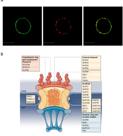
5.3 Complexity on the edge of nucleus: nuclear pores ... 49
Synopsis ... 55
Results ... 57
1. HIV-1 integration in CD4+ T cells occurs in the nuclear periphery ... 57
2. HIV-1 localization at the nuclear periphery depends on efficient integration ... 63
3. HIV-1 recurrent integration genes (RIGs) localize in the outer shell of the nucleus ... 69
4. RIGs are transcriptionally active genes at the nuclear periphery ... 79
5. HIV RIGs are excluded from the nuclear Lamin Associated Domains (LADs) ... 81
6. HIV genome associates with the NPC ... 86
7. Role of Tpr in HIV-1 positioning ... 90
8. Transcriptionally active proviruses associate with the nuclear pore ... 92
Discussion ... 97
Materials and methods ... 110
Bibliography ... 116 Attachments
INTRODUCTION
1. HIV-1 discovery
Starting in June 1981, several young men affected by severe immunosuppression, opportunistic infections and unusual cancers were admitted as first cases of a new, unknown disease. One year later, more than 400 cases were reported to the Centers for Disease Control and Prevention (CDC) of the United States, and half of them were fatal; the new disease was termed Acquired Immuno Deficiency Syndrome (AIDS).
The infectious nature of this disease was highlighted in 1983, when a new retrovirus, termed LAV (Lymphadenopathy-Associated Virus) was isolated by Dr Luc Montagnier from lymph nodes of one of these patients (Barre-Sinoussi, Chermann et al. 1983). At the same time, Dr. Robert Gallo and Dr Jay Levy isolated a retrovirus from AIDS patients and some still healthy individuals in the risk groups, and termed it HTLV-III (Human T-Cell Leukemia Virus III)(Gallo, Sliski et al. 1983). In 1984, several publications consistently proved that HTLV III was the cause of AIDS (Alter, Eichberg et al. 1984, Gajdusek, Amyx et al. 1984, Gallo, Salahuddin et al. 1984, Popovic, Sarngadharan et al. 1984). In June 1984, Drs. Robert Gallo and Luc Montagnier held a joint press conference to announce that HTLV-III and LAV were almost certainly identical. Later on, the new retrovirus was termed HIV (Human Immunodeficiency Virus). On the same year, almost 7,000 cases were reported to CDC, with more than 2,000 deaths only in United States. This number exponentially increased, so that in August 1988, the number of AIDS patients exceeded 70,000 in the United States only; the CDC predicted that more than 1 million people was infected in that country, without being yet aware of it, and that a new case was
reported every 14 minutes. HIV infection became pandemic and spread worldwide, infecting at least 60 million people and causing more than 25 million deaths (http://www.unaids.org/).
But where exactly did HIV-1 originated? Scientists discovered that HIV-1 and a similar virus termed HIV-2 were the result of the transmission of viruses from primates to humans, that likely occurred in Africa in the 1920s (Sharp and Li 1988, Hahn, Shaw et al. 2000). Indeed simian immunodeficiency viruses (SIVs) were identified in different sub-Saharan primates, suggesting a cross-species infection; however these viruses do not cause disease in their host (Sharp, Bailes et al. 2001). Despite frequent human exposure to SIV-infected monkeys in Africa, only 11 cross-species transmission events have been identified, and only four of them have resulted in a significant human-to-human transmission. Indeed, phylogenetic analysis revealed that the main subtypes of HIV-1 (groups M and O) and HIV-2 (groups A and B) originated from four different infections of SIVs, from chimpanzees and sooty mangabeys respectively (Marx, Apetrei et al. 2004). Recently, it has been defined that such events occurred in Leopoldville (now Kinshasa) in Congo, essentially due to the consumption and manipulation of bushmeat, by which humans are directly exposed to animal blood and secretions. Demographic data proved that pandemic HIV-1 developed in the late ‘50s together with the exponential growth of urban population in West Central Africa and the expansion of fluvial travel and commerce along the Congo river; a subtype of the group M spread to Haiti, then to US and other Western countries (Sharp and Hahn 2011, Faria, Rambaut et al. 2014).
Over the last two decades, HIV has been deeply studied and characterized in unprecedented molecular detail, allowing the development of drugs that, even though unable tot eradicate HIV-1 infection, still can
successful control viral replication in the host and spread. Therefore, in the Western Countries, AIDS has been transformed from a lethal disease into a chronic one, that still represents a heavy burden on the health organizations. Moreover, despite this remarkable success, AIDS remains a devastating and fatal disease in all countries where drugs are not available, including most of the African continent and several Asian countries. The global expenditure for HIV-1 treatment and prevention could reach 35 billion dollars before 2031 (Report from Aids2031 Project, UNAIDS 2010).
2. HIV-1 genome and structure
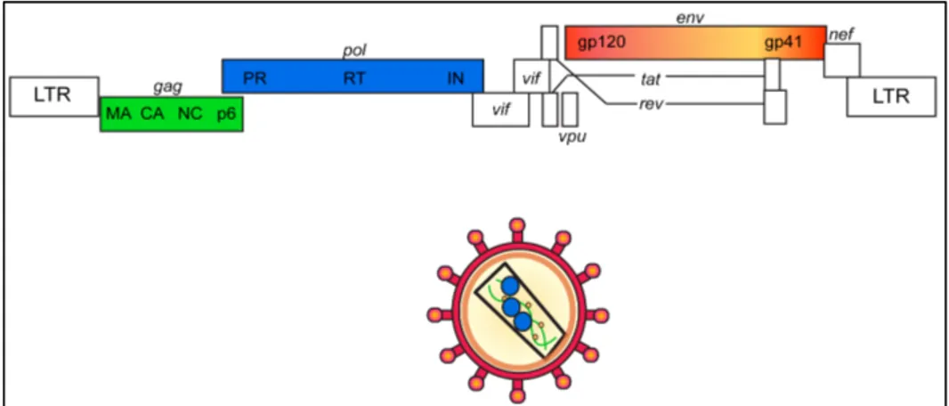
HIV-1 belongs to the Lentivirus genus, from the Retroviridae family. Lentiviruses (lente-, Latin for "slow”) are a group of RNA viruses characterized by a very long incubation period and by a particular tropism for cells of the monocyte/macrophage lineage. They cause chronic persistent infection in different mammalian species. Five main serogroups of lentiviruses have been identified, each corresponding to vertebrate hosts: primates (including HIV-1, HIV-2 and SIVs), felines (FIVs), horses (EIAVs), cattle and sheep/goats (CAEV, VISNA) (Gifford 2012). Not all lentiviruses are pathogenic in their natural hosts, but some of them cause immune system dysfunctions that can be lethal. The virions are composed of a spherical envelope that measures about 100 nm in diameter; the envelope encloses a rod-shaped nucleocapsid that contains both the viral enzyme and some viral enzymes. Once in the host cell, lentiviral RNA genomes are transformed into DNA by the viral reverse transcriptase; the DNA is then incorporated into the cellular genome by the integrase enzyme, thus becoming a provirus capable of replication by exploiting the host transcription machinery.
positive-stranded RNA (Alizon, Sonigo et al. 1984), containing nine genes, which encode for 19 proteins.
The Gag gene is translated into a p55 polyprotein that, after cleavage, originates p17, p24, p7 and p6. Viral protein p24 forms a capsid that surrounds and encloses the viral genome, which is further protected by nucleocapsd protein p7. The capsid is surrounded by a matrix composed of viral protein p17.
The Pol gene codes for the main viral enzymes, protease (PR), reverse transcriptase (RT), and integrase (IN), which are included in the capsid and are required for the subsequent steps of infection.
The matrix and the capsid are surrounded by an envelope, which is composed by a phospholipid bilayer derived directly from the cellular membrane, and enriched in two viral glycoproteins, gp120 and gp41, which are translated from the Env gene and derive from the cleavage of the gp160 polyprotein. The resulting virion is spherical and with a diameter of 120 nm.
The remaining 6 genes (tat, rev, nef, vif, vpr and vpu) are regulatory genes, which code for accessory proteins.
The Tat protein is an essential, strong transcriptional transactivator of the viral promoter, constituted by the Long Terminal Repeats (LTR, see chapter 3 for details) (Berkhout and Jeang 1989), which acts by binding to the TAR RNA element and activating transcription initiation and elongation. It is the first transcription factor interacting with RNA rather than DNA, and it is very similar to prokaryotic anti-termination factors (Ott, Geyer et al. 2011). The human positive transcription elongation factor b (P-TEFb) is an essential human cofactor for Tat transactivation (Mancebo, Lee et al. 1997). Rev is a protein necessary for HIV-1 expression; it promotes nuclear export and stabilization of viral unspliced mRNA, by binding to Rev-Responsive Element contained in the env region (Chang and Sharp 1990, Pollard and Malim 1998).
Vif plays a role to antagonize the restriction factor APOBEC3G (Sheehy, Gaddis et al. 2003, Stopak, de Noronha et al. 2003), whereas Vpu enhances the release of new virions, by counteracting cellular protein tetherin, able to inhibit this process (Neil, Zang et al. 2008).
Additional proteins auxiliary for a efficient viral propagation are Vpr, that probably plays a role in the nuclear import of preintegration complexes, and Nef, that acts in downregulating both CD4 and MHC molecules from the cell surface and it is essential for efficient viral spread and disease progression in vivo (Malim and Emerman 2008).
3. HIV-1 life cycle: an overview
As other primate lentiviruses, HIV-1 targets cells from the hematopoietic / lymphoid lineage, mainly T helper lymphocytes and macrophages, and at a lesser extent also microglial and dendritic cells; such cells share the feature
of expressing the CD4 receptor and CC-chemokine receptor 5 (CCR5) (Dragic, Litwin et al. 1996) or CXC chemokine receptor 4 (CXCR4) (Feng, Broder et al. 1996). It has been shown that other non hematopoietic, CD4 negative cell types can be infected in vitro (with a very low efficiency) but the significance of such observation in the in vivo pathogenesis is considered to be scarce (Dittmar, McKnight et al. 1997, Clapham and McKnight 2001). The CD4 receptor interacts with viral envelope protein gp120 (Kwong, Wyatt et al. 1998), and such interaction promotes the binding to the mentioned co-receptors eventually leading to viral entry mostly by direct fusion with the plasma membrane (mediated by gp41)(Chan and Kim 1998), or by endocytosis followed by glycoprotein- and dynamin-dependent fusion with intracellular compartments (Miyauchi, Kim et al. 2009). Such process is extremely conserved among primate lentiviruses.
The binding with one of the coreceptors CCR5 or CXCR4 is crucial for efficient membrane fusion (Bleul, Wu et al. 1997, Dittmar, McKnight et al. 1997, Kozak, Platt et al. 1997). Interestingly, individuals that are homozygous for a mutant CCR5 allele (CCR5D32) were found to remain uninfected even after frequent exposures to HIV (Samson, Libert et al. 1996). This discovery is linked to the “Berlin patient”, remaining nowadays the only individual to have been cured of HIV infection, after having received an allogenic hematopoietic stem cells transplant from a homozygous CCR5D32 (Hutter, Nowak et al. 2009).
Figure 2. Scheme of the main steps of HIV-1 replication.
Following cell entry, the cooperation of viral proteins (p17 Matrix, Nef and Vif) with host proteins leads to the uncoating of the nucleocapsid, which allows the viral genome to be extruded out from the capsid and released into the cytosol (Dvorin and Malim 2003). Once uncoating is completed, the viral RNA, RT, IN, p17, p7, Vpr and a plethora of cellular cofactors all assemble together into the so-called reverse transcription complex. The reverse transcription starts in the cytoplasm and is completed in the nucleus; the so-called "Long Terminal Repeats” (LTRs) are formed as duplication of the U3, R and U5 sequences at both 5’ and 3’ ends of the final cDNA
(Gilboa, Mitra et al. 1979) (see paragraph 4.1 for details). However, the exact mechanism leading from viral entry to reverse transcription is still matter of debate; indeed, it has been indeed hypothesized that uncoating might occur simultaneously with the reverse transcription process. Moreover this would occur not in the cytoplasm but rather in the proximity of the nuclear pore complexes (Bichel, Price et al. 2013).
After or during reverse transcription (see paragraph 4.1), both viral (IN, p17, p7, RT and Vpr) and cellular proteins associate with the double-stranded viral DNAs in the so-called Pre-Integration Complex (PIC) (Farnet and Haseltine 1991, Miller, Farnet et al. 1997). The cellular component of the PICs has been deeply studied in the last years through a proteomic approach (Raghavendra, Shkriabai et al. 2010), leading to a long list of cofactors that are included in the PICs even though not always the role for their presence has been determined. Also the molecular mechanism of the PIC entry into the nucleus is still not completely understood; however it has been clearly demonstrated that cellular protein partners are crucial for the nuclear entry of the PIC and viral integration (see paragraph 4.2.1 for major details).
The early steps of viral replication appear to be the main targets for the "restriction factors", namely host proteins exerting an innate anti-viral response.
A recent and comprehensive analysis of such factors is presented in the table http://www.retrovirology.com/content/10/1/106/table/T1 (Abden-Mohsel et al. 2013). The most characterized restriction factors for HIV-1 infection are the TRIM5α, APOBEC3 and SAMHD1.
• TRIM5α blocks HIV-1 infection in simian cells (Stremlau, Owens et al. 2004) at the reverse transcription and nuclear import steps; TRIM5α recognizes motifs within the capsid proteins and interferes with the uncoating process, therefore preventing successful reverse transcription and
transport to the nucleus of the viral genome. Retroviral resistance to TRIM5α can be generated by sequences in the viral capsid protein (Sebastian and Luban 2005, Stremlau, Perron et al. 2006).
• Apolipoprotein B mRNA-editing enzyme 3 family (APOBEC3) can also suppress HIV-1 replication (Sheehy, Gaddis et al. 2002, Sheehy and Erthal 2012). It is a family of cytidine deaminases that hypermutates the viral genomes, impairing viral replication. A deaminase-independent inhibition by APOBEC was also reported (Sasada, Takaori-Kondo et al. 2005, Nguyen, Gummuluru et al. 2007). Vif interacts with APOBEC3G by triggering its degradation via the proteasomal pathway (Donahue, Vetter et al. 2008). • SAM domain and HD domain-containing protein 1 (SAMHD1) has also been described as an additional restriction factor for HIV-1. SAMHD1 is a phosphohydrolase that can convert nucleotide triphosphate to a nucleoside and triphoshate. In doing so, SAMHD1 depletes the pool of nucleotides available for viral cDNA synthesis by reverse transcriptase, and in this way prevents viral replication (Goldstone, Ennis-Adeniran et al. 2011, Laguette, Sobhian et al. 2011, Ayinde, Casartelli et al. 2012, Baldauf, Pan et al. 2012, Descours, Cribier et al. 2012, Kim, Nguyen et al. 2012). SAMHD1 is also able to restrict other retroviruses, such Murine Leukemia Virus (MLV), SIV, EIAV, FIV, showing a broad antiretroviral activity (Gramberg, Kahle et al. 2013). SAMHD1 is counteracted by the viral protein Vpx, which forms a complex with a E3 ubiquitin ligase, which in turn induces ubiquitination and proteasomal degradation of SAMHD1. Therefore, the dNTP pool is restored and the block to reverse transcription is released (Hrecka, Hao et al. 2011, Ahn, Hao et al. 2012, Hofmann, Logue et al. 2012).
Integration of the proviral genome into host chromosomal DNA is one of the defining features of retroviral replication (Coffin, Hughes et al. 1997).
To access chromatin, some retroviral PICs require nuclear-envelope disassembly during cell division, whereas HIV-1 and other lentiviruses that infect non-dividing cells can directly cross the nuclear envelope to enter the nucleus. In order to integrate their cDNA into the most preferable chromosomal targets, their PICs are transported through the Nuclear Pore Complexes (NPCs) by active translocation (Suzuki and Craigie 2007).
Once inside the nucleus, integration of viral genome into the host DNA is mediated by the IN protein, with the support of several cellular partners and post-translational modifications (see chapter 4.2.1). Although HIV-1 integration is not site-specific, several lines of evidence have indicated that it is not completely random and favors portion of chromatin with particular features. Mainly, HIV-1 prefers to integrate inside actively transcribing genes, possibly to maximize the exploitation of the cellular machinery needed for transcription (see chapter 4.2.2) (Jordan, Defechereux et al. 2001, Schroder, Shinn et al. 2002, Bushman, Lewinski et al. 2005).
After integration into the host genome, the provirus behaves as any human RNA Pol II-transcribed gene: transcription proceeds downstream to the promoter and enhancer elements encoded in the 5’ LTR, to the polyadenylation site within the 3’ LTR (see chapter 4.3.1). Regulation of HIV-1 gene expression is defined by a complex cross-talk between the local chromatin landscape, host transcription factors, and the viral trans-activating factor Tat (Malim, Fenrick et al. 1989, Fujinaga, Cujec et al. 1998, Marzio, Tyagi et al. 1998, Lusic, Marcello et al. 2003) (see chapter 4.3.2-3). HIV transcription is classically subdivided into an early and a late phase. In the early phase of transcription, in the absence of Rev and Tat regulatory proteins, transcription is triggered by host transcription factors bound to the LTR; the RNA splicing machinery quickly splices mRNA and only Rev, Tat and Nef proteins are produced (Kim, Byrn et al. 1989). In the subsequent step of transcription, Tat boosts initiation and elongation of viral
transcription (Berkhout, Silverman et al. 1989), leading to the production of a single mRNA, which is then processed to generate over thirty different viral transcripts. In the early phase of transcription, short, multiply spliced transcripts (encoding for Tat, Rev and Nef) are produced, while later, singly spliced RNAs and long unspliced RNAs become predominant (for an extensive review, see (Karn and Stoltzfus 2012)). The first short transcripts, which do not contain unspliced exons, exploit the cellular transport of mature cellular mRNAs into the cytoplasm (Cullen 1998); instead, Rev coordinates the export to the cytoplasm of unspliced and partially spliced mRNAs (Malim, Hauber et al. 1989, Pollard and Malim 1998, Greene and Peterlin 2002).
After the production of new viral genomes and the synthesis of several viral proteins, three main steps need to be executed: the assembly of the virion, when essential components for infectivity are packed together; the budding, when the virion acquires its lipid envelope through the fusion with the cellular membrane; and the maturation, when the virion becomes infectious and it is released from the cell (Sundquist and Krausslich 2012). These steps require a multiplicity of cellular pathways and partners, but are coordinated by the viral Gag precursor protein (p55) that guides the entire process. p55 leads this process after moving to the host cell plasma membrane, within membrane microdomains corresponding to the so called lipid rafts, specific membrane sub-domains enriched in cholesterol (Gheysen, Jacobs et al. 1989, Karacostas, Nagashima et al. 1989, Freed 1998, Nguyen and Hildreth 2000, Zheng, Plemenitas et al. 2003). Here, Gag simultaneously triggers membrane binding (through its N-terminal component, MA), virion assembly (through the central domain, CA which will constitute the capsid in the mature virion), and RNA packaging (by the NC domain) (Benjamin, Ganser-Pornillos et al. 2005, Briggs, Riches et al.
2009, Jouvenet, Simon et al. 2009).
At the same time, Env is synthesized as a precursor of about 850 aminoacids and later it is processed as an integral membrane protein. Indeed it undergoes first post-translational processing that includes signal peptide cleavage, folding and trimerization (Haim, Salas et al. 2013). Later it follows the cellular secretory pathway through the endoplasmic reticulum where it gets glycosylated. The resulting gp160 Env precursor is transported to the Golgi where host proteases cleave it into the gp120 (surface) and gp41 (membrane) subunits (Willey, Bonifacino et al. 1988, Stein and Engleman 1990). The trimeric complexes are then inserted into the cell membrane in trimeric complexes (Decroly, Vandenbranden et al. 1994, Bugelski, Maleeff et al. 1995). Gag initially assembles spherical particles, which will bud from the membrane through the interaction with components of the cellular Endosomal Sorting Complex Required for Transport (ESCRT) and the apoptosis-linked-gene 2 interacting protein (Alix) (Martin-Serrano, Zang et al. 2001, Strack, Calistri et al. 2003, Martin-Serrano and Marsh 2007, Martin-Serrano and Neil 2011). After budding, PR is activated and cleaves p55 into its separate components MA, CA, NC and p6 proteins, forming a mature infectious virion (Gottlinger, Sodroski et al. 1989, Peng, Ho et al. 1989, Schatzl, Gelderblom et al. 1991, Kaplan, Zack et al. 1993, Bharat, Davey et al. 2012, Meng, Zhao et al. 2012).
In this context, Tetherin and calcium-modulating cyclophilin Ligand 1 (CAML-1) inhibit viral release; these restriction factors are counteracted by viral Vpu, although its exact mechanism is still under investigation (Neil, Zang et al. 2008, McNatt, Zang et al. 2013).
Interestingly, lipid rafts, where assembly and budding start, are the same compartment in which the myristoylated form of the Nef protein is incorporated, being this a prerequisite for the biological activity of the protein itself (Geyer, Fackler et al. 2001, Zheng, Plemenitas et al. 2003).
Nef promotes the release of more infectious virions by a variety of molecular mechanisms, including: activation of TCR-activated pathways that stimulate HIV transcription (Wang, Kiyokawa et al. 2000, Simmons, Aluvihare et al. 2001, Fenard, Yonemoto et al. 2005); decrease in the expression of CD4 on the cell surface (Lama, Mangasarian et al. 1999, Glushakova, Munch et al. 2001); reduction in the expression of MHC I and thus the visibility of the infected cells to CD8+ cytotoxic T-cells (Collins, Chen et al. 1998); block of apoptosis (Fackler and Baur 2002). The overall result of these activities is to prolong the life of the infected cell to optimize viral replication (Greenway, Holloway et al. 2003).
4. HIV-1 nuclear biology
4.1 Reverse transcription and nuclear import
Following cell-receptor entry, HIV-1 starts reverse transcription of its genome. The viral capsid is able to provide the right environment for this process to occur, but the virus must uncoat its core to enter the nucleus. So far, it is not clear where uncoating takes place: whereas the reverse transcription complex has been visualized in the cytoplasm (McDonald, Vodicka et al. 2002, Hulme, Perez et al. 2011), some studies suggest that uncoating occurs at the NPCs, in parallel with the completion of reverse transcription itself within the capsid (Arhel, Souquere-Besse et al. 2007). This might reduce the exposure of viral RNA/DNA to the cytoplasmic environment and therefore to degradation. Moreover, coupling uncoating with reverse transcription might speed up the entire process, lowering the risk of activating the innate immunity pathways, which are able to sense both the cytosolic viral genomes and viral capsid (Towers, Hatziioannou et al. 2003, Pertel, Hausmann et al. 2011, Schaller, Ocwieja et al. 2011).
Once retro-transcribed, the viral DNA integrates into the host genome; integration is a key feature of retroviruses (Temin 1976), and it ensures expression of viral genes for the subsequent production of new viral particles. In order to integrate, retroviral PICs have to access the nucleus. Some retroviruses, such as MLV, require the nuclear envelope breakdown during mitosis, to get in contact with cellular chromosomes. On the contrary, lentiviruses, including HIV, can enter the nucleus in non-dividing cells, by passing through the NPC (Greene and Peterlin 2002, Bukrinsky 2004). The molecular processes underlining the PIC entry into the nucleus remains elusive. It appears that a redundancy of mechanisms exist, at least in part mediated by the Nuclear Localization Signals (NLS) that have been identified in most of the viral proteins of the PIC (Bouyac-Bertoia, Dvorin et al. 2001).
Importantly, the viral capsid is implicated in the HIV-1 nuclear import. M. Yamashita and M. Emerman have indeed demonstrated that the capsid is crucial for the capacity of HIV-1 to infect non-dividing cells. In particular, substituting the HIV-1 capsid with MLV capsid in the HIV-1 virion, the virus containing the latter is not anymore able to infect non-dividing cells. On the opposite, if the HIV-1 capsid is inserted into a MLV virion, this is able to infect non-dividing cells in a HIV-1-like manner (Yamashita and Emerman 2004). Indeed, the HIV-1 capsid is able to interact with several nucleoporines involved in nuclear import, including Nup153, TNPO3 and Nup358/RANBP2 (Christ, Thys et al. 2008, Lee, Ambrose et al. 2010) (for a comprehensive review, see (Suzuki and Craigie 2007)). Capsid mutants, such as CA N74D, affect its uncoating speed (Yamashita, Perez et al. 2007), and therefore lead to unsuccessful reverse transcription and missed interaction with Nup153, TNPO3 and Nup358. However, such mutants appear to still reach the cellular genome through other, not well-determined import pathways.
Cleave and Polyadenylation Factor 6 (CPSF6) is a host restriction factor preventing HIV-1 nuclear entry by targeting the capsid; the N74D capsid mutant is able to evade the interaction with CPSF6 (Lee, Ambrose et al. 2010). Interestingly, a novel HIV-1 restriction factor, interferon-induced Myxovirus Resistance 2 (MX2) protein, was recently described as inhibitor of HIV-1 capsid-dependent nuclear import (Goujon, Moncorge et al. 2013, Kane, Yadav et al. 2013). However MX2 and N74D mutant might be involved into different pathways: indeed the N74D mutant reduced but did not eliminate the sensitivity to overexpression of MX2 (Kane, Yadav et al. 2013).
4.2 Integration
4.2.1 HIV Integrase (IN) and its cofactors
Once inside the nucleus, integration of viral DNA into the cellular genome is mediated by the IN protein, which binds the end of the viral DNA and tethers it to chromatin, where it catalyzes the joining reaction within the host chromosome.
IN is a 32 kDa protein containing three different domains (Engelman 1999, Krishnan, Li et al. 2010, Krishnan and Engelman 2012). The N-terminal domain (NTD, 1-50 residues), important for the IN multimerization and enzymatic activity (Khan, Mack et al. 1991, Schauer and Billich 1992, Zheng, Jenkins et al. 1996, Lee, Xiao et al. 1997), consists of three alpha-helices with coordination of a single Zn2+ ion stabilizing the structure (Carayon, Leh et al. 2010). The catalytic core domain (CCD, 51-212 residues) is the most conserved among all retroviral INs. It consists of five stranded alpha-sheets with six surrounding helices; this structure resembles the one of the transposase from the bacteriophage Mu, the Rnase H domain of RT enzyme and the RuvC protein of E. Coli, all belonging to a varied
superfamily of polynucleotidyl transferases (Rice and Mizuuchi 1995). The key catalytic site is the highly conserved D,D(35)E motif, consisting in three conserved acidic amino acids (Asp-64, Asp-116, and Glu-152) with a conserved spacing of 35 amino acids between the second and the third (Engelman and Craigie 1992). The CCD interacts with the target DNA (Heuer and Brown 1997). Mutation of the D,D(35)E motif inhibits IN enzymatic activity (Kulkosky, Jones et al. 1992, Leavitt, Shiue et al. 1993). The C-terminal domain (CTD, 213-288 residues) is needed for proper multimerization, and binds DNA in a non specific manner, possibly representing the first determinant of chromosomal DNA recognition during integration (Engelman, Hickman et al. 1994) (Lu, Ghory et al. 2005).
Immediately after reverse transcription is completed, IN complex is bound to the LTR DNA ends, forming the so called intasome (Wei, Mizuuchi et al. 1997), which will undergo different events before integration is completed (Maertens, Hare et al. 2010, Krishnan and Engelman 2012). In the first step, the so-called stable synaptic complex (SSC) is transformed into the cleaved donor complex (CDC) by the removal of two nucleotides from each 3’ end of viral DNA (3’ processing) (Pauza 1990, Vink, van Gent et al. 1991, Vink, Yeheskiely et al. 1991).
In the second step (DNA strand transfer), the CDC is engaged to the target DNA after the attack of the 3’ ends to a pair of phosphodiester bonds in the chromatin acceptor, which are separated by 5 nucleotides. At this step, the intasome is called target capture complex (TCC) (Engelman, Mizuuchi et al. 1991). Finally, IN catalyzes the link of the 3’ ends to the 5’ phosphates of the cleaved host DNA, whereas the 5’ ends of the viral genome remain unjoined (strand transfer complex, STC) (Brown, Bowerman et al. 1987, Fujiwara and Mizuuchi 1988, Brown, Bowerman et al. 1989). Afterwards, cellular enzymes with gap repair functions remove two nucleotides on each of the 5’ ends of the viral DNA and trigger the ligation of the 5’ ends to the
target DNA (Katz, Mack et al. 1992, Yoder and Bushman 2000).
Alternatively, if the viral DNA does not get integrated, it may follow three different fates. The ends of the viral DNA might be joined to form a 2-LTR circle by the non-homologous end joining cellular pathway (NHEJ) (Li, Yoder et al. 2000); the viral genome might result in a single LTR circle through the action of factors of the cellular homologus DNA recombination (Brown, Bowerman et al. 1987, Kilzer, Stracker et al. 2003); viral DNA might auto-integrate into itself producing a rearranged circular structure (Farnet and Haseltine 1991). Notwithstanding that none of these variants produce infectious virus, they seem to be transcriptionally active (Stevenson, Haggerty et al. 1990, Stevenson, Stanwick et al. 1990), leading to selected transcription of tat and nef genes before integration (Wu and Marsh 2003). The persistence of these forms of circular DNA before integration is related to the phenomenon referred to as pre-integration latency (Coiras, Lopez-Huertas et al. 2009, Sloan and Wainberg 2011).
Even if IN alone is sufficient to promote in vitro integration (Craigie, Fujiwara et al. 1990, Katz, Merkel et al. 1990, Bushman and Craigie 1991, Sinha, Pursley et al. 2002), for efficient integration in the cellular context, the protein requires several host factors, most of which already recruited into the PIC before entering the nucleus (for review see: (Turlure, Devroe et al. 2004, Van Maele, Busschots et al. 2006)).
PICs isolated from HIV-1 infected cells can integrate their endogenous viral cDNA into a target DNA substrate in vivo (Ellison, Abrams et al. 1990, Farnet and Haseltine 1990, Khiytani and Dimmock 2002). Given that the integrating activity of the PICs was disrupted by treating them with high concentration of salts, and that extracts of uninfected cells restored their activity, cellular cofactors had to be involved. The first protein to reconstitute the activity of salt-treated PICs was the high mobility group
protein A1 (HMGA1) (Farnet and Bushman 1997). HMGA proteins are non-histone chromatin-associated proteins that participate in several nuclear processes; in particular, HMGA1 is able to induce local conformational changes in DNA strands, affecting gene expression (for a review see: (Jiang and Pugh 2009)). Inside the PICs, HMGA1 interacts with viral DNA but not with IN and it may help in bridging the viral ends together within the intasome (Farnet and Bushman 1997, Hindmarsh, Ridky et al. 1999, Henderson, Bunce et al. 2000).
Salt treatment of extracted PICs led to the discovery of another cellular cofactor (Lee and Craigie 1994). Indeed, high salt concentrations induced an increase in autointegration events in MLV, by removing Barrier-to-Autointegration Factor (BAF) (Chen and Engelman 1998, Lin and Engelman 2003). This protein, involved in nuclear envelope assembly and organization (Margalit, Brachner et al. 2007), is included in the PICs of both MLV and HIV. As HMGA1, BAF interacts with DNA and not with IN, and it may help compacting the viral DNA and so making it inaccessible for premature integration into itself (Suzuki and Craigie 2002).
Other cellular factors can bind directly IN protein. The first to be discovered was INtegrase Interactor 1 (INI1) that appeared to be able to stimulate integration activity in vitro 10 to 20 folds (Kalpana, Marmon et al. 1994). The importance of this factor, however, was later questioned (Boese, Sommer et al. 2004). More recently, it was reported that defects in IN-INI1 interaction impairs integration in HEK293T and HeLa cells (Mathew, Nguyen et al. 2013). In considering these findings, it appears worth taking into account that INI1 is a component of the Switch/Sucrose Non Fermentable (SWI/SNF) chromatin remodeling complex (for a review see: (Euskirchen, Auerbach et al. 2012)); therefore, rather than acting directly on integrase activity, INI1 might help targeting integration into SWI/SNF-remodeled regions of the genome (Kalpana, Marmon et al. 1994, Lesbats,
Botbol et al. 2011).
One of the most studied cofactors of HIV-1 IN is the product of the
PSIP1 gene, LEDGF/p75 (Maertens, Cherepanov et al. 2003), a
transcriptional coactivator implicated in cell differentiation and cellular response to stress (Llano, Morrison et al. 2009). In the context of lentiviral integration, LEDGF/p75 acts by tethering IN to chromatin through its two main domains: an N-terminal PWWP (Pro-Trp-Trp-Pro) chromatin-binding domain and a C-terminal IN-binding domain (Cherepanov, Maertens et al. 2003, Llano, Saenz et al. 2006, Llano, Vanegas et al. 2006, Turlure, Maertens et al. 2006, Busschots, Voet et al. 2007). Depletion of LEDGF/p75 reduces efficiency of HIV-1 integration by 10 fold (Vandekerckhove, Christ et al. 2006), but does not affect IN catalysis (Shun, Raghavendra et al. 2007), suggesting that the LEDGF/p75 function in chromatin engagement is crucial (Busschots, Vercammen et al. 2005, Emiliani, Mousnier et al. 2005) (see paragraph 4.2.2 for details).
Other IN interactors include Polycomb Protein EED (Violot, Hong et al. 2003), HSP60 (Parissi, Calmels et al. 2001), nuclear pore component NUP153 (Woodward, Prakobwanakit et al. 2009), and gap repair protein Rad18 (Mulder, Chakrabarti et al. 2002). A role for emerin in HIV-1 integration was described (Jacque and Stevenson 2006), but it was later disproved by two other studies (Shun, Daigle et al. 2007, Mulky, Cohen et al. 2008).
Moreover, IN is affected by several post-translational modifications. Acetylation by the Histone Acetyl Transferase (HAT) p300 increases its affinity for the DNA template (Cereseto, Manganaro et al. 2005). However, once acetylated, IN can be bound by KRAB-Associated Protein 1 (KAP1), a protein belonging to the TRIM family of antiviral proteins that induces IN deacetylation through the formation of a protein complex that includes
HDAC1 deacetylase; therefore KAP1 may be considered as a restriction factor (Allouch, Di Primio et al. 2011).
Ubiquitination reduces IN stability (Mousnier, Kubat et al. 2007), but is prevented by phosphorylation by c-Jun N-terminal kinase (JNK) and subsequent prolin-isomerization by the Peptidyl-Prolyl cis-trans Isomerase NIMA-interacting 1 protein (Pin1) (Manganaro, Lusic et al. 2010). Finally, IN sumoylation has been also proposed to be necessary in the pre-integration processes (Zamborlini, Coiffic et al. 2011).
4.2.2 Integration site selection
IN requires specific sequences at the end of the viral cDNA, whereas cellular DNA sequences that serve as integration target sites show only weak primary sequence specificity (Stevens and Griffith 1996, Carteau, Hoffmann et al. 1998, Wang, Ciuffi et al. 2007). Yet, integration into the host genome is not random: preferences for particular regions do exist and are different among retroviruses.
Based on a positive correlation between integration frequency and DNase I-hypersensitive sites, early studies of MLV integration suggested that open chromatin might favor this process (Vijaya, Steffen et al. 1986, Rohdewohld, Weiher et al. 1987). The first genome-wide study on the HIV-1 integration target site selection, performed a decade ago using high-throughput DNA sequencing, confirmed that HIV-1 strongly favors integration within active transcription units of the human genome, in gene-rich regions (Schroder, Shinn et al. 2002). The authors also found a group of genes that were upregulated following HIV-1 infection (confirming the results by Corbeil et al. (Corbeil, Sheeter et al. 2001)) and were more often targeted by the integration process. Since then, HIV-1 integration site selection has been studied in many cell types and integration in active transcription units was observed in all cases (Mitchell, Beitzel et al. 2004,
Lewinski, Bisgrove et al. 2005, Barr, Ciuffi et al. 2006, Lewinski, Yamashita et al. 2006, Brady, Agosto et al. 2009). Moreover, HIV integration appears to favor Alu elements (potentially because these repetitive sequences are enriched in gene-rich regions) (Kumar, Mehta et al. 2007), and to avoid the CpG islands present in the gene promoter regions (Mitchell, Beitzel et al. 2004).
Integration of the SIV shows a similar pattern of integration into active genes, suggesting that lentiviruses might have similar target preferences (Crise, Li et al. 2005). In contrast, MLV favors integration at promoter regions (Mitchell, Beitzel et al. 2004), transcription start sites (Wu, Li et al. 2003), DNase-sensitive regions and CpG islands (Lewinski, Yamashita et al. 2006).
To explain target site selection, different models have been proposed. A first model is based on the concept that open chromatin is more accessible to the PIC (“chromatin accessibility model”). This notion is supported by all the information gathered so far about flanking sequences and epigenetic features by genome-wide studies, as HIV-1 integrants are mostly found in active transcription units. However, should chromatin be the sole determinant of target selection, all retroviruses should integrate into the same accessible hotspots, which is not the case. As an example, MLV, but not HIV-1, prefers DNase-sensitive regions, considered as markers for open chromatin. Moreover, in vitro experiments have shown that nucleosome-packed DNA not only does not obstruct integration, but rather naked DNA is disfavored (Pryciak, Sil et al. 1992, Pryciak and Varmus 1992). Therefore, chromatin accessibility cannot be the main determinant of target site selection.
A second model to explain integration site preferences follows the discovery of a cellular protein (LEDGF/p75, the product of the PSIP1 gene) capable of binding both PIC-associated HIV-1 IN (Cherepanov, Maertens et
al. 2003) and chromatin (Maertens, Cherepanov et al. 2003). According to this model, LEDGF/p75 tethers the PIC to the chromatin regions at which it associates (De Rijck, Bartholomeeusen et al. 2010, Ferris, Wu et al. 2010, Gijsbers, Ronen et al. 2010, Gijsbers, Vets et al. 2011).
LEDGF/p75 was originally described as a transcriptional co-activator, p75 (Ge, Si et al. 1998); later, it was proven to be identical to a protein found in a screen for factors involved in lens epithelial cell growth and survival, namely Lens Epithelium-Derived Growth Factor (LEDGF) (Singh, Kimura et al. 2000, Nishizawa, Usukura et al. 2001); hence the double name LEDGF/p75 that is commonly used. LEDGF/p75 is involved in the modulation of stress-related gene expression (Llano, Morrison et al. 2009) and is ubiquitously expressed.
LEDGF/p75 binds tightly lentiviral IN proteins (from HIV, SIV and FIV) (Busschots, Vercammen et al. 2005) through a C-terminal IN-binding domain that interacts with the core domain of IN (Cherepanov, Maertens et al. 2003, Llano, Saenz et al. 2006, Llano, Vanegas et al. 2006, Busschots, Voet et al. 2007). On the other hand, LEDGF/p75 binds DNA through a N-terminal PWWP (Pro-Trp-Trp-Pro) chromatin-binding domain (Turlure, Maertens et al. 2006). A recent study based on the DamID (DNA adenine methyltransferase identification) technology unraveled the genome-wide distribution of LEDGF/p75, showing its association with transcription start sites (TSSs) of active transcription units, Pol2-bound genomic regions and active chromatin markers (H3 and H4 acetylation, H3K4 monomethylation), confirming the correlation between LEDGF/p75 genome-wide distribution and HIV-1 integration (De Rijck, Bartholomeeusen et al. 2010).
LEDGF/p75 depletion from cells does not abolish lentiviral integration completely (Vandekerckhove, Christ et al. 2006), but rather it results in diminished integration in the transcriptional units (Ciuffi, Llano et al. 2005) and a more random HIV integration pattern (Schrijvers, Vets et al.
2012). By combining integration site sequencing with transcriptional profiling of a LEDGF/p75 knock-down cell line, it was found that HIV-1 preferentially targets LEDGF/p75 modulated genes in the control cells, whereas this preference was abolished in the knocked-down cells (Ciuffi, Llano et al. 2005). The importance of LEDGF tethering function is reinforced by a retargeting study, where the PWWP domain was replaced by CBX1 domain, which associates preferentially with pericentric heterochromatin and intergenic regions. The chimeric LEDGF/CBX1 protein was able to redirect lentiviral integration into the CBX1-bound heterochromatic regions (Gijsbers, Ronen et al. 2010).
However, LEDGF/p75 depletion does not abolish completely HIV-1 integration in transcription units, suggesting that probably other factors may be involved in this process. More recently, Hepatoma-derived growth factor related protein (HRP-2) was found as a co-factor of HIV-1 IN in LEDGF/p75-depleted cells. Nevertheless, even if integration frequency into transcription units was reduced in cells in which both LEDGF/p75 and HRP-2 was knocked down, still, the integration distribution was not fully random (Schrijvers, De Rijck et al. 2011, Schrijvers, Vets et al. 2012).
Taking into account that retroviruses differ in their ability to infect dividing or non-dividing cells, a third model to explain integration site selection can also be envisaged, based on the interaction of the PIC with nuclear pore proteins. MLV PICs require the disassembly of the nuclear membrane to enter the nucleus, whereas HIV PICs are able to pass the NPC, therefore not requiring cell division. The involvement of NPC proteins in HIV-1 nuclear import and/or integration has so far been reported for a number of different nucleoporins. For example, Nup62 was recently shown to interact with HIV-1 IN and to contribute to chromatin binding and efficient integration (Ao, Jayappa et al. 2012). Other nucleoporins, such as
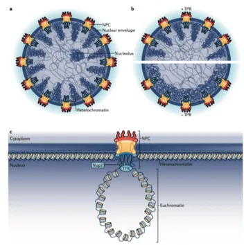
Nup85, Nup107, Nup133, and Nup160, which were found in genome-wide screenings for human genes that affect HIV-1 infection, have a less defined role in the viral life cycle (Brass, Dykxhoorn et al. 2008, Zhou, Xu et al. 2008). Two of the nucleoporins also identified in these screenings, Nup153 and Nup358/RanBP2, were explored in the context of both nuclear import and viral integration. Nup153, a protein of the inner nuclear basket, was shown to interact with HIV-1 IN (Woodward, Prakobwanakit et al. 2009) and Vpr (Varadarajan, Mahalingam et al. 2005) proteins. More recently, Nup153 was demonstrated to be responsible for the PIC nuclear import (Di Nunzio, Danckaert et al. 2012) via a viral capsid-dependent mechanism (Matreyek and Engelman 2011). Nup153 depletion from the cells reduced the tendency of HIV-1 to integrate into gene dense regions (Koh, Wu et al. 2013). Despite the ascertained role of nucleoporins in HIV-1 biology, the mechanisms by which these proteins could direct target site selection still remains elusive.
Interestingly, integrity of the capsid and the nuclear import pathway also appears to be important later for integration efficiency and gene targeting. Multiple evidence has been collected to support this conclusion: coumermycin-A1, a drug targeting HIV-1 capsid, impairs integration (Vozzolo, Loh et al. 2010); certain mutations, including N74D, impart different integration patterns into host chromosomes compared to wild type virus (Ocwieja, Brady et al. 2011, Schaller, Ocwieja et al. 2011); small amounts of capsid itself were detected inside the nuclear space (Zhou, Sokolskaja et al. 2011). It has been hypothesized that uncoating might occur inside the nucleus, through the NPC. According this hypothesis, TNPO3 might promote uncoating and displace residual capsid elements by facilitating their export (Zhou, Sokolskaja et al. 2011)
4.2.3 Integration and transcription
The choice of integration sites has strong consequences for basal viral transcription: indeed, this is sensitive to the chromosomal environment and proviruses can be transcriptionally repressed when integrated in heterochromatic regions. Thus, a strong correlation is expected to exist between integration site selection and the propensity of the virus to undergo latency, namely being maintained in a silent and repressed state (Jordan, Defechereux et al. 2001, Jordan, Bisgrove et al. 2003). Latent proviruses did show a slight preference for gene deserts, as opposed to active proviruses that are more likely found into short intergenic regions; since gene deserts are thought to be compacted into heterochromatin, this might contribute to silencing. However, genome-wide analysis of latent integrants showed no striking difference with actively transcribing proviruses, being poorly-expressed proviruses still integrated into highly poorly-expressed host genes (Lewinski, Bisgrove et al. 2005, Liu, Dow et al. 2006). In conclusion, based on these data, HIV-1 always integrates into actively transcribing genes; the transcriptional fate of the integrants is then determined successively by the specific chromatin microenvironment and, overall, by the activation state of the infected cell (for more information about transcriptional silencing and latency; see paragraph 4.3.4).
4.3 Transcription. 4.3.1 LTR
After integration into the host genome, the HIV-1 provirus starts transcribing viral mRNA upon activation of its LTR, which acts like a promoter (Rosen, Sodroski et al. 1985). LTRs are generated in their symmetrical configuration during the process of reverse transcription and appear as “repeats” only in the viral cDNA. From a functional point of view,
the LTR can be divided into one of four main regions: the core promoter region, the enhancer region, the modulatory region, the Trans-Activation-Responsive region (TAR).
1. The core promoter region, encompassing the TSS, exerts a positive basal effect on transcription. It contains a TATA box and three tandemly arranged binding sites for the constitutively expressed Sp1 transcription factor (Jones, Kadonaga et al. 1986). Both elements are necessary for basal level of LTR-driven RNA synthesis. As in other eukaryotic cellular promoters, the TATA box is specifically bound by the TBP (TATAA-Binding Protein) subunit of TFIID; mutations of this region result in a marked decrease of both basal transcription and viral replication (Garcia, Harrich et al. 1989). After binding of TFIID on TATA box, TFIIB is recruited, and in turn recruits RNA Pol II to the promoter, definitively establishing the location of the TSS.
2. The enhancer region mediates the transcriptional inducibility of the provirus in response to a variety of stimuli which trigger cellular activation and proliferation (Lusic, Marcello et al. 2003) (Siekevitz, Josephs et al. 1987, Chinnadurai 1991). Located upstream of the core promoter, the enhancer encompasses two partially overlapping binding sites for the inducible transcription factor NF-kB (Nabel and Baltimore 1987) and for STAT5 (Signal Tranducer and Activator of Transcription 5) (Selliah, Zhang et al. 2006), respectively. Noteworthy, also NFAT members can bind the same region of the NF-kB consensus sites, and likely play an important role, particularly in T cells (Kinoshita, Chen et al. 1998). NF-kB has been shown to stimulate both basal and Tat-mediated expression in activated T-cells (Nabel and Baltimore 1987, Siekevitz, Josephs et al. 1987).
3. The modulatory region, formerly called “negative regulatory element” contains several positive and negative regulatory elements, critical for modulating HIV-1 gene expression in response to various stimuli. This region is conserved among isolated HIV strains, and is bound by several cellular proteins such as LEF-1, Ets-1, USF, NFAT, c-Myb and COUP-TF (reviewed in (Pereira, Bentley et al. 2000)). The modulatory region has also been proposed to contain a Negative Regulatory Element (NRE), the deletion of which increases LTR-driven transcription and viral replication (Rosen, Sodroski et al. 1985).
4. The Trans-Activation-Responsive region (TAR) encompasses the 5’-terminal (nucleotides +1 to +59, numbering the TSS as +1) of all viral RNAs. Its importance was defined in concurrence with the findings of Sodroski and colleagues, who showed that LTR-driven expression was dependent on a 86-aminoacids viral product, a transactivating factor they named Tat, a 14 kDa protein conserved in the genomes of all primate lentiviruses (Sodroski, Patarca et al. 1985). To fulfill its functions, Tat binds a hairpin structure present at the 5’ end of the nascent viral RNA, named as TAR; this region functions as an RNA sequence rather than as a DNA element. It folds into a highly stable, nuclease-resistant stem-bulge-loop structure which is essential for Tat-mediated LTR transactivation (Berkhout, Silverman et al. 1989) (Berkhout and Jeang 1989, Selby, Bain et al. 1989), as it is suggested by the fact that mutations that destabilise the stem by disrupting base-pairing abolish Tat-stimulated transcription. Furthermore, the TAR element was found to be functional only when placed in the 3’ to the HIV-1 promoter and in the correct
orientation and position (Selby, Bain et al. 1989).
4.3.2 Tat
Tat is required for in vivo viral replication (Jeang, Xiao et al. 1999). Unique case among transcriptional activators, Tat functions via RNA rather than DNA promoter elements (Berkhout, Silverman et al. 1989) (Berkhout and Jeang 1989). After binding to TAR, Tat binds specifically to the cellular P-TEFb complex (Marshall and Price 1995, Zhu, Pe'ery et al. 1997, Zhou, Chen et al. 1998) (for comprehensive reviews on P-TEFb see (Bres, Yoh et al. 2008)). P-TEFb contains a cyclin component, cyclin T1, which can form a stable complex with CDK9, Tat and TAR RNA. The formation of the P-TEFb/Tat/TAR ternary complex is essential for recruitement of the processive RNA Pol 2 machinery at the LTR promoter (Bieniasz, Grdina et al. 1998, Zhou, Chen et al. 1998).
At the beginning of the transcription cycle, CDK7-mediated phosphorylation on Serine 5 of RNA Pol II CTD facilitates promoter clearance, but, shortly after initiation, the progression of Pol II is stalled by two negative elongation factors, namely NELF (Negative Elongation Factor) and DSIF (Dichloro-1-b-D-RibofuranosylBenzimidazole riboside (DRB)-Sensitivity-Inducing Factor) (for a review, see: (Ott, Geyer et al. 2011)). To overcome this checkpoint, Tat recruits P-TEFb to the stalled Pol II, forming the stable ternary complex with Tat/TAR/Cyclin T1. P-TEFb phosphorylates serines at the second position (Ser2) within each of the heptapeptide repeats that constitute the C-terminal domain (CTD) of the largest subunit of Pol II. The phosphorylated CTD serves as a platform for the assembly and functioning of different transcription and RNA processing factors. P-TEFb also phosphorylates DSIF and NELF, causing NELF dissociation and the conversion of DSIF into a positive elongation factor, finally allowing the production of full-length transcripts (reviewed in (Barboric and Peterlin 2005)).
It has recently been shown that, in addition to the CycT1/CDK9 core complex, Tat also associates with transcription factors/cofactors ELL2, AFF4, ENL and AF9 that together with some additional proteins form the so-called SEC (Super elongation complex). SEC has been shown to be involved also in activation of basal HIV-1 transcription in the absence of Tat, an event important for the reactivation from latency (He, Liu et al. 2010) (Sobhian, Laguette et al. 2010, Ott, Geyer et al. 2011).
4.3.3 Transcription and chromatin
It is well established that the HIV-1 promoter is structurally remodeled in order to allow efficient transcription (Verdin, Paras et al. 1993, El Kharroubi, Piras et al. 1998). Immediately after integration into the cell genome, and independently from the integration site, the proviral DNA, similar to cellular genes, is organized into a chromatin structure. The chromosomal integration packages the proviral DNA into specifically positioned nucleosomes (Verdin, Paras et al. 1993) (Van Lint, Emiliani et al. 1996).
The 5’ LTR, independent from the integration site, is incorporated into two distinct nucleosomes, nuc-0 and nuc-1, precisely positioned with respect to cis-acting regulatory elements, and separated by a nuclease-hypersensitivity region (Verdin, Paras et al. 1993). This arrangement undergoes remodeling, when transcription from HIV-LTR is activated (Verdin, Paras et al. 1993, Marcello, Lusic et al. 2004). Interestingly, several genome-wide studies of nucleosome positioning have revealed that most RNA Pol II-transcribed genes carry a similar chromatin conformation, with nucleosomes precisely positioned at promoters that are remodeled during transcription activation by covalent epigenetic modifications on both DNA and histones (for a review see (Schones, Cui et al. 2008)).
partners that eventually stimulate transcription after rearrangement of the chromatin structure (Lusic, Marcello et al. 2003) (Marcello, Lusic et al. 2004). Indeed, after HIV-1 integration into chromatin, several transcription factors can recruit HDACs to the proviral LTR under basal conditions; once recruited, HDACs are able to deacetylate histone proteins locally, and lead to transcriptional silencing (Margolis, Somasundaran et al. 1994, Jiang, Espeseth et al. 2007, Marban, Suzanne et al. 2007). To counteract this mechanism, the viral Tat protein is able to induce the remodeling of the nucleosome arrangement downstream of the transcription-initiation site by recruiting to the LTR the transcriptional coactivator p300 and the closely related CREB-binding protein (CBP), (Marzio, Tyagi et al. 1998), P300/CBP-associated factor P/CAF (Benkirane, Chun et al. 1998, Lusic, Marcello et al. 2003), GCN5 (Lusic, Marcello et al. 2003), all three having histone acetyltransferase activity. Consequently, acetylation of both histones H3 and H4 occurs at discrete nucleosomal regions before the onset of viral mRNA transcription, relieving chromatin repression (Lusic, Marcello et al. 2003). HDAC inhibitor treatment is sufficient to activate silent, latent HIV (Van Lint, Emiliani et al. 1996, Savarino, Mai et al. 2009).
4.3.4 Transcriptional silencing: latency
Most of HIV-1 integrants are productive, and viral replication is completed within days. However, a small population of resting memory CD4+ cells can enter a transcriptionally inactive state, or “latent state”, as long as these cells remain quiescent (Perelson, Essunger et al. 1997). Latent reservoirs have an extremely long half-life and cannot be cleared even after several decades of antiviral therapy (Siliciano, Kajdas et al. 2003). Indeed, upon cessation of highly active antiretroviral therapy (HAART), viral replication rapidly resumes (Chomont, El-Far et al. 2009). Therefore, latency represents the main obstacle for HIV-1 purge, and understanding the mechanisms controlling silencing and reactivation of the provirus may have
strong implications for the study of HIV-1 pathogenesis.
HIV-1 latency was both documented in HIV-1 infected patients (Chun, Finzi et al. 1995, Finzi, Hermankova et al. 1997) and reproduced in T cell cultures. The latent reservoirs are established within days after acute HIV infection, and continuously repopulated during active viral replication; approximately 1 out of 1x10⁶ CD4+ T cells harbors a replication-competent
but transcriptionally silent provirus (Chun, Carruth et al. 1997, Chun, Engel et al. 1998). Latency can occur both pre- and post-integration. Pre-integration latency is mainly due to incomplete reverse transcription (Zack et al. 1990); restriction factor APOBEC3G is able to inhibit both reverse transcription and then integration by modifying the linear cDNA substrate. (Bishop, Verma et al. 2008, Sloan and Wainberg 2011). Unintegrated latent genomes are labile and decay within 2 days, therefore it can be excluded that pre-integration latency is originating the long-term persistence of latently infected cell populations (Pierson, Zhou et al. 2002). A more recent study, however, has also identified a preintegration, inducible reservoir in HIV+ patients (Petitjean, Al Tabaa et al. 2007).
On the other hand, post-integration latency has been clearly detected in HAART- treated HIV+ patients; post-integration latency may be established in T cells infected during the decline of a T cell activating event (Chun, Carruth et al. 1997). In these memory CD4+ T cells, the provirus, which is successfully integrated into the host genome, is silenced but its transcription can be reactivated by a variety of stimuli, such as antigens, cytokines, mitogens or chemicals like phorphol esters (Chomont, El-Far et al. 2009).
As previously mentioned, remodeling of chromatin conformation is an important determinant of regulation of HIV gene expression (Hakre, Chavez et al. 2011). Histones on nuc-0 and nuc-1 in the HIV LTR are constitutively deacetylated in cellular models of latency, and HDAC inhibitors are able to
reactivate silent proviruses (Van Lint, Emiliani et al. 1996). As already mentioned in paragraph 4.3.3, HDACs can be recruited to the viral promoter through different transcriptional repressors such as Ying Yang 1 (YY-1)(Margolis, Somasundaran et al. 1994), late SV40 factor (LSF) (Coull, Romerio et al. 2000), the p50 subunit of NF-kB complex (Williams, Chen et al. 2006), the c-promoter binding factor CBF-1 (Tyagi and Karn 2007), the COUP-TF interacting protein CTIP2 (Marban, Suzanne et al. 2007). Also, transcription factors are able to recruit HDACs such as AP-4 (Imai and Okamoto 2006), c-Myc and Sp1 (Jiang, Espeseth et al. 2007). Deacetylated nucleosomes create a repressive environment that discourages binding of the transcriptional machinery to HIV-1 LTR.
This information highlights the importance of HDACs in establishing and maintaining HIV latency, and has prompted the start of clinical trials entailing the use of the HDAC inhibitor valproic acid in order to stimulate the latent reservoir and thus eradicate the remaining infected cells with normal therapeutics – with the so-called “shock and kill therapy” (Demonte, Quivy et al. 2004, Lehrman, Hogue et al. 2005, Routy 2005). However, adding valproic acid to HAART-treated patient did not ablate the latent HIV reservoir in patients (Savarino, Mai et al. 2009, Routy, Angel et al. 2012, Routy, Tremblay et al. 2012).
Another class I HDAC inhibitor, vorinostat (also known as suberanilohydroxamic acid and abbreviated as SAHA) was shown to induce virus production in vitro from resting CD4+ T-cells of HIV+ patients on ART with levels of plasma HIV RNA below the detection limit (Archin, Espeseth et al. 2009, Edelstein, Micheva-Viteva et al. 2009). This was reproduced in patients, where vorinostat was able to disrupt latency and HIV RNA expression was increased of about 5-fold upon vorinostat treatment (Archin, Liberty et al. 2012, Archin, Vaidya et al. 2012). Later, Rasmussen and colleagues performed a comparative analysis of the effect of various
HDACs inhibitors, each of which displaying a different degree in the stimulation of HIV-1 expression in latently infected cell lines. According their findings, the HDAC inhibitor panobinostat was the most potent, even in a very low concentration range, followed by givinostat, belinostat, vorinostat and valproic acid (Rasmussen, Schmeltz Sogaard et al. 2013). Entinostat is another HDAC inhibitor that is nowadays matter of investigation (Wightman, Lu et al. 2013).
Histone methylation also regulates post-integration latency. The histone methyltransferase (HMT) Suv39H1 is attracted to the viral promoter via CTIP-2 (Marban, Suzanne et al. 2007) and is able to add tri-methylation on histone H3 lysine 9 on silent HIV proviruses; this event recruits heterochromatin protein 1 HP1 thereby initiating or maintaining the formation of heterochromatin (du Chene, Basyuk et al. 2007). Similarly, another HMT, G9a, is able to deposit repressive di-methylation at histone H3 lysine 9, inhibiting basal and induced HIV-1 gene expression (Imai, Togami et al. 2010). These findings have suggested the possible use of HMT inhibitors in anti-HIV therapy, maybe in combination with HDAC inhibitors (Bernhard, Barreto et al. 2011, Bouchat, Gatot et al. 2012). Recently a third HMT, G9a-like protein GLP, was found to have a role similar to G9a (Ding, Qu et al. 2013).
Our group also provided evidence for an important role of Promyelocytic Leukemia Protein nuclear bodies (PML NBs) in the regulation of HIV-1 latency through the control of G9a and H3K9me2 deposition (Lusic, Marini et al. 2013). Indeed, we were able to show that silent HIV-1 provirus resides in close proximity to PML NBs; the presence of PML protein, able to interact with G9a, ensures the deposition of the repressive H3K9me2 along the viral genome. Upon activation, PML NBs are displaced from HIV-1 proviruses via an actin-dependent mechanism that set the virus free from
transcriptional repression. Arsenic, a drug triggering selectively PML degradation, is able to strongly transactivate the viral promoter, also in a primary model of latency (Lusic, Marini et al. 2013, Ott and Verdin 2013). Tri-methylation of histone H3 lysine 27 and H2A ubiquitylation via Polycomb group protein may also play a crucial role in epigenetic silencing accounting for HIV-1 latency. Indeed, the knockdown of HMT enhancer of Zeste 2 (EZH2), a key component of Polycomb repressive complex 2 (PRC2), induced strong reactivation of the proviruses in a T cell model of latency (Freed 1998).
Finally, DNA methylation on HIV LTR may also be involved in post-integration latency. DNA methylation is a common repressive epigenetic mark that preferentially is deposited on CpG-rich sequences. The HIV promoter is methylated in latently-infected cells (Bednarik, Cook et al. 1990, Schulze-Forster, Gotz et al. 1990, Chavez, Kauder et al. 2011) and this correlates with its transcriptional repression: induction of viral gene expression results in demethylation of the LTR (Ishida, Hamano et al. 2006), whereas methylated CpG islands recruit the methyl-CpG-binding protein MBD2, that in turn can recruit HDACs (Kauder, Bosque et al. 2009). It has been shown that inhibition of cytosine methylation abrogates HDACs recruitment, contributing to viral silencing (Kauder, Bosque et al. 2009). However, according another study, such treatment provokes the opposite effect and it reactivates viral transcription (Pion, Jordan et al. 2003). Therefore, it has been suggested that the HIV-1 DNA methylation may be not uniform over all the integration events, but rather it might be affected by the local context depending on each integration site; according to such hypothesis, considering a population of cells with different integrants (each of them with a different methylation state) the global effect will result to be limited (Pion, Jordan et al. 2003).
HIV DNA methylation is a rare event in HIV+ aviremic patients, suggesting that the effective role of methylation in latency is limited (Blazkova, Murray et al. 2012); another study performed in the same year, showed enrichment of DNA methylation in long-term non-progressor patients, suggesting that this modification may contribute to LTR silencing instead (Palacios, Perez-Pinar et al. 2012). Further work is clearly needed to better define this issue. All together, the findings collected so far underline the importance of chromatin modeling and epigenetic regulation in the establishment and control of viral latency, and highlights the importance of these studies for the future development of viral eradication strategies.
5. Nuclear organization
As already mentioned, a key step in HIV-1 life cycle is its proper integration into the cellular genome, after entering the nucleus; chromatin structure is important both for integration site selection and, later, for the regulation of gene expression. However, the nucleus is not a homogenous organelle, but is extremely organized in subdomains and compartments. Thus, the actual position in the nuclear space of protein factors and DNA domains also needs to be taken into account when considering molecular regulation of HIV integration and expression (Figure 3).
Figure 3. Nuclear organization.
The three-dimensional organization of the genome in the nucleus started to be revealed after the improvement of a few technologies, such as Fluorescent in situ Hybridization (FISH), Chromatin Immuno-Precipitation (ChIP) and its derivatives (ChIP on chip, ChIP-seq), Chromosome Conformation Capture (3C) and, more recently, the Dam-ID technique.
5.1 The concept of chromosome territory
The idea that each chromosome may occupy a specific territory in the nucleus is very ancient, dating back at the beginning of the 20th century when Theodor Boveri first hypothesized that, during interphase, chromosomes keep a peculiar positioning in the nuclear space; actually,
Boveri was the first to introduce the term of chromosome territory (CT) (Boveri 1909). It was only in the '70s that Thomas Cremer confirmed experimentally the existence of CTs, by using in situ hybridization techniques that later allowed detecting specific portions of the genome by laser confocal microscopy (Eils, Dietzel et al. 1996, Croft, Bridger et al. 1999) (Cremer, Cremer et al. 1982).
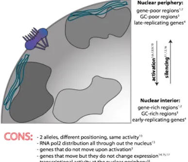
Specific parts of chromatin can be arranged in a non-random fashion, either in terms of radial distances or of neighborhood and proximity to other nuclear structures. Entire CTs were confirmed to keep specific positions of the nuclear space, and these positions were also found to differ strongly between different cell types, adding a new layer of complexity (Mayer, Brero et al. 2005). Whether radial-positioning matters is still under debate, with various studies arguing in favor and other against this possibility. The supporting and denying evidence is summarized in Figure 4.
Figure 4. Summary of the main findings about the relationship between radial gene