1
UNIVERSITA’ DI SALERNO
Facoltà di Farmacia
Dipartimento di Scienze Farmaceutiche
DOTTORATO IN SCIENZE FARMACEUTICHE X CICLO- 2008/2011
“Exploring the chemical diversity
in marine organisms: new molecules for
pharmaceutical applications”
Dr.ssa Genoveffa Nuzzo
2
INDEX
INTRODUCTION: Marine products chemistry 5
Aim of the work 11
CHAPTER 1: Phylum Cnidaria 14
1.1. Alcyonium antarcticum 1.1.1. Isolation procedure 1.1.2. Structure determination 1.1.3. Stereochemical assignment
1.1.4. Biological and ecological activities evaluation 15 17 19 24 25 1.2 Parazoanthus axinellae 1.2.1. Isolation procedure 1.2.2. Structure determination
1.2.3. Biological and ecological activities evaluation
26 28 28 31
CHAPTER 2: Phylum Chordata 33
2.1. Pseudodistoma crucigaster 2.1.1. Isolation procedure 2.1.2. Structure determination 2.1.3. Stereochemical assignment
2.1.4. Biological and ecological activities evaluation 35 37 38 43 46
CHAPTER 3: Phylum Porifera 48
3.1. Haliclona fulva 3.1.1. Isolation procedure 3.1.2. Structure determination 3.1.3. Stereochemical assignment 49 51 52 63
3 3.1.4. Biological and ecological activities
evaluation
63
CHAPTER 4: Phylum Mollusca 65
4.1. Placida dendriticaIsolation procedure 4.1.2. Biological and ecological activities
evaluation 68 70 71 4.2. Peltodoris atromaculata 4.2.1. Isolation procedure 4.2.2. Structure determination 4.2.3. Stereochemical assignment
4.2.4. Biological and ecological activities evaluation 73 75 76 81 82 4.3. Aldisa Andersoni 4.3.1. Isolation procedure 4.3.2. Structure determination
4.3.3. Biological and ecological activities evaluation
82 83 84 87
CHAPTER 5: Experimental section 89
5.1. General methods 5.1.1. Biological material 5.1.2. Extraction and isolation 5.1.3. Spettroscopic data
89 89 89 91 5.2. Experimental section for Chapter 1
5.2.1. Alcyonum antarcticum 5.2.2. Parazoanthus axinellae
92 92 94 5.3. Experimental section for Chapter 2
5.3.1. Pseudodistoma crucigaster
95 95 5.4. Experimental section for Chapter 3
5.4.1. Haliclona fulva
99 99 5.5. Experimental section for Chapter 4
5.5.1. Placida dendritica 5.5.2. Peltodoris atromaculata 5.5.3. Aldisa andersoni 102 102 103 104
4
CHAPTER 6: General Conclusion 106
List of publications and poster communications to international symposium
112
5
I
NTRODUCTION
:
Marine products chemistry
It has long been recognized that natural product structures have the characteristics of high chemical diversity, biochemical specificity and other molecular properties that make them favourable as lead structures for drug discovery. These qualities make them different from libraries of synthetic and combinatorial compounds.1
Moreover, the chemical diversity that characterizes the natural molecules makes the exploration of their biological properties not only a major source of new compounds that could be used for the production of drugs, but also a useful tool for the discovery of new mechanisms of action. For these reasons, the chemistry of natural substances has been making significant progress in recent decades.
In addition, the growing interest in natural molecules has been also favoured by the development of modern biochemical techniques and genetics, the advent of new techniques for purification and structural determination as well as a series of biological assays capable of highlighting the nature and the possible drug activity.
The ocean, which hosts approximately 87% of the Earth’s life, offers huge potential for the discovery of pharmaceutical products. The vast ocean, which has an area of about 360 million km2, possesses incredible resources of novel compounds for investigation by natural product chemists, playing a leading role in drug discovery.
6
Unlike terrestrial organisms, marine organisms have to adapt to extreme environmental conditions such as high pressure, high salt concentration, low nutrient concentration, low but steady temperature (except the high temperature near underwater volcanoes and the extremely low temperature in polar regions), limited sunlight, and low oxygen content. To acclimatize to these conditions, marine organisms possess unique characteristics that differentiate them from terrestrial organisms in many aspects, such as metabolism, behaviour, information transfer, and adaptation strategy. These differences are responsible for the diversity in the secondary metabolism of marine organisms.2
Moreover, the coexistence of an enormous number of species that interact each other and with the environment in different ways has resulted in organisms which produce chemically diverse compounds with a wide variety of possible ecological roles. These include -but are not limited to-: a) toxins, which can reduce predation , larval settlement and overgrowth by neighbouring organisms; b) compounds reducing palatability or nutrient uptake in predators; and c) compounds which direct larval settlement and reproduction. Thus, among marine organisms, the chance of finding bioactive compounds is remarkably higher because many of these compounds are involved in their chemical defence, which is essential for the survival of sessile organisms, often lacking any physical defence from their predators.3
7
The isolation of new substances from marine environment, which often exhibit unusual and complex molecular architecture, never identified in terrestrial organisms, seems to suggest the existence of a separate "Chemistry of the Sea." The diversity of organisms in the marine environment has inspired researchers for many years to identify novel marine natural products that could eventually be developed into therapeutics. By 1974, two marine-derived natural products (cytarabine, Ara-C and vidarabine, Ara-A) were part of the pharmacopeia used to treat human disease.4
From 1984 to 2002, the study of marine natural products and the ever-increasing number of new identified metabolites has been well documented in the annual reviews by Faulkner,5,6 that is presently continued by Blunt and co-workers.7,8 These reviews provide statistics on new compounds of marine origin broadly grouped by the organisms from which they are isolated, while giving details of reported biological activities. For the year of 2008 alone, marine natural product research resulted in the isolation of 1065 new compounds whereas in 2009, 13 marine natural products were in human clinical trials expecting to be approved as therapeutic agents.8,9 Much of the research into marine natural products is focused on finding and assessing compounds with exploitable biological activities, for example those with antitumor, antibiotic (as applied to many forms of life) and bio-modulating properties. An excellent
8
annual review series on the pharmacology of these compounds has been published by Mayer and co-workers for the last 11 years.10,11
There are currently three Food and Drug Administration (FDA)-approved drugs in the US Pharmacopeia, namely cytarabine (Cytosar-U®, Depocyt®), vidarabine (Vira-A®) and ziconotide (Prialt®). Currently, trabectedin (Yondelis®) has been approved by the European Agency for the Evaluation of Medicinal Products (EMEA), and is completing key Phase III studies in the US for approval (Figure 1). Concomitantly numerous other marine natural products or derivatives thereof are in different phases of clinical trials (Table 1).
9
Figure 1: Marine natural products or derivatives thereof approved for use by the FDA or EMEA, their biological source, chemical structures and treatment usage.
10
Table 1: The odyssey of marine pharmaceuticals: a current pipeline perspective (Alejandro M.S. Mayer et al., TRENDS in
Pharmaceuical Sciences 31, 2010, 255-265).
Status Compound name Trademark Marine
organism
Chemical class Disease area
Approved Cytarabine, Ara-C Vidarabine, Ara-A Ziconotide
Trabectedin (ET-743) (EU Registered only)
Cytosar-U® Vira- A® Prialt® Yondelis® Sponge Sponge Cone snail Tunicate Nucleoside Nucleoside Peptide Alkaloid Cancer Antiviral Pain Cancer
Phase III Eribulin Mesylate(E7389) Soblidotin (TZT 1027) NA NA Sponge Bacterium Macrolide Peptide Cancer Cancer Phase II DMXBA (GTS-21) Plinabulin (NPI-2358) Plitidepsin Elisidepsin PM1004 Tasidotin (ILX-651) Pseudopterosins NA NA Aplidin® Irvalec® Zalypsis® NA NA Worm Fungus Tunicate Mollusc Nudibranch Bacterium Soft coral Alkaloid Diketopiperazine Depsipeptide Depsipeptide Alkaloid Peptide Diterpene glycoside Cognition Schizophrenia Cancer Cancer Cancer Cancer Cancer Wound healing Phase I Bryostatin 1 Hemiasterlin (E7974) Marizomib(Salinosporamide A; NPI-0052) NA NA NA Bryozoa Sponge Bacterium Polyketide Tripeptide Beta-lactone-gamma lactam Cancer Cancer Cancer
11 Aim of the work
It is increasingly recognized that the oceans preserve a huge number of natural products and novel chemical entities, with biological activities that may be useful in the quest for finding drugs with greater efficacy and specificity for the treatment of many human diseases.12,13
In this light, the aim of my project was to isolate and characterize novel molecules from marine organisms with regard to the identification of new “lead compounds” for pharmaceutical applications. The organisms considered for this study were selected by using two different strategies. The first one was based on enhancement of the taxonomic diversity. In this process, an emphasis was placed on collecting specimens related to - but differing from - those known to contain bioactive natural products. The second approach was to evaluate ecological factors such as costumer pressure, growth form (e.g. thin encrusting), level of resource competition, presence or absence of biofouling, etc., and relate this to the expression of the secondary metabolism. Some invasive species have chemical defences, which may enhance their invasion success, so as many marine organisms are soft bodied and have a sedentary life style necessitating chemical means of defence. Therefore, they have evolved the ability to synthesize or to obtain from marine microorganisms bioactive compounds that help them in deterring predators, keep competitors at bay or paralyze their prey.
12
The work presented in this thesis can be divided in two parts. The first includes Chapters 1 and 2, dealing with the chemical studies of species known to be rich in bioactive natural compounds. The second part including Chapter 3 and 4 is based on the chemical investigation of organisms selected by ecological observations.
Each Chapter is dedicated to a different phylum. In particular,
Chapter 1 reports the results of the chemical analysis of two organisms of the phylum Cnidaria, the Antarctic soft coral Alcyonium antarcticum and the Mediterranean sea anemone Parazoanthus axinellae;
Chapter 2 describes the chemical study of the Mediterranean ascidian Pseudodistoma crucigaster, belonging to the phylum Chordata;
Chapter 3 deals with the chemical investigation of a member of the phylum Porifera, the Mediterranean sponge Haliclona fulva;
Chapter 4 is dedicated to the phylum Mollusca and includes the chemical studies of opisthobranchs Placida dendritica and Aldisa andersoni from the Indian Ocean and of the nudibranch Peltodoris atromaculata from the Mediterranean Sea.
The research work has been conducted at the Institute of Biomolecular Chemistry (ICB) of CNR, Pozzuoli, Naples, and for a limited period of three months at the University of Athens. The biological material has been collected by marine biologists of the ICB research group in the frame of distinct collection campaigns by scuba
13
diving. The lipophilic extracts obtained have been fractionated by utilizing chromatographic techniques whereas the structure elucidation of pure compounds has been carried out by an extensive use of spectroscopic methods.
14
C
HAPTER
1: Phylum Cnidaria
Cnidaria is a phylum containing over 9,000 species of animals found exclusively in aquatic and mostly marine environments.
The phylum is classified into four main classes: 1) Anthozoa, including sea anemones, corals, sea pens; 2) Scyphozoa, containing jellyfishes; 3) Cubozoa, that comprises box jellies; 4) and Hydrozoa, a diverse group that embraces all the freshwater cnidarians as well as many marine forms.
The class Anthozoa is in turn subdivided into two subclasses Octocorallia and Hexacorallia, encompassing corals (e.g. Alcyonium antarcticum) and sea anemones (e.g. Parazoanthus axinellae), respectively.
15
1.1.
Alcyonium antarcticum
Alcyonium antarcticum is a soft coral belonging to the order
Alcyonacea reported from Sub Antarctic and Antarctic zones. The Antarctic benthic community has been regarded with major interest only recently, due to the interest of the scientists. In spite of the low temperature, the pronounced seasonality and limitation of food reserves, the Antarctic ecosystem appears very rich and stable. To date, there are only about 300 natural products (excluding fatty acids and sterols) described from Antarctic marine organisms, many of which are not found in congeners from temperate and tropical regions. Cnidarians represent an ecologically important group in Antarctic benthic community and they are recognized to be rich in natural products with interesting biological properties.14 There are about 270 species of Antarctic cnidarians described, but until now only eight of them have been studied. The most studied Antarctic cnidarians belong to the group of the soft corals (Order Alcyonacea) and include Clavularia frankliniana, Alcyonium paessleri and Gersemia antarctica.15 These three species are chemically defended,16 although they have structural skeletal elements (sclerites). In fact, chemo-ecological experiments showed that extracted tissues are not ichthyo-deterrent compared to non-extracted tissues, suggesting that sclerites have no apparent effect in deterring potential predatory fish.
16
This indicates that chemical compounds, removed during the organic extraction process, are responsible for predator deterrence. Organic extracts of Alcyonium paessleri and Gersemia antarctica have also been found to possess antifouling and antimicrobial activities.
Chemical studies on soft corals of the genus Alcyonium demonstrated that they are especially rich in terpenes and steroids.17 Recently, the investigation of the lipophilic extract of the soft coral Alcyonium grandis led to the isolation of nine new sesquiterpenes belonging to the chemical class of illudalanes, which showed strong ichthyotoxic activity against predators.17b Cytotoxic18,19 and antispasmotic20 activities have been reported for alcyopterosins, illudalane sesquiterpenes isolated from the sub-Antarctic deep sea soft coral Alcyonium paessleri.18 Moreover, interesting DNA-binding properties have been described for alcyopterosins and their synthetic analogues.21,22
In addition, in 2004, Mellado et al. have found in the Antarctic octocoral Anthomastus bathyproctus polyoxygenated steroids exhibiting cytotoxic activity against some human tumour cell lines.23
The work here described is the first chemical investigation of the soft coral A. antartictum, collected in Terra Nova Bay Antarctica and has resulted in the isolation of two new bicyclic sesquiterpenes, alcyonicene (1) and deacetoxy-alcyonicene (2),24 along with three other known compounds,
4-methyl-2-[(E)-2-methyl-6-methyleneocta-17
2,7-dienil]-furan25, pregnenolone and pregnenolone acetate26, and pukalide27.
The new compounds 1 and 2 exhibit the rare bulgarane skeleton previously described only for metabolites of essential oils from Mentha piperita28 and Juniperus oxycedrus.29
1.1.1. Isolation procedure
The frozen soft coral A. antarcticum (dry weight, 112 g), collected in January 2002 during the XVII Italian Campaign in Antarctica off Terra Nova Bay (Stazione M. Zucchelli), was chopped and then extracted exhaustively with Me2CO (400 mL x 4) using ultrasound.
18
After removing the organic solvent under reduced pressure, the aqueous residue was subsequently extracted with Et2O (200 mL x 4) to obtain an oily residue of 3.8 g. The ethereal extract was submitted to the first fractionation step, by silica-gel column chromatography, to give five fractions: fr. I (550 mg), fr. II (20 mg), fr. III (890 mg), fr. IV (810 mg), and fr. V (400 mg). These fractions were subsequently purified as described in Table 2 to obtain the pure compounds.
The known metabolites were identified by comparison of their NMR and mass spectral data with those reported in the literature.25-27 The structures of compounds 1 and 2, which exhibited a rare bulgarane skeleton30, never described from the marine environment, were determined by an extensive use of spectroscopic methods.
19
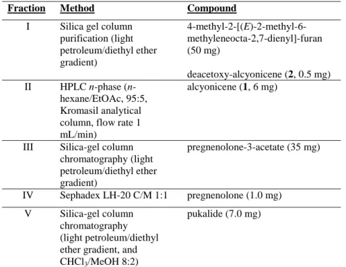
Table 2: Purification procedures of the various fractions.
Fraction Method Compound I Silica gel column
purification (light petroleum/diethyl ether gradient) 4-methyl-2-[(E)-2-methyl-6-methyleneocta-2,7-dienyl]-furan (50 mg) deacetoxy-alcyonicene (2, 0.5 mg) II HPLC n-phase (n-hexane/EtOAc, 95:5, Kromasil analytical column, flow rate 1 mL/min)
alcyonicene (1, 6 mg)
III Silica-gel column chromatography (light petroleum/diethyl ether gradient) pregnenolone-3-acetate (35 mg) IV Sephadex LH-20 C/M 1:1 pregnenolone (1.0 mg) V Silica-gel column chromatography (light petroleum/diethyl ether gradient, and CHCl3/MeOH 8:2)
pukalide (7.0 mg)
1.1.2. Structure determination
The molecular formula C17H26O2 of compound 1, named alcyonicene, was established by the analysis of the sodiated molecular peak at m/z 285.1824 [M+Na]+, obtained from the HRESIMS spectrum of the sample. This molecular formula indicated five degrees of unsaturations.
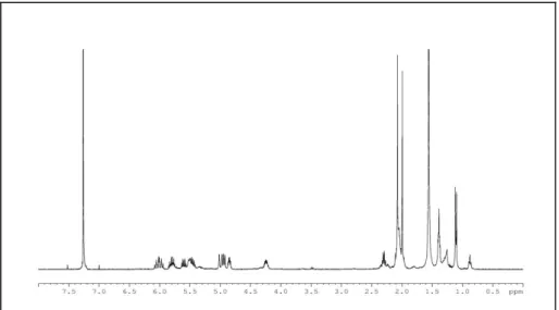
Analysis of the 1H NMR spectrum of 1 (Figure 2), showed a series of methyl signals, at high-field region, that were consistent with the presence of a terpenoid compound.
20
Figure 2: 1H NMR spectrum of alcyonicene (1)
In particular, two signals at 1.82 (br s, H3-14) and 1.05 (d, J = 7.3 Hz, H3-11), each integrating for three protons, were attributed to a vinyl methyl and to a secondary sp3 methyl, respectively. In addition, the low-field region of the 1H NMR spectrum showed a 1H multiplet at 5.15 (ddd, J = 4.7, 10.8, and 10.8 Hz, H-2) that was attributed to a proton linked to an oxygenated carbon, as confirmed by its HSQC correlation at C 70.2. Moreover, the spectrum contained four 1H broad singlets at 4.91 (H-13a), 4.89 (H-13b), 4.75 (H-15a), and 4.46 (H-15b) that suggested the presence of two exomethylene groups.
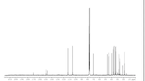
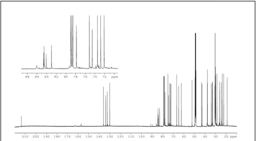
The 13C NMR spectrum of 1 (Figure 3) disclosed five sp2 and twelve sp3 carbons.
21
Figure 3: 13C NMR spectrum of alcyonicene (1)
Four olefinic carbon signals [( 149.5, s, 10), (147.5, s, C-12), (112.9, t, C-13), (105.2, t, C-15)] were recognized to belong to the two exomethylene groups, whereas one sp2 carbon was attributed to the carboxyl of the acetyl group, confirmed by an intense IR band at 1731 cm-1. The presence of an acetyl group was also deduced by methyl signals 21.5 (COCH3) in the 13C NMR spectrum and a 3H acetyl singlet at 2.01 (COCH3) in the 1H NMR spectrum. The remaining two unsaturations required by the molecular formula were thus attributed to two rings. Thus compound 1 had a bicyclic sesquiterpene skeleton.24,31-33
The 1H-1H COSY experiment showed the presence in the molecule of a single spin system, H-1/H2-9 sequence (Table 3),
22
according to the decaline framework of a cadinene carbon skeleton. 28-34
Analysis of the HMBC spectrum of 1 confirmed this hypothesis and aided us to assign all the proton and carbon values, as reported in Table 3.
Compound (2), named deacetoxy-alcyonicene, was isolated only in trace amount from the extract. The NMR spectra of 2 showed proton and carbon resonances very similar to those of 1, indicating the presence of the same carbon framework. The only difference was the lack of the acetoxy substituent at C-2 in compound 2 with respect to compound 1. Accordingly, in deacetoxy-alcyonicene (2), C-2 was a methylene rather than an oxygenated methine (in 2: C 23.9, H 1.65/1.41; in 1: C 70.2, H 5.15). Analysis of the EIMS spectrum showed the molecular peak at m/z 204, confirming the molecular formula C15H24. Careful analysis of 2D NMR experiments as well as comparison of the spectroscopic data of the related main metabolite 1 led us to assign the 1H and 13C NMR values of 2 (Table 3).
23 Table 3: NMR spectroscopic data for alcyonicene (1) and deacetoxy-alcyonicene (2).
1 2 Position 13C,a mb 1H,c m HMBC 13C,a mb 1H,c m 1 46.9, CH 2.40, m H-2, H-6 42.3, CH 2.08, br t (11.5) 2 70.2, CH 5.15ax, ddd (4.7,10.8,10.8) H-1, H2-3 23.9, CH2 1.65, m/1.41, m 3 37.0, CH2 1.92eq, m, 1.48ax, ddd (4.4,10.8,15.8) H-2, H3-11, H-4,H2-5 n.d. 1.41, m 4 27.5, CH 2.13, m, H3-11 27.4, CH 2.04, m 5 35.7, CH2 1.66ax, ddd (4.4,13.1,17.5) 1.25eq, m H3-11 n.d. 1.25, m 6 39.5, CH 1.78, m H-1, H-7 38.6, CH 1.70, m 7 44.7, CH 2.31eq, br dd (5.3, 5.3) H-6, H2-8, H2-13, H3-14 45.4, CH 2.20, br t (5.18) 8 32.8, CH2 1.86, m 1.72, m H-7 31.9, CH2 1.83, m 1.71, m 9 33.5, CH2 2.38, m 2.18, ddd (3.9,13.1,13.1) H2-8, H2-15 32.6, CH2 2.40, ddd (2.9,13.1,13.1) 2.17, dt (3.9,13.1) 10 149.5, C --- H2-15, H-1, H2-9 151.5, C 11 18.2, CH3 1.05, d (7.3) H-4 17.9, CH3 0.97, s 12 147.5, C --- H2-13, H3-14 147.7, C 13 112.9, CH2 4.91, br s 4.89, br s H3-14 112.5, CH2 4.89 br s 4.85 br s 14 26.6, CH3 1.82, s H-7, H2-13 26.2, CH3 1.81, s 15 105.2, CH2 4.75, br s 4.46, br s H2-9, H-1 104.3, CH2 4.69, br s 4.58, br s OAc 170.7, C 21.5, CH3 --- 2.01, s H3-17, H-2 a
Bruker 300 MHz, values are reported in ppm referred to CDCl3 (c 77.4); b
Assignments deduced by DEPT sequence; c Bruker 400 MHz,
24
1.1.3. Stereochemical assignment
Analysis of the vicinal proton coupling constants (Table 3), NOE difference experiments and the 13C NMR values allowed the establishment of the relative stereochemistry of alcyonicene (1). In particular, irradiation of the proton at 5.15 (H-2), in the 1H-1H homodecoupling experiments, simplified the signal at 2.40 (H-1) to a large doublet (11.6 Hz) suggesting the presence of a trans-diaxial relationship of the two angular protons according to a trans-fused ring.
The relative configuration at C-4 was suggested by the high-field shifted value of C-11 ( 18.2), consistent with an axial orientation of the methyl at C-4. This was in agreement with the spectroscopic data reported in the literature for related cadinene models exhibiting at C-4 either the equatorial methyl (i.e. cadinane:
C-11 23.0531) or the axial methyl (i.e. xenitorin A: C-11 18.332; 8-epi-xenitorin A: C-11 18.133). Furthermore, diagnostic NOE effects were observed among H3-11, H-6 and H-2 thus inferring the axial orientation for all of them (Figure 4). The relative configuration of C-7 was deduced by the multiplicity of the H-C-7 signal ( 2.31, br dd, J = 5.3 and 5.3 Hz), consistent with its equatorial orientation. This suggestion was further supported by a series of NOE effects observed between H-7 and H-6, H-8ax and H-5eq confirming the proposed stereochemistry (Figure 4).
25 Figure 4: NOE correlations for alcyonicene (1)
Alcyonicene (1) was thus characterized as possessing a trans-fused decaline system with the isopropenyl chain at C-7 axially oriented, as occurs in the bulgarane subgroup of the cadinene sesquiterpene class.30,34
Subsequently, with the aim to establish the absolute stereochemistry, compound 1 was hydrolyzed in the corresponding alcohol derivative to which the modified Mosher’s method could be applied. Unfortunately, every attempt was unsuccessful due to the rapid degradation of 1 under different hydrolysis conditions. Thus the absolute stereochemistry remained undetermined.
The relative stereochemistry of deacetoxy-alcyonicene 2 was suggested to be the same as 1 by both the similarity of the NMR values and biogenetic considerations.
1.1.4 . Biological and ecological activities evaluation
The ecological properties of alcyonicene 1 as well as of known compounds 4-methyl-2-[(E)-2-methyl-6-methyleneocta-2,7-dienyl]-furan, pregnenolone, pregnenolone-3-acetate and pukalide were
26
preliminarily evaluated by conducting assays with Carassius auratus35 and Gambusia affinis.36
The assay against the mosquito fish G. affinis is indicative to establish the ichthyotoxic properties of the samples tested. According to literature procedures,36 all the isolated metabolites were assayed at 10 ppm, but no significant activity was observed.
In addition, feeding-deterrence tests against the gold fish C. auratus were conducted according to literature procedures.35 Among the compounds tested, pukalide was feeding-deterrent at a concentration of 50 μg/cm2
. A similar ecological activity has been previously reported for a derivative of pukalide, isolated from a soft coral and its prey, the aeolid mollusc Phyllodesmium guamensis.37
All compounds were also tested in antimicrobial assays against Escherichia coli DH5a and Staphylococcus aureus ATCC6538P.38 No significant activity was evidenced at 100 µg/mL.
1.2.
Parazoanthus axinellae
Parazoanthus axinellae is a sea anemone belonging to the order Zoanthidea. The chemical investigation
on this animal started during my stage at the University of Athens. Despite evidence of their rich natural product chemistry,39 relatively few chemical studies of zoanthids have been so far reported.
27
Colonial sea anemones of the genus Parazoanthus have been identified in almost all the oceans, and they often have been described as epibionts of marine sponges belonging to Agelas or Axinella genera. As sponges are known to exude toxic compounds, these zoanthids must have developed adaptative tools to minimize effects of such toxins.
P. axinellae has been described to posses three groups of compounds: fluorescent guanidine alkaloids of the zoanthoxanthin families,40-45 ecdysteroids46 and hydantoins alkaloids.47
The specimens analyzed in this study were collected along the Greek coast. The chemical investigation resulted in the isolation of the known parazoanthines A-E 47 along with two new compounds 3 and 4 also belonging to this class of compounds.
28
1.2.1. Isolation procedure
The sea anemone P. axinellae (dry weight 118 g), collected in 2008, was extracted three times with CH2Cl2/MeOH 1:1. An aliquot of the extract (~10 g) was fractionated by VLC, using C18-reverse-phase silica gel and a gradient of MeOH/H2O until only MeOH. A chromatographic profile (TLC) of the recovered fractions displayed the presence of strong UV-visible spots, mainly in the first fraction. Due to the complexity of this mixture, the purification of compounds was obtained by subsequent chromatographic steps including MPLC and then HPLC (RP-amide column, MeOH/H2O gradient). parazoanthines –F (3), –A, –B, –G (4), –C, –D and –E were obtained in order of decreasing polarity.
The structures of parazoanthines –F and –G were determined by means of spectroscopic methods whereas the known metabolites were identified by comparison of their NMR and mass spectral data with those reported in the literature.47
1.2.2. Structure determination
Analysis of NMR spectra of both compounds 3 and 4 revealed a close resemblance with those of co-occurring known parazoanthines suggesting the same structural framework.
The analysis of the ion peak in the HRESIMS spectrum of parazoanthine F at m/z 318.1555 [M+H]+ led us to deduce the
29
molecular formula C15H19N5O3. The 1H NMR spectrum (Figure 5) of parazoanthine F contained a series of proton signals, at low-field region, that indicated the presence of an aromatic compound. A para-substitueted phenolic moiety was easily recognized due to the characteristic signals at δH 6.69 (2H, d, J 8.0 Hz, H-16 and H-20) and 7.00 (2H, d, J = 8.0 Hz, H-17 and H-19) in the 1H NMR spectrum, and at δC 126.0 (C, 15), 132.6 (CH, 16 and 20), 116.5 (CH, C-17 and C-19), and 157.5 (C, C-18) (Table 4) in the 13C NMR spectrum.
Figure 5:1
H NMR spectrum of parazoanthine F
An additional unsaturation was evidenced by the presence of the 1H NMR signals at δH 5.54 (1H, t, J = 7.5 Hz, H-6), and 13C resonances at δC 129.4 (C, C-5), and 114.3 (CH, C-6). The NMR data
30
were very similar to those of known parazoanthine B; the difference was in the presence of two methylenes [C-13 (δC 58.2; 2H, δH 4.32, t, J = 4.5 Hz, H2-13) and C-14 (δC 40.7; 1H, δH 3.10, overlapped; 1H, δH 2.95, dd, J = 14.4 and 4.5 Hz, H2-14)] in parazoanthine F (3) rather the double bond [C-13 (δC 116.5; 1H, δH 6.95, d, J = 15.0 Hz), C-14 (δC 121.7; 1H, δH 7.42, d, J = 15.0 Hz)] in parazoanthine B.
Table 4: NMR spectroscopic data for parazoanthine F and G (3, 4). 3 4 Position 13C,a mb 1H,c m 13C,a mb 1H,c m 2 168.1, C --- 174.2 --- 4 161.7, C --- 157.2 --- 5 129.4, C --- 55.7, CH 4.20, t (5.0) 6 114.3, CH 5.54, t (8.0) 29.7, CH2 1.92, m 1.81, m 7 27.8, CH2 2.25, m 24.9, CH2 1.75, m 1.66, m 8 41.3, CH2 3.15, t (7.5) 41.9, CH2 3.26, t (7.0) 10 158.2, C --- 162.0 --- 13 58.2, CH2 4.32, t (4.5) 117.1, CH 7.03, d (15.0) 14 40.7, CH2 3.10, overlapped 2.95, dd (14.4, 4.5) 121.4, CH 7.48, d (15.0) 15 126.0, C --- 129.6, C --- 16-20 132.6, CH 6.69, d (8.0) 128.2, CH 7.37, d (8.0) 17-19 116.5, CH 7.00, d (8.0) 115.1, CH 6.92, d (8.0) 18 157.5, C --- 163.1, C --- CH3-O 56.9, CH3 3.78, s a
Bruker 300 MHz, values are reported in ppm referred to CD3OD (c 49.0); b Assignments deduced by DEPT sequence;
c Bruker 400 MHz, values are reported in ppm referred to CH3OH (H 3.34)
The COSY and HMBC correlations confirmed the sequence of the alkyl chain (Figure 6).
31 Figure 6: Key COSY and HMBC correlations of 3
Compound 4, named parazoanthine G, had the molecular formula C16H21N5O3, deduced from HRESIMS spectrum (m/z 332.2881 [M+H]+). This information suggested the addition of a methylene unit compared to parazoanthine A, and the close NMR data (Table 4) suggested a strong similarity between both compounds. The replacement of the hydroxy group in parazoanthine A by a methoxy group in parazoanthine G (4) was supported by the 1H NMR signal at δH 3.78 (3H, s, CH3-O) and the 13C NMR resonance at δC 56.9, CH3, CH3-O) and was further confirmed by the key CH3-O/C-18 HMBC correlation.
1.2.3. Biological and ecological activities evaluation
All compounds were tested on different human cancer cell lines (Hs683, U373, U251, A549, MCF7, SKMEL28, PC3) to evaluate in vitro growth inhibitory concentration, but none of them exhibited significant bioactivity.
32
The main metabolites, the known parazoanthines A and B, were tested in vitro Scratch Wound Assay, using different cell lines. Mouse B16F10 melanoma, and human A549 non-small-cell lung cancer (NSCLC) and Hs683 glioma cells were grown until confluence and then a scratch has been performed as detailed in Mathieu V et al. 2005.48 The data indicate that 100 µM of parazoanthine B decreased A549 NSCLC migration, while parazoanthine A did not. Parazoanthine B also induced weak anti-migratory effects on Hs683 glioma cells, while parazoanthine A did not. Parazoanthine B did not display anti-migratory effects on B16F10 melanoma cells because they grew too rapidly in presence of 10% serum. Indeed, the wound healing process was already completed in the control condition at 100% about 12 hrs after having performed the scratch.
New experiments are ongoing with less proliferating B16F10, A549 and Hs683 cancer cells, e.g. in culturing them for 48 hrs in presence of 1% serum only.
Parazoanthines were also analyzed in antimicrobial assays against Escherichia coli DH5a and Staphylococcus aureus ATCC6538P. The experiments were performed in triplicate at concentration of 100 µg/mL, but no significant activity was evidenced.
33
C
HAPTER
2:
Phylum Chordata
Chordates are animals which are either vertebrates or one of several closely related invertebrates. This phylum consists of three subphyla: Urochordata, represented by tunicates, Cephalochordata, represented by lancelets, and Craniata, including vertebrata.
Tunicates, also known as ascidians, are a group of underwater saclike marine invertebrate filter feeders, characterized by a tough "tunic" outer made of polysaccharides.
Ascidians are found all over the world and, usually, are sessile animals. They remain firmly attached to substrata such as rocks and shells. There are 2,300 species of ascidians and three main types: solitary ascidians, social ascidians, that form communities through the aggregation of each other at their bases, and colonial ascidians that consist of several small individuals (called zooids) forming colonies up to several meters in diameter.
Tunicates are the natural prey of many animals, including molluscs, flatworms, rock crabs, sea stars, fish and sea otters. They are able to chemical defend themselves by maintaining an extremely high concentration of vanadium in the blood, having a very low pH of the tunic (due to an acid in easily-ruptured bladder cells), and /or producing secondary metabolites harmful to predators and invaders. Some of these metabolites are toxic to cells and are of potential use in pharmaceutical chemistry.
34
Ascidians are renowned for their great chemical diversity and, during the last 25 years, they have been shown to produce an array of cytotoxic molecules. The high potential of these organisms as a new source of antitumor compounds is demonstrated by the fact that among the first six marine derived compounds that have reached clinical trials as antitumor agents, three are derived from ascidians.49 Ecteinascidin-743 (ET-743/trabectedin), isolated from the Caribbean tunicate Ecteinascidia turbinata, represents a significant milestone in the development of marine derived drugs. Almost 40 years after its discovery and 17 years after the publication of its structure, it became the first marine-derived anticancer drug to reach the market. However, ET-743 was not the unique lead anticancer agent found from marine ascidians.50 Two closely related compounds - didemnin B and aplidine (Figure 7) isolated from the tropical Trididemnum solidum and the Mediterranean Aplidium albicans, respectively - have been extensively investigated for more than 20 years; although clinical trials for didemnin B were stopped in the mid-1990s. Differently, phase II clinical trials with aplidine are ongoing in indications that include metastatic melanoma, multiple myeloma, non-Hodgkin’s lymphoma, acute lymphoblastic leukaemia, prostate cancer and bladder cancer.
35 Figure 7: Didemnin B from Trididemnum solidum and aplidine (also known as dehydrodidemnin B) from Aplidium albicans
2.1. Pseudodistoma crucigaster
Among tunicates, the genus
Pseudodistoma has been found to be rich in cytotoxic alkaloids,51 linear52-54 and cyclic55-59 amino alcohols, tryptophan-related compounds,60,61 alkyl amines62,63 and nucleosides.64,65
The Mediterranean Pseudodistoma crucigaster52 has been reported to contain linear erythro 2-amino-alken-3-ols, crucigasterins (i.e., crucigasterin 277). These molecules are closely related to the sphingosines,66 basic building blocks of sphingolipids and glycosphingolipids, that are constituents of cell walls and membranes
36
and play diverse roles in biological processes such as cell growth, cell differentiation, and the immune response.67
In this study, we have analyzed specimens of Mediterranean P. crucigaster, collected off the coasts of Sardinia. The chemical investigation has led to the structural elucidation of five novel unsaturated amino alcohols, crucigasterins A-E (5-9),68 isolated along with related known compound 10, previously reported from South African Pseudodistoma sp.53
These molecules, which exhibit different carbon chain length and oxidation degree, are all characterised by the threo-configuration of the amino alcohol moiety, in contrast with the erythro-stereochemistry of previously reported crucigasterins.52
All compounds have been characterised as diacetyl derivatives
37
derivative of known compound 10a,53 which previously was not fully characterized, has been also made.
2.1.1. Isolation procedure
The colonial ascidian P. crucigaster (dry weight, 5.2 g), collected along the Sardinia coast, was extracted exhaustively with Me2CO using ultrasound. After filtration and evaporation in vacuum of the organic solvent, the residue was subsequently extracted with Et2O and BuOH to obtain two gummy residues (0.783 g and 0.976 g, respectively). The ethereal extract was fractionated by a silica gel column chromatography using a gradient eluent system with increasing polarity. A preliminary NMR analysis of the recovered fractions revealed that the most polar fraction was constituted by a complex mixture of unsaturated amino alcohols. Any effort to separate this mixture was unsuccessful, also due to the degradation of the compounds observed during the purification steps.
Thus, the fraction was acetylated with acetic anhydride and pyridine (2 h, room temperature). The acetylated mixture was easily fractionated by reverse-phase HPLC (MeOH/H2O 8:2) to give six pure metabolites, the diacetyl derivatives 5a-10a of five novel molecules, named crucigasterins A-E (5-9), and the diacetyl derivative 10a of known related compound 10. Compound 10a was identified by comparison of spectroscopic data with the literature53 even though only a partial NMR assignment had been reported. However, the
38
proton and carbon resonances of 10a were fully assigned, by interpretation of 2D-NMR spectra, in Tables 5 and 6.
2.1.2. Structure determination
Analysis of the NMR spectra of compounds 5a-9a revealed a close structural relationship with compound 10a indicating the presence in all the molecules of the same 2-amino-3-hydroxy-alkyl residue. The structural differences among them were in either the alkyl chain length or in the number and/or geometry of the double bonds.
Diacetyl crucigasterin A (5a) was considered first. The 2-amino-3-hydroxy moiety was easily recognized by sequentially correlated proton signals at 1.10 (3H, d, J = 6.7 Hz, H3-1), due to the terminal methyl, and at 4.25 (1H, dq, J = 3.8 and 6.7 Hz, H-2) and 4.85 (1H, dt, J = 3.8 and 6.7 Hz, H-3), that were attributed to two methines linked to the amino and the hydroxyl group, respectively (Figure 8). Moreover, the 13C NMR spectrum displayed carbon signals at 18.5 (q, C-1), 46.7 (d, C-2) and 75.9 (d, C-3), that confirmed this assignment.
39
Figure 8: 1H NMR spectrum of diacetyl crucigasterin A (5a)
In addition, the carbon resonances indicated the presence of three double bonds (Table 6), one of which had to be in the terminal position. Accordingly, the 1H NMR spectrum displayed in the low-field region seven olefinic signals (Table 5), which were assigned to a conjugated diene system and to the terminal vinyl moiety by analysis of the 1H-1H COSY and HSQC spectra. The geometries of 5 and 7 double bonds were suggested to be E,E by the coupling constants (J5,6=J7,8=14.6 Hz) and further supported by the 13C chemical shift values of the allylic methylene carbons C-4 ( 35.0) and C-9 (32.3) (Tables 5 and 6).
41
Diacetyl crucigasterin B (6a) had the same molecular formula (C18H31NO3Na) of known metabolite 10a. Analysis of 1H and 13C NMR spectra (Tables 5 and 6) indicated the presence of an identical structural sequence: they differed only in the geometry of 5 double bond. In particular, the proton coupling constant J5,6=10.0 Hz and the carbon values of C-4 (d 29.5) and C-7(d 27.3) clearly indicated that 6a was the 5Z-isomer of 10a.
Diacetyl crucigasterin C (7a), with the molecular formula C22H33NO3, contained a highly unsaturated C18 alkyl chain. The 1H NMR spectrum showed signals accounting for 11 olefinic protons (Table 5) according to the presence of five vinyl unsaturations (four
42
disubstituted and one terminal double bond). The terminal vinyl moiety was conjugated with one of the disubstituted double bonds, whereas the remaining three were isolated. Analysis of the 1H-1H COSY spectrum revealed that the 6H signal at 5.42-5.33 was coupled with three bis-allylic methylene groups at 2.97 (2H, app. t, J = 5.6 Hz, H2-14), 2.84 (2H, app. t, J = 5.6 Hz, H2-8 or H2-11) and 2.79 (2H, app. t, J = 5.6 Hz, H2-11 or H2-8), and to an allylic methylene at
2.02 (2H, m, H2-5). This latter signal was additionally correlated with the methylene multiplet at 1.61 (2H, m, H2-4), which in turn showed cross-peaks with the carbinol proton of 2-amino-3-hydroxy moiety. The 13C NMR values of bis-allylic and allylic methylenes (Table 6) suggested the Z geometry of all the disubstituted double bonds.
Diacetyl crucigasterin D (8a) was characterized by two additional CH2 units in the alkyl chain with respect to 6a and 10a. The 1
H and 13C NMR spectra exhibited signals attributable to two disubstituted double bonds [C 134.1(C-6),130.5 (C-9), 128.2 (C-10), 124.4 (C-5); H 5.51 (H-6), 5.41-5.28 (H-5, H-9 and H-10)], in addition to the signals due to the 2-amino-3-hydroxy moiety (Tables 5 and 6). Analysis of 1H-1H COSY correlations was diagnostic to establish the position of the two double bonds at C-5 and at C-9. The geometry of 5,6 double bond was assigned as E by the proton coupling constant J5,6=14.0 Hz and the 13C NMR chemical shift values of allylic methylene groups [ 35.0 (C-4) and 32.7 (C-7)], whereas the
43
geometry of 9,10 double bond was suggested to be Z by the chemical shift values of the remaining allylic methylenes [ 27.1 (C-8) and 27.3 (C-11)].
Diacetyl crucigasterin E (9a) had the molecular formula C22H37NO3, consistent with a C18 alkyl chain with three double bonds. In fact, the 1H NMR spectrum of 9a showed three olefinic signals (Table 5), a signal at 2.76 (2H, app. t, 5.8 Hz), which was due to a bis-allylic methylene group, and multiplets centred at 2.24 (2H, m, H2-4), 2.10 (2H, m, H2-8), 2.06 (2H, m, H2-7) and 2.02 (2H, m, H2 -14), which were attributed to four allylic methylene groups. The 1 H-1
H COSY experiment helped to locate the three double bonds. The geometry of the three double bonds was suggested by the 13C NMR values (Table 6) of bis-allylic and allylic methylenes resonating at 25.7 (C-11), 27.0 (C-8), 27.2 (C-14), 32.6 (C-7), and 35.0 (C-4) consistent with the Z stereochemistry for 9 and 12 double bonds and the E stereochemistry for 5 double bond.
2.1.3. Stereochemistry assignment
With the aim to establish the relative configuration of the chiral centers C-2 and C-3 in the novel crucigasterins, a series of NOE difference experiments were recorded on the oxazolidinone derivative (11) of crucigasterin A (5), according to the procedure applied for related molecules.53,54 In order to prepare the oxazolidinone, the
44
diacetyl derivative 5a (1 mg) was treated with NaOH (4 M) at 70 °C for 20 h, to obtain the free aminoalcohol (0.8 mg, 5), that was then reacted with 1,10-carbonyldiimidazole (3 mg) in 1 mL of CH2Cl2 and 100 mL of DMF at 0 °C for 19 h under argon atmosphere. The solution was extracted with water and the organic layer was dried under nitrogen yielding 0.5 mg of the oxazolidinone 11.
The NOE effects observed between H-3 and H3-1 and between H-2 and H2-4 suggested that the substituents at both C-2 and C-3 were on the opposite face of oxazolidinone ring. Consequently, 2 and H-3 were suggested to be in a threo relationship in the acyclic starting compound 5. This suggestion was definitively confirmed by a comparative analysis of NMR data of diacetyl crucigasterin A (5a) with those reported in the literature for synthetic threo and erythro 2-acetamido-3-acetoxy-5E,7E-tetradecadiene models.69 A good agreement of carbon NMR resonances was observed between 5a and the threo-isomer, in particular with regards to diagnostic 13C chemical shift value of C-1 (threo 18.49; erythro 14.93; 5a 18.5). On this basis, the relative configuration of 5a, and consequently of 5, was assigned to be threo (2R*,3R* or 2S*,3S*). Consequently, the threo
45
stereochemistry of the 2-amino-3-hydroxy moiety of all the other crucigasterins was assigned by the indicative 13C NMR value of the terminal methyl C-1.
The next step was to assign the absolute stereochemistry of crucigasterins by applying the Mosher method to suitable derivatives. Unfortunately, during the methanolysis to obtain the hydroxyl substrates, a significant degradation was observed to occur under the reaction conditions for most compounds preventing the formation of the desired hydroxyl derivates. However, the reaction was successfully applied on the known co-occurring compound 10a, the absolute stereochemistry of which had not been reported in the previous paper.54
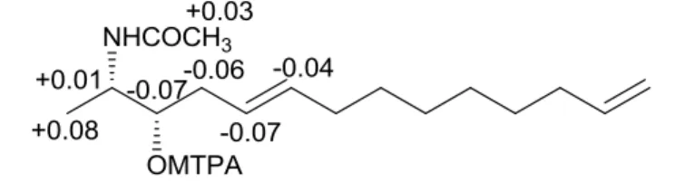
The O-deacetyl derivative 10b, obtained by treatment with Na2CO3 in anhydrous MeOH of 10a, was treated with (R)- and (S)-MTPA chlorides to get the (S)- and (R)-(S)-MTPA esters 10c and 10d, respectively. The (Sester-Rester) values observed (Figure 9) for the
signals of protons close to the hydroxyl group at C-3 indicated the S configuration as depicted in formula 10b. Consequently, the absolute stereochemistry of naturally occurring metabolite 10 was assigned as 2S,3S.
46
Figure 9: Chemical shifts differences (Sester-Rester) between (S)- and (R)-MTPA
derivatives of compound 10a
The same configuration was suggested for the other crucigasterins based on biogenetic considerations. This suggestion was supported by the optical rotation values of diacetyl derivatives of crucigasterins (5a-10a), which were all laevorotatory according to the 2S absolute configuration.69 Particularly diagnostic was the comparison of the []D value of diacetyl derivative of crucigasterin A (5a) {[]D -20.3 (c 0.03, CH3OH)} with those reported for synthetic (2R,3R)-{[]D +20.7 (c 1.66, CH3OH)} and (2S,3S)-{[]D -20.6 (c 0.32, CH3OH)} 2-acetamido-3-acetoxy-5E,7E-tetradecadiene enantiomers69,70 further confirming the 2S,3S absolute configuration for 5a and for all co-occurring metabolites.
2.1.4. Biological and ecological activities evaluation
Selected diacetyl-crucigasterins have been tested for antibacterial and antifungal activities, exhibiting moderate activity against E. coli and C. albicans, respectively.
47
Antifungal assay was performed by the broth macrodilution method following the guidelines of the National Committee for Clinical Laboratory Standards (NCCLS) document M27-P.71, 72 MIC was defined as the lowest concentration of the compound that completely inhibited the growth of the test organism.
The antibacterial assay was performed by using the same method as the antifungal test, only differing in the assay medium and in the incubation temperature.
In particular, the diacetyl-crucigasterins B (6a) and E (9a) were found to be active at 50 g/mL against both E. coli and Candida albicans, and at 100 g/mL against E. coli, respectively.
48
C
HAPTER
3:
Phylum Porifera
Among the organisms that inhabit the seas, Porifera, commonly known as sponges, comprising the most primitive multicellular animals. These sessile organisms have world-wide distribution and live anchored to substrates, such as rocks, seaweed, shells and crabs. Sponges could have solitary life or make dense colonies that become important habitats for animals and plants: their cavities can host several symbionts as small crustaceans, insect larvae, algae, cyanobacteria, etc.
Porifera are filter-feeders that use flagellated cells to pump water into their canal systems. They can be encrusting or erect, assuming different morphologies according to the environmental characteristics (substrate, currents, waves). Schematically, the body of a sponge may be considered a sort of bag, all perforated (hence the name Porifera) from inhalants thin pores (orifices), with a large opening said exhaling pore or osculum and an inner chamber called gastral cavity or spongocele.
Depending on the different endoskeleton, phylum Porifera is divided into four classes: 1) Calcispongiae, that are characterized by calcareous skeletons, generally small or tabular; 2) Hexactinellida (Hyalospongiae), also known as vitreous sponges due to their siliceous skeleton, with a funnel or cylindrical shape; 3) Demospongiae (mostly sponges species, 90%), including animals whose skeleton is composed of siliceous spicules, which in some forms are partially or fully
49
replaced by skeletal elements consisting of a special protein called spongin; 4) Sclerospongiae, a small group of sponges, mostly tropical, whose skeleton consists of calcium carbonate crystals on a network of organic fibbers.
Even sponges are primitive organisms, they have proved to be a rich source of secondary metabolites. It has been repeatedly suggested73-75 that they are protected by toxic or noxious chemicals, i.e. allomones. Many of these molecules, often characterized by complex molecular architectures, are known to possess interesting biological activities.76,77
3.1. Haliclona fulva
Haliclona fulva is an encrusting and orange desmosponge with a globular shape. A previous chemical study on this sponge has reported the isolation of a series of
bioactive polyacetylenes,78-80 closely related to petroformines, a family of long chain polyacetylenes isolated from another Mediterranean sponge, Petrosia ficiformis.81 All these compounds display linear alkin chain of 46 carbons with a characteristic 1-yn-3-ol-4-ene moiety at each terminus. In closely related haplosclerid sponges,82 similar polyacetylenes usually only have a mono-terminal 1-yn-3-ol-4-ene moiety - sometimes modified - and a considerably shorter alkyl chain.83-87
50
The biological interest of polyacetilene molecules prompted us to chemically reinvestigate H. fulva. The work here described reports the isolation and spectroscopic elucidation of nine high molecular weight polyoxygenated acetylenes, fulvynes A-I (12-20),88 from the butanolic extract of a specimen of the sponge collected in the Gulf of Naples.
The new compounds are characterized by a long linear alkyl chain bearing a residue of propargylic acid, a terminal acetylenic moiety, a diacetylenic carbinol and several hydroxyl and keto groups. Previous isolated metabolites – fulvinol and renierines - have been detected in the ether extract of the sponge under investigation along with a mixture of additional non-polar polyacetylenes. These latter metabolites have been also found in the lipophilic extract of the nudibranch Peltodoris atromaculata, which was observed grazing on
51
H. fulva. The structure elucidation of these compounds will be describe in the next Chapter, dedicated to the phylum Mollusca.
3.1.1. Isolation procedure
The sponge H. fulva (dry weight, 42 g), collected along the coast of Procida Island (Gulf of Naples) at a depth of ~ 40 m, was chopped and then extracted with Me2CO using ultrasound. After filtration and evaporation of the organic solvent in vacuo, the residue was subsequently extracted with Et2O and BuOH. Both organic phases were dried to give two gummy residues (1.46 g and 2.48 g, respectively) which were tested in preliminary antimicrobial and antifungal assays at a fixed concentration (50 µg/mL). The butanol extract showed a good activity against the gram positive B. subtilis whereas it showed weak against E. coli and C. albicans , with respect to the ethereal extract. Therefore, part of the bioactive butanol extract (1.2 g) was thus subjected to Sephadex LH-20 chromatography in MeOH to give eight fractions (I-VIII). Preliminary 1H NMR analysis of these fractions showed that fraction III (0.228 g) contained a mixture of polyacetylenic compounds. An aliquot of this fraction (0.120 g) was further purified on RP-amide semipreparative HPLC column with a gradient of H2O/MeOH/TFA (from 29:70:0.1 to 100% MeOH, flow 2.0 mL min-1) to afford, in order of retention time, pure fulvynes A-I (12-20). Preliminary 1H NMR analysis revealed that all isolated molecules were polyhydroxylated acetylenes. In particular,
52
comparison of the spectra with literature data showed strong similarities of the structural framework of fulvynes with those of osirisynes89,90 and haliclonyne,91 polyacetylene compounds isolated from other Haliclona species.
The structures of compounds 12-20 were determined by an extensive use of spectroscopic techniques.
3.1.2. Structure determination
Among the fulvynes, compound 14, named fulvyne C, was the most abundant metabolite. As deduced from the sodiated molecular ion peak at m/z 851.4879 [M+Na]+ in the HRESIMS, fulvyne C (14) had the molecular formula C47H72O12, indicating 12 unsaturation degrees.
The 1H NMR spectrum of 14 (Figure 10) displayed signals due to four olefinic protons [H 5.44, dd (15.2, 7.0); 5.64, dt (15.2, 6.7); 5.79, dd (15.5, 5.4); 5.92, dd (15.5, 6.0)], an acetylenic proton at (H 2.93), nine oxygenated methines in the range H 5.14-347 and a series of methylene groups in the high-field region (H 2.52-1.30) (Table 7).
53
Figure 10: 1H NMR spectrum of fulvyne C (14)
The IR spectrum showed bands at 2237, 1696, and 1679 cm-1 suggesting the presence of triple bonds and carbonylic functions. The 13
C NMR spectrum (Figure 11) showed eight sp carbon resonances in the range at C 85.3-74.8, due to four triple bonds, and C=O signals at
C 157.2 and 213.6. Moreover, carbon resonances due to sp2 carbons of two double bonds in the range C 136.3-130.5, nine oxygen-bearing methines in the region of the field between C 78.4 and 52.5, and several methylene groups were also present (Table 8).
55
Figure 11: 13C NMR spectrum of fulvyne C (14)
All proton-bearing carbons were assigned by the HSQC experiment. Analysis of the 1H-1H COSY spectrum aided us to recognize the partial structures a-e in the long alkyl chain of 14 (Figure 12).
Figure 12. Partial structures of fulvyne C (14) as deduced by 1H 1H COSY correlations
56
The 1H-1H TOCSY experiment was essential to assemble fragments a-e. In particular, the oxymethine H-42 in partial structure a was correlated to the oxymethine H-38 in moiety b through an additional methylene H2-40 (H 1.55-1.51, C 22.6). The -carbonyl methylene H2-28 in fragment c showed correlations with methylene H2-31 in fragment b through methylene H2-30 (H 1.56-1.50, C 29.0), whereas the other -carbonyl methylene H2-26 had cross-peaks with the allylic methylene H2-22 in partial structure d through an additional methylene H2-24 (H 1.38-1.30, C 30.4). The observed correlations allowed to connect fragments a-d accounting for a C30 segment of the alkyl chain of 14. This implied that fragment e had to be linked to the remaining part of the molecule through nine additional methylenes thus satisfying the molecular formula. Analysis of the HMBC spectrum of 14 confirmed the proposed structure and enabled us to assign properly the acetylenic quaternary carbon resonances (Table 8). The geometries of the two double bonds were easily assigned as 20E, 43E by the coupling constant analysis of the olefinic protons (J = 15.2 and 15.5 Hz, respectively).
Fulvyne A (12) had the same molecular formula C47H72O12 as fulvyne C (14). Comparison of 1D and 2D proton NMR spectra of 12 with those of 14 disclosed a substantial structural analogy between the two compounds suggesting the presence of the same functional groups. In particular, the differences were limited to the partial structure b' where one of the two hydroxyl groups of the vicinal diol
57
(37-OH) in fulvyne C was shifted on the other side of the di-yne moiety at C-31 in 12 (Figure 13).
The remaining signals appeared to be the same as compound
14 thus confirming that the two polyacetylenes were positional
isomers.
Fulvyne B (13) was also an isomer of fulvyne A (12) and fulvyne C (14). The proton and carbon spectra of 13 displayed signals that were strongly reminiscent with those of 14 being consistent with the same partial structures a-e. Analysis of 2D NMR spectra, especially TOCSY experiment, clearly demonstrated that the sequence of fragments b, c, and d in fulvyne B (13) was different from that set in fulvyne C (14). In particular, the keto group was positioned far from the inner triple bonds. In fact, the methylene protons at H 2.26 (H2-31) were long-range correlated with the allylic oxymethine at H 4.00 (H-28), rather than one of the -carbonyl methylene as in compound 14. In addition, the -carbonyl methylene at H 2.47 (H2-21) had a clear correlation with the allylic methylene at H 2.07 (H2-25), consistent with the sequential connection in the chain of fragments b, d, and c, as depicted in formula 13.
60
Figure 13. Additional partial structures of fulvynes
The molecular formula C47H72O11 of fulvyne D (15), as deduced from the sodiated peak at m/z 835.4865 in the HRESIMS, indicated that 15 was lacking in an oxygen atom with respect to co-occurring 12-14. In addition, the 13C NMR spectrum evidenced the presence of eight oxygen-bearing carbons (Table 8) rather than nine oxymethines, such as in 12-14. Analysis of 2D NMR experiments of fulvyne D indicated that the partial structure b'' lacked the hydroxyl group in -position of di-yne moiety present in fulvyne C. In fact, the COSY spectrum showed that the bis propargyl alcohol proton at H 5.07 (H-34) was coupled with two methylene groups at H 2.25 (H2-31) and H 2.40 (H2-37), that had to be both linked to internal triple bonds. By TOCSY experiments, the fragments were assembled as in fulvyne B (13). Thus fulvyne D (15) was the 37-dehydroxy derivative of 13.
Fulvyne E (16) with the molecular formula C47H72O11 was isomeric with compound 15, from which it differs in the sequence of the fragments in the chain. In particular, analysis of the TOCSY spectrum showed a clear correlation of the methylene at H 2.27 (H2 -31) in fragment b'' with the -carbonyl methylene at H 2.52 (H2-28)
61
in partial structure c according to the location of the keto group in proximity of the bis propargylic alcohol as in fulvyne C (14). Thus fulvyne E (16) was the 37-dehydroxy derivative of 14.
Last four compounds, fulvynes F-I (17-20), were all characterized by the lack of a hydroxyl group in fragment e, showing a diol moiety close to propargylic acid residue (fragment e') (Figure 13). The molecular formula C47H72O11 of fulvyne F (17) was deduced from the sodiated molecular ion peak at m/z 835.4843 in the HRESIMS. 13C and 1H NMR data of 17 (Table 9 and 10) were quite similar to those of fulvyne A (12). Careful analysis of 2D NMR spectra and in particular of both COSY and TOCSY spectra of 17 confirmed the presence of the same sequence of fragments a, b', d, and c as fulvyne A, whereas a difference was detected in the terminal fragment containing the propargylic acid unit missing in one of the three hydroxyl groups. Thus fulvyne F (17) was the 6-dehydroxy derivative of fulvyne A (12).
Fulvyne G (18) was isomeric with fulvyne F (17). The NMR spectra displayed strong similarities with those of co-occurring fulvyne C (14) except for the spin system in the terminal acid part. As
17, fulvyne G was characterized by the presence of a di-hydroxyl
62
The molecular formula of fulvyne H (19) was deduced as C48H76O9 from the sodiated molecular ion at m/z 819.4990 in the HRESIMS. The spectroscopic data of 19 (Table 9 and 10) clearly evidenced that it was lacking in both the ketone and 6-OH groups, whereas the remaining part was identical with fulvyne D (15). Thus,
63
the alkyl chain connecting partial structures d and e' contained an additional methylene unit with respect to all other co-occurring fulvynes.
Finally, fulvyne I (20) had a molecular formula C47H72O10 as deduced from the sodiated molecular ion peak at m/z 819.4917. Comparison of NMR data of compound 20 with those of fulvyne E (16) indicated the presence of the same fragment sequence a, b", c, and d, whereas the terminal residue was constituted by fragment e' characterizing fulvynes F-H (17-19).
3.1.3. Stereochemical assignment
The relative configuration of contiguous chiral carbons of fulvynes as well as of isolated carbinol centres remained unassigned. Any attempt to obtain suitable derivatives for a stereochemical analysis were unsuccessful due to a rapid degradation of the compounds under different reaction conditions. Even the simple acetylation conducted under mild conditions resulted in the decomposition of the starting material.
3.1.4. Biological and ecological activities evaluation
Due to the antimicrobial activity directed mainly against the gram positive B. subtilis of the butanol extract, pure isolated compounds were tested in a more specific assay on a chloramphenicol
64
resistant B. subtilis strain (PY79), showing a good activity against this strain (Table 11). Fulvynes were tested also on a gram-positive human pathogenic strain, Staphylococcus aureus, and showed a moderate activity.
Cytotoxic activity has been reported for polyacetylenes from sponges,78 thus the main metabolite was also subjected to antiproliferative assays. In particular, fulvyne C (14), tested on different tumour cell lines (C6, H9c2, HeLa, 3T3-L1), showed selectivity for mouse pre-adipocyte cells line 3T3-L1, being active with an IC50 of 55.7 ± 11 μM (Table 12).