Circulating Levels of PAI-1 and
SERPINE1 4G/4G Polymorphism
Are Predictive of Poor
Prognosis in HCC Patients
Undergoing TACE
1Rosa Divella*, Antonella Daniele*, Ines Abbate*, Eufemia Savino*,
Porzia Casamassima*, Giancarlo Sciortino*,
Giovanni Simone†, Gennaro Gadaleta-Caldarola‡,
Vito Fazio§, Cosimo Damiano Gadaleta§,
Carlo Sabbචand Antonio Mazzocca¶ ,#
*Clinical Pathology Laboratory, Department of Experimental Oncology, Giovanni Paolo II National Cancer Institute, Bari, Italy;†Department of Pathology, Giovanni Paolo II National Cancer Institute, Bari, Italy;‡Hospital“Monsignor Raffaele Dimiccoli”, Barletta, Italy;§Unit of Interventional Radiology, Department of Critical Area and Surgery, Giovanni Paolo II National Cancer Institute, Bari, Italy;¶Interdisciplinary Department of Medicine, University of Bari School of Medicine, Bari, Italy;#IRCCS“S. de Bellis”, National Institute for Digestive Diseases, Bari, Italy
Abstract
Although several molecular markers have been proposed as prognostic of disease progression in Hepatocellular carcinoma (HCC), predictive markers of response to treatment are still unsatisfactory. Here, we propose a genetic polymorphism as a potential predictive factor of poor prognosis in HCC patients treated with transcatheter arterial chemoembolization (TACE). In particular, we show that the guanosine insertion/deletion polymorphism in the promoter region of SERPINE1 gene at the−675 bp position, named 4G/4G, predicts poor prognosis in a cohort of 75 patients with HCC undergoing TACE. By a combination of ELISA and SERPINE1 promoter study, we found that the presence of elevated plasma levels of plasminogen activator inhibitor-1 (PAI-1) in patients with 4G/4G genotype is significantly associated with reduced overall survival compared to patients with 5G/5G or 4G/5G genotype in HCC patients after TACE. Our analysis provided evidence that variation in SERPINE1 gene plays a role in defining the outcome in patients treated with TACE. In addition to a poor disease outcome, the 4G/4G variant represents an unfavorable predictive factor for response to chemotherapy as well.
Translational Oncology (2015) 8, 273–278
Introduction
Many Hepatocellular carcinomas are diagnosed in intermediate or advanced stages only when locoregional or palliative treatments are feasible. The long-term survival of Hepatocellular carcinoma (HCC) patients treated with locoregional or palliative treatments remains unsatisfactory because the treated tumor frequently maintains residual viability, leading to disease reactivation, although the survival rate can be good in properly selected patients and under standardized condi-tions [1,2]. Identifying biologic markers capable of predicting the response to treatment in HCC patients may facilitate the disease’s management. Serine protease (Serpins) inhibitors including plasmin-ogen activator inhibitor-1 (PAI-1) play an important role in regulating a wide array of diverse biologic activities, representing up to 2% to 10% of www.transonc.com
Address all correspondence to: Rosa Divella, PhD, Clinical Pathology Laboratory, Department of Experimental Oncology, Giovanni Paolo II National Cancer Institute, Viale Orazio Flacco, 65, 70100 Bari, Italy or Antonio Mazzocca, MD, PhD, Interdisciplinary Department of Medicine, University of Bari School of Medicine, Piazza G. Cesare, 11, 70124 Bari, Italy.
E-mails:[email protected](R. Divella), [email protected](A. Mazzocca)
1Conflict of interest statement: None declared.
Received 6 February 2015; Revised 7 May 2015; Accepted 7 May 2015
© 2015 The Authors. Published by Elsevier Inc. on behalf of Neoplasia Press, Inc. This is an open access article under the CC BY-NC-ND license (http://creativecommons.
org/licenses/by-nc-nd/4.0/).
1936-5233/15
circulating plasma proteins[3]. The serpine suicide inhibitors regulate coagulation (thrombosis and thrombolysis), neurotrophic factors, hormone transport, complement and inflammation, angiogenesis, hormone transport, and blood pressure among many other biologic reactions[4,5]. Select serpins have been associated with progression or remission of selected cancers, making them valuable for therapeutic or diagnostic use [6,7]. PAI-1, the main regulator of thrombolysis, displays the potential to either reduce or accelerate tumor growth; however, the blockade of PAI-1 has recently been reported to reduce cancer cell migration, proliferation, and survival through modulating the function of urokinase-type plasminogen activator receptor[8].
As a potential prognostic factor, the concept of germline variation imparting interindividual variability in tumor development, progres-sion, and metastasis is receiving increasing attention. In vitro studies suggest that cytokines, growth factors, and hormones can affect PAI-1; however, the genetic and environmental determinants of
SERPINE1 expression are not fully understood [9,10]. Gene
variability may also contribute to the level of SERPINE1 biosynthesis
[11]. The human SERPINE1 gene is located on chromosome 7. A guanosine insertion/deletion polymorphism in the promoter region of
SERPINE1 gene at the − 675 bp position, named 4G/5G
(rs1799889), has been reported [12]. Recent studies indicate that the protein encoded by the 4G-allele possesses higher activity than that encoded by the 5G-allele. This is because the 5G-allele contains an additional binding site for a DNA-binding protein that acts as a transcriptional repressor [13,14]. Studies carried out in different populations have consistently shown that individuals, homozygous for the 4G-allele, have significantly higher plasma SERPINE1 levels than those homozygous for the 5G-allele[15,16]. The role of PAI-1
as a predictive factor of outcome in patients with HCC, and in particular in patients treated with transcatheter arterial chemoemboliza-tion (TACE), is poorly investigated. In a previous study, we investigated the distribution of genotypes and the frequency of alleles of the 4G/5G polymorphism in patients with HCC and the influence of the 4G/5G polymorphism on the circulating levels of SERPINE1. In the present study, we extended this knowledge by assessing the prognostic significance and clinical impact of plasma levels of PAI-1 in HCC patients before (pre) and after (post) TACE treatment. In particular, we evaluated the clinical impact of the SERPINE14G/4G genotype on prognosis of patients with HCC undergoing chemoembolization. Materials and Methods
Cancer Stadiation
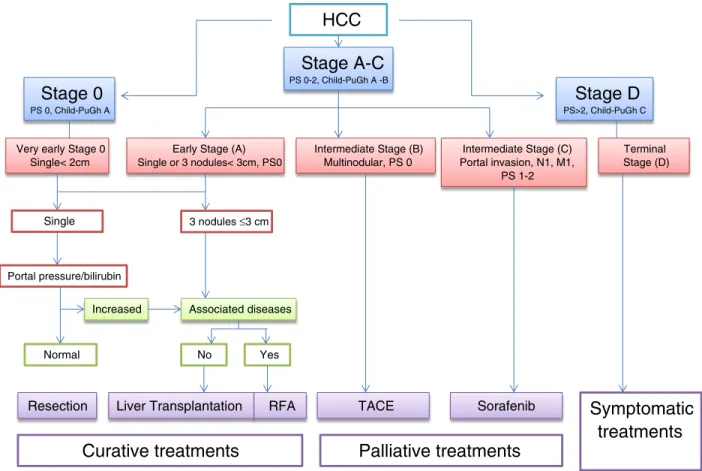
The Barcelona Clinic Liver Cancer (BCLC) tumor staging classification combines the stage of the liver disease, tumor stage, clinical performance, and treatment options for HCC. For unresectable HCC intermediate stage (BCLC stage B or Child-Pugh class A/B with large or multifocal HCC, no vascular invasion, or extrahepatic spread), the current standard treatment is TACE as reported inFigure 1.
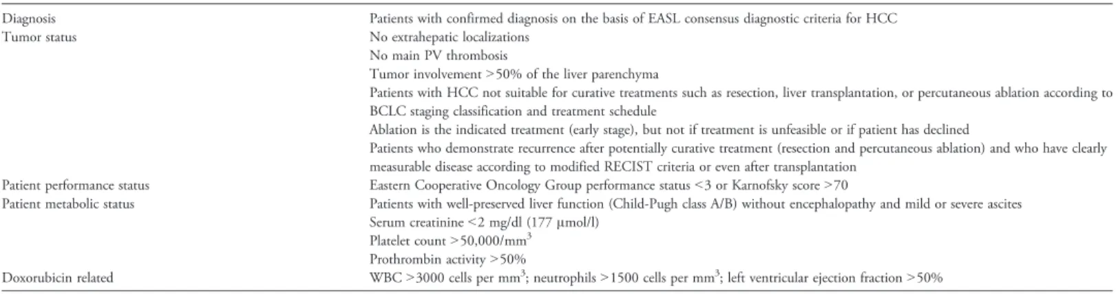
Inclusion Criteria for TACE Treatment
Criteria for the inclusion of HCC patients suitable for treatment with TACE are shown inTable 1, including patients with measurable inoperable HCC, histologic proven, multinodular HCC with intermediate grade stage A or B, with no vascular invasion or extrahepatic spread, and monofocal patients with HCC N 5 cm in advanced cirrhosis (stage C), who had not received prior systemic treatments for HCC.
HCC
Stage D
PS>2, Child-PuGh CStage 0
PS 0, Child-PuGh AStage A-C
PS 0-2, Child-PuGh A -BVery early Stage 0 Single< 2cm
Early Stage (A) Single or 3 nodules< 3cm, PS0 Terminal Stage (D) Intermediate Stage (B) Multinodular, PS 0 Intermediate Stage (C) Portal invasion, N1, M1, PS 1-2 Single 3 nodules ≤3 cm Portal pressure/bilirubin
Increased Associated diseases
Normal No Yes
Resection Liver Transplantation RFA
Curative treatments
TACE Sorafenib
Palliative treatments
Symptomatic
treatments
Patient Enrollment and Clinical Characteristics of the Tumor
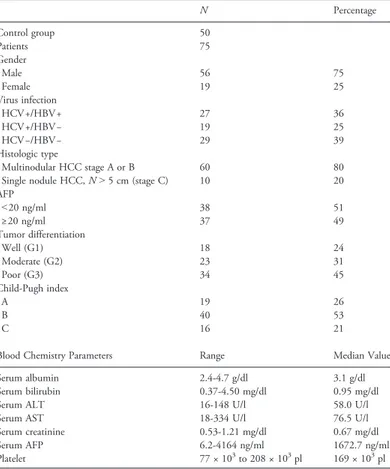
Using this protocol, from June 2007 to December 2010, TACE was performed in 75 consecutive HCC patients, 56 males (74.6%) and 19 females (25.4%), aged from 45 to 87 years (median, 73 years) enrolled at the Giovanni Paolo II National Cancer Institute (Bari, Italy). Information about gender, age, etiology, histologic diagnosis of cancer, score of liver disease according to Child-Pugh, serum levels of total albumin, bilirubin, transaminase ALT and AST, creatinine and α-fetoprotein at baseline, the Cancer of the Liver Italian Program score, portal thrombosis, the presence of liver metastasis identified by ultrasound with contrast medium, computed tomography (CT) scan of the abdomen, and the type of locoregional treatment (TACE) were collected from clinical charts of each patient (Table 2). No patient receiving TACE treatment showed extrahepatic metastases, patency of the portal vein, or altered liver function; the diagnosis of HCC was cyto-histologically confirmed by echo-guided fine needle aspiration biopsy.
Sample Collection
A written consent was obtained from all patients before enrollment in the study, and the Ethical Committee of the Giovanni Paolo II National Cancer Institute approved the protocol, which was in accordance with the ethical guidelines of the 1975 Declaration of Helsinki. From each participant to this study, 5 ml of peripheral blood was collected in a Vacutainer system with lithium-heparin. Whole blood samples (200μl) were collected for DNA extraction before any invasive procedures or therapy. For PAI-1 ELISA test, the remaining part of the sample was centrifuged, and plasma was immediately separated from the cellular fraction by centrifugation at 1500g for 10 minutes and stored in microtubes (aliquots of 200μl) and frozen and at−20°C. Patients agreed to provide two blood samples: one before TACE (pre) treatment and one after TACE (post) treatment, 4 to 6 weeks at the time of spiral CT.
TACE Procedure
Each patient was discussed in a team meeting to decide the appropriate approach. Asymptomatic patients who had multinodular HCC in the intermediate stage (A or B) with no vascular invasion or extrahepatic spread and monofocal patients with HCC, NN 5 cm, in advanced cirrhosis (stage C) were treated. This procedure has been previously described[17]. Briefly, treatment with TACE was performed with a standard protocol under general anesthesia by binding DC-Beads (Biocompatibles, Farnham, United Kingdom) to a total dose of doxorubicin of 100 mg/50 ml and injecting by percutaneously inserting a microcatheter into the femoral artery of the patient under fluoroscopic guidance (X-ray) that corresponds to
the artery of the liver. When applicable, the artery feeding the tumor was cannulated in a superselective approach. In the case of bilobar tumor involvement, the chemoembolic agent was injected two subsequent time points, after 30 and 60 days, starting with the lobe more extensively involved. Efficacy was evaluated by dynamic CT 2 to 3 days after each treatment session, 1 month, 3 months, and 6 months, and the sessions were repeated until an ablative margin was obtained.
SERPINE1 ELISA and SERPINE1 Promoter
4G/4G Polymorphism
These procedures have been previously described [7–18]. Plasma SERPINE1 concentrations were determined with ELISA (Imunobind Plasma PAI-1 ELISA; American Diagnostica GmbH, Pfungstadt, Germany) according to the manufacturer’s recommendations. For SER-PINE1 promoter 4G/4G polymorphism, briefly, after extraction of genomic DNA from whole blood with QIAamp DNA blood mini kit (Qiagen, Hilden, Germany), DNA was amplified for molecular detection of SERPINE1 promoter 4G/5G polymorphism by an allele-specific (polymerase chain reaction) analysis using specific primers as previously described[7–18]. The polymerase chain reaction was carried out in a final volume of 25μl, and the amplified DNA fragments were separated by a 5% polyacrylamide gel electrophoresis. Each study participant was classified into one of the three possible genotypes: 4G/4G, 4G/5G, or 5G/5G.
Statistical Analysis
For the continuous variables, data were analyzed with the unpaired t test as well as analysis of variance. The correlation between the PAI-1 plasma values before and after TACE treatment was evaluated using the Student's t test. A P value≤.05 indicates statistical significance. Overall survival (OS) was the end point of the survival analysis and was estimated with Kaplan-Meier curves and tested with the log-rank test. OS was defined as the time between the date of the blood sample drawing and the date of death or the last follow-up examination. A P value≤.05 indicates statistical significance. All statistical analyses were performed by Number Cruncher Statistical System–Power Analysis and Sample Size Software 2007 (NCSS-PASS, Kaysville, UT).
Results
Evaluation of SERPINE1 Plasma Levels before and after
TACE Treatment
To compare the circulating plasma levels of PAI-1 in patients with HCC before and after TACE, we carried out ELISAs on collected
Table 1. Indications for TACE in HCC Patients
Diagnosis Patients with confirmed diagnosis on the basis of EASL consensus diagnostic criteria for HCC Tumor status No extrahepatic localizations
No main PV thrombosis
Tumor involvementN50% of the liver parenchyma
Patients with HCC not suitable for curative treatments such as resection, liver transplantation, or percutaneous ablation according to BCLC staging classification and treatment schedule
Ablation is the indicated treatment (early stage), but not if treatment is unfeasible or if patient has declined
Patients who demonstrate recurrence after potentially curative treatment (resection and percutaneous ablation) and who have clearly measurable disease according to modified RECIST criteria or even after transplantation
Patient performance status Eastern Cooperative Oncology Group performance statusb3 or Karnofsky score N70
Patient metabolic status Patients with well-preserved liver function (Child-Pugh class A/B) without encephalopathy and mild or severe ascites Serum creatinineb2 mg/dl (177 μmol/l)
Platelet countN50,000/mm3
Prothrombin activityN50%
Doxorubicin related WBCN3000 cells per mm3; neutrophilsN1500 cells per mm3; left ventricular ejection fractionN50%
peripheral blood samples. We found significantly decreased concentra-tions of circulating PAI-1 after TACE (34.11 ± 30.5 ng/ml) compared with those before TACE treatment (42.76 ± 25.8 ng/ml, P = .014;
Figure 2).
Elevated Circulating Plasma Levels of PAI-1 Are Associated
with the 4G/4G SERPINE1 Polymorphism
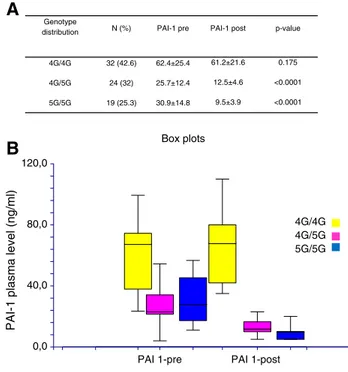
Since after TACE concentrations of PAI-1 remained elevated in a number of patients (N = 32), we sought to evaluate whether circulating plasma levels of PAI-1 were influenced by SERPINE1 genotype distribution before and after TACE. We therefore correlate 4G/5G polymorphism with the levels of PAI-1 in patients with HCC before and after TACE. We observed that genotype 4G/4G (N = 32 patients) is associated with higher levels of PAI-1 without difference before and after TACE (P = .175), whereas the alleles 4G/5G (N = 24) and 5G/5G (N = 19) are associated with high levels of PAI-1 in plasma of patients before TACE compared to plasma from patients after TACE (Pb .0001, respectively;Figure 3). Thus, the genotype influences the circulating plasma levels of PAI-1 in patients with HCC before and after TACE.
4G/4G SERPINE1 Genotype Represents an Adverse Prognostic
Factor for Patients with HCC Undergoing TACE Treatment
To evaluate whether SERPINE1 genotype has a prognostic role in these patients, we analyzed the OS with Kaplan-Meier analysis. The analysis shows that patients harboring the 4G/4G genotype have a poorer prognosis compared to 4G/5G and 5G/5G genotypes. The
median OS was 9, 18, and 25 months in 4G/4G, 4G/5G, and 5G/5G patient genotype groups, respectively (4G/4G vs 4G/5G, P = .05 and 4G/4G vs 5G/5G, P = .01, log-rank test;Figure 4). In addition, we analyzed the OS according to the PAI-1 plasma levels. We found that patients with elevated circulating levels of PAI-1 displays a poor prognosis compared to those with lower circulating levels (Figure 5). In fact, in patients with levels of PAI-1 above the cutoff value≥35.5 ng/ml (35.5 ng/ml = median value), the OS was 15 months versus 28 months (P = .001, log-rank test), which was in contrast to that observed in patients with levels lower than the cutoff valueb35.5 ng/ml (Figure 5). These findings corroborate our observations showing that HCC patients with 4G/4G genotype and therefore with higher PAI-1 levels have decreased OS.
Discussion
To our knowledge, this is the first report investigating the influence of SERPINE1 4G/4G polymorphism on the expression of plasma SERPINE1 protein in patients with HCC undergoing TACE to evaluate the prognostic significance in response to treatment. In particular, we have shown that elevated circulating PAI-1 levels and the presence of 4G/4G genotype influence the prognosis in HCC patients who have undergone TACE. In our prospective evaluation, we examined plasma concentrations of PAI-1 before TACE and, subsequently, 4 to 6 weeks later, after TACE in 75 patients with HCC. Analysis of collected data has confirmed that elevated plasma levels of PAI-1 were found in patients before starting therapy, which decreased (P = .01) after initiation of treatment. Surprisingly, in 32 patients, the plasma levels of PAI-1 remained elevated even after TACE and these patients had a worse prognosis than patients with decreased levels of PAI-1 after TACE. In our previous work, we found that the frequency of the SERPINE1 4G allele was significantly higher in patients with Hepatitis B virus (HBV) and Hepatitis C virus (HCV) co-infection (N = 32) than in those with no viral infection
[19]. In line with this evidence, in this study, we found that 26 of 32 HCC patients had viral etiology (HCV/HBV) and these patients were homozygous for the SERPINE1 4G allele with plasma level of SERPINE1 significantly elevated compared to HCC patients without viral infection (Pb .001, analysis of variance). Studies carried out in different populations have consistently shown that individuals homozygous for the 4G allele have significantly higher plasma
0,0 46,7 93,3 140,0 Box Plot P=0,014
PAI-1 plasma level (ng/ml)
PAI 1-pre PAI 1-post
Figure 2. Circulating plasma levels of PAI-1 assayed before (pre) and after (post) treatment with TACE.
Table 2. Clinical Characteristics of Patients
N Percentage Control group 50 Patients 75 Gender Male 56 75 Female 19 25 Virus infection HCV+/HBV+ 27 36 HCV+/HBV− 19 25 HCV−/HBV− 29 39 Histologic type Multinodular HCC stage A or B 60 80 Single nodule HCC, NN 5 cm (stage C) 10 20 AFP b20 ng/ml 38 51 ≥20 ng/ml 37 49 Tumor differentiation Well (G1) 18 24 Moderate (G2) 23 31 Poor (G3) 34 45 Child-Pugh index A 19 26 B 40 53 C 16 21
Blood Chemistry Parameters Range Median Value
Serum albumin 2.4-4.7 g/dl 3.1 g/dl
Serum bilirubin 0.37-4.50 mg/dl 0.95 mg/dl
Serum ALT 16-148 U/l 58.0 U/l
Serum AST 18-334 U/l 76.5 U/l
Serum creatinine 0.53-1.21 mg/dl 0.67 mg/dl
Serum AFP 6.2-4164 ng/ml 1672.7 ng/ml
SERPINE1 levels than those homozygous for the 5G allele[20]. In fact, we observed that the presence of 4G/4G genotype with elevated plasma levels of PAI-1 is significantly associated with reduced OS compared to 5G/5G and 4G/5G genotypes.
As a potential prognostic factor, the concept of “germline variation” imparting interindividual variability in tumor develop-ment, progression, and metastasis is receiving increasing attention. Our analysis provided evidence that variation in PAI-1 plays a role in defining individual patient prognosis. Owing to its association with poor disease outcome, the 4G/4G variant may represent an unfavorable predictive factor for response to chemotherapy as well
[21]. If the unfavorable effect of the 4G/4G variant can be confirmed, it may help identify no responders to chemotherapy, with curative intent [22]. These data are interesting because a major problem in prognostic outcome after locoregional treatment is the lack of histopathologic features such as microscopic vascular invasion and intrahepatic metastasis that are important prognostic factors after resection or transplantation. The 4G/5G polymorphism of the SERPINE1 gene has been extensively studied for associations with cardiovascular disease; however, few studies have been conducted regarding association with cancer[23,24]. Our working hypothesis is that, as the presence of the 4G allele results in a higher SERPINE1 transcription in response to cytokines or growth factors than the 5G allele, the 4G/5G polymorphism may influence circulating SER-PINE1 protein levels in patients with HCC through the action of cytokines released by tumor cells. The co-presence of 4G allele and viral infection may also exert an unfavorable influence on tumor progression. Therefore, circulating SERPINE1 may be useful as a prognostic predictor for TACE therapy, particularly in patients with 4G/5G SERPINE1 genotype or in patients homozygous for the 5G
allele. While the importance of polymorphisms of PAI-1 in relation to prognosis is growing, we suggest that PAI-1 may also be a potential therapeutic target especially for those patients with 4G/4G genotype. Our findings show that the genetic variation of PAI plays a role in the prognosis of HCC. In addition, genetic variations of PAI could help identify different models of outcome between patients with the same clinical features of the disease, thus giving a rationale for treatment based on a combination of genotype and tumor characteristics of a patient. In conclusion, our findings provide evidence that inherited variation influences the clinical outcome of HCC and indicate PAI-1 genetic variations as a prognostic marker. Further independent replication studies based on unbiased data sets are, however, necessary to confirm the general validity of our findings.
0,0 40,0 80,0 Box plots 4G/4G 4G/5G 5G/5G
PAI 1-pre PAI 1-post
PAI-1 plasma level (ng/ml)
Genotype
distribution N (%) PAI-1 pre PAI-1 post p-value
4G/4G 4G/5G 5G/5G 32 (42.6) 24 (32) 19 (25.3) 62.4±25.4 25.7±12.4 30.9±14.8 61.2±21.6 12.5±4.6 9.5±3.9 0.175 <0.0001 <0.0001
B
A
120,0Figure 3. (A) Genotype distribution in HCC patients before (pre) and after (post) treatment with TACE. (B) Circulating plasma levels of PAI-1 assayed pre-treatment and post-treatment with TACE in relation to SERPINE1 genotype.
0,000 0,250 0,500 0,750 1,000 0,0 17,0 34,0 51,0 68,0 Survivorship: S(t) Plot 4G/4G ---4G/5G ---5G/5G ---Months Survival Probabilities
Log Rank Test 4G/4G vs 5G/5G
p=0,01 4G/4G vs 4G/5G
P=0,05
Figure 4. Kaplan-Meier survival analysis showing the relationship between PAI-1 4G/4G genotype and prognosis from HCC.
0,000 0,250 0,500 0,750 1,000 0,0 17,0 34,0 51,0 68,0 Survivorship: S(t) Plot Overall Survival Cumulative product P=0,001
Figure 5. Differences in OS between HCC patients with low and high circulating levels of PAI-1.
References
[1] Abdel-Rahman O and Elsayed ZA (2013). Combination trans arterial
chemoembolization (TACE) plus sorafenib for the management of unresectable hepatocellular carcinoma: a systematic review of the literature. Dig Dis Sci 58(12), 3389–3396.
[2] Wang J, He XD, Yao N, Liang WJ, and Zhang YC (2013). A meta-analysis of
adjuvant therapy after potentially curative treatment for hepatocellular
carcinoma. Can J Gastroenterol27(6), 351–363.
[3] Zheng D, Chen H, Davids J, Bryant M, and Lucas A (2013). Serpins for
diagnosis and therapy in cancer. Cardiovasc Hematol Disord Drug Targets13(2),
123–132.
[4] Dupont DM, Madsen JB, Kristensen T, Bodker JS, Blouse GE, Wind T, and
Andreasen PA (2009). Biochemical properties of plasminogen activator
inhibitor-1. Front Biosci (Landmark Ed)14, 1337–1361.
[5] Lademann UA and Rømer MU (2008). Regulation of programmed cell death by
plasminogen activator inhibitor type 1 (PAI-1). Thromb Haemost 100(6),
1041–1046.
[6] Schmitt M, Mengele K, Napieralski R, Magdolen V, Reuning U, Gkazepis A,
Sweep F, Brünner N, Foekens J, and Harbeck N (2010). Clinical utility of level-of-evidence-1 disease forecast cancer biomarkers uPA and its inhibitor
PAI-1. Expert Rev Mol Diagn10(8), 1051–1067.
[7] Divella R, Lacalamita R, Tommasi S, Coviello M, Daniele A, Garrisi VM,
Abbate I, Simone G, Gadaleta C, and Paradiso A, et al (2008). PAI-1, t-PA and circulating hTERT DNA as related to virus infection in liver carcinogenesis.
Anticancer Res28(1A), 223–228.
[8] Van De Craen B, Declerck PJ, and Gils A (2013). The biochemistry, physiology
and pathological roles of PAI-1 and the requirements for PAI-1 inhibition in
vivo. Thromb Res130(4), 576–585.
[9] Roca C, Primo L, Valdembri D, Cividalli A, Declerck P, and Carmeliet P (2003).
Hyperthermia inhibits angiogenesis by a plasminogen activator inhibitor
1-dependent mechanism. Cancer Res63, 1500–1507.
[10] Kwaan HC and McMahon B (2009). The role of plasminogen-plasmin system in
cancer. Cancer Treat Res148, 43–66.
[11] Kutz SM, Hordines J, McKeown-Longo PJ, and Higgins PJ (2001).
TGF-β1-induced PAI-1 gene expression requires MEK activity and
cell-to-sub-strate adhesion. J Cell Sci114, 3905–3914.
[12] Grancha S, Estelles A, Tormo F, Falco C, Gilabert J, and Espana F (1999).
Plasminogen activator inhibitor-1 (PAI-1) promoter 4G/5G genotype and increased PAI-1 circulating levels in postmenopausal women with coronary artery
disease. Thromb Haemost81, 516–521.
[13] Loskutoff DJ, Sawdey M, Keeton M, and Scheiderman J (1993). Regulation of
PAI-1 gene expression in vivo. Thromb Haemost70, 135–137.
[14] Blasiak J, Smolarz B, Romanowicz-Macowska H, and Pertynski T (2000).
Polymorphisms of the promoter region of the plasminogen activator-1 (PAI-1)
gene in women with endometrial cancer. Pol J Gynaecol Invest3, 611–666.
[15] Dawson SJ, Wiman B, Hamsten A, Green F, Humphiries S, and Henney AM
(1993). The two allele sequences of a common polymorphism in the promoter of the plasminogen activator inhibitor-1 (PAI-1) gene respond differently to
interleukin-1 in HepG2 cells. J Biol Chem268, 10739–10745.
[16] Asselbergs FW, Pattin K, Snieder H, Hillege HL, van Gilst WH, and Moore JH
(2008). Genetic architecture of tissue-type plasminogen activator and
plasmin-ogen activator inhibitor-1. Semin Thromb Hemost34(6), 562–568.
[17] Daniele A, Divella R, Quaranta M, Mattioli V, Casamassima P, Paradiso A,
Garrisi VM, Gadaleta CD, Gadaleta-Caldarola G, and Savino E, et al (2014). Clinical and prognostic role of circulating MMP-2 and its inhibitor TIMP-2 in HCC patients prior to and after trans-hepatic arterial chemo-embolization. Clin
Biochem47(3), 184–190.
[18] Divella R, Mazzocca A, Gadaleta C, Simone G, Paradiso A, Quaranta M, and
Daniele A (2012). Influence of plasminogen activator inhibitor-1 (SERPINE1) 4G/5G polymorphism on circulating SERPINE-1 antigen expression in HCC
associated with viral infection. Cancer Genomics Proteomics9(4), 193–198.
[19] Festa A, D’Agostino R, Rich SS, Jenny NS, Tracy RP, and Haffner SM (2003).
Promoter (4G/5G) plasminogen activator inhibitor-1 genotype and plasminogen activator inhibitor-1 levels in blacks, Hispanics, and non-Hispanic whites: the
Insulin Resistance Atherosclerosis Study. Circulation107, 2422–2427.
[20] Palmirotta R, Ferroni P, Savonarola A, Martini F, Ciatti F, Laudisi A, Sini V, Del
Monte G, Guadagni F, and Roselli M (2009). Prognostic value of pre-surgical plasma PAI-1 (plasminogen activator inhibitor-1) levels in breast cancer. Thromb
Res124(4), 403–408.
[21] Yagmurdur MC, Atac FB, Tutar NU, Verdi H, Isiklar I, Ozdemir BH, Ozbek N,
Karakayali H, and Haberal M (2008). Prognostic value of the PAI-1 4G/5G
polymorphism in invasive ductal carcinoma of the breast. Int Surg93(3), 163–168.
[22] Kinik ST, Ozbek N, Yuce M, Yazici AC, Verdi H, and Atac FB (2008). PAI-1
gene 4G/5G polymorphism, cytokine levels and their relations with metabolic
parameters in obese children. Thromb Haemost99(2), 352–356.
[23] Kohler HP and Grant PJ (2000). Plasminogen-activator inhibitor type 1 and
coronary artery disease. N Engl J Med342(24), 1792–1801.
[24] Huang J, Sabater-Lleal M, Asselbergs FW, Tregouet D, Shin SY, Ding J,
Baumert J, Oudot-Mellakh T, Folkersen L, and Johnson AD, et al (2012). Genome-wide association study for circulating levels of PAI-1 provides novel