Translational Medicine @ UniSa - ISSN 2239-9747 2020, 21(7): 24-26
24 Università degli Studi di Salerno
Abstract - Non-atheromatous surgical lesions are estimated to represent at most 10% of all carotid procedures, most of which involve atheromatous lesions. Isolated tortuosity of the carotid vessels is sometimes treated surgically. The pathologies most frequently studied are extra-cranial carotid aneurysms, dissections, and fibromuscular dysplasia. Behcet’s disease only rarely affects the carotid trunk, but in view of its prevalence in our country of Algeria a short section will be devoted to it. A series of 57 patients treated for non-atheromatous carotid lesions is presented article. These cases were treated using both endovascular and conventional surgical techniques. A review of the literature shows that endovascular treatment is now replacing conventional surgery for most indications except carotid paraganglioma. Keywords: carotid lesions, fibromuscular dysplasia, carotid aneurysms, Behcet’s disease, carotid stent
I. INTRODUCTION
Atheromatous stenosis represents the majority of carotid lesions. Nevertheless, approximately 10% of surgical procedures on the carotid are performed for other causes [1]: kinkings, radiation-induced stenosis, restenosis and false aneurysms (FA) after endarterectomy, FA after closed trauma, carotid paraganglioma, dissections, fibromuscular dysplasia (FMD), extra-cranial arterial aneurysms (ECAA) and carotid complications of Behcet’s disease (BD). Occurrence of a stroke is the major risk in the evolution of these lesions. Improvements in equipment and techniques have led to endovascular treatment being used more and more frequently for these carotid lesions.
We treated 57 patients at our department for non-atheromatous carotid lesions. Among them, 6 were FA related to BD which is relatively frequent in our geographical region. Arterial complications during treatment and follow-up represent 7% of all our vascular Behcet cases. In 80% of these cases they took the form of FA with a high risk of recurrence after treatment. Localization in the carotid is relatively rare compared with other aortic, pulmonary and peripheral sites. Over the last 14 years we have operated 30 cases of arterial complications in BD, including 4 carotid
cases (13.3%). The occlusive, or very rarely aneurismal, lesions associated with Takayasu’s disease that are located preferentially in the supra-aortic trunks are excluded from our series, as are open traumatic carotid lesions.
The aim of this work is to present, through the study of our series and a review of the literature, the current state-of-the-art in the treatment of non-atheromatous carotid lesions.
II. PERSONAL SERIES
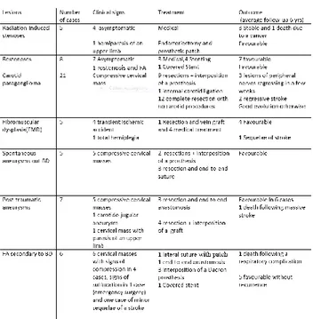
57 cases of non-atheromatous carotid lesions were treated in our department. The table below summarizes the data in the patient files (table 1).
table 1: summary of lesions, clinical signs, treatments and outcomes for our patients
For FA linked to BD, sutures are systematically reinforced by prosthetic pledgets, and anastomoses by bands of prosthetic tissue. Patients with BD receive mandatory dual therapy: steroid and immunosuppressive treatment.
III. DISCUSSION
HOW TO REPAIR NON-ATHEROMATOUS CAROTID LESIONS
Bouayed NM, Bouziane LA
Vascular Surgery Department University Hospital Hai Sabah, Oran, Algeria ([email protected])
Translational Medicine @ UniSa - ISSN 2239-9747 2020, 21(7): 24-26
25 Università degli Studi di Salerno
Isolated kinking of the carotids is rarely a surgical indication.
The 3 cases in our series of restenosis after endarterectomy which remained asymptomatic were simply kept under observation during follow-up. However, the others, and above all the symptomatic cases, are now treated by dilation and stenting2. FA caused by disinsertion of an angioplasty patch are treated with good results by implantation of a covered stent 3. Endovascular treatment by angioplasty and stenting has been extensively validated for the treatment of radiation-induced stenosis4. Carotid paraganglioma is treated surgically. In some cases pre-operative embolization makes it possible to reduce the size of some large tumours and reduce intra-operative bleeding.
Acute dissections are generally treated by anticoagulants however Ohta reported a series of 44 cases of carotid dissection treated by stenting with a favourable outcome in 83.7% of cases 5. We believe however that only the aneurismal complications of carotid dissection can benefit from surgery or an endovascular procedure.
FMD is a rare, segmental, non-atherosclerotic and non-inflammatory idiopathic arteriopathy affecting medium-sized vessels in women aged 30 to 50 6. Lesions of the internal carotid are rare and generally asymptomatic 7. However, this type of dysplasia may be complicated by dissections, dilation due to aneurysm and transient or permanent stroke 6,7. The prevalence of carotid FMD, as assessed by angiography, is only 0.5 – 0.7% 8.
Three types have been identified: type 1 giving rise to the typical “pearl necklace” appearance ; type 2, less frequent, giving rise to an image of a short focal stenosis (present in the only patient in our series who underwent surgery) or a long tubular lesion; and type 3 characterized by a dysplastic aneurysm 6.
Once the indication has been decided, carotid FMD is increasingly being treated by angioplasty and stenting with good long-terms results 8.
We have also treated 12 cases of extra-cranial carotid aneurysms (ECAAs) of which 5 were spontaneous and 7 post-traumatic.
ECAAs are rare. They represent between 0.4% and 4% of all peripheral aneurysms and only 0.1% to 2% of all carotid surgical procedures 1.
For ECAAs (true or false aneurysms), the surgical procedures involve resection with, according to the situation, a direct anastomosis, possible when there is elongation of the carotid, or interposition of a vein graft when the aneurysm is located at the level of the internal carotid, or interposition of a prosthesis when it is located at the level of the common carotid artery.
Resection of saccular aneurysms is often followed by an end-to-end anastomosis; sometimes, when the neck is narrow, a lateral suture is possible. ECAAs, when they are situated high up near the base of the skull, can now be treated using endovascular techniques. Many authors have reported series of ECAAs treated by covered stents with good results 9. Li published a compilation of several series totaling 224 cases where an ECAA was treated by implantation of covered stents. The initial success rate was 92.8%, with a 1.8% rate of established ischemic stroke and a mortality rate of 4.1%. The medium-term patency rate was 93.2% 10. One of our patients was treated using an endovascular approach. The ECAAs were accessible through a cervical approach in patients with low operative risk. Extra-cranial carotid FA secondary to BD are extremely rare. According to Berard only 32 cases have been reported in the literature [11]. Our experience is that the most effective surgical technique is resection with a lateral or end-to-end suture of the native artery reinforced by synthetic tissue pledgets, or with interposition of a PTFE prosthesis, always reinforced by a band of prosthetic tissue surrounding the artery at the level of the anastomosis. In all cases an antithrombotic is prescribed, combined with long-term dual therapy: prednisone at a dose of 1mg/Kg/day quickly reducing to a maintenance dose of 10 to 15mg/day and an immunosuppressive drug. We use azathioprine at a dose of 2.5mg /kg/day. This dual therapy is necessary to prevent local recurrence and the recurrence of other FA in other territories. A vein graft as an arterial substitute is not adequate because venous material itself can suffer degenerative modifications due to BD, leading to occlusion or FA in the graft 11. Endovascular treatment of FA in a context of BD has been reported but the very limited number of cases does not allow any conclusion to be drawn from the results 12. One of our BD patients was treated by implantation of a covered carotid stent with a good result but 50% of the stents placed in other peripheral sites became thrombotic within 18 months. Finally, a simple reinforced ligation of the internal carotid is used by some authors when the lesions are in a high position, not susceptible to easy repair and provided there is good collateral circulation as evidenced by a sufficient residual pressure 13.
IV. CONCLUSION
Non-atheromatous carotid lesions are infrequent. They should be treated when they are symptomatic: compressive signs and focal neurological accidents. However, an aneurysm, even asymptomatic, should
Translational Medicine @ UniSa - ISSN 2239-9747 2020, 21(7): 24-26
26 Università degli Studi di Salerno
be operated on if imaging studies reveal the presence of thrombus on its walls. Numerous surgical procedures are available and are adapted to the morphology and anatomy of specific lesions. However, it must be acknowledged that endovascular techniques are now tending to replace conventional surgery with results that are continually improving.
V. REFERENCES
[1] Thévenet A.- Chirurgie des carotides.Lésions carotidiennes non athéromateuses.- Editions Techniques – Encycl. Méd. Chir. (Paris, France). Techniques chirurgicales, Chirurgie vasculaire, 43144, 9-1990, 9 p.
[2] Midy D, Berard X, Becquemin JP, et al.;
Association Universitaire de Recherche en Chirurgie Vasculaire. Multicentric Retrospective Study of Endovascular Treatment for Restenosis after Open Carotid Surgery. Eur J Vasc Endovasc Surg. 2011 Aug 31.
[3] Abdelhamid MF, Wall ML, Vohra RK. Carotid artery pseudoaneurysm after carotid endarterectomy: case series and a review of the literature. Vasc Endovascular Surg. 2009 Dec;43(6):571-7. Epub 2009 Jul 29.
[4] Rimmer J, Giddings CE, Vaz F, et al.:
Management of vascular complications of head and neck cancer. J Laryngol Otol. 2011 Sep 5:1-5.
[5]- Ohta H, Natarajan SK, Hauck EF, et al.: Endovascular stent therapy for extracranial and intracranial carotid artery dissection: single-center experience. J Neurosurg. 2011 Jul;115(1):91-100.
[6] Touzé E, Oppenheim C, Trystram D, et al.: Fibromuscular dysplasia of cervical and intracranial arteries. Int J Stroke. 2010 Aug; 5(4):296-305.
[7] Mazza A, Zamboni S, Cuppini S, et al.: Internal carotid artery fibromuscular dysplasia in arterial hypertension: management in clinical practice.Blood Press. 2008; 17(5-6):274-7.
[8] Assadian A,Senekowitsch C,Assadian et al.: Combined Open and Endovascular Stent Grafting of Internal Carotid Artery Fibromuscular Dysplasia: Long Term Results. Eur J Vasc Endovasc Surg. 2005 Apr; 29(4):345-9
[9] Yi AC, Palmer E, Luh GY, et al.: Endovascular treatment of carotid and vertebral pseudoaneurysms with covered stents. AJNR Am J Neuroradiol. 2008 May; 29(5):983-7.
[10] Li Z, Chang G, Yao C, Guo L, et al.:
Endovascular stenting of extracranial carotid artery aneurysm: a systematic review. Eur J Vasc Endovasc Surg. 2011 Oct; 42(4):419-26.
[11] Berard X, Corpataux JM, Taoufiq H et al.: Don't trust a vein graft to treat carotid aneurysm in patients with Behçet disease. J Vasc Surg. 2010 Aug;
52(2):471-4.
[12] Ohshima T, Miyachi S, Hattori K et al.: A case of giant common carotid artery aneurysm associated with vascular Behçet disease: successfully treated with a covered stent. 2008 Mar;69(3):297-301.
[13] Sayed A, Elwan H, Fouad F, et al.: Behcet extracranial carotid aneurysms: is there still a role for ligation?2010 Jan;39(1):17-22.